GREEN SYNTHESIS OF ZNS NANOPARTICLES AND FABRICATION OF ZNS-CHITOSAN NANOCOMPOSITES FOR THE REMOVAL OF CR(VI) ION FROM WASTEWATER
←
→
Page content transcription
If your browser does not render page correctly, please read the page content below

Green Processing and Synthesis 2021; 10: 374–383
Research Article
Thokozani Xaba*
Green synthesis of ZnS nanoparticles and
fabrication of ZnS–chitosan nanocomposites for
the removal of Cr(VI) ion from wastewater
https://doi.org/10.1515/gps-2021-0026
received June 10, 2020; accepted November 24, 2020
1 Introduction
Abstract: A modified homogeneous precipitation method The extreme discharge of heavy metals into the environ-
has been used to synthesize ZnS nanoparticles. Starch ment due to industrialization and urbanization has cre-
and polyvinyl alcohol (PVA) were utilized as capping ated difficult challenges worldwide. The existence of
molecules, and later, the ZnS–PVA-capped nanoparticles heavy metal ions in the water systems is a major concern
were then incorporated with chitosan to form ZnS–chitosan due to their toxicity and non-biodegradability which can
nanocomposites for the removal of Cr(VI) ion from waste- cause problems to the environment [1]. The high con-
water. The optical measurements of the synthesized ZnS centrations of these heavy metals in the effluents may
nanoparticles showed the band gap which was blue- trigger hindrance with biological treatment processes at
shifted when compared with the bulk ZnS material. The the sewage treatment systems [2]. Among these heavy
crystalline structures were determined by X-ray diffrac- metals, chromium is graded as one of the top sixteen toxic
tion, and the crystalline sizes were estimated from the
pollutants that have harmful effects on human well-being
Scherer formula. XRD spectra confirmed the formation
[3]. A high concentration of chromium can cause a bad
of hexagonal phase for the uncapped ZnS nanoparticles
effect on human kidneys and livers [4]. A high quantity
with an average crystalline size of 3.71 nm whereas the
of chromium can also cause cancer in the intestinal and
starch- and PVA-capped ZnS nanoparticles showed the
lungs [5].
formation of cubic phase structures with crystalline sizes
Heavy metals can be treated from wastewater through
of 3.26 and 2.88 nm. The TEM image showed spherical
reverse osmosis, precipitation, ultra-filtration membrane
particles with regular morphologies and significantly
filtration, adsorption, co-precipitation, adsorption, sol-
narrow size distributions. The calculated average particle
vent extraction, and membrane process [6]. Among these
diameters were in good agreement with the estimated
techniques, the adsorption process has been proven to be
XRD result. The removal of Cr(VI) ion from wastewater
a highly effective method that has been used lately for the
was studied through the adsorption process. The effect
removal of heavy metals from waste streams. It has been
of pH, dosage, and contact time was investigated. More
proven to be cost-effective, long-lasting, renewal adsor-
than 95% of the metal ion recovery was achieved through
bent, and an easy method to operate when compared with
using ZnS–chitosan nanocomposites.
the other techniques [7].
Keywords: zinc sulfide nanoparticles, chitosan, nano- In recent years, various types of coagulants have
composites, Cr(VI) ions, adsorption shown potential applications in wastewater and water
treatment. Chitosan, which is a non-toxic linear high
molecular weight cationic polymer, has been used lately
as a coagulant in water treatment since it has an ability to
interact with the bacterial surface. It has been endorsed
as a potentially eco-friendly coagulant and flocculant due
to its natural biological characteristics and biodegrad-
ability [8]. The combination of nanomaterials with chito-
* Corresponding author: Thokozani Xaba, Department of Chemistry,
san to form the polymer nanocomposite can coactively
Vaal University of Technology, P/Bag X021, Vanderbijlpark, improve the antimicrobial effect of the polymer material.
South Africa, e-mail: thokozanix@vut.ac.za This unification usually improves the surface charge of
Open Access. © 2021 Thokozani Xaba, published by De Gruyter. This work is licensed under the Creative Commons Attribution 4.0
International License.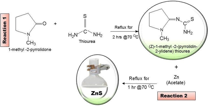
ZnS nanoparticles, ZnS–chitosan nanocomposites, and removal of Cr(VI) ion 375
the composite and also increases multiply sites for bond- from industrial effluents using nanotechnology was
ing with metal centers during wastewater treatment. reported by Mitra et al. [26] while Owalude and Tella
Nano-sized materials have attracted a great deal of [27] reported the removal of hexavalent chromium from
interest over the years in scientific societies due to their aqueous solutions by adsorption on modified groundnut
exceptional and interesting physical, chemical, and bio- hull. The modified groundnut shell was found to be the
logical properties [9]. Semiconducting materials, espe- better adsorbent of Cr(VI) ions when compared with the
cially the metal chalcogenides, have been studied due unmodified groundnut shell.
to their wide bandgap and their application in solar cells, Most of the technologies that have been used in water
optoelectronics, optical sensor devices, photolumines- treatment together with their starting materials are gen-
cence, etc. [10]. Among metal chalcogenides, zinc sulfide erally expensive, complicated, and time-consuming. To
(ZnS) has been studied and revealed significant proper- the best of my knowledge, there is no study that has been
ties for unique diverse applications in electrolumines- reported in the past for the removal of Cr(VI) from waste-
cence [11], lasers [12], light-emitting diodes (LEDs) [13], water using the ZnS–chitosan as an adsorbent that was
and bio-devices [14]. ZnS is an important II–VI chalco- prepared from a simple, cost-effective, and easy environ-
genide with a wide direct band gap of 3.77 eV for the mental method such as the homogeneous precipitation
wurtzite structure [15] as well as 3.72 eV for the zinc method. Thus, in the present study, the preparation of
blende structure [16]. ZnS is also regarded as a low-cost ZnS nanoparticles capped with starch and PVA via the
and non-toxic material with high resistance to photo- greener route and the preparation of ZnS–chitosan nano-
chemical degradation [17]. Several methods have been composites is reported. The prepared polymer nanocom-
reported for the synthesis of zinc sulfide nanoparticles posites and chitosan were used as adsorbents to remove
that include hydrothermal technique [18], microwave Cr(VI) ions from wastewater through a batch experiment.
irradiation [19], solvothermal [20], and wet chemical or The percentage removal was also determined. Factors
co-precipitation methods [10]. During the synthesis of such as the changes in pH of solutions, dosage, and con-
nanomaterials, it is important to use chemical processes tact time were investigated. The optical properties were
that eliminate the use of toxic and harmful substances. characterized with UV–Vis and PL. The structural and
Designing and utilizing green chemistry approaches for morphological properties have been studied using XRD
the synthesis of nanomaterials can help to protect the and TEM whereas the flame atomic absorption spectro-
environment. The homogeneous precipitation method is photometry (AAS) was utilized to measure the concentra-
regarded as an alternative technique that eliminates the tions of the solutions.
usage of non-hazardous substances [21,22].
Past research findings have proven that the combina-
tion of nanomaterials with the polymeric substance such
as chitosan to form the polymer nanocomposite can 2 Materials and methods
improve and increase the surface charge of the polymer
and eventually multiply the number of bonding sites that
2.1 Materials
can allow the metal ions to be attracted on the surface of
the nanocomposites during the adsorption process [23].
Thiourea, 1-methyl-2 pyrrolidone, zinc acetate dihydrate,
In our previous study, thiosemicarbazone ligand was suc-
starch, PVA, ammonium hydroxide, chitosan, chromium
cessfully used in the synthesis of ZnS nanoparticles and
salt, methanol, and acetone were reagents from Sigma-
the preparation of ZnS–polydadmac nanocomposites. The
Aldrich and were all used without further purification.
influence of the concentration of green capping agents as
stabilizers was studied [24]. Tiwari et al. [25] reported the
synthesis and optical properties of polymer-based ZnS
nanocomposites. PVA, starch, and hydroxypropylmethyl 2.2 Experimental
cellulose were used due to their non-toxicity, water solu-
bility, and biocompatibility. The effect of hydroxyl-func- 2.2.1 Synthesis of the zinc sulfide nanoparticles
tionalized polymers on ZnS and their optical properties
was studied. The results showed that hydroxyl-functiona- The (Z)-2-(1-methyl-pyrrolidin-2-ylidene) thiourea ligand
lized polymers were much effective at nucleating and sta- was prepared according to the method described pre-
bilizing ZnS nanoparticles when compared with the other viously [24,28]. ZnS nanoparticles were synthesized by
polymers. A review based on the removal of chromium mixing zinc acetate (5 mmol) in warm 50% methanol
376 Thokozani Xaba
(20 mL) with (20 mL) warm 50% methanolic solution of Measurements were taken using a glancing angle of inci-
the ligand (10 mmol) in a 100 mL two necked flask. The dence detector at an angle of 2θ values over 10 to 80
warm mixture was refluxed inside the water bath at 70°C degrees in steps of 0.0167 with a scan speed of 0.0452.
for an hour to produce a white solution. Exactly, 0.5% Transmission electron microscopy (TEM) was performed
starch or PVA solution was added into the white solution using a Tecnai F30 FEG TEM instrument at an accelerat-
to stabilize the nanoparticles. The pH of the solution was ing voltage of 300 kV. TEM samples were prepared by
adjusted to pH = 11 with ammonia hydroxide solution and placing 1 or 2 drops of ZnS nanoparticles dissolved in
further stirred for an hour. The synthesized ZnS nanopar- water/acetone mixture on lacey carbon copper grids to
ticles were separated from the solution using the centri- obtain TEM images. AAS analysis was collected from
fuge technique, washed three times with acetone, and the AA-7000 Shimadzu model coated GFA-7000 graphite
dried in an open-air. furnace atomizer.
2.2.2 Preparation of ZnS–chitosan nanocomposites
3 Results and discussion
The synthesized ZnS–PVA-capped nanoparticles (∼3 mg)
were dispersed in 10 mL of distilled water. The filtered
3.1 Zinc sulfide nanoparticles
nanoparticle solution was then transferred into a small
beaker with 50 mL of 0.5% chitosan solution that was
previously prepared from dilute acetic acid. The beaker The substituted thiourea ligand was prepared from the
with the mixture was then sealed with a foil and placed reaction of 1-methyl-2-pyrrolidone with thiourea. The
inside an ultra-sonic bath for 4 h to ensure complete uni- ligand was then used to synthesize zinc sulfide nanopar-
fication. The prepared ZnS nanocomposite solution was ticles through the homogeneous precipitation method
then used in water treatment. as represented in Scheme 1. The incorporation of ZnS
nanoparticles into chitosan to form ZnS–chitosan nano-
composites has been explored. The nanocomposites were
2.2.3 Batch adsorption experiments then used in water treatment to remove Cr(VI) ions from
wastewater.
A stock solution of heavy metal (1,000 ppm) was pre-
pared by dissolving 2.83 g of Cr(VI) salt in 1 L of distilled
water. The desired concentrations ranging from 20 to 3.1.1 Optical properties
100 ppm were obtained by the dilution method. For each
experiment of adsorption, 20 mL of the ion metal solution The optical absorption spectra of ZnS nanoparticles have
was shaken at 250 rpm in a plastic bottle. The pH of the been carried out using UV–Vis spectroscopy. Figure 1a
solution was adjusted to the desired value by adding shows the optical absorption profile with the band edges
0.1 M NaOH or 1.0 M HCl. Batch adsorption studies were at 299, 275, and 256 nm for the (i) uncapped, (ii) starch-,
carried out using the thermostat shaker at room tempera- and (iii) PVA-capped ZnS nanoparticles. It was observed
ture at a speed rate of 250 rpm. that as the capping molecule is introduced into the nano-
particles, the spectra were blue-shifted which is an
indication of the decrease in sizes of the particles. To
determine the band gap of the ZnS nanoparticles, a plot
2.3 Characterization of absorbance square versus energy (eV) was done. The
band gap energies were estimated by extrapolating the
UV-1800 Shimadzu spectrophotometer and Gilden fluor- steepest part of the curve. The results are represented in
escence spectrometer were used to measure the optical Figure 1b. The band gap of the ZnS nanoparticles was
properties of ZnS nanoparticles. The nanoparticles were observed at 4.09, 4.35, and 4.34 eV, which were blue-
dissolved in distilled water, and the solution was placed shifted when compared with the bulk ZnS material [29,30].
in a quartz cuvette with a path length of 1 cm. XRD pat- Photoluminescence is an instrumental technique that
terns of the samples were obtained from a Phillips X’Pert was used to study the luminescence properties of the ZnS
chemistry research diffractometer using secondary mono- nanomaterials and is presented in Figure 1c. The emission
chromated Cu Kα radiation (λ = 1.54060 Å) at 40 kV/30 mA. spectra show the maximum peaks at 311 nm (3.99 eV) for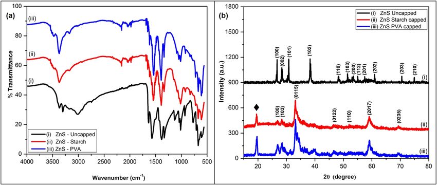
ZnS nanoparticles, ZnS–chitosan nanocomposites, and removal of Cr(VI) ion 377
Scheme 1: Preparation of the (Z)-2-(1-methyl-pyrrolidin-2-ylidene) thiourea ligand and the synthesis of ZnS nanoparticles.
Figure 1: Absorption (a), Tauc plot (b), and emission spectra (c) of ZnS uncapped (i), starch- (ii), and PVA- (iii) capped nanoparticles.
the uncapped ZnS nanoparticles (Figure 1c(i)) while both capping agents and O–H stretching of the water molecules
capped ZnS nanoparticles reveal maxima peaks at 309 nm [28,31,32]. The remaining peaks of the uncapped ZnS
(4.01 eV) in Figure 1c(ii,iii). nanoparticles in Figure 2a(i) were shifted toward the lower
frequency when compared with the absorption peaks of
the capped ZnS nanoparticles. The absorption bands
3.1.2 Structural properties between 660–674 cm−1 and 598–612 cm−1 from the FTIR
spectra of the uncapped, starch-, and PVA-capped ZnS
Figure 2a shows the FTIR spectra of the uncapped, starch-, nanoparticles are attributed to the zinc sulfide bond [33].
and PVA-capped ZnS nanoparticles. The spectral mea- XRD patterns of the synthesized ZnS nanoparticles
surements were carried out in the range between 500 are shown in Figure 2b. The diffraction peaks of the
and 4,000 cm−1 at room temperature. The FTIR spectra of uncapped nanoparticles in Figure 2b(i) show the 2θ values
all ZnS nanoparticles show the same absorption band at located at 26.72°, 28.42°, 30.85°, 38.38°, 48.33°, 51.41°,
about 3,366 and 3,012 cm−1 for the uncapped ZnS nanopar- 53.53°, 55.00°, 57.56°, 61.12°, 70.45°, and 75.05° which cor-
ticles, which correspond to the O–H vibrations of the respond to (100), (002), (101), (102), (110), (103), (200),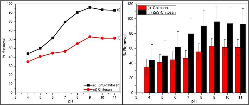
378 Thokozani Xaba
Figure 2: FTIR spectra (a) and X-ray diffraction patterns (b) of ZnS uncapped (i), starch- (ii), and PVA- (iii) capped nanoparticles.
(112), (201), (202), (203), and (210), respectively. These ZnS nanoparticles may be due to the particle aggregation
planes are indexed to a hexagonal phase which matches which might be caused by potential environmental fac-
with JCPDS card No: 79-2204 whereas Figure 2b(ii,iii) of tors since the nanoparticles were not protected by cap-
the starch- and PVA-capped ZnS nanoparticles show the ping molecules [35].
2θ values located at 26.90°, 28.41°, 33.25°, 36.86°, 46.73°,
59.18°, and 69.35° which correspond to (100), (103), (0115),
(0122), (110), (2017), and (0235), respectively, which can be 3.2 Adsorption studies
indexed to cubic phase with the JCPDS card number:
01-072-9259. These results are in consistent with the Parameters such as pH, adsorbent dosage, and contact
reported results [24]. The diffraction peaks at 19.67° and time can play an important role in the removal of Cr(VI)
19.76° correspond to the starch and PVA, respectively from wastewater. The initial concentration of the Cr(VI)
The crystalline sizes of the synthesized ZnS nano- solution of 100 ppm (20 mL) was used throughout and the
particles were calculated using the Debye–Scherrer formula Whatman filter paper No. 42 was utilized to filter the
in Eq. 1 [34]: solutions. Flame atomic absorption spectrophotometry
0.9(λ) (AAs) was used to analyze the adsorbed amount of
D= , (1) Cr(VI) by the nanocomposites from wastewater and the
β cos θ
percentage removal of Cr(VI) was calculated using Eq. 2
where D is the particle size in nm, 0.9 is a Scherrer’s [36]:
constant, λ is the wavelength of X-rays, θ is the Bragg
C0 − C1
diffraction angle, and β is the full-width at half-maximum % Removal = × 100% (2)
C0
(FWHM) of the diffraction peak corresponding to the
maximum peaks. The average particle sizes of the nano- where C0 (ppm) is the initial metal ion concentration and
particles were found to be 3.71, 3.26, and 2.88 nm for the C1 (ppm) is the final metal ion concentration in the
uncapped, starch-capped, and PVA-capped ZnS nano- solution.
particles, respectively.
Figure 3 shows TEM nanographs of the synthesized
ZnS nanocrystallines. The TEM images for all the nano- 3.2.1 Effect of pH
particles showed spherical-shaped particles and uniform
sizes with average diameters of 3.71 ± 0.653, 3.49 ± 0.383, The study based on the effect of pH in adsorption is a very
and 2.71 ± 0.423 nm for the uncapped and capped ZnS essential factor since it regulates the adsorbent’s surface
nanoparticles. These results were in good agreement with charges. Previous reports confirm that the binding sites of
the sizes determined from XRD analysis by Scherrer’s the metal cation and adsorbent become protonated at low
equation. The large particle size value of the uncapped pH values. It had been reported that repulsion occurs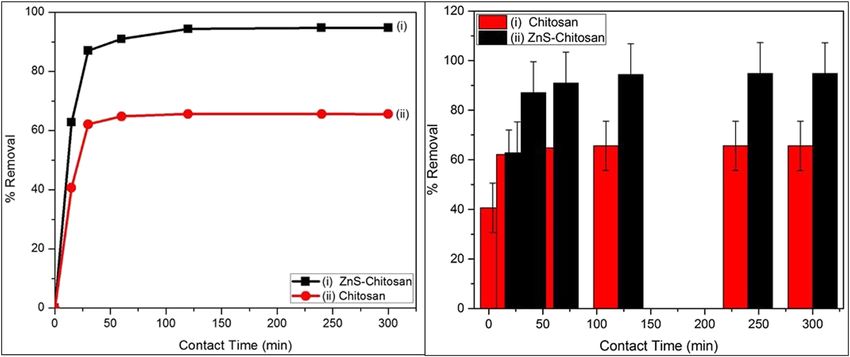
ZnS nanoparticles, ZnS–chitosan nanocomposites, and removal of Cr(VI) ion 379
Figure 3: TEM image of ZnS uncapped (a), starch- (b), and PVA- (c) capped nanoparticles.
between the metal cation and the adsorbent at higher pH. at the pH between 4 and 8, the percentage removal of
The binding spots begin to deprotonate and generate dif- Cr(VI) is low for both the ZnS–chitosan and chitosan. As
ferent functional groups available for binding [37]. the pH is increased from 8 to 11, the percentage removal
The effect of pH of the solution was varied using the of the metal cation was also increased to a maximum
initial concentration of 100 ppm for Cr(VI) solution. The percentage of 95.99% for ZnS–chitosan and a maximum
percentage removal of the Cr(VI) ions at different pH percentage of 62.96% for pure chitosan. The optimum
values is represented in Figure 4. The results show that pH = 9 for both adsorbents was observed.
Figure 4: Effect of pH on adsorption of Cr(VI) ion using chitosan (i) and ZnS–chitosan nanocomposites (ii) as absorbents.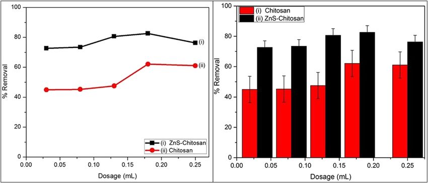
380 Thokozani Xaba
Figure 5: Effect of dosage on adsorption of Cr(VI) ion using chitosan (i) and ZnS–chitosan nanocomposites (ii) as absorbents.
3.2.2 Effect of adsorbent dosages transferred. Each sample was treated with 0.18 mL nano-
composites and shaken with thermo shaker in different
Effect of adsorbent dosage in Figure 5 was carried out at time intervals of 15, 30, 60, 120, 240, and 300 min, respec-
pH = 9 as it showed a greater percentage removal of metal tively. The results in Figure 6 show that Cr(VI) ion removal
ions. It was noted that the percentage removal was is increasing with an increase in contact time. The results
increasing to 59.79% and 80.91% when the amount of project the removal capacity of the metal ion to 65.60%
chitosan or ZnS–chitosan nanocomposites raised from and 94.79% when chitosan and ZnS–chitosan nanocom-
0.03 to 0.25 mL. About 0.18 mL was found to be the maxi- posites are used as absorbents. The equilibrium was
mum adsorbent dosage. reached at 240 min by both absorbents.
3.2.3 Effect of contact time 3.2.4 Mechanism of Cr(VI) ion removal
The effect of contact time is the most important parameter Generally, the adsorption of heavy metals depends on the
for economical wastewater treatment systems [23]. In six surface area and pore structure of the adsorbent [38]. The
different containers, 20 mL of the Cr(VI) ion solution was surface area of the nanocomposites consists of amino and
Figure 6: Effect of contact time on adsorption of Cr(VI) ion using chitosan (i) and ZnS–chitosan nanocomposites (ii) as absorbents.ZnS nanoparticles, ZnS–chitosan nanocomposites, and removal of Cr(VI) ion 381
Scheme 2: The formation of nanocomposites and the adsorption of heavy metal on chitosan-based nanocomposites.
hydroxyl groups that are designed to bind with the Cr(VI) ZnS–chitosan nanocomposite was used as an adsorbent.
ions. Adsorption of Cr(VI) ion onto chitosan and nano- The obtained data may be useful in designing and fabri-
composites depends on the amino and hydroxyl groups cating an economical wastewaters treatment that pos-
[39]. The lone pair that is available in the nitrogen and sesses a high concentration of chromium(VI) ions.
oxygen atoms are existing to the empty atomic orbitals of
the Cr(VI) ion, which forms coordination complexes on the Acknowledgment: The author would like to acknowledge
surface of the material [40] as represented by Scheme 2 the Vaal University of Technology for the support.
below by Xaba et al. [23].
Funding information: The study was funded by the National
Research Foundation (TTK13071722088: “Thuthuka Grant
4 Conclusion Holder”).
The ZnS nanoparticles were synthesized through the Author contributions: Thokozani Xaba confirms sole
homogeneous precipitation method. The optical proper- responsibility for the following: study conception and
ties of the capped nanoparticles showed a blue shift in design, data collection, analysis and interpretation of
wavelength when compared with the uncapped ZnS nano- results, and manuscript preparation.
particles. The XRD studies show a cubic phase for the
capped nanoparticles whereas the uncapped nanoparti- Conflict of interest: The author states no conflict of
cles projected hexagonal phase crystal structures. TEM interest.
images for the synthesized ZnS nanoparticles showed
spherical-shaped particles. These results corroborated Data availability statement: The datasets generated or
well with the XRD results. The highest percentage removal analyzed during the current study are available from
of Cr(VI) ion from wastewater was achieved when the corresponding author on reasonable request.382 Thokozani Xaba
References [17] Dilpazir S, Siddiq M, Iqbal A. Synthesis of zinc sulphide
nanostructures by Co-precipitation: effects of doping on
[1] Gupta VK, Gupta M, Sharma S. Process development for the electro-optical properties. Kenkyu J Nanotechnol Nanosci.
removal of lead and chromium from aqueous solution using 2015;1:34–9.
red mud–an aluminum industry waste. Water Res. [18] Hoa TTQ, Vu LV, Canh TD, Long NN. Preparation of ZnS nano-
particles by hydrothermal method. J Phys.
2001;35:1125–34.
2009;187:012081–7.
[2] Tahir SS, Rauf N. Removal of Fe(II) from the wastewater of a
[19] Zhao Y, Hong JM, Zhu JJ. Microwave-assisted self-assembled
galvanized pipe manufacturing industry by adsorption onto
ZnS nanoballs. J Cryst Growth. 2004;270(3–4):438–45.
bentonite clay. J Environ Manag. 2004;73(4):285–92.
[20] Park J, Joo J, Kwon SG, Jang Y, Hyeon T. Synthesis of mono-
[3] Gardea-Torresdey JL, Tiemann KJ, Armendariz V, Bess-
disperse spherical nanocrystals. Angew Chem Int Ed.
Oberto L, Chianelli RR, Rios J, et al. Characterization of Cr(VI)
2007;46(25):4630–60.
binding and reduction to Cr(III) by the agricultural by products
[21] Díaz-Cruz C, Alonso Nuñez G, Espinoza-Gómez H, Flores-
of Avena monida (Oat) biomass. J Hazard Mater.
López LZ. Effect of molecular weight of PEG or PVA as reducing-
2000;80(1–3):175–88.
stabilizing agent in the green synthesis of silver-nanoparti-
[4] Mungasavalli DP, Viraraghavan T, Jin YC. Biosorption of chro-
cles. Eur Polym J. 2016;83:265–77.
mium from aqueous solution by pretreated Aspergillus niger:
[22] Xaba T, Moloto MJ, Moloto N. The effect of water-soluble
batch and column studies. Colloids Surf A Physicochem Eng
capping molecules in the “Green” synthesis of CdS nanopar-
Asp. 2007;301(1–3):214–23.
ticles using the (Z)-2-(pyrrolidin-2-ylidene)thiourea ligand.
[5] Ofudje EA, Awotula AO, Oladipo GO, Williams OD.
Mater Lett. 2015;146:91–5.
Detoxification of chromium(VI) ions in aqueous solution via
[23] Xaba T, Moloto MJ, Nchoe O, Nate Z, Moloto N. Synthesis of
adsorption by raw and activated carbon prepared from
silver sulfide nanoparticles through homogeneous precipita-
sugarcane waste. Covenant J Phys Life Sci. 2014;2(2):110–22.
tion route and the preparation of the Ag2S-chitosan nano-
[6] Li Q, Zhai J, Zhang W, Wang M, Zhou J. Kinetic studies of
composites for the removal of iron(II) ion from wastewater.
adsorption of Pb(II), Cr(III) and Cu(II) from aqueous solution by
sawdust and modified peanut husk. J Hazard Mater. Chalcogenide Lett. 2017;14(8):337–46.
2007;141(1):163–7. [24] Xaba T, Moloto MJ, Al-Shakban M, Malik AM, Moloto N,
[7] Beh CL, Chuah TG, Nourouzi MN, Choong TSY. Removal of O’Brien P. The influence of concentration of green capping
heavy metals from steel making waste water by using electric agents and ammonium solution as an “activator and stabi-
arc furnace slag. E-J Chem. 2012;9(4):2557–64. lizer” in the synthesis of ZnS nanoparticles for the preparation
[8] Renault F, Sancey B, Badot P-M, Crini G. Chitosan for coagu- of the polymer nanocomposites. Green Process Synth.
lation/flocculation processes – an eco-friendly approach. 2017;6(2):173–82.
Eur Polym J. 2009;45(5):1337–48. [25] Tiwari A, Khan SA, Kher RS, Dhoble SJ, Chandel ALS. Synthesis
[9] Azam A, Ahmed AS, Oves M, Khan MS, Memic A. Size-depen- characterization and optical properties of polymer‐based ZnS
dent antimicrobial properties of CuO nanoparticles against nanocomposites. Luminescence. 2016;31(2):428–32.
Gram-positive and -negative bacterial strains. Int J Nanomed. [26] Mitra S, Sarkar A, Sen S. Removal of chromium from industrial
2012;7:3527–35. effluents using nanotechnology: a review. Nanotechnol
[10] Palve AM. Deposition of zinc sulfide thin films from Zinc(II) Environ Eng. 2017;2(1):1–11.
thiosemicarbazones as single molecular precursors using [27] Owalude SO, Tella AC. Removal of hexavalent chromium from
aqueous solutions by adsorption on modified groundnut hull.
aerosol assisted chemical vapour deposition technique. Front
Beni-Suef Univ J Basic Appl Sci. 2016;5(4):377–88.
Mater. 2019;6:46.
[28] Xaba T, Magagula J, Nchoe OB. Green Synthesis of Cu2S
[11] Tang W, Cameron DC. Electroluminescent zinc sulphide
nanoparticles from (Z)-1-methyl-2-(pyrrolidin-2-ylidene)
devices produced by sol-gel processing. Thin Solid Films.
thiourea ligand for the preparation of Cu2S-Chitosan nano-
1996;280(1–2):221–6.
composites for the removal of Cr(VI) ion from wastewater.
[12] Biswas S, Ghoshal T, Kar S, Chakrabarti S, Chaudhuri S.
Mater Lett. 2018;229:331–5.
ZnS nanowire arrays: synthesis, optical and field emission
[29] Murugadoss G, Rajamannan B, Ramasamy V. Synthesis,
properties. Cry Growth Des. 2008;8(7):2171–6.
characterization and optical properties of water-soluble
[13] Luo Y, Duan G, Ye M, Zhang Y, Li G. Poly(ethylene glycol)-
ZnS:Mn2+ nanoparticles. J Lumin. 2010;130(11):2032–9.
mediated synthesis of hollow ZnS microspheres. J Phys
[30] Lakshmi PVB, Raj KS, Ramachandran K. Synthesis and char-
Chem C. 2008;112(7):2349–52.
acterization of nano ZnS doped with Mn. Cryst Res Technol.
[14] Lin KB, Su YH. Photoluminescence of Cu:ZnS, Ag:ZnS, and
2009;44(2):153–8.
Au:ZnS nanoparticles applied in Bio-LED. Appl Phys B.
[31] Vetrone F, Boyer JC, Capobianco JA. Yttrium oxide nanocrys-
2013;113(3):351–9.
tals: luminescent properties and applications. Am Sci Publ Ed.
[15] Ong HC, Chang RPH. Optical constants of wurtzite ZnS thin
2004;10:725–65.
films determined by spectroscopic ellipsometry. Appl Phys
[32] Divya Rao M, Pennathur G. Facile bio-inspired synthesis of zinc
Lett. 2001;79(22):3612–314.
sulfide nanoparticles using chlamydomonas reinhardtii cell
[16] Tran TK, Park W, Tong W, Kyi MM, Wagner V, Summers CJ.
free extract: optimization, characterization and optical prop-
Photoluminescence properties of ZnS epilayers. J Appl Phys.
1997;81(6):2803–9. erties. Green Process Synth. 2016;5(4):379–88.ZnS nanoparticles, ZnS–chitosan nanocomposites, and removal of Cr(VI) ion 383
[33] Rema Devi BS, Raveendran R, Vaidyan AV. Synthesis and [37] Hadi AG. Removal of Fe(II) and Zn(II) ions from aqueous solu-
characterization of Mn2+-doped ZnS nanoparticles. Pramana. tions by synthesized chitosan. Int J Chemtech Res.
2007;68(4):679–87. 2016;9(4):343–9.
[34] Panda SK, Antonakos A, Liarokapis E, Bhattacharya S, [38] Kuchta B, Firlej L, Maurin G. Modeling of adsorption in nano-
Chaudhuri S. Optical properties of nanocrystalline SnS2 thin pores. J Mol Model. 2005;11(4–5):293–300.
films. Mater Res Bull. 2007;42(3):576–83. [39] Yu K, Ho J, McCandlish E, Buckley B, Patel R, Li Z, et al. Copper
[35] Korshed P, Li L, Ngo DT, Wang T. Effect of storage conditions on ion adsorption by chitosan nanoparticles and alginate micro-
the long-term stability of bactericidal effects for laser gener- particles for water purification applications. Colloids Surf A
ated silver nanoparticles. Nanomaterials (Basel). Physicochem Eng Asp. 2013;425:31–41.
2018;8(4):218. [40] Futalan CM, Kan C-C, Dalida ML, Pascua C, Wan M-W. Fixed-
[36] Milonjic SK, Boskovic MR, Ceranic TS. Adsoprtion of uranium bed column studies on the removal of copper using chitosan
(VI) and zirconium(IV) from acid solutions on silica gel. Sep Sci immobilized on bentonite. Carbohydr Polym.
Technol. 1992;27:1643–53. 2011;83(2):697–704.You can also read