Hombre en la tercera edad con taquiarritmia sostenida de QRS variable y estrecho Elderly man with sustained tachyarrhythmia of variable ...
←
→
Page content transcription
If your browser does not render page correctly, please read the page content below
Hombre en la tercera edad con taquiarritmia sostenida de QRS
                      variable y estrecho

   Elderly man with sustained tachyarrhythmia of variable
      Electrocardiographic Pattern and narrow QRS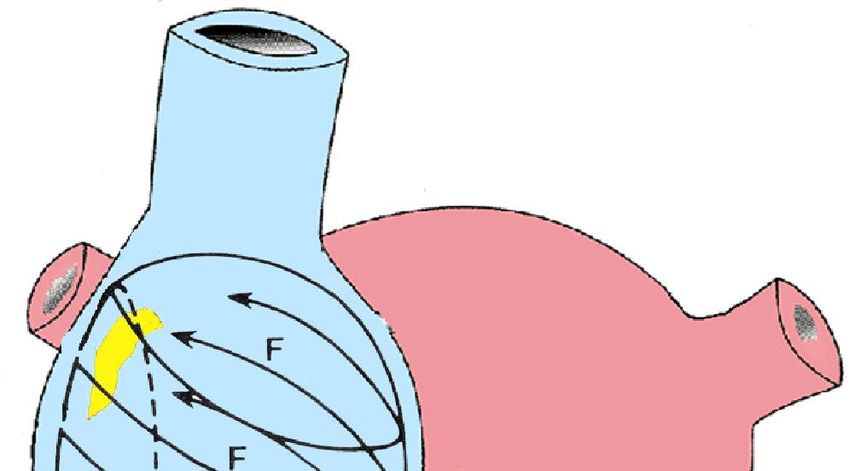
68 yr old, white man, born in Paraíba, living in Fortaleza-CE, Brazil; married,
painter.
The patient said to feel burning retrosternal pain, with no irradiation,
accompanied by cold sweating, general discomfort and quick, regular and
prolonged palpitations, starting on the afternoon of Nov 10, 2013. He denies
experiencing dyspnea.
He mentioned a similar but less intense episode, 3 months ago.
BP=140/90
Medications in use: Valproic acid 500 mg 2 x day due to simple absence seizures,
Salicylic Acid 100mg/d and Atenolol 50mg/d
He was medicated with IV amiodarone

Questions
1. What is the mechanism of the tachycardia?
2. What happened after amiodarone?
3. What to do now?

Warm regards,

Raimundo Barbosa- Barros M.D.
68 años , hombre, blanco, nacido en Paraíba, residente en Fortaleza-CE, Brasil, casado,
pintor.
Paciente referiendo dolor retroesternal en quemazón, sin irradiación, acompañado de
sudoración fria, malestar general y palpitaciones rápidas regulares y prolongada
comenzada en la tarde del día 10.11.13. Niega disnea.
 Refiere un episodio similar de menor intensidad, hace 3 meses.
PA=140/90
Medicamentos en uso:
Acido valpróico 500 mg 2 x día por cuadro de quadros de ausência simples
AAS 100mg/d
Atenolol 50mg/d
Fue medicado con amiodarona EV

Preguntas
1. ¿Cuál es el mecanismo de la taquicardia?
2. ¿Qué pasó después de amiodarona?
3. ¿Qué hacer ahora?

Un abrazo

Raimundo Barbosa- Barros
Admisión -1 Admission-1
Admisión -2 Admission-2
30 minutos después de la amiodarona/ 30 minutes after amiodarone
2 horas después de la administración de amiodarona endovenosa
ECG preformed 2 hours after intravenous administration of amiodarone
Colleagues opinions
Hi
Thanks for sharing the case.
ECG#1: Narrow complex and regular tachycardia. Very difficult to see P waves:
Differential diagnosis include: Atrial tachycardia, Typical AVNRT and Orthodromic
AVNRT (the 3 more frequent diagnosis).
ECG#2: It seems the same tachycardia with left anterior fascicular block aberrancy
ECG#3: After amiodarone, it seems atrial tachycardia with 2:1 conduction
 One other possibility is a dual tachycardia with one of the 2 first ECGs being VT rather
than atrial tachycardia (involving the posterior fascicle).
Management: full EP study with both atrial and ventricular stimulation. Ablation of the
source if AT/

Best wishes

Dr Baranchuk, Adrian MD FACC
Assistant Professor of Medicine Cardiac Electrophysiology and Pacing
Director, EP Training Program Kingston General Hospital FAPC 3, 76 Stuart Street
K7L 2V7, Kingston ON Queen's University Ph: 613 549 6666 ext 3801
Fax: 613 548 1387 barancha@kgh.kari.net
ECG #1 shows rapid narrow complex tachycardia with short RP configuration and
negative P wave in aVL.
ECG # 2 shows LAFB? Amiodarone effect with NCT and P wave negative in Leads I
and V6 at the same rate.
ECG #3 shows atrial fibrillation at a slow rate again probable amiodarone effect.
Possibilities Orthodromic AVRT* over left sided AP or A.T. from the LA.
* impulses traveling in an anterograde manner through the AV node and in a retrograde
manner through the accessory pathway; this is called orthodromic AVRT.

Melvin M. Scheinman, Professor of Medicine:
Department of Cardiac Electrophysiology, University of California San Francisco, San
Francisco, California, USA.
Address:
UCSF
Electrophysiology Service
500 Parnassus Avenue
San Francisco, CA 94143-1354
Telephone/FAX/E-mail:
Phone: (415) 476-5706
Fax: (415) 476-6260
email: scheinman@medicine.ucsf.edu
Hi everybody
1. ECG-1 first tracing Looks like atrial tachycardia with 1/1 AV conduction at a rate of
     around 250 bpm and ascending ST depression.
2. ECG-2 second tracing: is the same except for a left anterior fascicular block.
3. ECG-3 third tracing after amiodarone: the AV conduction is now 2/1 with a
     ventricular rate of around 130 bpm

It does not looks like an typical atrial flutter.

                                      Management
Best solution, after exclusion of thrombus in the left appendage, is to investigate the
tachycardia and the circuit/focus location.
Other possibility: waiting some hours that amiodarone stopped the tachycardia or
performed DC shock or esophageal overdrive.

Then ruling out underlying significant coronary stenosis.

Philippe Maury M.D.
 Department of Cardiology, University Hospital Rangueil, 31059 Cedex 09 Toulouse,
France.
Tel.: +33 (0) 5 61 32 30
 mauryjphil@hotmail.com,
Spanish Trazado 1: Flutter auricular con conducción AV 1:1 Trazado 2 Flutter auricular con conducción AV 1:1 + posiblemente de origen isquémico Trazado 3 Reentrada nodal Le daria adenosina IV Marcelo Chambo Trace 1: atrial flutter with 1:1 AV conduction Trace 2 atrial flutter with 1:1 AV conduction + possibly ischemic in orirgin Trace 3: Nodal Reentry I would give adenosine IV Marcelo Chambo. ---------------------------------------------------------------------------------------------------------- Flutter con conducción 1:1 Flutter con conducción 2:1 Marigel La suerte favorece sólo a la mente preparada. Isaac Asimov
Spanish
Estimado Raimundo y Potro:
1. TPSV por reentrada intranodal. Observo ondaP en DI, aVL y V1. El eje electrico
   ligeramente desviado a la izquierda. Luego del comienzo de la infusión de
   amiodarona, no afecta el ciclo de la taquicardia se desvía el eje eléctrico a la
   izquierda, con r inicial y cambio en la transición precordial en V3, bien podría
   deberse a fenómeno de fatiga de de la división anterosuperior. No me impresiona la
   taquicardia se expresión de isquemia miocardica.
2. En el tercer electrocardiograma aún continua con el ciclo de la taquicardia, pero solo
   han transcurrido 30 minutos de la infusión de amiodarona. Si se encuentra
   compensado aguardaría la amiodarona se distribuya y se corta la taquicardia. Si se
   encuentra descompensado o sintomático, o retorna taquicardia reentrante de alta
   frecuencia, infusión de adenosina y por ultimo cardioversión eléctrica.

Le colocaría un acceso central y tendría un marcapasos transitorio en la cabecera del
paciente, recibe atenolol, infusión de amiodarona me impresiona que cuando revierta la
taquicardia, padecerá de bradicardia extrema y siendo una complicación esperable por
la terapéutica, preveer que pueda necesitar la colocación de urgencia de un marcapasos
transitotio.
Un cordial saludo.
Martín Ibarrola
English
Dear Raimundo and Potro,
•PSVT by intranodal reentry. I see P wave in DI, aVL and V1. The electrical axis
slightly shifted to the left. After the start of amiodarone infusion, it does not affect
the tachycardia cycle, the electrical axis is shifted to the left, with initial r and
change in precordial transition in V3, which could very well be due to fatigue
phenomenon of the anterosuperior fascicle. It doesn’t seem that the tachycardia is
the expression of myocardial ischemia.
•In the third electrocardiogram, the tachycardia cycle still continues, but only 30
minutes elapsed since the amiodarone infusion. If the patient is compensated, I
would wait for the amiodarone to spread and the tachycardia to end. If the patient
is decompensated or symptomatic, or reentrant tachycardia of a high rate returns,
adenosine infusion and finally, electrical cardioversion.
I would place a central access and I would place a transient pacemaker in the
headboard of the patient. He receives atenolol, amiodarone infusion; it seems to
me when the tachycardia reverts, he will suffer extreme bradycardia and with this
being an expected complication by the therapy, you should foresee the need to
place an emergency transient pacemaker.
Kind regards,
Martín Ibarrola
Spanish
hola a todos los colegas del foro: en este caso opino que es una taquicardia por
reentrada intranodal aunque no descartaria que fuera un fluter con conduccion 1:1. De
todas formas me inclino por la primera.
Conducta de ser posible haria estudio electrofisiologico con intención de ablación. En
caso de no ser posible como el atenolol no lo protegio me inclinaria por verapamil via
oral aunque se pudiera valororar una droga 1c como la propafenona intuyendo (desde
luego despues de complemantarios al respecto) que no tenga cardiopatia isquémica.
 Un saludo afectuoso y a esperar por los que de verdad saben.
Yamir from Cuba
                                         English
Hello to all the colleagues in the forum,
In this case, it is my opinion that this is intranodal reentrant tachycardia, although
I would not rule out a 1:1 conduction flutter. Anyway, I lean toward the first
option.
If possible, the management should be electrophysiology study with ablation
intention. If not possible, since atenolol did not protect the patient, I would lean
toward oral verapamil, although a 1c drug could be considered, such as
propafenone, with the intuition (of course, after supplementary studies about it)
that there is no ischemic heart disease.
Warm regards and let’s wait for those who really know.
Yamir - Cuba
Analisis del caso del Dr Raimundo Barbosa Barros y del Profesor Andrés Pérez -Riera M.D.Ph.D. Me parece que es una taquicardia por reentrada nodal “AV nodal reentrant tachycardia” (AVNRT). El tercer ECG muestra un bloqueo fascicular anterosuperior izquierdo dependiente de la taquicardia prolongada, o por la administración de amiadarona.El ECG de base sugiere una hipertrofia o isquemia basal del ventrículo izquierdo expresado por una onda T negativa en aVL y TIII.>TI en un corazón horizontal con una ligera desviación de eje frontal a la izquierda.Como tratamiento específico empezaria con verapamil 5 mm en 10cc glucosa 5% , 2 cc cada minuto y constante control del ritmo A los 3 minutos la arritmia habitualmente desapareceLa amiodarona lleva mas tiempo y no es especifica . Esta droga es mas especifica para las las taquicardias de reentrada extranodales ”AV reentrant tachycardia”Por que yo recomiendo este tratamiento ? Porque un bolus de 5 mg de verapamil o adenosina, provocan bloqueos muy prologados de la parte superior de nodulo auriculo- ventricular acompaniadas con arritmias ventriculares que provocan palpitaciones y angustia al medico tratante ,y bastante peligroso cuando el paciente es un enfermo cardiaco o una jovencita con hypotension. Con el protocolo que yo recomiendo no hay ninguna complicacion y ningun transtorno arritmico despues de la reversion y tampoco descenso de la presion periferica ,y si ocurre se puede imediatamente suspender el tratamiento con poca droga inyectadaEsta es mi experiencia personal y no pase' mas angustias con este problema de taquicardias rapidas de este tipo Que hacer despues del tratamiento / Si es la primera vez no hacer nada y se repite ablacion esta muy indicada un fraternal abrazo Samuel Sclarovsky - Israel
Englsih
Analysis of the case by Dr. Raimundo Barbosa Barros and Professor Andrés Pérez-Riera, M.D.,
Ph.D.: Dear friends of the forum, It seems to me this is AV nodal reentrant tachycardia (AVNRT).
The third ECG shows left anterior fascicular block dependent on prolonged tachycardia, or the
administration of amiodarone.
Baseline ECG suggests basal hypertrophy or ischemia in the LV, expressed by negative T wave in
aVL and TIII>TI in a horizontal heart with mild shift of the frontal axis to the left.
As specific treatment, I would start with verapamil 5 mm in glucose 10 cc 5%, 2cc per minute
and constant rhythm control. After 3 minutes, the arrhythmia usually disappears.
Amiodarone requires more time and is not specific. This drug is more specific for AV reentrant
tachycardias.
Why do I recommend this treatment? Because 5 mg of verapamil or adenosine in bolus cause
very prolonged blocks in the upper part of the atrioventricular node, accompanied by
ventricular arrhythmias that cause palpitations and distress to the attending physician, and quite
dangerous when the patient is a cardiac patient or a young lady with hypotension. With the
protocol I recommend, there is no complication and arrhythmic disorder after reversion and
neither peripheral pressure decrease; if it happens the treatment can be interrupted
immediately with a little drug injected.
This is my personal experience and I was no longer distressed with this rapid tachycardias
problem.
What to do after the treatment? If it is the first time, do nothing, and repeat ablation is quite
indicated.
Warm regards,
Samuel Sclarovsky
Final conclusions
ECG-1 Admission-1 : Narrow-complex QRS Paroxysmal Supraventricular
Tachycardia(PSVT) regular rapid (heart rate 214 bpm), P’ is separate of the QRS
complex in aVL lead: Ortodromic Circus Movemet Tachycardia.

                             P                          P-waves

ECG-2 Admission-2: Narrow-complex QRS Paroxysmal Supraventricular
Tachycardia(PSVT) regular rapid with aberrancy(LAFB-pattern) Aberrancy is common
in CMT differently of AVNRT. Because presence of adverse signs (, chest pain, heart
rate >200 bpm), we choose IV amiodarone.

ECG-3 : Tachycardic irregular rhythm: atrial fibrillation?
ECG-4 ECG preformed 2 hours after intravenous administration of amiodarone: Sinus
rhythm with short PR interval and delta wave insinuation
aVL
What to do after the treatment? RFCA is indicated. Always before Transoesophageal
echocardiography for prevention of thromboembolic accidents(61) Additionally, coronary
computed tomography (CT) scanTheoretical considerations of Narrow QRS-Tachycardias
Paroxysmal Supraventricular Tachycardia (PSVT) is an episodic condition with an
abrupt onset and termination.
SVT in general is any tachyarrhythmia that requires atrial and/or atrioventricular
(AV) nodal tissue for its initiation and maintenance. It is usually a narrow-complex
tachycardia that has a regular, rapid rhythm; exceptions include atrial fibrillation
(AF) and multifocal atrial tachycardia (MAT). Aberrant conduction during SVT
results in a wide-complex tachycardia. (Second ECG.)
SVT is a common clinical condition that occurs in persons of all age groups, and
treatment can be challenging.
Electrophysiologic studies are often needed to determine the source of the
conduction abnormalities.
Manifestations of SVT are quite variable; patients may be asymptomatic or they
may present with minor palpitations or more severe symptoms. Results from EPSs
have helped to determine that the pathophysiology of SVT involves abnormalities
in impulse formation and conduction pathways. The most common mechanism
identified is reentry. (1-4)
Rare complications of paroxysmal SVT include myocardial infarction, congestive
heart failure, syncope, and sudden death.
Classification
The development of intracardiac electrophysiologic studies has dramatically
changed the classification of SVT, with intracardiac recordings having identified
the various mechanisms involved in the condition. Depending on the site of origin
of the dysrhythmia, SVT may be classified as an atrial or AV tachyarrhythmia.
Another way to separate the arrhythmias is to classify them into conditions having
either regular or irregular rhythms(5;6)
Atrial tachyarrhythmias include the following:
    1. Sinus tachycardia
    2. Inappropriate sinus tachycardia (IST)
    3. Sinus nodal reentrant tachycardia (SNRT)
    4. Atrial tachycardia
    5. Multifocal atrial tachycardia
    6. Atrial flutter
    7. Atrial fibrillation
AV tachyarrhythmias include the following:
    1. AV nodal reentrant tachycardia (AVNRT)
    2. AV reentrant tachycardia (AVRT)
    3. Junctional ectopic tachycardia (JET)
    4. Nonparoxysmal junctional tachycardia (NPJT)
Etiology SVT and paroxysmal SVT(PSVT) are triggered by a reentry mechanism. This may be induced by premature atrial or ventricular ectopic beats. Other triggers include hyperthyroidism and stimulants, including caffeine, drugs, and alcohol. PSVT is observed not only in healthy individuals; it is also common in patients with previous myocardial infarction, mitral valve prolapse, rheumatic heart disease, pericarditis, pneumonia, chronic lung disease, and current alcohol intoxication. (7-10) Digoxin toxicity also may be associated with PSVT. (9-11) Atrial tachyarrhythmias Sinus tachycardia Sinus tachycardia is the most common regular SVT. It has an accelerated sinus rate that is a physiologic response to a stressor. It is characterized by a heart rate faster than 100 beats per minute (bpm) and generally involves a regular rhythm. 100 bpm to 160 bpm (exceptionally 180 bpm). During the day, newborn babies: >180 bpm; between 1 week and 3 months: >220 bpm; between 3 months and 2 years: >170 bpm; from 2 years to 10 years: >110 bpm Underlying physiologic stresses such as hypoxia, hypovolemia, fever, anxiety, pain, hyperthyroidism, and exercise usually induce sinus tachycardia. (12;13) Certain drugs, such as stimulants (eg, nicotine, caffeine), medications (eg, atropine, salbutamol), recreational drugs (eg, cocaine, amphetamines, ecstasy), and hydralazine, can also induce the condition. Treatment involves addressing the basic underlying stressor.
Inappropriate Sinus Tachycardia(IST) Concept: rare arrhythmia, hard to control and with sinus origin, characterized by persistently high, chronic tachycardic rhythm (HR over 100 bpm during the day), with more than a 3-month evolution without underlying secondary cause (Primary Sinus Tachycardia), enhanced and exaggerated adrenergic response in the face of minimal physical activities and physiological stress with inappropriate response to drugs with sinus “bradycardizing” action and sensitive to sinus node modification and close regions via catheter by radiofrequency energy. It is an accelerated baseline sinus rate in the absence of a physiologic stressor. In this situation, healthy adults may have an elevated resting heart rate and an exaggerated heart rate response to even minimal exercise. This tachyarrhythmia is observed most commonly in young women without structural heart disease. (14, 15, 10) The underlying mechanism of IST may be hypersensitivity of the sinus node to autonomic imput or an abnormality withing the sinus node and/or autonomic input Etiology: controversial. The following has been proposed: 1) beta-adrenergic hypersensitivity 2) depression of efferent reflects. Clinical-electrocardiographic characteristics 1. Permanently high HR( >100 bpm) without justification. 2. P wave with sinus morphology, SÂP and normal polarity, which indicates sinus command or close. 3. Absence of vagal physiologic decrease of HR during sleep, with values always over 90 bpm in this period. 4. Exaggerated increase of heart rate faced with physiological stress. 5. Absence or poor response to bradycardizing drugs: -blockers, amiodarone, verapamil or digitalis. 6. 1:1 A-V conduction. 7. Palpitations and/or pre-syncope clearly
related with rest or minimal physical efforts. 8. Clinical exclusion of secondary causes
of sinus tachycardia: E.g. hyperthyroidism, pheochromocytoma and diabetes with
autonomous dysfunction. 9. Holter study with medium HR over 90 bpm, with an
almost normal HR during sleep and >100 bpm during the day. 10. Ergometer test:
exaggerated response to HR at minimal effort. Within the first 90 seconds of the Bruce
protocol, HR is observed over 130 bpm.
Management
1) Pharmacological: beta-blockers in high doses are the choice When they are
contraindicate nondihydropyridine calcium antagonists or ivabradine. IST patients
treated with ivabradine showed both HR normalization and quality-of-life improvement
maintained in the long-term follow-up. Stopping ivabradine after 1 year unexpectedly
showed that HR remained in the normal limits in 80% of the patients.(59)
If drugs fail:
2) Radiofrequency Cateter Ablation(RFCA). Regrettably, not in all the cases the return
to normal rates implies abolishing the symptoms. Rate of post-radiofrequency relapses:
20% to 25% of the cases. Combined epicardial/endocardial sinus node ablation is a
viable approach for patients with severely symptomatic IST after a failed endocardial
attempt (60)
Sinus nodal reentrant tachycardia
SNRT is frequently confused with IST. SNRT is due to a reentry circuit, either in or
near the sinus node. Therefore, it has an abrupt onset and offset. The heart rate is
usually 100-150 bpm, and electrocardiographic tracings usually demonstrate a normal
sinus P wave morphology. (14;15;10)
Atrial tachycardia
Atrial tachycardia is an arrhythmia originating in the atrial myocardium. Enhanced
automaticity, triggered activity, or reentry may result in this rare tachycardia.
(16;17;18;19;20).
The heart rate is regular and is usually 120-250 bpm.
The P-wave morphology is different from the sinus P waves and is dependent on the
site of origin of the tachycardia. Because the arrhythmia does not involve the AV node,
nodal blocking agents, such as adenosine and verapamil, are usually unsuccessful in
terminating this arrhythmia.
Atrial tachycardia has also been associated with digoxin toxicity via the triggered
mechanism
Multifocal Atrial Tachycardia (MAT) or caotic tachycardia
Multifocal atrial tachycardia is a tachyarrhythmia that arises within the atrial tissue; it is
composed of 3 or more P-wave morphologies and heart rates. This arrhythmia is fairly
uncommon; it is typically observed in elderly patients with chronic obstructive
pulmonary disease(COPD: 80% of the cases). The heart rate is greater than 100 bpm,
and electrocardiographic findings typically include an irregular rhythm,usually
transitory and recurrent, which may be misinterpreted as atrial fibrillation. Treatment
involves correcting the underlying disease process.. (20-22) Magnesium and verapamil
may sometimes be effective.
Prevalence: infrequent: 0.05% to 0.13% of hospital admittances.
1)COPD: 80%: triggered by intercurrent infection and/or excessive dose of xantines.
2)Diabetes mellitus is associated very frequently. 3) Hypertensive heart disease.
4) Coronary heart disease: both chronic coronary insufficiency and acute infarction. In
the latter it is triggered by high levels of catecholamines. 5) Post-operative of general
anesthesia: in general by hypoxia. 6)         hypopotassemia. 7)      hypomagnesemia. 8)
Pulmonary embolism. 9)          Pulmonary neoplasia.10)        Valvular heart diseases.11)
Digitalis intoxication: rare. 12) No heart disease: in patients dehydrated and with
electrolytic imbalance.
MAT ELECTROCARDIOGRAHIC
                     CHARACTERIZATION

          P       P      P P           P

1) P waves with at least three different morphologies in the same lead.
2) Irregular P-P.
3) Presence of isoelectric line.
4) Rate of P between 100 and 130 bpm.
5) Usual AV conduction is 1:1.
6) PP, PR and RR intervals variable in duration.
7) Preceded or followed by atrial fibrillation and flutter, ectopic atrial tachycardia,
sinus tachycardia.
8) It usually progresses into atrial fibrillation, and it may be confused with the latter
by RR irregularity.
9) Possible presence of intermittent periods of concomitant AF or atrial flutter.
MALE PATIENT, 70 YEARS OLD, SEVERE EMPHYSEMA, COPD,
RIGHT CORONARY INSUFFICIENCY:
CHRONIC COR PULMONALE AND USE OF DIGOXIN
P P P P
P P P
V1
Comments: peaked P waves: “P pulmonale”, P-P cycles with marked variation,
different P waves not conduced to the ventricles with Wenckebach-type AV block,
indicating digitalis intoxication, variable P morphology, slow atrial rate between
100 and 140 bpm.
Electrocardiographic characterization.DIFFERENTIAL DIAGNOSIS OF MAT
MULTIFOCAL ATRIAL
ATRIAL FIBRILLATION
TACHYCARDIA
PP, PR and RR intervals: Variable. Variable.
P wave: Present and with three or Absent. Possible presence
more morphologies in the of “f” waves that present a
same lead and rate between rate between 400 and 700
100 and 130 bpm. bpm.
Atrial event voltage: The P wave may be It may not exist, be of small
increased, indicating voltage (“fine”) or of voltage
chamber enlargement. greater than 1 mm
(“coarse”).
AV conduction: Usually 1:1 There is “concealed
conduction” phenomenon in
the junction.
Differential diagnosis with AF.Atrial fibrillation(AF) CONCEPT: atrial arrhythmia characterized by continuing, fragmented, chaotic, very fast and disorganized electrical activity, which causes at one and the same moment, the appearance of “islands” of depolarized cell groups, in depolarization phase and repolarized, which spread in different directions without effective atrial contraction of the biatrial chamber (loss of atrial mechanical activity) that is translated into surface ECG by absence of P waves and substituting them, small high rate waves, which are irregular and with variable voltages, called fibrillatory or “f” waves, or in certain cases only baseline. AF is an extremely common arrhythmia arising from chaotic atrial depolarization. The atrial rate is usually 300-600 bpm, while the ventricular rate may be 170 bpm or more. Electrocardiographic findings characteristically include an irregular rhythm with fibrillatory atrial activity. This arrhythmia is associated with the following condition(12;23;11): Rheumatic heart disease, hypertension, ischemic heart disease, pericarditis, thyrotoxicosis, alcohol intoxication, mitral valve prolapse and other disorders of the mitral valve and digitalis toxicity When AF occurs in young or middle-aged patients in the absence of structural heart disease or any other apparent cause, it is called lone or idiopathic, cryptogenic, lone, or isolated AF. : it is defined as the AF in absence of any etiologic factor, associated to normal ventricular function by echocardiogram. True lone AF does not possess positive familial background. AF of familial/genetic origin: it may be confused with the previous variety, because a cardiac or extra-cardiac cause is not found. Just the identification of the gene permits us to reckon it. Thus far, mutations have been identified in several chromosomes such us 10q22-q24, 11p15.5, KCNQ1, KCNE1 and
others.
Congenital short QT syndrome: it has a high tendency to atrial fibrillation, because
there is an increase in the function of the potassium rectifier channels. Three mutations
were described: in potassium channels: HERG, KCNQ and KCNJ2
Relevant factors in atrial fibrillation
I) Type of underlying disease when it is present
II) Medium ventricular heart rate
III) Prevention of recurence\Potential of thromboembolism
Atrial flutter

CONCEPT: atrial tachyarrhythmia not very frequent and nearly always present,
accompanied by organic substrate, which has as electrophysiological mechanism in
most of the cases, a macro-reentry circuit in circle that covers the whole RA; more
rarely by a unifocal or multifocal atrial focus with very high shock, or exceptionally by
focal micro-reentry in the RA.
There are always intra-atrial or inter-atrial dromotropic alterations, with a minimal
extension being necessary in the movement of the circle, refractoriness dispersion and
autonomous tone variations. Atrial flutter
Atrial flutter may be a transitional rhythm and can progress to atrial fibrillation.
Electrocardiographic findings of typical atrial flutter include negative sawtooth flutter
waves in leads II, III, and aVF. AV conduction is most commonly 2:1, which yields a
ventricular rate of approximately 150 bpm. (11;12;23)
The ECG is characterized by the typical atrial “F” waves with sawtooth or picket-fence
appearance, frequently observed better in DII, DIII, aVF and V1, with median atrial HR
of 250-350 bpm (the HR of atypical or type II flutter is 350 to 450 bpm), characteristic
absence of isoelectric line between F waves, variable degrees of AV block or rarely, 1:1
conduction.
Ventricular HR is usually half of the atrial HR (i.e., 150 beats/min). A ventricular rate
significantly slower in absence of drugs, suggests normal AV conduction.
Causes of atrial flutter
1.    Coronary heart disease: Chronic; AMI: 03% (5.3% of the cases).
2.    COPD
3.    Rheumatic mitral and/or tricuspid valve disease.
4.    Severe aortic stenosis
5.    Bronchopneumonia
6.    Acute pulmonary embolism: transitory character
7.    Bronchogenic carcinoma
8.    Thyrotoxicosis: transitory. Frequent rate of ventricular response 1:1. Ventricular
      HR is 300 bpm
9.    Congenital heart diseases: E.g.: ASD not operated > 40 years old. Ebstein anomaly
10.   Post-operative of heart surgery. E.g.: Mustard surgery for TGA correction
11.   Digitalis intoxication (exceptional)
12.   As part of the “brady-tachy” syndrome
13.   Myocarditis
14.    Pericarditis
15.   As part of WPW syndrome (frequent AV 1:1 conduction with syncope and possible
      sudden death)
16.    Mitral valve prolapse syndrome
17.    Hypertensive heart disease
18.   Alcoholism.
ELECTROPHYSIOLOGICAL
MECHANISMS OF ATRIAL FLUTTER
A) DROMOTROPIC MECHANISMS
1) Macro-reentry around fixed or functional, anatomical or surgical barriers.
2) Micro-reentry.
B) AUTOMATIC MECHANISMS
1) Unifocal atrial focus.
2) Multifocal atrial focus with very high shock.
Mechanisms.DROMOTROPIC MECHANISMS BY MACRO-
REENTRY IN ATRIAL FLUTTER
FLUTTER TYPE I OR CLASSICAL: intercaval
macro-reentry: counterclockwise circular motion,
descending by the RA free wall and ascending by the SVC “MOTHER CIRCUS WAVE”
interatrial septum: “mother circus wave”.
DAUGHTER
WAVES
INTERCAVAL COUNTER-
MACROREENTRY CLOCKWISE
INTERATRIAL
FREE SEPTUM
WALL
CAUDOCRANEAL
CRANEOCAUDAL
IVC
III II
F waves of negative polarity
in II, III and aVF
without baseline
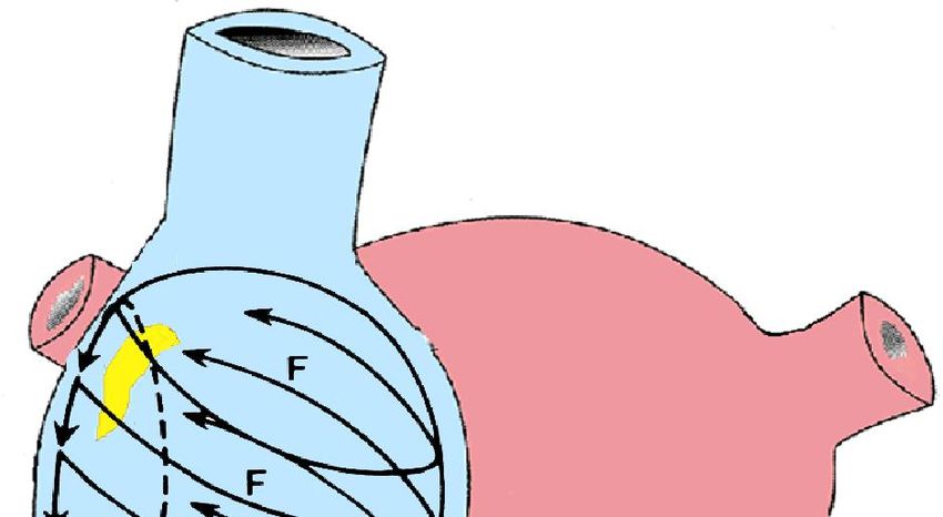
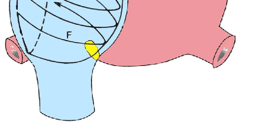
Outline of macro-reentry in atypical flutter.DROMOTROPIC MECHANISMS BY MACRO-
REENTRY IN ATRIAL FLUTTER
TYPE II FLUTTER OTHER
NAMES:
Atypical, rare, antidromic, type
B, rapid, left atrial. Macro- CW
ROTATION
reentry in the junction of the
right appendage with the RA
near the atrioventricular sulcus.
Clockwise circular motion
descending by the septum and
ascending by the RA free wall.
F F
F WAVES OF POSITIVE POLARITY IN II, DII AND aVF
AND WITH A MUCH GREATER RATE
WITHOUT BASELINE
Atrial rate of 345 bpm
Differences between flutter type I and type II.Dromotropic Mechanism by Macro-reentry in atral flutter
TYPE I COMMON OR TYPE II, ATYPICAL OR RARE
CLASSICAL
Rate of F waves: 240 to 339 bpm 340 to 433 bpm.Over 300 bpm
MOST FREQUENT Intercaval macro-reentry, Macro-reentry in the junction of
MECHANISM: counterclockwise circular motion, the right appendage with the RA
descending by the RA free wall and near the atrioventricular sulcus.
ascending by the interatrial septum: Clockwise circular motion
“mother circus wave”. descending by the septum and
ascending by the RA free wall.
Polarity of F waves Negative polarity in II, III and aVF Positive polarity in II, III and aVF
without baseline. without baseline.
Drugs that may be used Antagonists of Na2+ channels, Blockers of K+ channels: class III
except lidocaine, mexiletine and drugs, particularly Amiodarone
tocainide. Class IA drugs: (200 to 400 mg/day) and Sotalol
Quinidine, (600 to 800 mg/day); (80 to
Procainamide (200 to 400 mg 4/6h) 160 mg/day).
and Disopyramide (300 to
400 mg/day) and IC: Propafenone
(400 to 900 mg/day) and
Flecainide: single dose of 300 mg.
Ibutilide.
Response to artificial Good Null
transesophageal pacingCLINICS OF ATRIAL FLUTTER
History: palpitations, fatigue, poor tolerance to exercise, dyspnea, angina,
dizziness and syncope.
Physical examination: heart rate approximately 150 bpm in most cases,
regular or slightly irregular pulse, frequent hypotension, left CHF, syncope.
History and physical examination.ELECTROCARDIOGRAPHIC CHARACTERIZATION
1) Characteristics of atrial activation.
2) Absence of isoelectrical “plateau” between F waves.
3) AV conduction: ventricular rate depends on AV nodal conduction; more
frequently between 150 bpm and 220 bpm.
4) Intraventricular conduction that determines QRS complex duration. When
it is conducted with aberrance, QRS is prolonged.
Electrocardiographic characterization.CHARACTERISTICS OF ATRIAL ACTIVATION IN ATRIAL FLUTTER
ATRIAL DEPOLARIZATION
SAWTOOTH APPEARANCE
ATRIAL REPOLARIZATION
ABSENCE OF ISOELECTRICAL
“PLATEAU”
BETWEEN F WAVES
Waves with sawtooth or picket fence appearance called F waves, with a heart rate between 250 and
350 bpm, observed better in the inferior wall and V1 with slowly descending and rapidly ascending ramp.
These waves resemble an inverted P wave, followed by an ascending ramp: “Tp” waves.
Electrocardiographic characterization.CHARACTERISTICS OF ATRIAL
ACTIVATION IN ATRIAL FLUTTER
In the common form, the polarity of F waves is negative in inferior leads II,
III and aVF: caudocephalic atrial activation: type I, common or classical
flutter.
The DII or V1 leads should be recorded separately, in prolonged tracings
(from 15s to 20s) to establish the relationship between F waves and QRS
complexes.
During atrial flutter with 2:1 conduction, the ‘’F’’ waves may be masked in
the DII leads and be very visible in V1.
Electrocardiographic characterization.CHARACTERISTICS OF ATRIAL
ACTIVATION IN ATRIAL FLUTTER
CAUSES OF F WAVES OF LOW RATE FLUTTER
1) Great mega-atria show atrial rate sometimes lower than 200 bpm.
2) Use of IA drugs: drugs of intermediate kinetics (Quinidine,
procainamide, disopyramide and ajmaline) reduce Vmax. and
extend action potential.
3) IC: they are drugs of slow kinetics (Propafenone, Flecainide,
Encainide, Moricizine and Lorcainide).
4) Class III drugs: K+ channel block (prolongation of action potential in
phase 3) (Amiodarone).
Electrocardiographic characterization.CHARACTERISTICS OF ATRIAL
ACTIVATION IN ATRIAL FLUTTER
CAUSES OF F WAVES OF FLUTTER OF HIGH RATE
1) Flutter in children: median HR of 300 bpm with 1:1 response.
2) Flutter of ventricular pre-excitation.
3) Type II flutter of Wells: rate of F between 340 and 433 bpm: it cannot
be interrupted with pacing.
Electrocardiographic characterization.TYPES OF ATRIOVENTRICULAR
CONDUCTION IN ATRIAL FLUTTER
A) REGULAR: 2:1 – 1/2
1:1 – suggests ventricular pre-excitation
3:1 – 1/3
4:1 – 1/4
B) IRREGULAR
C) ABSENT: with complete AV block – the ventricular rate is usually low and
independent from atrial rate.
AV conduction in flutter.REGULAR ATRIAL FLUTTER WITH 1:1
AV CONDUCTION
300 bpm
F
1:1 AV conduction (rare) is a medical emergency. The ventricular rate near 300 bpm must be treated
immediately. 1:1 AV conduction may be found in the following circumstances:
A) Pre-excitation of WPW type, because the stimulus is conduced in anterograde fashion by the
anomalous pathway;
B) Atrial flutter secondary to hyperthyroidism;
C) Flutter of the pediatric group;
D) Consequence of initial use of IA class drugs (quinidine, procainamide or disopyramide) by atrial
slowing and by vagolytic anti-cholinergic action in the AV junction that this group of drugs causes,
especially if the drugs were used without administering dixogin, calcium antagonists or -blockers
previously in order to control the rate of ventricular response.
Examples of 1:1 flutter.REGULAR ATRIAL FLUTTER
WITH 2:1 AV CONDUCTION
150 bpm
F F
300 bpm
The most frequent ratio in non-treated patients is 2:1 with atrial and ventricular rate of
300/150 bpm respectively; this ratio is due to the physiological interference in the junction.
If the ventricular response rate is regular and constant (e.g.: always 2:1) the F-R interval
will be too, varying between 260 and 460 ms.
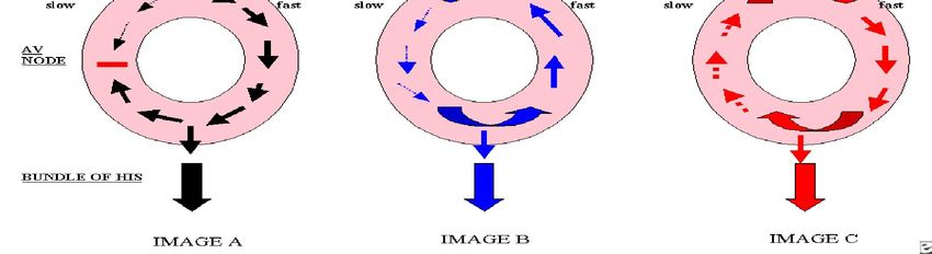
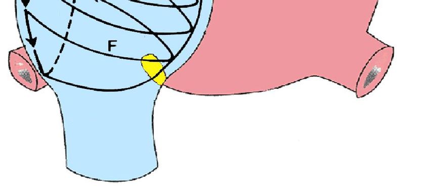
Example of 2:1 flutter.AV tachyarrhythmias AV nodal reentrant tachycardia The most common cause of paroxysmal SVT is AVNRT. AVNRT is diagnosed in 50- 60% of patients who present with regular narrow QRS tachyarrhythmia. (3;23;24). The heart rate is 120-250 bpm and is typically quite regular. AVNRT may occur in healthy, young individuals, and it occurs most commonly in women.(25) Most patients do not have structural heart disease. However, occasionally these individuals may have an underlying heart condition such as rheumatic heart disease, pericarditis, myocardial infarction, mitral valve prolapse, or preexcitation syndrome. (3;24;25) An understanding of the electrophysiology of AV nodal tissue is very important in comprehending the mechanism of AVNRT. In most people, the AV node has a single conducting pathway that conducts impulses in an anterograde manner to depolarize the bundle of His. In certain cases, AV nodal tissue may have 2 conducting pathways with different electrophysiologic properties. One pathway (alpha) is a relatively slow conducting pathway with a short refractory period, while the second pathway (beta) is a rapid conducting pathway with a long refractory period. The coexistence of these functionally different pathways serves as the substrate for reentrant tachycardia. (3;23;24;9)Electrophysiologic studies have demonstrated dual AV nodal pathways in 40% of patients. The onset of AVNRT is triggered by a premature atrial impulse. A premature atrial impulse may reach the AV node when the fast pathway (beta) is still refractory from the previous impulse but the slow pathway (alpha) may be able to conduct. The premature impulse then conducts through the slow pathway (alpha) in an anterograde manner; the fast pathway (beta) continues to recover
because of its longer refractory period.
After the impulse conducts in an anterograde manner through the slow pathway (alpha),
it may find the fast pathway (beta) recovered. The impulse then conducts in a retrograde
manner via the fast (beta) pathway. If the slow pathway (alpha) has repolarized by the
time the impulse completes the retrograde conduction, the impulse can reenter the slow
pathway (alpha) and initiate AVNRT.

Image A displays the slow pathway and the fast pathway, with a regular impulse being conducted through the atrioventricular node. Image B
displays a premature impulse that is conducted in an anterograde manner through the slow pathway and in a retrograde manner through the fast
pathway, as is seen in typical atrioventricular nodal tachycardia. Image C displays the premature impulse conducting in a retrograde manner
through the pathway and the impulse reentering the pathway with anterograde conduction, which is seen commonly in patients with atypical
atrioventricular nodal tachycardia.
Importantly, note that AVNRT does not involve the ventricles as part of the reentry
circuit; the necessity of perinodal atrial tissue to the circuit is controversial. Because the
impulse typically conducts in an anterograde manner through the slow pathway and in a
retrograde manner through the fast pathway, the PR interval is longer than the RP
interval. Thus, in patients with typical AVNRT, the P wave is usually located at the
terminal portion of the QRS complex. (3;23;11;24;9)
In patients with atypical AVNRT, anterograde conduction is via the fast pathway, while
retrograde conduction is via the slow pathway. For these atypical patients, the RP
interval is longer than the PR interval. (24;26;23;25;3;9;27;11)
AV Reentrant Tachycardia (AVRT)
AVRT is the second most common form of paroxysmal SVT. The incidence rate of
AVRT in the general population is 0.1-0.3%. AVRT is more common in males than in
females (male-to-female ratio of 2:1), and patients with AVRT commonly present at a
younger age than do patients with AVRT. AVRT is associated with the Ebstein anomaly,
although most patients with AVRT do not have evidence of structural heart disease.
AVRT occurs in the presence of accessory pathways, or bypass tracts. Accessory
pathways are errant strands of myocardium that bridge the mitral or tricuspid valves.
(24;28;9;10) AVRT results from the presence of 2 or more conducting pathways;
specifically, the AV node and 1 or more bypass tracts. In a normal heart, only a single
route of conduction is present. Conduction begins at the sinus node, progresses to the
AV node, and then to the bundle of His and the bundle branches. However, in AVRT, 1
or more accessory pathways connect the atria and the ventricles. The accessory
pathways may conduct impulses in an anterograde manner, a retrograde manner, or
both. ( 29;30;24;31;28;32;9;10) When impulses travel down the accessory pathway in
an anterograde manner, ventricular preexcitation results. This produces a short PR
interval and a delta wave, as is observed in persons with WPW syndrome. Importantly,
note that not all accessory pathways conduct in an anterograde manner. Concealed
accessory pathways are not evident during sinus rhythm, and they are only capable of
retrograde conduction. A reentry circuit is most commonly established by impulses
traveling in an anterograde manner through the AV node and in a retrograde manner
through the accessory pathway; this is called orthodromic AVRT.
A reentry circuit may also be established by a premature impulse traveling in an
anterograde manner through a manifest accessory pathway and in a retrograde manner
through the AV node; this is called antidromic AVRT. While the orthodromic AVRT is
typically a narrow-complex tachycardia, antidromic AVRT inscribes a bizarre, wide-
complex tachycardia.(33;34;35)
Patients with WPW syndrome can develop AF and atrial flutter . The rapid non
decremental conduction via the accessory pathways can result in extremely rapid rates,
which can degenerate to VF and cause SCD. Patients with preexcitation syndromes
with AF must not be administered an AV nodal blocking agent; these agents can further
increase conduction via the accessory pathway, which increases the risk of ventricular
fibrillation and death. ( 36;37;7;38;8;39).
ECG Recognition
Usually initiated by atrial premature beat, heart rate 170 to 250 bpm, regular rhythm or
slightly irregular in casses of changing conduction velocities through the AV node, P
wave buried within the QRS complex; either distorts the end of the QRS complex or is
not seen at all, QRS complex narrow but often disotrted by the P’wave, causing a
pseudo S wave in the inferior leads and V6 and/or pseudo r’ in lead V1.
Usually benign and self-limiting. Easily terminated by a vagal maneuver(patient shuld
be taught several valgal maneuvers. Occasionally refractory to vagal maneuvers and
referred for RFCA.
Treatment: RFCA of slow atrionodal pathway
Junctional ectopic tachycardia and nonparoxysmal junctional tachycardia

JET and NPJT are rare; they presumably arise because of increased automaticity,
triggered activity, or both. They are usually observed following valvular surgery, after
myocardial infarction, during active rheumatic carditis, or with digoxin toxicity. These
tachycardias are also observed in children following congenital heart surgery.
Electrocardiographic findings include a regular narrow QRS complex, although P
waves may not be visible. Patients with AV dissociation have also been described(9;
40;41).
Epidemiology
Occurrence in the United States
The incidence of paroxysmal SVT is approximately 1-3 cases per 1000 persons. The
incidence rate of the WPW pattern on electrocardiographic tracings is 0.1-0.3% in the
general population, although not all patients develop SVT.(7-10)
In a population-based study, the prevalence of paroxysmal SVT was 2.25 cases per
1000 persons, with an incidence of 35 cases per 100,000 person-years. (43) AVNRT is
more common in patients who are middle aged or older, while adolescents are more
likely to have SVT mediated by an accessory pathway.
Paroxysmal SVT is observed not only in healthy individuals; it is also common in
patients with previous myocardial infarction, mitral valve prolapse, rheumatic heart
disease, pericarditis, pneumonia, chronic lung disease, and current alcohol
intoxication. (7-10) Digoxin toxicity also may be associated with paroxysmal SVT.
(9;10;11)
Sex-related demographics
Most series of RFCA reflect a higher proportion of female patients with AVNRT than
male patients. This may reflect a true higher incidence in women, or it may reflect the
sample of patients who are referred (or choose) to undergo extensive evaluation and/or
RFCA.
In a population-based study, the risk of developing paroxysmal SVT was twice as high
in women as it was in men. (43)
Age-related demographics The prevalence of paroxysmal SVT increases with age.
AVNRT is seen more commonly in persons who are middle aged or older, while
adolescents usually have SVT from an accessory pathway.(43) The relative frequency of
tachycardia mediated by an accessory pathway decreases with age. Prognosis: Patients
with symptomatic WPW syndrome have a small risk of SD. Otherwise, prognosis in
paroxysmal SVT is dependent on any underlying structural heart disease; patients with
a structurally normal heart have an excellent prognosis. Morbidity and mortality
Paroxysmal SVT may start suddenly and last anywhere from seconds to days. Patients
may or may not be symptomatic, depending on their hemodynamic reserve and heart
rate, the duration of the paroxysmal SVT, and coexisting diseases. Paroxysmal SVT
can result in HF, pulmonary edema, myocardial ischemia, and/or myocardial AMI
secondary to an increased HR in patients with poor Lvfunction. (9;10;11) In fact, one
study found that one third of patients with SVT experienced syncope or required
cardioversion.(44) Incessant SVT can cause tachycardia-induced cardiomyopathy.
Patients with WPW syndrome may be at risk for cardiac arrest if they develop AF or
atrial flutter in the presence of a rapidly conducting accessory pathway (ie, a pathway
with a short anterograde refractory period). Extremely rapid ventricular rates during AF
or atrial flutter can cause deterioration to VF. This complication is unusual and occurs
primarily in patients who have had prior symptoms due to WPW syndrome. SCD may
be the initial presentation of WPW syndrome, but how often this occurs is unclear. In
the absence of manifest preexcitation (ie, WPW syndrome), the risk of sudden death
with paroxysmal SVT is extremely small.
History: Because symptom severity depends on the presence of structural heart disease
and on the hemodynamic reserve of the patient, individuals with paroxysmal SVT may
present with mild symptoms or severe cardiopulmonary complaints. Their frequency
rates are as follows(42;44): Palpitations (>96%), dizziness(75%), shortness of breath
(47%), syncope ( 20%), chest pain (35%), fatigue ( 23%), diaphoresis ( 17%), and
nausea ( 13%). Palpitations and dizziness are the most common symptoms reported by
patients with SVT. Chest discomfort may be secondary to a rapid heart rate, and it
frequently subsides with the termination of the tachycardia. Persistent SVT may lead to
tachycardia-induced cardiomyopathy. History should include time of onset, any
triggers, any previous episodes or arrhythmia, and previous treatment. A detailed past
medical and cardiac history and a complete list of all medications should be obtained.
Patients who are hemodynamically unstable should be resuscitated immediately with
cardioversion. An ECG should be performed as soon as possible. Many patients with
frequent episodes of paroxysmal SVT tend to avoid activities such as exercising and
driving due to past episodes of syncope or near-syncope.
Physical examination: Pertinent findings are generally limited to the patient’s
cardiovascular and respiratory systems. Patients often appear quite distressed.
Tachycardia may be the only finding in persons who are otherwise healthy and have
significant hemodynamic reserve. Patients who have limited hemodynamic reserve may
be tachypneic and hypotensive. Crackles may be auscultated secondary to HF. An
S3 may be present, and large jugular venous pulsations may also be visualized.
(9;44;10).
Paroxysmal Supraventricular Tachycardia Differential Diagnoses
1. Atrial Fibrillation
2. Atrial Flutter
3. Atrial Tachycardia
4. Atrioventricular Nodal Reentry Tachycardia (AVNRT)
5. Sinus Node Dysfunction
6. Ventricular Fibrillation
7. Ventricular Tachycardia.
Approach Considerations
Laboratory studies: A cardiac enzyme evaluation should be ordered for patients with
chest pain, patients with risk factors for myocardial infarction, and patients who are
otherwise unstable and present with heart failure, hypotension, or pulmonary edema.
Young patients with no structural heart defects have a very low risk of myocardial
infarction.
Other laboratory tests include the following: Electrolyte levels - Should be checked
because electrolyte abnormalities can contribute to paroxysmal SVT
Complete blood count (CBC) - Helps to assess whether anemia is contributing to the
tachycardia or ischemia Thyroid studies - The results are rarely diagnostic of
hyperthyroidism Digoxin level - Obtain for patients on digoxin, because paroxysmal
SVT is one of the many dysrhythmias that can be caused by supratherapeutic levels of
this drug.
Electrophysiology Electrophysiologic studies have dramatically changed the diagnosis
of SVT. Intracardiac recordings have helped to map accessory pathways and reentry
circuits in patients, and they have also assisted cardiologists and electrophysiologists in
understanding the mechanisms behind these tachyarrhythmias.
At present, electrophysiologic studies are generally performed in combination with
radiofrequency catheter ablation.

Chest radiography
Obtain a chest radiograph to assess for the presence of pulmonary edema and
cardiomegaly. Infections such as pneumonia, which in certain cases are associated with
paroxysmal SVT, can also be confirmed with findings from this imaging method.
(9;40;10;41;11).Congenital heart defects such as Ebstein anomaly of the tricuspid valve
can be suspected.

Transthoracic echocardiography
A transthoracic echocardiogram may be helpful if structural or congenital heart disease
is suggested.

Magnetic resonance imaging
Cardiac magnetic resonance imaging (MRI) can be useful, especially if a congenital
heart disease is being considered.
Electrocardiography
Electrocardiographic findings permit classification of the tachyarrhythmia, and they
may allow a precise diagnosis. P waves may not be visible; when present, they may be
normal or abnormal, depending on the mechanism of atrial depolarization. (9;10;35)
Electrocardiographic characteristics of the various SVTs are as follows:
Sinus tachycardia – HR > 100 bpm; P waves similar to sinus rhythm
Inappropriate sinus tachycardia (IST) - Findings similar to sinus tachycardia; P
waves similar to sinus rhythm
Sinus nodal reentrant tachycardia (SNRT) - P waves similar to sinus rhythm; abrupt
onset and offset
Atrial tachycardia - HR 120-250 bpm; P-wave morphology different from sinus
rhythm; long RP interval (in general); AV block does not terminate tachycardia
Multifocal atrial tachycardia - Heart rate 100-200 bpm; 3 or more different P-wave
morphologies
Atrial flutter- Atrial rate of 200-300 bpm; flutter waves; AV conduction of 2:1 or 4:1
Atrial fibrillation - Irregularly irregular rhythm; lack of discernible P waves
AV nodal reentrant tachycardia (AVNRT) - HR of 150-200 bpm; P wave located
either within the QRS complex or shortly after the QRS complex; short RP interval in
typical AVNRT and long RP interval in atypical AVNRT
AV reentrant tachycardia (AVRT) - HR of 150-250 bpm; narrow QRS complex in
orthodromic conduction and wide QRS in antidromic conduction; diagnosis excluded
by AV block during SVT; P wave after QRS complex
Following the termination of the tachycardia, an ECG should be performed during the
sinus rhythm to screen for WPW syndrome. Echocardiography and/or Holter
monitoring also may be useful. These tests can help to assess the frequency and
duration of SVT episodes, although they have a low yield. Echocardiography may be
helpful in screening for structural or congenital heart disease.
Characterizing a patient’s SVT by comparing the RP interval to the PR interval is
helpful. Long RP tachycardias result when atrial activity precedes the QRS complex. In
short RP tachycardias, atrial activity occurs with or shortly after ventricle excitation,
and the P wave is found within the QRS complex or shortly after the QRS
complex(9;10; 40;41). The classifications of SVTs based on the RP interval are as
follows:
Short RP tachycardias – Typical AVNRT, AVRT, junctional ectopic tachycardia (JET),
and nonparoxysmal junctional tachycardia (NPJT)
Long RP tachycardias – Sinus tachycardia, SNRT, atrial tachycardia, atrial flutter,
atypical AVNRT, and a permanent form of junctional reciprocating tachycardia
Two consecutive P waves without an intervening QRS complex may be due to atrial
tachycardias, in some cases, AVNRT, but they are unlikely to be due to AVRT.
Vagal maneuvers and nodal blocking agents like adenosine work in AVNRT in some
cases, but not in atrial tachycardias. Blocking the tachycardia with adenosine or vagal
maneuvers may assist in diagnosing the rhythm as well as treating it.
Approach Considerations
Patients with symptomatic WPW syndrome should be told of the potential for cardiac
arrest. In general, these patients should not be treated longitudinally with calcium
channel blockers or digoxin unless the pathway is known to be of low risk (long
anterograde refractory period). This is because of the potential for more rapid
ventricular rates should AF or atrial flutter occur.
Patients with preexcited AF should not be treated with intravenous AV nodal blocking
agents, such as adenosine, beta-blockers, calcium channel blockers, and digoxin.
Rather, if the patient is hemodynamically stable, intravenous procainamide should be
administered. If the patient is unstable, direct current cardioversion should be
performed.
Most of the patients who present with paroxysmal supraventricular tachycardia
(paroxysmal SVT) have AVNRT or AVRT. These arrhythmias depend on AV nodal
conduction and therefore can be terminated by transiently blocking this conduction.
Electrical cardioversion
Electrical cardioversion is the most effective method for restoring sinus rhythm.
Synchronized cardioversion starting at 50J can be used immediately in patients who are
hypotensive, have pulmonary edema, have chest pain with ischemia, or are otherwise
unstable.If AF has been present for longer than 24-48 hours, defer cardioversion until
the patient has been adequately anticoagulated to prevent thromboembolic
complications. (40; 36; 45;46;47;48;49;41)
Inpatient care: Patients who require cardioversion, are unstable, and have comorbid
illnesses should be admitted to the hospital. Patients who are young, healthy, and
asymptomatic may be discharged and advised to have a follow-up examination with
their primary physician or cardiologist. If the patient is having more frequent episodes
of paroxysmal SVT and medical therapy is not successful or desired, then RFCA
should be proposed.
Diet and activity: Dietary changes depend on underlying medical problems. Changes
in physical activity depend on underlying cardiac problems and other comorbidities.
Consultations: A cardiologist should be consulted for patients with frequent episodes
of paroxysmal SVT, syncope, and/or preexcitation syndromes. Consultation with a
cardiologist should also be obtained for patients in whom medical management has
failed. An electrophysiologist should be consulted for patients considered for RFCA.
Pediatric patients should be referred to a pediatric electrophysiologist.
Transfer: Patient transfer to a center with RFCA is reasonable if this therapy is
planned. Alternatively, patients can be discharged home and scheduled for outpatient
procedures. Exceptions include patients with syncope, profound symptoms, or
preexcited atrial fibrillation or atrial flutter.
Monitoring: Patients treated medically should be monitored regularly. Patients cured
with RFVC are typically seen once in a follow-up examination following the procedure,
then as needed for recurrent symptoms.
The first-line treatment in hemodynamically stable patients, vagal maneuvers, such as
breath-holding and the Valsalva maneuver (ie, having the patient bear down as though
having a bowel movement), slow conduction in the AV node and can potentially
interrupt the reentrant circuit.
Carotid massage is another vagal maneuver that can slow AV nodal conduction.
Massage the carotid sinus for several seconds on the nondominant cerebral hemisphere
side. This maneuver is usually reserved for young patients. Due to the risk of stroke
from emboli, auscultate for bruits before attempting this maneuver. Do not perform
carotid massage on both sides. A Valsalva maneuver, if performed properly by the
patient, can frequently avert an attack.
Short-Term Pharmacologic Management
When SVT is not terminated by vagal maneuvers, short-term management involves intravenous
adenosine or calcium channel blockers. Adenosine is a short-acting drug that blocks AV node
conduction; it terminates 90% of tachycardias due to AVNRT or AVRT. Adenosine does not
usually terminate atrial tachycardia, although it is effective for terminating SNRT.[40, 36, 45, 50, 48, 41]
Typical adverse effects of adenosine include flushing, chest pain, and dizziness. These effects are
temporary because adenosine has a very short half-life of 10-20 seconds. (49)
Other alternatives for the acute treatment of SVT include calcium channel blockers, such as
verapamil and diltiazem, as well as beta-blockers, such as metoprolol or esmolol. Verapamil is a
calcium channel blocker that also has AV blocking properties. It has a longer half-life than
adenosine and may help to maintain sinus rhythm following the termination of SVT. It is also
advantageous for controlling the ventricular rate in patients with atrial tachyarrhythmia.
(19;51;54;46; 10;48;49; 11)
You can also read