Hypoplastic left heart syndrome: surgical therapy
←
→
Page content transcription
If your browser does not render page correctly, please read the page content below
Aus dem Deutschen Herzzentrum München des Freistaates Bayern

         Klinik an der Technischen Universität München

  Hypoplastic left heart syndrome: surgical therapy
                         Dissertation

           zum Erwerb des Doktorgrades der Medizin

               an der Medizinischen Fakultät der

           Ludwig-Maximilians-Universität München

                         vorgelegt von
                Dr. sc. Jelena Pabst von Ohain

                              aus
                        Zagreb, Kroatien

                             Jahr
                             2021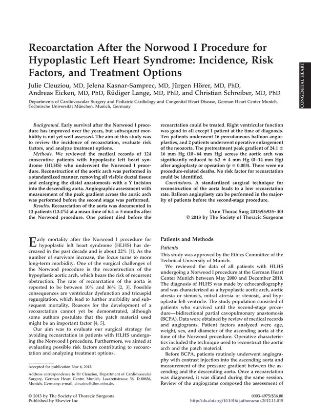
Mit Genehmigung der Medizinischen Fakultät der

                    Ludwig-Maximilians-Universität zu München

Erster Gutachter:                Prof. Dr. Jürgen Hörer

Zweiter Gutachter:               Priv. Doz. Dr. Sebastian Michel

Dritter Gutachter:               Prof. Dr. Nikolaus Haas

Dekan:                           Prof. Dr. med. dent. Reinhard Hickel

Tag der mündlichen Prüfung:      08.09.2021

Affidavit

                                       Eidesstattliche Versicherung

Pabst von Ohain, Jelena
________

_________________________________________________________________
Name, Vorname

Ich erkläre hiermit an Eides statt, dass ich die vorliegende Dissertation mit dem Titel:
Hypoplastic left heart syndrome: surgical therapy

selbständig verfasst, mich außer der angegebenen keiner weiteren Hilfsmittel bedient und alle
Erkenntnisse, die aus dem Schrifttum ganz oder annähernd übernommen sind, als solche
kenntlich gemacht und nach ihrer Herkunft unter Bezeichnung der Fundstelle einzeln
nachgewiesen habe.

Ich erkläre des Weiteren, dass die hier vorgelegte Dissertation nicht in gleicher oder in ähnlicher
Form bei einer anderen Stelle zur Erlangung eines akademischen Grades eingereicht wurde.

München, 09.09.2021                                                 Jelena Pabst von Ohain
__________________________                                          __________________________________
Ort, Datum                                                          Unterschrift Doktorandin

                                                        5
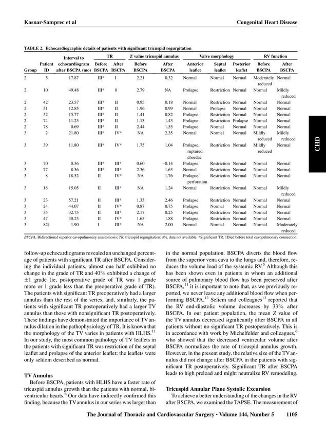
Table of contents
Affidavit ................................................................................................................................................... 5
TABLE OF FIGURES................................................................................................................................... 9
ABBREVIATIONS..................................................................................................................................... 11
ORIGINAL PUBLICATIONS TO THE TOPIC OF PROMOTION ................................................................... 13
AUTHOR CONTRIBUTION STATEMENT .................................................................................................. 15
   Contribution to Paper I...................................................................................................................... 15
   Contribution to Paper II..................................................................................................................... 15
INTRODUCTION ..................................................................................................................................... 17
   Congenital heart disease ................................................................................................................... 17
   Functionally single ventricle .............................................................................................................. 17
   Hypoplastic left heart syndrome ....................................................................................................... 19
       Norwood Operation ...................................................................................................................... 21
       Bidirectional superior cavopulmonary anastomosis ..................................................................... 22
       Total cavopulmonary connection .................................................................................................. 23
   Influence of bidirectional superior cavopulmonary anastomosis on the systemic right ventricle and
   the systemic tricuspid valve .............................................................................................................. 24
   Re-coarctation after the Norwood operation ................................................................................... 24
ZUSAMMENFASSUNG ........................................................................................................................... 25
   Unloading of right ventricle by bidirectional superior cavopulmonary anastomosis in hypoplastic
   left heart syndrome patients promotes remodeling of systemic right ventricle but does not
   improve tricuspid regurgitation ........................................................................................................ 25
   Recoarctation after the Norwood I procedure for hypoplastic left heart syndrome: incidence, risk
   factors, and treatment options ......................................................................................................... 29
ABSTRACT .............................................................................................................................................. 31
   Unloading of right ventricle by bidirectional superior cavopulmonary anastomosis in hypoplastic
   left heart syndrome patients promotes remodeling of systemic right ventricle but does not
   improve tricuspid regurgitation ........................................................................................................ 31
   Recoarctation after the Norwood I procedure for hypoplastic left heart syndrome: incidence, risk
   factors, and treatment options ......................................................................................................... 33
PAPER I .................................................................................................................................................. 35
PAPER II ................................................................................................................................................. 45
REFERENCES .......................................................................................................................................... 53
DANKSAGUNG ....................................................................................................................................... 57
CURRICULUM VITAE .............................................................................................................................. 59
   Publications ....................................................................................................................................... 61

                                                                              7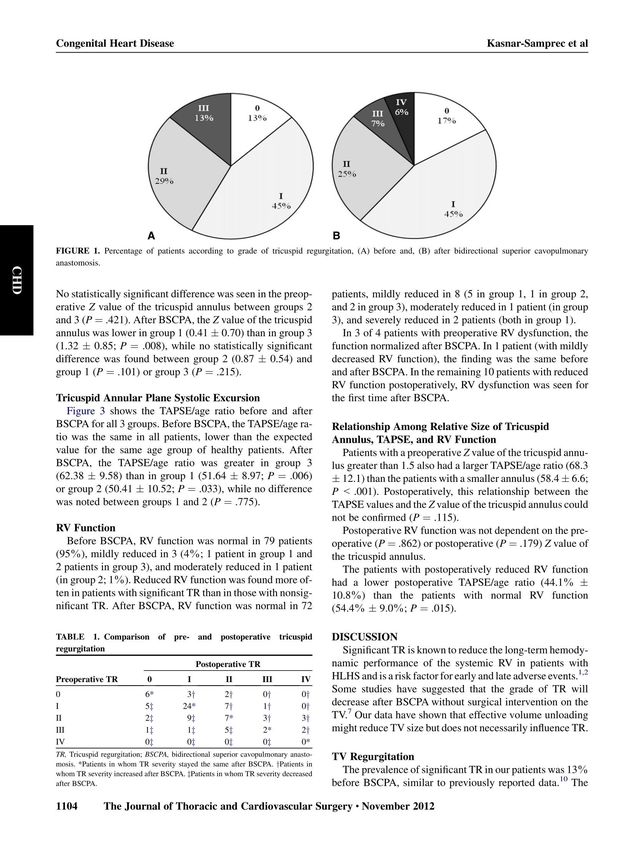
TABLE OF FIGURES

Figure 1. Hypoplastic left heart syndrome .............................................................................. 20

Figure 2. Norwood procedure.................................................................................................. 21

Figure 3. Bidirectional superior cavopulmonary anastomosis ................................................ 22

Figure 4. Total cavopulmonary connection ............................................................................. 23

                                                               9
ABBREVIATIONS
BSCPA bidirectional superior cavopulmonary anastomosis
CHD congenital heart disease
HLHS hypoplastic left heart syndrome
TCPC total cavopulmonary connection
11ORIGINAL PUBLICATIONS TO THE TOPIC OF PROMOTION
Paper I
Jelena Kasnar-Samprec*, Andreas Kühn*, Jürgen Hörer, Manfred Vogt, Julie Cleuziou, Rüdiger
Lange, Christian Schreiber: Unloading of right ventricle by bidirectional superior
cavopulmonary anastomosis in hypoplastic left heart syndrome patients promotes
remodeling of systemic right ventricle but does not improve tricuspid regurgitation. The
Journal of thoracic and cardiovascular surgery 09/2012; 144(5):1102-9.
DOI:10.1016/j.jtcvs.2012.08.012
* Equally contributing authors
Paper II
Julie Cleuziou, Jelena Kasnar-Samprec, Jürgen Hörer, Andreas Eicken, Rüdiger Lange, Christian
Schreiber: Recoarctation After the Norwood I Procedure for Hypoplastic Left Heart Syndrome:
Incidence, Risk Factors, and Treatment Options. The Annals of thoracic surgery 01/2013;
95(3):935-40. DOI:10.1016/j.athoracsur.2012.11.015
13AUTHOR CONTRIBUTION STATEMENT
Contribution to Paper I
Collection and assembly of data
Data curation
Formal analysis
Validation
Writing – original draft
The co-author Andreas Kühn, MD, who contributed equally to the publication, analysed all of
the echocardiograms of the enrolled patients, and contributed significantly to data collection
as well as reviewing and editing.
Contribution to Paper II
Collection and assembly of data
Writing – review & editing
15INTRODUCTION
Congenital heart disease
Congenital heart disease (CHD) accounts for 28% of all major congenital anomalies.1 The
worldwide incidence of CHD is estimated to 9 per 1,000 live births;2 moderate and severe
forms are present in about 6 per 1,000 live births.3 Amongst all birth defects, CHD is the
leading cause of illness and death in the infant age.4 However, the advances in diagnostic
methods, medical, interventional and surgical therapy of infants and children with CHD have
resulted in one of the largest survival increments in medical history.5-8 In developed countries,
more than 90% of CHD patients are expected to survive the complete childhood.9, 10 Survival
to adulthood varies with the severity of CHD: 96-98% of patients with mild and moderate CHD
and 56% of patients with severe CHD survive to adult ages.11 The prevalence of patients with
CHD in developed countries increased by >50% from 2000 to 2010.12
Functionally single ventricle
Approximately 25% of the infants with CHD are born with a so-called „critical CHD“4 and need
of surgical or interventional treatment in the neonatal and infant age. Part of these patients
has a functionally univentricular heart. This term includes all hearts in which one of the
ventricles is anatomically or functionally inadequate, and the other, dominant ventricle has to
support both the systemic and pulmonary circulation. Another common attribute of these
hearts is that surgical therapy in the form of a biventricular correction is not achievable.13
The group of functionally univentricular hearts includes many different anatomical variations.
Generally, it is possible to differentiate the dominant ventricle into the left, right or
undifferentiated ventricle.14 An anatomically single ventricle, without even a rudimentary part
of the second ventricle, is very rare.15, 16
Many different anatomical variants of the functionally single ventricle are described, the most
common being tricuspid atresia, double inlet left ventricle, pulmonary atresia with an intact
interventricular septum, unbalanced common atrioventricular canal and the hypoplastic left
heart syndrome.
17Physiologically, the dominant ventricle supplies the systemic and pulmonary circulation in
parallel. This parallel circulation poses a strain on the functionally single ventricle, and it leads
to hypoxemia due to mixing of the oxygen-saturated and oxygen-desaturated blood. The
function of the single ventricle and the competence of the atrioventricular valves play a
significant role in the univentricular circulation.
The clinical picture of the newborns depends primarily on the amount of blood in the
pulmonary circulation.17 In this way, the functionally univentricular hearts can be divided into
three groups, depending on the balance between the systemic and pulmonary circulation in
the newborn age:
- balanced systemic and pulmonary circulation
- few clinical symptoms13, 18
- surgical treatment in the newborn age is not necessary
- restrictive pulmonary circulation
- main symptom is cyanosis
- a systemic-to-pulmonary shunt in the newborn age
- excessive pulmonary circulation
- mainly symptoms of heart failure
- without the obstruction of the systemic circulation a pulmonary artery
banding in the newborn age
- with obstruction of the systemic circulation a complex Norwood
operation in the newborn age
The surgical therapy of all types of the functionally univentricular heart has a common
objective: separating the pulmonary and systemic bloodstream while using the pump function
of the dominant ventricle for the systemic circulation. The surgical strategy includes three
steps:
- Step I: initial palliation in the newborn age for insuring unhindered systemic outflow
as well as systemic and pulmonary venous return, and controlling the pulmonary blood
flow13
18- Step II: partial separation of the pulmonary and systemic bloodstreams by creating a
superior cavopulmonary anastomosis, between 3-6 months of age
- Step III: complete separation of the pulmonary and systemic circulation by connecting
the inferior vena cava to the pulmonary artery and completing the so-called Fontan-
circulation, in the age of 18-48 months
Hypoplastic left heart syndrome
First described by Noonan and Nadas in 1958,19 hypoplastic left heart syndrome (HLHS) is the
most common type of a functionally univentricular heart,20 with an incidence of 2 to 3 per
10,000 births,21, 22 accounting for 1 – 3% of all CHD.23 HLHS is also one of the most complex
and surgically challenging CHD.
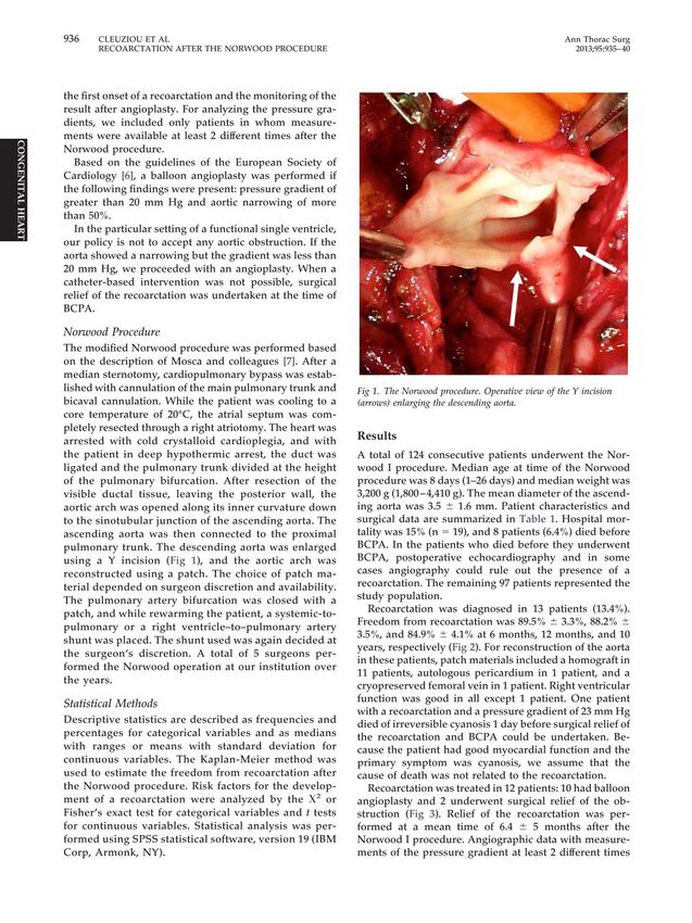
HLHS consists of a variety of cardiac anomalies characterised by a hypoplastic left ventricle,
atresia or stenosis of the aortic and mitral valve, and hypoplastic ascending aorta and aortic
arch Figure 1.24
Newborns with HLHS are dependent on the persisting arterial duct, and the open atrial
communication Figure 1.23 The oxygenated blood from the pulmonary veins flows from the left
atrium over the open atrial septum into the right atrium („left-to-right shunting“) and mixes
there with the desaturated blood from the systemic veins. The “mixed” blood flows then
through the dominant right ventricle in the main pulmonary artery. A part of the blood volume
goes again in the pulmonary circulation. The other part of the blood flows over the persistent
arterial duct in the aorta and facilitates the retrograde perfusion of the upper body part and
the antegrade perfusion of the lower body part.17
19Figure 1. Hypoplastic left heart syndrome
© 2014 Children's Hospital of Philadelphia
https://www.chop.edu/conditions-diseases/hypoplastic-left-heart-syndrome-hlhs
The systemic tricuspid valve is dysplastic approximately one-third of patients with HLHS, more
commonly so in the presence of a non-atretic mitral valve.25 Bileaflet tricuspid valve is present
in up to 12% of patients; quadricuspid valve, clefts, and accessory orifices are less common. 25
The size of the tricuspid annulus is usually on the upper range of normal or above.
The theoretical work on surgical therapy of the HLHS started in the ‘60s and ‘70s26, 27 with
several unsuccessful surgical attempts.28, 29 The first successful surgical treatment was
performed by Norwood in the early ‘80s.30-32
20Norwood Operation
The Norwood operation is usually performed during the first ten days of life. To avoid possible
complications, the surgery should not be delayed beyond the 2-3 weeks of life.33, 34
The goals of the Norwood operation Figure 2 are:
- unobstructed systemic circulation
- using the pulmonary valve for the systemic outflow
- augmenting the aorta
- unobstructed mixing of the blood from the pulmonary and the systemic veins
- surgical widening of the atrial septal communication
- optimal pulmonary blood flow for enabling the growth of the pulmonary arteries
- systemic-to-pulmonary shunt implantation
Figure 2. Norwood procedure
© 2014 Children's Hospital of Philadelphia
https://www.chop.edu/treatments/staged-reconstruction-heart-surgery
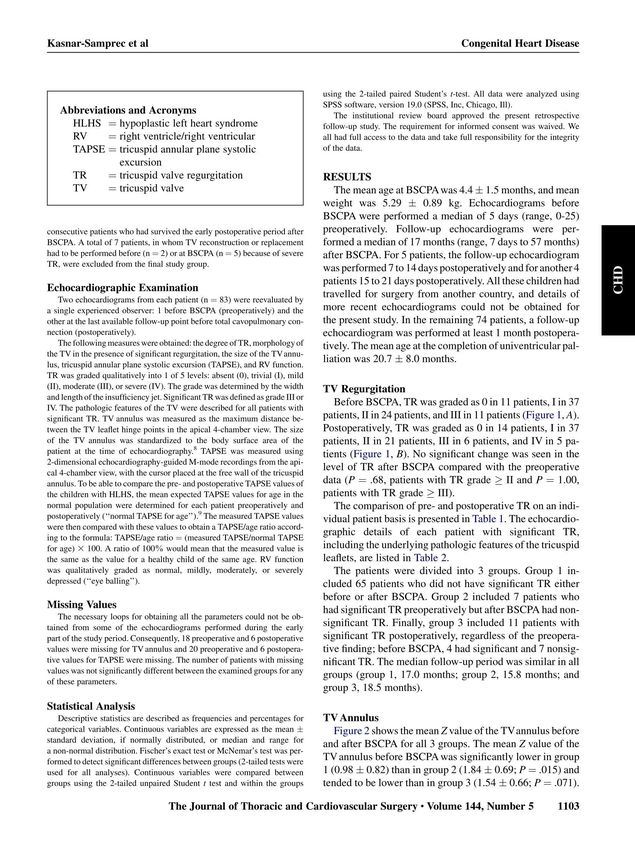
21Bidirectional superior cavopulmonary anastomosis
The bidirectional superior cavopulmonary anastomosis (BSCPA) is performed in the age of 3-
6 months.35 The timing for BSCPA differs significantly among centres, but it should not be
performed while the pulmonary vascular resistance exceeds 2-3 Wood units.36, 37
During this operation, the superior vena cava is separated from the right atrium and sutured
to the pulmonary artery Figure 3. The systemic-to-pulmonary shunt is divided. After BSCPA,
unsaturated blood from the upper part of the body enters the pulmonary circulation without
circulating through the heart.
Figure 3. Bidirectional superior cavopulmonary anastomosis
© 2014 Children's Hospital of Philadelphia
https://www.chop.edu/treatments/staged-reconstruction-heart-surgery
The goals of the BSCPA are:
- partial separation of the pulmonary and systemic bloodstream
- providing sufficient "low pressure" pulmonary perfusion
- decline of the volume load of the right ventricle
22Total cavopulmonary connection
Total cavopulmonary connection (TCPC) is performed in the age of 18 to 36 months.38 Some
centres perform Fontan completion at an older age with comparable outcomes.39
The goal of the TCPC is the complete separation of the pulmonary and systemic circulation.
This is achieved by disconnecting the vena cava inferior from the right atrium and suturing it
to the pulmonary artery Figure 4. In this way, the desaturated blood from the lower body also
flows to the pulmonary circulation without entering the heart.
Figure 4. Total cavopulmonary connection
© 2014 Children's Hospital of Philadelphia
https://www.chop.edu/treatments/staged-reconstruction-heart-surgery
23Influence of bidirectional superior cavopulmonary anastomosis on the systemic right
ventricle and the systemic tricuspid valve
High-grade of tricuspid valve insufficiency is a known risk factor for early and late morbidity
and mortality40, 41 in the patients with HLHS. The function of the tricuspid valve is influenced
by the interaction of volume overload and consequent enlargement of the systemic right
ventricle and the tricuspid annulus, morphological anomalies of the valve leaflets, and the
pump-function of the systemic right ventricle.42
The construction of BSCPA changes the origin of the pulmonary bloodstream and decreases
the volume of blood entering the systemic right ventricle.43 The reduction of the ventricular
volume slows down the dilation of the tricuspid valve annulus.44 It has been reported that the
grade of insufficiency of the atrioventricular valve in patients with a functionally univentricular
heart can decrease after BSCPA, without simultaneous valve surgery.45
We aimed to evaluate the effect of BSCPA and consequent reduction of volume load on the
systemic right ventricle and the systemic tricuspid valve in patients with HLHS.
Re-coarctation after the Norwood operation
Early mortality following the Norwood operation in patients with HLHS has declined in the
past years to approximately 10-22%.46 Serious complications which might occur after the
Norwood operation are excessive or inadequate pulmonary blood flow, impairment of
coronary artery perfusion, and neoaortic arch obstruction.47 The reported rate of re-
coarctation of the aorta is 10-36%.48, 49 Re-coarctation is associated with decreased ventricular
function and tricuspid regurgitation, and consequent morbidity and mortality.50 Recurrent or
residual aortic arch obstruction may be technique and materials dependent.51-53
Our study aimed to determine the incidence, evaluate the risk factors, and analyse therapy
options of re-coarctation after Norwood operation in patients with HLHS.
24ZUSAMMENFASSUNG
Unloading of right ventricle by bidirectional superior cavopulmonary anastomosis in
hypoplastic left heart syndrome patients promotes remodeling of systemic right
ventricle but does not improve tricuspid regurgitation
Jelena Kasnar-Samprec*, Andreas Kühn*, Jürgen Hörer, Manfred Vogt, Julie Cleuziou, Rüdiger
Lange, Christian Schreiber
J Thorac Cardiovasc Surg. 2012 Nov;144(5):1102-8.
* Equally contributing authors
Ziel: Das Ziel unserer Studie war die Beurteilung des Effekts der bidirektionalen superioren
cavopulmonalen Verbindung (BSCPA) zur Verringerung der Volumenbelastung des
systemischen rechten Ventrikels und der systemischen Trikuspidalklappe bei Patienten mit
hypoplastischem Linksherz Syndrom (HLHS).
Patienten und Methoden: Neunzig aufeinanderfolgende Patienten mit HLHS, die die
frühpostoperative Periode nach BSCPA überlebt hatten, wurden analysiert. Zwei
Echokardiogramme wurden durch einen erfahrenen Spezialisten bewertet: eines vor der
BSCPA und das zweite beim letzten Follow-Up vor der totalen cavopulmonalen Anastomose.
Die folgenden Parameter wurden beurteilt: Grad der Trikuspidalklappeninsuffizienz, die
Morphologie der Trikuspidalklappe bei Klappeninsuffizienz, der Durchmesser des
Trikuspidalklappenannulus, tricuspid annular plane systolic excursion (TAPSE) und die
rechtsventrikuläre Pumpfunktion.
Ergebnisse: In den Echokardiogrammen, die im Median fünf Tage vor der BSCPA durchgeführt
wurden, gab es keine Trikuspidalklappeninsuffizienz bei 11 Patienten, minimale bei 37
Patienten, geringgradige bei 24 Patienten und mittelgradige bei 11 Patienten. In den
Echokardiogrammen, die im Medianalter von 17 Monate durchgeführt wurden, gab es keine
Trikuspidalklappeninsuffizienz bei 14 Patienten, minimale bei 37 Patienten, geringgradige bei
21 Patienten, mittelgradige bei 6 Patienten und hochgradige bei 5 Patienten. Der Unterschied
zwischen der Trikuspidalklappeninsuffizienz vor und nach der BSCPA war statistisch nicht
signifikant. Die meisten Patienten mit einer mittelgradigen und hochgradigen
25Trikuspidalklappeninsuffizienz hatten eine morphologisch auffällige Trikuspidalklappe, mit
einem Prolaps des anterioren Segels und/oder einer Restriktion des posterioren Segels.
Der Durchmesser des Trikuspidalklappenannulus ist nach der BSCPA in Patienten mit
mittelgradigen und hochgradigen Trikuspidalklappeninsuffizienz gleichgeblieben; bei den
verbliebenen Patienten ist er kleiner geworden.
Vor der BSCPA war das Verhältnis von TAPSE zu Alter bei allen Patienten ähnlich. Nach der
BSCPA war das Verhältnis von TAPSE zu Alter bei Patienten mit mittelgradiger oder
hochgradiger Trikuspidalklappeninsuffizienz größer als beim Rest der Patienten.
Die Funktion des systemischen rechten Ventrikels war normal in 95% der Patienten vor der
BSCPA und 87% der Patienten nach der BSCPA. Die postoperative Pumpfunktion des rechten
Ventrikels war nicht abhängig vom Durchmesser des Trikuspidalklappenannulus.
Kommentar: In unserer Studie hat sich nach BSCPA der Grad der
Trikuspidalklappeninsuffizienz nicht geändert. Die relative Größe des
Trikuspidalklappenannulus hat sich verkleinert, bedingt wahrscheinlich durch die verringerte
Volumenbelastung nach der BSCPA. Bei Patienten mit mittelgradiger und hochgradiger
Trikuspidalklappeninsuffizienz hat sich der Annulus nicht verändert und war größer als beim
Rest der Patienten. Die Segel der systemischen Trikuspidalklappe waren nur selten normal bei
Patienten mit mittelgradiger und hochgradiger Klappeninsuffizienz.
Nach der BSCPA war TAPSE nur bei Patienten mit weniger als moderater
Trikuspidalklappeninsuffizienz signifikant kleiner. TAPSE ist direkt proportional zur Vorlast des
rechten Ventrikels: diese ist vor der BSCPA höher als danach. TAPSE ist in Präsenz einer
mittelgradigen oder hochgradigen Trikuspidalklappeninsuffizienz größer, weil der Unterschied
zwischen systolischem und diastolischem rechtsventrikulären Volumen größer ist.
In unserer Studie war die Prävalenz einer eingeschränkten rechtsventrikulären Pumpfunktion
nach der BSCPA größer als präoperativ. Eine mögliche Erklärung dafür ist, dass der
morphologisch rechte Ventrikel beim HLHS den systemischen Kreislauf unterstützen muss.
Um eine Erklärung für dieses Phänomen zu finden, müssten weitere Studien die genaue
Morphologie und Funktion des systemischen rechten Ventrikels und dessen Interaktion mit
dem vorhandenen hypoplastischen linken Ventrikel klären.
26Schlussfolgerung: Unsere Ergebnisse haben gezeigt, dass weniger Volumenbelastung durch
BSCPA den Durchmesser der systemischen Trikuspidalklappe verringern kann, ohne dass es
zwingend zu einer Verringerung der Insuffizienz der Trikuspidalklappe führt. Man kann
vermuten, dass sich der Grad der Trikuspidalklappeninsuffizienz nach der BSCPA ohne
chirurgische Behandlung nicht ändern wird. Eine Trikuspidalklappenrekonstruktion sollte
deswegen während der BSCPA in Betracht gezogen werden.
27Recoarctation after the Norwood I procedure for hypoplastic left heart syndrome:
incidence, risk factors, and treatment options
Julie Cleuziou, Jelena Kasnar-Samprec, Jürgen Hörer, Andreas Eicken, Rüdiger Lange, Christian
Schreiber.
Ann Thorac Surg. 2013 Mar;95(3):935-40.
Ziel: Das Ziel unserer Studie war die Feststellung der Inzidenz, die Evaluierung der
Risikofaktoren, sowie die Analyse der Therapieoptionen für die Recoarctation nach einer
Norwood Operation bei Patienten mit hypoplastischem Linksherz Syndrom (HLHS).
Patienten und Methoden: In dieser Studie wurden 124 HLHS Patienten mit durchgeführter
Norwood Operation analysiert. Die folgenden Faktoren waren in der Risikoanalyse
eingeschlossen: Alter, Gewicht, Geschlecht, Durchmesser der nativen Aorta, chirurgische
Technik und Patchmaterial für die Rekonstruktion der Aorta. In der Angiographie vor der
bidirektionalen superioren cavopulmonalen Anastomose (BSCPA) wurde der Druckgradient
zwischen der Aorta ascendens und der Aorta descendens, sowie die Notwendigkeit einer
Dilatation der Recoarctation festgestellt.
Ergebnisse: Eine Recoarctation ist bei 13% der Patienten, durchschnittlich 6.4±5 Monate nach
der Norwood Operation diagnostiziert worden. Die geschätzte Freiheit von der Recoarctation
nach der Norwood Operation lag bei 89.5% nach 6 Monate, 88.2% nach 12 Monate und 84.9%
nach 10 Jahren.
In der univariaten Analyse hat keiner der analysierten morphologischen oder chirurgischen
Faktoren das Einstehen einer Recoarctation beeinflusst.
Der durchschnittliche Druckgradient bei Patienten mit einer Recoarctation lag bei 24±16
mmHg und damit signifikant höher als bei den Patienten ohne Recoarctation. Ein Patient
verstarb vor der Therapie der Recoarctation. Bei den restlichen 12 Patienten mit
Recoarctation wurde eine Ballondilatation bei 10 Patienten und eine Operation bei 2
Patienten durchgeführt. Nach der interventionellen oder chirurgischen Therapie hat sich der
Druckgradient signifikant auf 6.3±4 mmHg verringert.
29Kommentar: In unserer Studie war die Inzidenz einer Recoarctation nach einer Norwood
Operation bei Pateinten mit HLHS mit 13% niedrig.
Obwohl das Patchmaterial einer der vermuteten Risikofaktoren für eine Recoarctation ist,
konnte das unsere Studie nicht bestätigen. Keiner der analysierten morphologischen oder
chirurgischen Faktoren hat das Einstehen einer Recoarctation signifikant beeinflusst.
Die Therapieoptionen für eine Recoarctation sind interventionelle (Ballondilatation, Stent-
Implantation) und chirurgische Prozeduren. Nach Ballondilatationen sind keine
Komplikationen entstanden. Bei einigen Patienten musste aber eine Re-Dilatation vor der
totalen cavopulmonalen Anastomose durchgeführt werden. Es traten keine Komplikationen
nach chirurgischer Revision der Recoarctation auf.
Schlussfolgerung: Eine niedrige Rate der Recoarctation nach der Norwood Operation bei HLHS
Patienten ist durch Standardisierung der chirurgischen Technik mit einer kompletten
Resektion des Ductus-Gewebe und einer zusätzlichen Erweiterung der distalen Anastomose
erreichbar. Eine Recoarctation kann bei der Feststellung der Diagnose durch
Ballonangioplastie erfolgreich behandelt werden. Eine chirurgische Therapie der
Recoarctation ist selten erforderlich.
30ABSTRACT
Unloading of right ventricle by bidirectional superior cavopulmonary anastomosis in
hypoplastic left heart syndrome patients promotes remodeling of systemic right
ventricle but does not improve tricuspid regurgitation
Jelena Kasnar-Samprec*, Andreas Kühn*, Jürgen Hörer, Manfred Vogt, Julie Cleuziou, Rüdiger
Lange, Christian Schreiber
J Thorac Cardiovasc Surg. 2012 Nov;144(5):1102-8.
* Equally contributing authors
Objective: Our study aimed to evaluate the effect of bidirectional superior cavopulmonary
anastomosis (BSCPA) and consequent reduction in volume load on the systemic right ventricle
and the systemic tricuspid valve in patients with hypoplastic left heart syndrome (HLHS).
Patients and methods: Ninety consecutive patients with HLHS, who were early-survivors after
BSCPA were included in the study. Two ultrasound examinations from each patient were re-
evaluated by an experienced paediatric cardiologist: one prior to BSCPA and the second prior
to completing the Fontan circulation. The degree of tricuspid regurgitation, the morphology
of the systemic tricuspid valve, the diameter of the tricuspid annulus, tricuspid annular plane
systolic excursion (TAPSE), and the right ventricular function were noted.
Results: In the echocardiograms performed in median five days before BSCPA, tricuspid
regurgitation was absent in 11 patients, trivial in 37 patients, mild in 24 patients, and
moderate in 11 patients. In the echocardiograms performed in age of median 17 months,
tricuspid regurgitation was absent in 14 patients, trivial in 37 patients, mild in 21 patients,
moderate in 6 patients, and severe in 5 patients. The change in grade of tricuspid regurgitation
before and after BSCPA was not statistically significant. Most of the patients with a moderate
and severe tricuspid regurgitation had a structurally abnormal tricuspid valve, most commonly
a prolapse of the anterior leaflet and/or a restriction of the posterior leaflet.
The diameter of the tricuspid valve annulus stayed the same after BSCPA in patients with
moderate and severe tricuspid regurgitation; in the remaining patients, it decreased.
31Prior to BSCPA, the TAPSE/age ratio was similar in all patients. After BSCPA, the TAPSE/age
ratio was larger in patients with postoperative moderate or severe tricuspid regurgitation,
than in the remaining patients.
The function of the systemic right ventricle was normal in 95% of the patients before BSCPA
and 87% of the patients after BSCPA. The postoperative function of the right ventricle was not
dependent on the diameter of the tricuspid annulus.
Comment: In our study, the grade of the tricuspid valve regurgitation did not change after the
BSCPA. The diameter of the tricuspid annulus decreased, most likely due to a reduction of the
volume load. In patients with moderate or severe tricuspid regurgitation, the annulus did not
change and was larger than in the remaining patients. The leaflets of the systemic tricuspid
valve were only rarely described as normal in patients with moderate or severe regurgitation.
TAPSE was significantly reduced after BSCPA only in patients without moderate or severe
tricuspid regurgitation.
In our cohort, more patients had a reduced function of the systemic RV after BSCPA than
before the operation. A partial explanation is that a morphologically right ventricle must
sustain the systemic blood flow in patients with HLHS. Additional investigation of the
morphology and function of the systemic right ventricle and its interaction with the
hypoplastic left ventricle are necessary to explain this result.
Conclusion: Our study suggests that the reduction in volume load reduces the size of the
systemic tricuspid annulus, without necessarily influencing the valve regurgitation. It can be
assumed that the degree of tricuspid regurgitation won’t change after the BSCPA without
valve surgery. Therefore, tricuspid valve repair should be carefully discussed prior to
performing BSCPA.
32Recoarctation after the Norwood I procedure for hypoplastic left heart syndrome:
incidence, risk factors, and treatment options
Julie Cleuziou, Jelena Kasnar-Samprec, Jürgen Hörer, Andreas Eicken, Rüdiger Lange, Christian
Schreiber.
Ann Thorac Surg. 2013 Mar;95(3):935-40.
Objective: Our study aimed to determine the incidence, evaluate risk factors, and analyse
therapy options of re-coarctation after Norwood operation in patients with hypoplastic left
heart syndrome (HLHS).
Patients and methods: One hundred twenty-four consecutive patients with HLHS who
underwent the Norwood operation were included in the study. The following factors were
included in the risk analysis: age, weight, gender, the diameter of the native ascending aorta,
the surgical technique and the patch material. The difference in the pressure in the ascending
and the descending aorta, as well as the need for dilatation of a re-coarctation, was noted on
the angiography before bidirectional superior cavopulmonary anastomosis (BSCPA).
Results: Re-coarctation was diagnosed in 13% of the patients at a mean time of 6.4±5 months
following the Norwood operation. Freedom from re-coarctation was 89.5% at six months,
88.2% at 12 months and 84.9% at ten years.
In the univariate analysis, none of the examined morphologic or surgical factors influenced
the development of a re-coarctation.
The mean pressure difference in patients with a re-coarctation was 24±16 mmHg, and
significantly higher than in patients without a re-coarctation. One patient demised before the
re-coarctation could be treated. In the remaining 12 patients, balloon angioplasty was
performed in 10 patients while two required surgical relief of the obstruction. After the
interventional or surgical procedure, the difference in pressures significantly decreased to
6.3±4 mmHg.
Comment: In our cohort, the incidence of re-coarctation after the Norwood operation in HLHS
patients was low, with 13%.
33Although the patch material is one of the implicated risk factors for developing a re-
coarctation after a Norwood operation in the HLHS patients, our study could not confirm this.
All of the examined patient characteristics and surgical factors did not significantly influence
the recurrence of an aortic arch obstruction.
Treatment options for re-coarctation include interventional (balloon angioplasty, stent
implantation) and surgical procedures. No complications occurred after balloon angioplasty.
However, several patients had to undergo a re-dilatation prior to total cavopulmonary
connection. No complications occurred after the surgical relief of the re-coarctation
Conclusion: A low frequency of re-coarctation following the Norwood procedure in HLHS
patients can be accomplished using a standardised surgical technique which includes
complete resection of the arterial duct and a careful broadening of the anastomosis at the
distal end of the descending aorta. A re-coarctation can be treated successfully by balloon
angioplasty at the time of the diagnosis. Surgical treatment of the re-coarctation is rarely
required.
34PAPER I
35PAPER II
45REFERENCES
1. Dolk H, Loane M, Garne E and European Surveillance of Congenital Anomalies Working
G. Congenital heart defects in Europe: prevalence and perinatal mortality, 2000 to 2005.
Circulation. 2011;123:841-9.
2. van der Linde D, Konings EE, Slager MA, Witsenburg M, Helbing WA, Takkenberg JJ and
Roos-Hesselink JW. Birth prevalence of congenital heart disease worldwide: a systematic
review and meta-analysis. Journal of the American College of Cardiology. 2011;58:2241-7.
3. Hoffman JI and Kaplan S. The incidence of congenital heart disease. Journal of the
American College of Cardiology. 2002;39:1890-900.
4. Oster ME, Lee KA, Honein MA, Riehle-Colarusso T, Shin M and Correa A. Temporal
trends in survival among infants with critical congenital heart defects. Pediatrics.
2013;131:e1502-8.
5. Perloff JK. Congenital heart disease in adults. A new cardiovascular subspecialty.
Circulation. 1991;84:1881-90.
6. Khairy P, Ionescu-Ittu R, Mackie AS, Abrahamowicz M, Pilote L and Marelli AJ. Changing
mortality in congenital heart disease. Journal of the American College of Cardiology.
2010;56:1149-57.
7. Marelli AJ, Mackie AS, Ionescu-Ittu R, Rahme E and Pilote L. Congenital heart disease
in the general population: changing prevalence and age distribution. Circulation.
2007;115:163-72.
8. Diller GP, Kempny A, Alonso-Gonzalez R, Swan L, Uebing A, Li W, Babu-Narayan S, Wort
SJ, Dimopoulos K and Gatzoulis MA. Survival Prospects and Circumstances of Death in
Contemporary Adult Congenital Heart Disease Patients Under Follow-Up at a Large Tertiary
Centre. Circulation. 2015;132:2118-25.
9. Moons P, Bovijn L, Budts W, Belmans A and Gewillig M. Temporal trends in survival to
adulthood among patients born with congenital heart disease from 1970 to 1992 in Belgium.
Circulation. 2010;122:2264-72.
10. Warnes CA. The adult with congenital heart disease: born to be bad? Journal of the
American College of Cardiology. 2005;46:1-8.
11. Bhatt AB, Foster E, Kuehl K, Alpert J, Brabeck S, Crumb S, Davidson WR, Jr., Earing MG,
Ghoshhajra BB, Karamlou T, Mital S, Ting J, Tseng ZH and American Heart Association Council
on Clinical C. Congenital heart disease in the older adult: a scientific statement from the
American Heart Association. Circulation. 2015;131:1884-931.
12. Marelli AJ, Ionescu-Ittu R, Mackie AS, Guo L, Dendukuri N and Kaouache M. Lifetime
prevalence of congenital heart disease in the general population from 2000 to 2010.
Circulation. 2014;130:749-56.
13. Khairy P, Poirier N and Mercier L-A. Univentricular Heart. Circulation. 2007;115:800-
812.
5314. Vanpraagh R, Ongley PA and Swan HJ. Anatomic types of single or common ventricle
in man. morphologic and geometric aspects of 60 necropsied cases. Am J Cardiol.
1964;13:367-86.
15. Wilkinson JL, Becker AE, Tynan M, Freedom R, Macartney FJ, Shinebourne EA, Quero-
Jiménez M and Anderson RH. Nomenclature of the univentricular heart. Herz. 1979;4:107-12.
16. Cook AC and Anderson RH. The functionally univentricular circulation: anatomic
substrates as related to function. Cardiology in the young. 2005;15 Suppl 3:7-16.
17. Schumacher G, Hess J and Bühlmeyer K. Klinische Kinderkardiologie. Heidelberg:
Springer Verlag; 2008.
18. Schmid C and Asfour B. Leitfaden Kinderherzchirurgie. Heidelberg: Steinkopf Verlag;
2009.
19. Noonan JA and Nadas AS. The hypoplastic left heart syndrome; an analysis of 101
cases. Pediatric clinics of North America. 1958;5:1029-56.
20. Reller MD, Strickland MJ, Riehle-Colarusso T, Mahle WT and Correa A. Prevalence of
congenital heart defects in metropolitan Atlanta, 1998-2005. The Journal of pediatrics.
2008;153:807-13.
21. Karamlou T, Diggs BS, Ungerleider RM and Welke KF. Evolution of treatment options
and outcomes for hypoplastic left heart syndrome over an 18-year period. The Journal of
thoracic and cardiovascular surgery. 2010;139:119-26.
22. Fixler DE, Nembhard WN, Salemi JL, Ethen MK and Canfield MA. Mortality in first 5
years in infants with functional single ventricle born in Texas, 1996 to 2003. Circulation.
2010;121:644-50.
23. Barron DJ, Kilby MD, Davies B, Wright JG, Jones TJ and Brawn WJ. Hypoplastic left heart
syndrome. Lancet. 2009;374:551-64.
24. Tchervenkov CI, Jacobs JP, Weinberg PM, Aiello VD, Béland MJ, Colan SD, Elliott MJ,
Franklin RC, Gaynor JW, Krogmann ON, Kurosawa H, Maruszewski B and Stellin G. The
nomenclature, definition and classification of hypoplastic left heart syndrome. Cardiology in
the young. 2006;16:339-68.
25. Stamm C, Anderson RH and Ho SY. The morphologically tricuspid valve in hypoplastic
left heart syndrome. Eur J Cardiothorac Surg. 1997;12:587-92.
26. Sinha SN, Rusnak SL, Sommers HM, Cole RB, Muster AJ and Paul MH. Hypoplastic left
ventricle syndrome. Analysis of thirty autopsy cases in infants with surgical considerations. Am
J Cardiol. 1968;21:166-73.
27. Deely WJ, Ehlers KH, Levin AR and Engle MA. Hypoplastic Left Heart Syndrome:
Anatomic, Physiologic, and Therapeutic Considerations. American Journal of Diseases of
Children. 1971;121:168-175.
28. Doty DB and Knott HW. Hypoplastic left heart syndrome. Experience with an operation
to establish functionally normal circulation. The Journal of thoracic and cardiovascular
surgery. 1977;74:624-30.
5429. Mohri H, Horiuchi T, Haneda K, Sato S, Kahata O, Ohmi M, Ishizawa E, Kagawa Y, Fukuda
M, Yoshida Y and Shima T. Surgical treatment for hypoplastic left heart syndrome: case
reports. The Journal of thoracic and cardiovascular surgery. 1979;78:223-8.
30. Norwood WI, Kirklin JK and Sanders SP. Hypoplastic left heart syndrome: experience
with palliative surgery. Am J Cardiol. 1980;45:87-91.
31. Norwood WI, Lang P, Casteneda AR and Campbell DN. Experience with operations for
hypoplastic left heart syndrome. The Journal of thoracic and cardiovascular surgery.
1981;82:511-9.
32. Norwood WI, Lang P and Hansen DD. Physiologic repair of aortic atresia-hypoplastic
left heart syndrome. The New England journal of medicine. 1983;308:23-6.
33. Alsoufi B, Manlhiot C, Al-Ahmadi M, Al-Halees Z, McCrindle BW, Mousa AY, Al-Heraish
Y and Kalloghlian A. Older children at the time of the Norwood operation have ongoing
mortality vulnerability that continues after cavopulmonary connection. The Journal of thoracic
and cardiovascular surgery. 2011;142:142-147 e2.
34. Sames-Dolzer E, Hakami L, Innerhuber M, Tulzer G and Mair R. Older age at the time
of the Norwood procedure is a risk factor for early postoperative mortality†. Eur J
Cardiothorac Surg. 2015;47:257-61.
35. Jaquiss RD, Siehr SL, Ghanayem NS, Hoffman GM, Fedderly RT, Cava JR, Mussatto KA
and Tweddell JS. Early cavopulmonary anastomosis after Norwood procedure results in
excellent Fontan outcome. Ann Thorac Surg. 2006;82:1260-5.
36. Bridges ND, Jonas RA, Mayer JE, Flanagan MF, Keane JF and Castaneda AR. Bidirectional
cavopulmonary anastomosis as interim palliation for high-risk Fontan candidates. Early
results. Circulation. 1990;82:Iv170-6.
37. Cnota JF, Allen KR, Colan S, Covitz W, Graham EM, Hehir DA, Levine JC, Margossian R,
McCrindle BW, Minich LL, Natarajan S, Richmond ME and Hsu DT. Superior cavopulmonary
anastomosis timing and outcomes in infants with single ventricle. The Journal of thoracic and
cardiovascular surgery. 2013;145:1288-96.
38. Feinstein JA, Benson DW, Dubin AM, Cohen MS, Maxey DM, Mahle WT, Pahl E,
Villafañe J, Bhatt AB, Peng LF, Johnson BA, Marsden AL, Daniels CJ, Rudd NA, Caldarone CA,
Mussatto KA, Morales DL, Ivy DD, Gaynor JW, Tweddell JS, Deal BJ, Furck AK, Rosenthal GL,
Ohye RG, Ghanayem NS, Cheatham JP, Tworetzky W and Martin GR. Hypoplastic left heart
syndrome: current considerations and expectations. Journal of the American College of
Cardiology. 2012;59:S1-42.
39. Salazar JD, Zafar F, Siddiqui K, Coleman RD, Morales DL, Heinle JS, Rossano JW, Mossad
EB and Fraser CD, Jr. Fenestration during Fontan palliation: now the exception instead of the
rule. The Journal of thoracic and cardiovascular surgery. 2010;140:129-36.
40. Sano S, Huang SC, Kasahara S, Yoshizumi K, Kotani Y and Ishino K. Risk factors for
mortality after the Norwood procedure using right ventricle to pulmonary artery shunt. Ann
Thorac Surg. 2009;87:178-85.
5541. Elmi M, Hickey EJ, Williams WG, Van Arsdell G, Caldarone CA and McCrindle BW. Long-
term tricuspid valve function after Norwood operation. The Journal of thoracic and
cardiovascular surgery. 2011;142:1341-7.e4.
42. Ohye RG, Gomez CA, Goldberg CS, Graves HL, Devaney EJ and Bove EL. Tricuspid valve
repair in hypoplastic left heart syndrome. The Journal of thoracic and cardiovascular surgery.
2004;127:465-72.
43. Jacobs ML, Rychik J, Rome JJ, Apostolopoulou S, Pizarro C, Murphy JD and Norwood
WI, Jr. Early reduction of the volume work of the single ventricle: the hemi-Fontan operation.
Ann Thorac Surg. 1996;62:456-61.
44. Michelfelder EC, Kimball TR, Pearl JM, Manning PB and Beekman RH, 3rd. Effect of
superior cavopulmonary anastomosis on the rate of tricuspid annulus dilation in hypoplastic
left heart syndrome. Am J Cardiol. 2002;89:96-8.
45. Mahle WT, Cohen MS, Spray TL and Rychik J. Atrioventricular valve regurgitation in
patients with single ventricle: impact of the bidirectional cavopulmonary anastomosis. Ann
Thorac Surg. 2001;72:831-5.
46. Hornik CP, He X, Jacobs JP, Li JS, Jaquiss RD, Jacobs ML, O'Brien SM, Peterson ED and
Pasquali SK. Complications after the Norwood operation: an analysis of The Society of Thoracic
Surgeons Congenital Heart Surgery Database. Ann Thorac Surg. 2011;92:1734-40.
47. Bartram U, Grünenfelder J and Van Praagh R. Causes of death after the modified
Norwood procedure: a study of 122 postmortem cases. Ann Thorac Surg. 1997;64:1795-802.
48. Chang AC, Farrell PE, Jr., Murdison KA, Baffa JM, Barber G, Norwood WI and Murphy
JD. Hypoplastic left heart syndrome: hemodynamic and angiographic assessment after initial
reconstructive surgery and relevance to modified Fontan procedure. Journal of the American
College of Cardiology. 1991;17:1143-9.
49. Zellers TM. Balloon angioplasty for recurrent coarctation of the aorta in patients
following staged palliation for hypoplastic left heart syndrome. Am J Cardiol. 1999;84:231-3,
a9.
50. Soongswang J, McCrindle BW, Jones TK, Vincent RN, Hsu DT, Kuhn MA, Moskowitz WB,
Cheatham JP, Kholwadwala DH, Benson LN and Nykanen DG. Outcomes of transcatheter
balloon angioplasty of obstruction in the neo-aortic arch after the Norwood operation.
Cardiology in the young. 2001;11:54-61.
51. Ashcraft TM, Jones K, Border WL, Eghtesady P, Pearl JM, Khoury PR and Manning PB.
Factors affecting long-term risk of aortic arch recoarctation after the Norwood procedure. Ann
Thorac Surg. 2008;85:1397-401.
52. Bautista-Hernandez V, Marx GR, Gauvreau K, Pigula FA, Bacha EA, Mayer JE, Jr. and del
Nido PJ. Coarctectomy reduces neoaortic arch obstruction in hypoplastic left heart syndrome.
The Journal of thoracic and cardiovascular surgery. 2007;133:1540-6.
53. Burkhart HM, Ashburn DA, Konstantinov IE, De Oliveira NC, Benson L, Williams WG and
Van Arsdell GS. Interdigitating arch reconstruction eliminates recurrent coarctation after the
Norwood procedure. The Journal of thoracic and cardiovascular surgery. 2005;130:61-5.
56DANKSAGUNG
Ich möchte mich bei Herrn Professor Jürgen Hörer, Herrn Professor Robert Dalla Pozza sowie
Herrn PD Sebastian Michel für die freundliche Unterstützung bedanken.
Ein ganz besonderer Dank gilt meiner Familie, meinem Ehemann Walter und unseren Kindern
Emil, Laura und Isabell, die mich alle mit Liebe und Geduld unterstützt haben.
57CURRICULUM VITAE
Name Dr. Jelena Pabst von Ohain
Geburtsname Kašnar-Šamprec
Geburtsdatum 01. November 1977
Geburtsort Zagreb, Kroatien
Familienstand verheiratet, 3 Kinder
Aktuelle Tätigkeit
Leitende Oberärztin
Sektion für Chirurgie angeborener Herzfehler und Kinderherzchirurgie, LMU Klinikum
Telefon: +49 89 440077277
E-Mail: j.pabst.von.ohain@med.uni-muenchen.de
Studium
2002 – 2009 Postdiplomstudium an der Fakultät der Naturwissenschaften der Universität
von Zagreb
1996 - 2002 Medizinische Fakultät der Universität von Zagreb
Werdegang
seit 10/2019 LMU Klinikum
Sektion für Chirurgie angeborener Herzfehler und Kinderherzchirurgie
2007 – 2019 Deutsches Herzzentrum München
Klinik für Chirurgie angeborener Herzfehler und Kinderherzchirurgie,
Klinik für Herz- und Gefäßchirurgie
2004 – 2007 Kinderkrankenhaus, Zagreb, Kroatien
Klinik für Kinderchirurgie
59Qualifikationen
2015, 2020: Zertifikat Chirurgie angeborener Herzfehler
Deutsche Gesellschaft für Thorax-, Herz- und Gefäßchirurgie
2014 Fachärztin für Herzchirurgie
Bayerische Landesärztekammer
2013, 2021 Prüfärztin in klinischen Prüfungen nach AMG und MPG
Trainingszentrum Management klinischer Studien
2009 PhD: “Mechanismus von Antikrebs-Aktivitäten von N-sulfonylcitozyne
Derivaten”
Fakultät der Naturwissenschaften der Universität von Zagreb
2006 Zertifikat ECFMG/USMLE,
Educational Commission for Foreign Medical Graduates, USA
2003 Lizenz als Doktor der Medizin
Kroatische Ärztekammer
2002 Dr. med. (Univ. Zagreb)
Medizinische Fakultät der Universität von Zagreb
Mitgliedschaften
European Congenital Heart Surgeons Association
Deutsche Gesellschaft für Thorax-, Herz- und Gefäßchirurgie
Deutsche Gesellschaft für Pädiatrische Kardiologie und Angeborene Herzfehler
Bayerische Landesärztekammer
60Publications
First, last and shared authorship
Pabst von Ohain J, Tonino E, Kaemmerer H, Cleuziou J, Ewert P, Lange R, Hörer J. German Heart
Centre Munich - 45 Years of Surgery in Adults with Congenital Heart Defects: From Primary
Corrections of Septal Defects and Coarctation to Complex Reoperations. Cardiovasc Diagn
Ther. 2021 Apr;11(2):492-502.
Pabst von Ohain J, Sarris G, Tobota Z, Maruszewski B, Vida V, Hörer J. Risk Evaluation in Adult
Congenital Heart Surgery: Analysis of the Society of Thoracic Surgeons – Congenital Heart
Surgery Database Risk Models on Data from the European Congenital Heart Surgeons
Association – Congenital Database. Eur J Cardiothorac Surg. 2021 May 31:ezab229.
Horer J*, Kasnar-Samprec J*, Cleuziou J, Strbad M, Wottke M, Kaemmerer H, et al. Mortality
Following Congenital Heart Surgery in Adults Can Be Predicted Accurately by Combining
Expert-Based and Evidence-Based Pediatric Risk Scores. World J Pediatr Congenit Heart Surg.
2016;7(4):425-35.
Munsterer A*, Kasnar-Samprec J*, Horer J, Cleuziou J, Eicken A, Malcic I, et al. Treatment of
right ventricle to pulmonary artery conduit stenosis in infants with hypoplastic left heart
syndrome. Eur J Cardiothorac Surg. 2013;44(3):468-71.
Kasnar-Samprec J, Horer J, Bierwirth H, Prodan Z, Cleuziou J, Eicken A, et al. Pulmonary
stenosis is a predictor of unfavorable outcome after surgery for supravalvular aortic stenosis.
Pediatr Cardiol. 2012;33(7):1131-7.
Kasnar-Samprec J*, Kuhn A*, Horer J, Vogt M, Cleuziou J, Lange R, et al. Unloading of right
ventricle by bidirectional superior cavopulmonary anastomosis in hypoplastic left heart
syndrome patients promotes remodeling of systemic right ventricle but does not improve
tricuspid regurgitation. J Thorac Cardiovasc Surg. 2012;144(5):1102-8.
Kasnar-Samprec J, Ratkaj I, Miskovic K, Pavlak M, Baus-Loncar M, Kraljevic Pavelic S, et al. In
vivo toxicity study of N-1-sulfonylcytosine derivatives and their mechanisms of action in
cervical carcinoma cell line. Invest New Drugs. 2012;30(3):981-90.
61Schreiber C*, Kasnar-Samprec J*, Horer J, Eicken A, Cleuziou J, Prodan Z, et al. Ring-enforced
right ventricle-to-pulmonary artery conduit in Norwood stage I reduces proximal conduit
stenosis. Ann Thorac Surg. 2009;88(5):1541-5.
Kašnar-Šamprec J, Glavaš-Obrovac Lj, Pavlak M, Mihaljević I, Mrljak V, Štambuk N, Konjevoda
P, Žinić B. Synthesis, Spectroscopic Characterization and Biological Activity of N-1-
Sulfonylcytosine Derivatives. Croatica Chemica Acta 2005;78(2):261-7.
* equally contributing authors
Co-authorship
Vitanova K, Leopold C, von Ohain JP, Wolf C, Beran E, Lange R, et al. Reasons for Failure of
Systemic-to-Pulmonary Artery Shunts in Neonates. Thorac Cardiovasc Surg. 2019;67(1):2-7.
Ono M, Burri M, Balling G, Beran E, Cleuziou J, Pabst von Ohain J, et al. Predicted clinical factors
associated with the intensive care unit length of stay after total cavopulmonary connection. J
Thorac Cardiovasc Surg. 2019;157(5):2005-13 e3.
Cleuziou J, Vitanova K, Pabst von Ohain J, Ono M, Tanase D, Burri M, et al. Incidence and Risk
Factors for Right Ventricular Outflow Tract Obstruction after the Arterial Switch Operation.
Thorac Cardiovasc Surg. 2019;67(1):37-43.
Vitanova K, Leopold C, Pabst von Ohain J, Wolf C, Beran E, Lange R, et al. Risk Factors for Failure
of Systemic-to-Pulmonary Artery Shunts in Biventricular Circulation. Pediatr Cardiol.
2018;39(7):1323-9.
Schleihauf J, Cleuziou J, Pabst von Ohain J, Meierhofer C, Stern H, Shehu N, et al. Clinical long-
term outcome of septal myectomy for obstructive hypertrophic cardiomyopathy in infants.
Eur J Cardiothorac Surg. 2018;53(3):538-44.
Reiter K, Balling G, Bonelli V, Pabst von Ohain J, Braun SL, Ewert P, et al. Neutrophil gelatinase-
associated lipocalin reflects inflammation and is not a reliable renal biomarker in neonates
and infants after cardiopulmonary bypass: a prospective case-control study. Cardiol Young.
2018;28(2):243-51.
62Ono M, Cleuziou J, Pabst von Ohain J, Beran E, Burri M, Strbad M, et al. Atrioventricular valve
regurgitation in patients undergoing total cavopulmonary connection: Impact of valve
morphology and underlying mechanisms on survival and reintervention. J Thorac Cardiovasc
Surg. 2018;155(2):701-9 e6.
Ono M, Beran E, Burri M, Cleuziou J, Pabst von Ohain J, Strbad M, et al. Long-term outcome
of preadolescents, adolescents, and adult patients undergoing total cavopulmonary
connection. J Thorac Cardiovasc Surg. 2018;156(3):1166-76 e4.
Ono M, Beran E, Burri M, Cleuziou J, Pabst von Ohain J, Rohlig C, et al. Outcomes of a total
cavopulmonary connection in patients with impaired ventricular function. Eur J Cardiothorac
Surg. 2018;54(1):55-62.
Lange R, Cleuziou J, Krane M, Ewert P, Pabst von Ohain J, Beran E, et al. Long-term outcome
after anomalous left coronary artery from the pulmonary artery repair: a 40-year single-centre
experience. Eur J Cardiothorac Surg. 2018;53(4):732-9.
Horer J, Roussin R, LeBret E, Ly M, Abdullah J, Marzullo R, et al. Validation of the grown-ups
with congenital heart disease score. Heart. 2018;104(12):1019-25.
Horer J, Belli E, Roussin R, LeBret E, Ly M, Abdullah J, et al. Evaluation of the Adult Congenital
Heart Surgery Mortality Score at Two European Centers. Ann Thorac Surg. 2018;105(5):1441-
6.
Georgiev S, Tanase D, Ewert P, Meierhofer C, Hager A, von Ohain JP, et al. Percutaneous
pulmonary valve implantation in patients with dysfunction of a "native" right ventricular
outflow tract - Mid-term results. Int J Cardiol. 2018;258:31-5.
Bambul Heck P, Pabst von Ohain J, Kaemmerer H, Ewert P, Hager A. Quality of life after surgical
treatment of coarctation in long-term follow-up (CoAFU): Predictive value of clinical variables.
Int J Cardiol. 2018;250:116-9.
Vitanova K, Cleuziou J, Schreiber C, Gunther T, Pabst von Ohain J, Horer J, et al. Long-Term
Outcome of Patients With Complete Atrioventricular Septal Defect Combined With the
Tetralogy of Fallot: Staged Repair Is Not Inferior to Primary Repair. Ann Thorac Surg.
2017;103(3):876-80.
63Vitanova K, Cleuziou J, Pabst von Ohain J, Burri M, Eicken A, Lange R. Recoarctation After
Norwood I Procedure for Hypoplastic Left Heart Syndrome: Impact of Patch Material. Ann
Thorac Surg. 2017;103(2):617-21.
Tanase D, Ewert P, Georgiev S, Meierhofer C, Pabst von Ohain J, McElhinney DB, et al. Tricuspid
Regurgitation Does Not Impact Right Ventricular Remodeling After Percutaneous Pulmonary
Valve Implantation. JACC Cardiovasc Interv. 2017;10(7):701-8.
Padalino MA, Frigo AC, Comisso M, Kostolny M, Omeje I, Schreiber C, et al. Early and late
outcomes after surgical repair of congenital supravalvular aortic stenosis: a European
Congenital Heart Surgeons Association multicentric study. Eur J Cardiothorac Surg.
2017;52(4):789-97.
Ono M, Burri M, Cleuziou J, Pabst von Ohain J, Beran E, Strbad M, et al. Impact of early Fontan
completion on postoperative outcomes in patients with a functional single ventricle. Eur J
Cardiothorac Surg. 2017;51(5):995-1002.
Georgiev S, Balling G, Ruf B, Ackermann K, von Ohain JP, Schreiber C, et al. Early postoperative
extubation of unstable patients following total cavopulmonary connection: impact on
circulation and outcome. Cardiol Young. 2017;27(5):860-9.
Bambul Heck P, Pabst von Ohain J, Kaemmerer H, Ewert P, Hager A. Survival and cardiovascular
events after coarctation-repair in long-term follow-up (COAFU): Predictive value of clinical
variables. Int J Cardiol. 2017;228:347-51.
Bambul Heck P, Pabst von Ohain J, Kaemmerer H, Ewert P, Hager A. Arterial Hypertension after
Coarctation-Repair in Long-term Follow-up (CoAFU): Predictive Value of Clinical Variables. Int
J Cardiol. 2017;246:42-5.
Bambul Heck P, Eicken A, Kasnar-Samprec J, Ewert P, Hager A. Early pulmonary arterial
hypertension immediately after closure of a ventricular or complete atrioventricular septal
defect beyond 6months of age. Int J Cardiol. 2017;228:313-8.
Ono M, Vogt M, Cleuziou J, Kasnar-Samprec J, Burri M, Strbad M, et al. Improved Exercise
Performance in Patients With Tricuspid Atresia After the Fontan-Bjork Modification With
Pulsatile Systolic Pulmonary Flow. Ann Thorac Surg. 2016;101(3):1012-9.
64You can also read