Introduction to NMR spectroscopy - I.Phan & J. Kopp Swiss Institute of Bioinformatics
←
→
Page content transcription
If your browser does not render page correctly, please read the page content below
Introduction to
  NMR spectroscopy

                     Swiss Institute of Bioinformatics
                     I.Phan & J. Kopp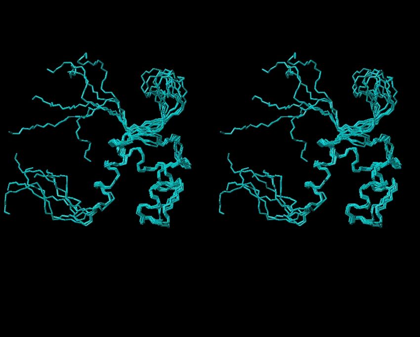
NMR: the background
Complex technique. Requires knowledge in:
Mathematics
Physics
Chemistry
Biology
(Medicin)
Involves a lot of computing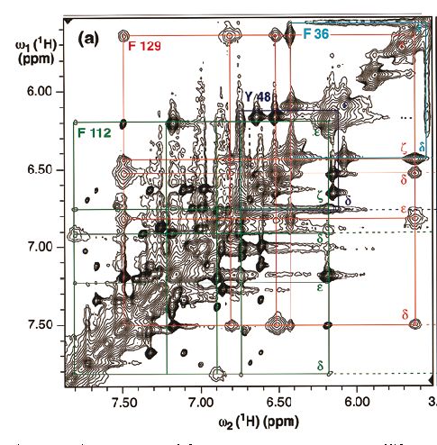
N.M.R.
Nuclear Magnetic Resonance
spectroscopy
imaging
solid-state
....and much more
NMR applications

                             dynamics
      binding

                structure

  in-vivo

                                    folding
                  kinetics

 imaging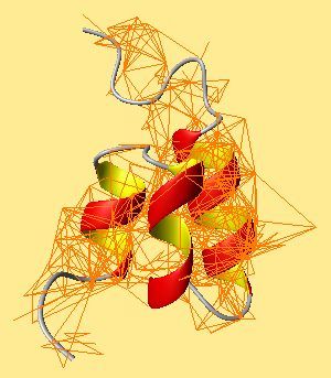
Can NMR be used for: Medical diagnosis? Drug-design? Computing?
NUCLEAR Magnetic Resonance NMR can detect atoms with a nuclear spin 1/2
Some nuclei with spin 1/2
   Isotope     Natural
              abundance   12
                            C and 16O do not have a spin
                 (%)      2
                            H, 14N have spin 1
     1
      H         99.98

     13
         C      1.11

    15
         N      0.37
                               Is this good news for
     19
          F     100            solving the structure
                               of proteins by NMR?
     31
          P     100
NUCLEAR Magnetic Resonance
Can NMR be used to detect:
Ozone?
Salt?
Butter?
A brain tumor?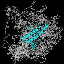
Nuclear MAGNETIC Resonance
Nuclei with spin ½ behave like a magnet
Placed in a constant magnetic field, they will
align with that field
B
H H
How do we create a constant magnetic field?NMR instrumentation sample tube
goes in here
The spectrometer
NOTE:
super-conducting the protein
magnet is in solution
[1mM in water]
RF field generatorNMR is not sensitive
Signals from the nuclei are measured in parts per
million [ppm] of the static field strength
NMR experiments require:
concentrated samples
strong fields = (very) big magnetsProtein structure file in the Protein Data Bank
Structure data in the Protein Data Bank
* X-ray crystallography : over 80%
* NMR : about 16%The cost of NMR
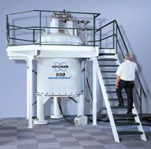
Superconducting magnet : no resistance, no
current loss
Requires cooling to almost absolute zero
Liquid helium
(+ Liquid nitrogen for insulation)
Strong magnetic field
Requires special infrastructure
Minimize field disturbance
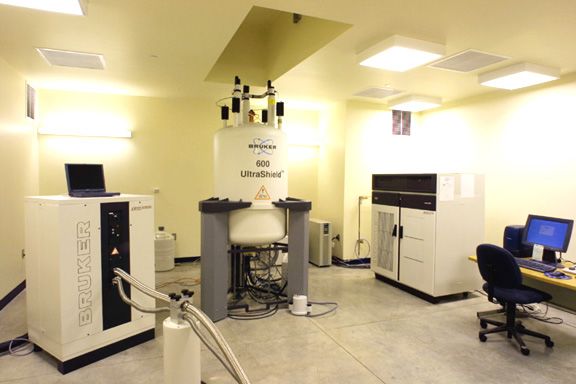
NMR is a very expensive technique!A typical NMR lab
radio frequency unit
superconducting magnetNuclear Magnetic RESONANCE
sound
wave
to
frequency
transformation
H H
HH
RF field
H
H HHNuclear Magnetic RESONANCE
sound
wave
to
frequency
transformation
H H
HH
RF field
H
H HH In an NMR experiment, the energy input to make the nuclei
resonate is produced by a Radio Frequency (RF) PULSE
RF field: magnetic field B1 perpendicular to the constant
magnetic field B0
Constant magnetic field B0
Protein sample
I
Apply and detect a
current in the coil
pulse
current
in coil
time
tWhat is the effect of the pulse?
The PULSE
rotates the bulk magnetisation M0
around x-axis
B0
The angle of rotation
is proportional to the duration
of the RF pulse time tNMR experiments
Depending on the length of the pulses and delay
between pulses, different effects are measured
Variety of NMR experiments: variety of spectra
Depending on the frequency of the RF pulse,
different nuclei can be detected
Proton 1H – NMR
15N – NMR spectra
13C – NMR spectra
...etc...How is an NMR signal detected?
After the pulse, the nuclei return to their ground
energy state
The nuclei precess back to their start position
Precessing induces a current that is detected by a
coil in the NMR spectrometer
As the nuclei return to equilibrium, the induced
current decreases back to zero = Free Induction
Decay (FID)The NMR signal: a Free Induction
Decay (FID)
The FID signal
I
time
Current induced by one precessing spin decays after RF pulseNuclear Magnetic RESONANCE
One spin: one bar magnet
Many spins: bulk magnetisation
Depending on the length of the RF pulse, the
bulk magnetisation of an ensemble of spins will
flip at a different angle with respect to the static
field (B0)
After the pulse, each spin precesses individually
and gives rise to an FID
?FID for an ensemble of spins Current induced by precessing spins decays after RF pulse
Nuclear Magnetic RESONANCE

                       sound
                                wave
                                to
                                frequency
                                transformation

           H     H
               HH
RF field
            H
H HHNMR signal processing
NMR spectrum: a superposition of signals
One signal: FID of one nucleus
Interpretation is made easier by a simple
mathematical formula that transforms of the FID
from the time domain to the frequency domain
Fourier transformationFourier Transformation
I FT
time
resonance
frequency
FT
time
frequencyRichard Ernst (ETHZ) Nobel 1991
NMR structures
How do we get a protein 3D structure with
NMR?
NMR experiments
Data collection
Spectrum assignment
Structure calculation
Method assessment: is NMR really worth the effort?NMR experiments
1D NMR
'hit, measure' (1D pulse sequence)
RF FT
t11H NMR experiment
1D NMR
1 peak for each proton in a distinct environment within
the protein
height ∝ number of structurally identical H ( -CH3)
position (shift) ∝ electronegativity of surrounding
Minute differences in shifts: measured in Part Per Million of the field
width ∝ protein size
size expressed in terms of tumbling (correlation) time
-> there is an experimental limit to the size of the proteins one can
determine by NMR ! we need tricks, more tricks....1
H NMR spectrum of ethanol
Electronegative group: proton signal is shifted compared to the referenceH NMR shifts in different molecular environment
1
What does it mean for NMR spectra of: lipids? Sugars? DNA? Proteins?1H NMR spectra of proteins
Problem Number 1: overlap. The larger the
protein, the more protons, the worse the overlap
Solutions:
- higher field, stronger magnet?
- 2D experiments
- 15N, 13C labelling1H NMR spectra of proteins
Problem number 2: in NMR terms, a large
protein [ > 250 residues] means also besides
overlapping peaks:
faster decaying FID
broader lines
poorer sensitivity
No practical solution for this problem (?).NMR experiments
2D NMR
'hit, wait, hit, measure' (2D pulse sequence)
crosspeaks
RF RF FT
t2 t1
A slice from above gives
a map of crosspeaks.The diagonal contains the 1-dimensional spectrum Off diagonal cross peaks contain new information
NMR experiments
Useful 2D NMR techniques
H-COSY: through-bond connections visible if protons are at most 3
bonds apart
H H peak on 2D map with
position f(H),f(H)
C C
H-NOESY: through-space connections visible if d < 6Å
d
C
H
H
CHeteronuclear experiments
When overlap is too bad for solving a structure
by 1H NMR alone
Label protein with 15N and 13C
Expensive, time-consuming, bad yields
...but 15N and 13C resonate at completely different
frequencies from 1H
Multi-dimensional experiments
... and much more...Heteronuclear experiments
15N-1H COSY
H H
N C 15
N
1
HNMR assignment
Assignment of spectra
method developped by K. Wuethrich
first protein 3D-structure solved by NMR in 1983
many protons, even in small proteins
complex problem
relatively simple solution
arguably the most fastidious stage of protein structure
determination by NMRKurt Wuethrich (ETHZ) Nobel 2002
NMR assignment
Assignment of spectra (K. Wuethrich)
map individual amino acids using COSY spectrum
set of 2D peaks particular for each side-chain 'spin-system',
or relative arrangement of protonsCOSY amino acid patterns
H H
C C
peak on 2D map with
position f(H),f(H)
Do you see potential
problems for proteins?Degeneracy of COSY patterns
Some amino acids share
the same spin system
... for DNA it's even worse!NMR assignment
Assignment of spectra (K. Wuethrich)
map individual amino acids using COSY spectrum
set of 2D peaks particular for each side-chain 'spin-system',
or relative arrangement of protons
locate individual amino acids within the sequence
using NOESY spectrum (sequential assignment)
through-space connections from HA(i) to HN(i+1)NOESY sequential assignment NOESY gives through-space connections: peak on 2D map with position f(H),f(H) if distance d < 6Å
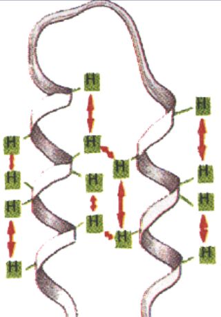
More from the NOESY spectrum Secondary structure patterns
Nuclear Overhauser Effect
Through-space connections, as given by the
NOESY experiment, are the key to solving a
protein structure by NMR
Long-range interactions give the fold of the
protein chain
d
C
H
H
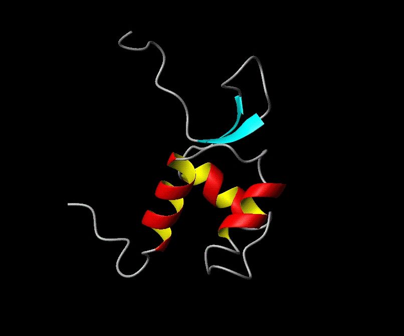
CPositioning of two helices
relative to each other
adjacent beta-strandsNMR assignment
Assignment of spectra (K. Wuethrich)
map individual amino acids using COSY spectrum
set of 2D peaks particular for each side-chain 'spin-system',
or relative arrangement of protons
locate individual amino acids within the sequence
using NOESY spectrum (sequential assignment)
through-space connections from HA(i) to HN(i+1)
compile list of all other peaks arising from through-
space connections (NOEs)
-> set of pairwise distance restraintsNMR distance restraints
NMR structure calculation
How to get from the NOEs to the 3D model?
d3
d1
pairwise distances
d2
?
atomic coordinates (x,y,z)
(x,y,z) (x,y,z)NMR structure calculation
Distance geometry
solves the triangular inequality
Simulated annealing (Michael Nilges)
fancy monte-carlo simulation ("travelling salesman
problem")
Torsion angle dynamics (Peter Güntert)
hybrid method in dihedral angle spaceMichael Nilges (EMBL)
XPLOR structure calculation and molecular
dynamics algorithmPeter Guentert (ETHZ)
DYANA structure calculation programme with
integrated automatic assignment protocolNMR structures What you see...
NMR structures ...is not what you get from NMR!
NMR structures
NMR-derived distance restraints (NOEs) are
upper-limits ("d < 6 Å")
transformation of distances to coordinates gives many
solutions
NMR relies on cooperativity of distance restraints:
the more restraints per residue, the better defined the
structure
one NOE set produces a family of structures:
loops: few experimental restraints -> bad definition -> "fuzzy"
core: lots of long-range restraints -> good definition -> "compact"NMR structure determination
Difficulties
protein in solution: protein has to be soluble
insensitive method: requires high concentrations of proteins
overlap: direct determination of 3D structures for small proteins only
(150-200 residues)
Advantages
no chemical modification necessary
protein in solution: no crystal packing artefacts, allows direct binding
experiments, hydrodynamic and folding studies
assignment of labile regions possible: no gaps in structureDetecting unstructured loops
NMR spectrum
spectrum shows no long-distance interactions but
sequential assignment is possible
backbone is free to adopt a range of conformations:
greater variation in structure coordinates for loop
residues
Crystallography
electron density map shows nothing at all
structures will have gaps for residues in mobile loopsPrion protein by NMR
Not explorable
by XRayRibosome structure by XRay
Impossible task
for NMRNMR applications
SAR: "shot-gun" approach to drug design
Exploring Fibrils by solid-state NMR
Protein folding mechanism
NMR of proteins in bicelles (semi-crystalline
state)
TROESYStructure Activity Relationship by NMR
Drug design
method developed by Abbott Laboratories
Aim:
discover high-affinity ligands for proteins
Example application:
antiviral agent against the human papillomavirusQuantum computing by NMR
Computers
molecules
Information
atomic nuclei state
Programming
radio-frequency pulseQuantum computing by NMR from bits: 0 1 to qubits: |0,0> |0,1> |1,0> |1,1>
Quantum computing by NMR
WHY?
atoms change energy states very quickly
-- much more quickly than even the fastest computer processors.
each qubit can take the place of an entire processor
-- 1,000 ions of barium could take the place of a 1,000 processor
computer.Quantum computing by NMR
WHAT sort of problem a quantum computer
would be able to solve in principle?
Large-scale cryptography
modelling and indexing very large databasesQuantum computing by NMR
WHO wants to build a quantum computer?
IBM/MIT/Berkeley/ Stanford
Isaac Chuang (IBM) Neil Gershenfeld (MIT)
“Enabling technology” for NMR-based quantum computing; scale up
to 10-40 qubits
Harvard/ MIT/Los Alamos
David Cory (Harvard)
Quantum algorithms and NMR-based systems
Oxford University
David Deutsch, Jonathan Jones
Ion-trap and NMR implementations; quantum information theoryfinally...the R.Ernst view of
NMR
medicin
R
biology
chemistry M
physics
N
mathematicsYou can also read