Investigation of the Biofidelity of the MIL-Lx Foot - The Injury ...
←
→
Page content transcription
If your browser does not render page correctly, please read the page content below
Investigation of the Biofidelity of the MIL-Lx Foot
                       J. E. de Lange1, L. Wong2, and C. E. Quenneville1,3
 1
     School of Biomedical Engineering; 2 McMaster Integrated Science Program; 3Department of
                 Mechanical Engineering. McMaster University, Hamilton, Canada

                                            ABSTRACT

Foot/ankle injuries from frontal automotive collisions are frequent and debilitating. Injury risk to
this region is assessed using Anthropomorphic Test Devices with instrumentation at the tibia, so
a biofidelic foot is essential for correctly transmitting load and properly quantifying injury risk to
the foot/ankle region. While post-mortem human subject testing is advantageous as it provides
realistic injury responses, it is expensive, highly variable and does not collect internal loading
data. Anthropomorphic Test Devices, however, collect load data and are easily operated in
industry. This study combined post-mortem human subject feet with an Anthropomorphic Test
Device tibia to examine the response of feet while collecting industry-relevant metrics. The
Military Lower Extremity, six post-mortem human subject lower legs, and six adapted specimens
containing the Military Lower Extremity “tibia” shaft and the post-mortem human subject feet
were equipped with an instrumented boot and axially impacted at 5 m/s, representing a frontal
automotive collision. No significant differences were found on the plantar surface among all
specimens, suggesting this may be a feasible method of evaluating foot response. An assessment
of the biofidelity of the Military Lower Extremity foot was conducted, and significant differences
were found in the proximal tibia load cells when comparing the adapted specimens with the
Military Lower Extremity, suggesting its foot is overly stiff. Further, significantly higher midfoot
region forces were recorded in the intact legform as compared to the Military Lower Extremity,
which may be the result of a stiffer mechanical ankle joint. These data can be used for informing
a new design of an Anthropomorphic Test Device foot, thus improving lower limb safety
assessments. The method developed herein may also be used to conduct injurious foot/ankle testing
in the future, to accurately quantify injury risk to this region that is directly transferrable to
industry testing.

                                        INTRODUCTION

        Up to 10% of all non-minor (Abbreviated Injury Scale, AIS 2+) injuries in automobile
collisions occur to the foot/ankle complex (Crandall et al., 1996), with axial loading responsible
for injuries with the most significant long-term impairment (Funk et al., 2002, Yoganandan et al.,
1996). Although there has been a decrease in the overall frequency of injuries related to car crashes,
foot and ankle injuries continue to increase in both severity and incidence (Richter et al., 2001).
Foot/ankle injuries are impactful, as they involve the disruption of many articular surfaces and
have poor vascularization for healing, often leading to post-traumatic osteoarthritis (Dischinger et

                                                   1
                      2021 The Ohio State University Injury Biomechanics Symposium
                                *This paper has not been peer-reviewed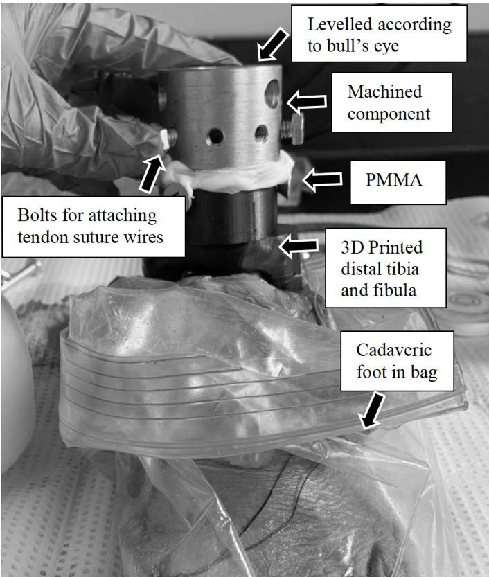
al., 2004). Typical methods of injury risk assessment in the automotive industry include the use of
Anthropomorphic Test Devices (ATDs), with injury risk to the foot and ankle usually grouped and
evaluated using load cells in the tibia. This means the foot/ankle region is often overlooked when
assessing injury risk.

         While ATDs are valuable tools with load cells to collect forces and act in a repeatable
manner, they do not undergo injuries. In contrast, PMHS testing is advantageous as it allows for
the identification of fracture limits, locations and mechanisms. However, it does not allow
collection of internal load data. Researchers have attempted to measure fracture forces in PMHS
testing by implanting load cells proximal to the tibia (e.g., Yoganandan et al., 1996) or in the tibia
itself (e.g., Funk et al., 2002). This is challenging to assess fracture risk and compare with ATD
measurements and the addition of load cells can alter stress concentrations, potentially varying the
injury mechanism as a result of impact. Each component of an ATD should be evaluated
independently, as they are acting in series. Relating PMHS data to load cells in the ATD is valuable
information that would allow for direct translation into industry how much force the foot can
withstand. The need for a transfer function (by testing the ATD under parallel conditions to those
that pose a specific level of risk to PMHS specimens) between ATDs and corresponding injury
risk could thus be eliminated.

        Two primary lower leg ATD models exist, the Hybrid III 50th Male (HIII), and the Military
Lower Extremity (MIL-Lx, Humanetics Innovative Solutions, Plymouth, MI, USA). The response
of the MIL-Lx has been investigated both at the lower leg level (McKay, 2010) and as an isolated
tibia (Quenneville and Dunning, 2012) and is generally accepted as having a more biofidelic
response. Although this ATD was designed for blast events, it has potential for being a useful tool
in the automotive industry for high-force crashes (McKay and Bir, 2009). As such, this ATD leg
was the focus herein. While the biofidelity of the MIL-Lx tibia has been evaluated and compared
to other surrogates, few studies have investigated the foot/ankle response specifically (Chirvi et
al., 2017). While the isolated MIL-Lx tibia (no foot) has previously been shown to have very good
agreement with non-fracture post-mortem human subject (PMHS) tibia data (R2 = 0.83), an
extensive comparative response including the foot/ankle has not been conducted (Quenneville and
Dunning, 2012). Investigation of the foot/ankle response is important, as load is transmitted
through this region to the tibia, where injury risk is assessed. If the foot/ankle does not transmit
the load correctly, tibia safety assessments may be incorrect. As part of ongoing efforts to design
biofidelic ATDs, it is important to examine the response of the MIL-Lx foot to either validate the
current ATD model or provide data for an improved design.

        The purpose of this study was first to compare the axial response of intact PMHS lower
legs to the MIL-Lx at impact velocities and durations similar to those experienced by the lower
extremity in frontal automotive collisions to compare the overall response. Secondly, to develop a
method to mount PMHS feet onto the MIL-Lx tibia shaft to facilitate the investigation of the foot
response while collecting the industry-relevant metrics of peak axial force (Fz). The objective was
to quantify the differences among lower legs to investigate the biofidelity of the MIL-Lx foot.

                                                   2
                      2021 The Ohio State University Injury Biomechanics Symposium
                                *This paper has not been peer-reviewed
METHODS

         All specimens were fitted with an instrumented boot while tested, which was equipped
with eight piezoresistive sensors covering the plantar surface of the foot to quantify impact force
at this location (Figure 1). The construction of the instrumented boot is detailed by Acharya and
colleagues (2019) and has previously been used to comparatively assess ATD models and evaluate
the effects of ankle posture (de Lange and Quenneville, 2019). In each of the tests, the boot was
tightened and laced firmly over each foot by the same investigator each time. It was tightened in
between impact events and all sensors were zeroed after it was fitted to account for any pre-impact
loading on the foot.

 Figure 1: Piezoresistive sensors were employed on the insole of the boot and covered the main
loading regions of the insole. Sensors were grouped together to form the forefoot loading region,
             highlighted in blue, the midfoot in orange, and the hindfoot, in yellow.

         All impact testing was completed using a pneumatic impacting apparatus (e.g., Martinez
et al., 2018). Impulse was transmitted to the plantar surface of the foot via an ankle positioner,
which was mounted on low-friction linear bearings (Figure 2). Ballast weight was secured to the
suspension jig to bring the total mass of each specimen to 12.9 kg, the total mass of a 50th
percentile male leg (Bull, 2019), in an effort to simulate natural linear inertial properties.

                                                  3
                     2021 The Ohio State University Injury Biomechanics Symposium
                               *This paper has not been peer-reviewed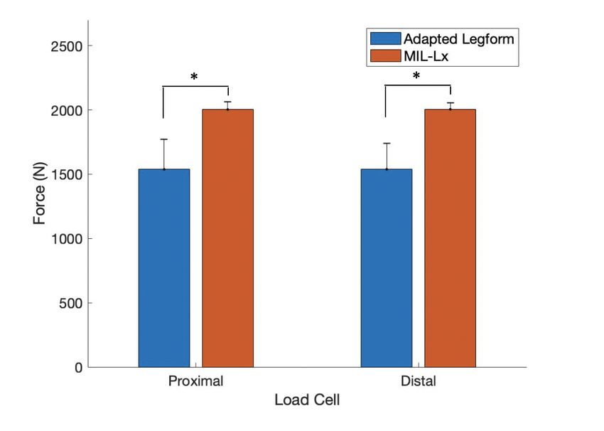
Figure 2: The MIL-Lx in the instrumented boot suspended in the pneumatic impacting apparatus.

MIL-Lx Experimental Testing

       The MIL-Lx was fitted with the instrumented boot, and a settling impact was conducted to
ensure the foot was positioned in the boot. The MIL-Lx was then axially impacted four times. It
was supported within the pneumatic impacting apparatus at the knee clevis.

Intact Post-Mortem Human Subject Experimental Testing

       Six intact lower leg specimens sectioned at the tibial plateau were tested (Table 1). The
specimens were x-rayed prior to impacting, and an orthopaedic surgeon declared there were no
pre-existing injuries.

                        Table 1: Characteristics of Lower Legs Tested
   Legform           Age (Years)                   Sex                Foot Length (cm)
   MIL-Lx               N/A              Male representative                26.1
  Specimen 1             69                      Female                     22.2
  Specimen 2             69                       Male                      27.3
  Specimen 3             95                       Male                      27.3
  Specimen 4             95                       Male                      26.7
  Specimen 5             77                      Female                     22.2
  Specimen 6             77                      Female                     21.6

                                                  4
                     2021 The Ohio State University Injury Biomechanics Symposium
                               *This paper has not been peer-reviewed
Specimens were dissected 2” distal to the tibial plateau and potted at the knee using dental
cement in a section of 4”-diameter circular polyvinyl chloride (PVC) piping, to support the
specimens while testing and ensure proper axial alignment. Consistent alignment was ensured
through the use of a laser level projected along the tibial ridge, and the bone was embedded to the
full depth of the PVC pipe (2”). All specimens were thawed for a minimum of 12 hours before
testing. The foot was inserted into a plastic bag, then into the instrumented boot and mounted in
the impacting chamber in a neutral ankle posture. It was secured proximally by fixing the PVC
pipe to the ballast plate.
Adapted Legform Experimental Testing
In order to characterize the response of the natural foot for defining stiffness requirements
for the MIL-Lx, isolated PMHS feet were tested. Each intact PMHS was disarticulated at the
tibiotalar joint and x-rayed in the anterior-posterior and lateral views after disarticulation. An
orthopaedic surgeon again evaluated the x-rays and declared no injuries had occurred during
dissection or intact testing. The PMHS feet were then mounted onto the MIL-Lx tibia shaft.
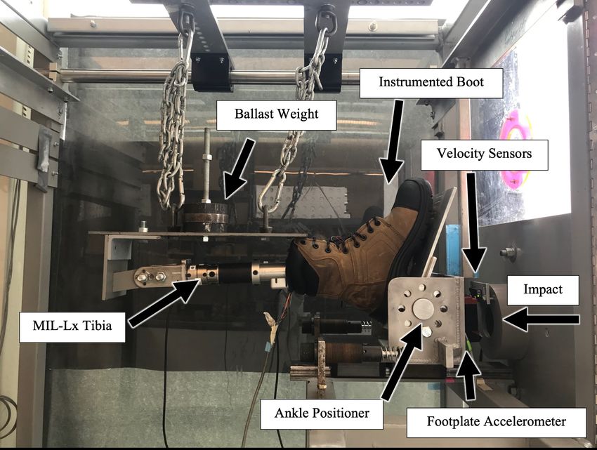
There were several challenges associated with mounting a PMHS foot to an ATD tibia.
First, soft tissue support was necessary to facilitate proper alignment of the foot with respect to the
artificial tibia shaft, in an effort to replicate natural joint motion during initial positioning. To
address this, surrounding tendons and ligaments that play an important role in ankle joint stability
(Campbell et al., 2014, Golanó et al., 2016) were sutured with a Krakow stitch (Krakow et al.,
1986). A second challenge with attaching a PMHS foot to the ATD shaft was keeping load
transmission between the talus and ATD shaft natural to limit abnormal stress concentrations. In
an effort to do so, the distal tibia and fibula of each specimen were optically scanned (Artec Eva,
Artec 3D, Hamm, Luxembourg) and 3D-printed to replicate natural bone geometry. Finally, to
address challenges associated with aligning the MIL-Lx tibia shaft at a 90° angle to the plantar
surface of the foot (neutral posture), a machined component was designed to secure the 3D printed
component to the MIL-Lx ATD shaft. A ball joint was created in the 3D printed tibia to allow the
MIL-Lx to rest at a 90° angle to the plantar surface of the foot and once precisely levelled,
polymethylmethacrylate (PMMA, Simplex P Bone Cement, Stryker, MI, United States) was used
to fill the gap between the 3D printed component and the steel attachment (Figure 3). The
machined component had a series of eight threaded holes around the circumference of the part to
allow for tendon attachment. The sutures were secured tightly such that there was visible tension
in each of the suture wires, as recommended by an orthopaedic surgeon, and performed by the
same researcher each time.
5
2021 The Ohio State University Injury Biomechanics Symposium
*This paper has not been peer-reviewedFigure 3: Components facilitating the attachment of the MIL-Lx tibia shaft to a PMHS foot.
Impact Conditions
All impacts were intended to be delivered at a velocity of 5.0 (± 0.5) m/s and an impact
duration of 20 (± 5) ms, intended to be in the range of realistic impact conditions experienced by
the lower leg resulting from a frontal collision (McKay and Bir, 2009, Crandall et al., 1998). A
settling impact was performed at the start of each testing sequence to seat the foot within the boot
(impact mass 3 kg, kinetic energy of 20-25 J). In order to increase impact energy while duration
and velocity remained constant, the projectile mass was increased. An impact mass of 6 kg and
impact energy of 80 J was used for all comparative impacts. This impact was intended to be at a
sub-failure level.
Data Analysis
The testing procedure was controlled, and data were collected, using a custom-written
LabVIEW (National Instruments, Austin, TX, USA) program. All data, including the two 5-axis
load cells (Fx, Fy, Fz, Mx, My) in the upper and lower tibia and the eight instrumented boot sensors
were recorded at 50 kHz. The data collected from the sensors on the instrumented boot were
assessed for peak force and distribution of force along the plantar surface of the foot. Sensors were
grouped into three regions for data presentation and analysis: the forefoot, midfoot and hindfoot.
Tibia load cell data were dual-pass filtered using a second-order Butterworth low-pass filter
with a cutoff frequency of 1,250 Hz, in accordance with industry standards (Society of Automotive
Engineers, 2003). Impact duration was considered to have begun 1 ms before the boot hindfoot
sensor (Sensor 1) decreased to 10% of the peak voltage and concluded 1 ms after the voltage fell
6
2021 The Ohio State University Injury Biomechanics Symposium
*This paper has not been peer-reviewedbelow 10% of the peak voltage. Changes in sensor voltage on the instrumented boot were
converted to force readings according to a previously developed calibration protocol (de Lange,
2020). The mean and standard deviation of each metric were also calculated.
A one-way Analysis of Variance (ANOVA) with post hoc Tukey test was conducted on
both the net boot forces and regional forces for all three leg representations. An unpaired t-test was
conducted to compare the load cell peak axial forces between the adapted leg form and the MIL-
Lx, for both the proximal and distal load cells. An unpaired t-test was also conducted to compare
impact velocities and durations among lower legs. Each of these tests had a significance threshold
of α = 0.05.
RESULTS
Impact velocities ranged from 5.4 to 6.4 m/s, and impact durations were 21 ± 0.4 ms (Table
2). No significant differences were found among impact velocities (p = 0.37); however, impact
durations were significantly higher in the adapted specimens as compared to the intact PMHS
specimens (p = 0.02). The kinetic energies of impacts were significantly higher in the MIL-Lx in
comparison to the intact specimens (p = 0.03). X-rays pre- and post-impact confirmed there was
no damage to the specimens at any stage of the process. No statistical differences were found
among lower leg representations for the net boot forces (representing the complete reaction force
from the plantar surface of the foot), suggesting that overall reaction force at the plantar surface
was comparable among legforms (p = 0.48). In comparison to the net boot forces, the MIL-Lx
proximal tibia measured forces 38% lower than the plantar surface, and similarly the adapted
legform read forces 66% lower in the proximal tibia rather than the plantar surface of the foot.
Table 2: Data measured during impacts, presented as mean ± SD. All impacts were delivered
with a 6 kg impact mass, resulting in an average kinetic energy of 78 ± 15 J for all impacts.
Impact Information Measured Outputs
Specimen Velocity Impact Total Insole Peak Proximal
(m/s) Duration (ms) Force (N) Force (N)
MIL-Lx 5.4 ± 0.1 19.6 ± 3.1 2764 ± 592 2003 ± 60
Intact Specimens 4.9 ± 0.7 17.2 ± 4.2 3139 ± 871 N/A
Adapted Legform 5.1 ± 0.6 24.7 ± 4.0 2553 ± 889 1538 ± 236
Results from the unpaired t-test indicated durations were significantly higher (p = 0.008)
in the adapted specimens than the MIL-Lx. The MIL-Lx also measured significantly higher forces
than the adapted surrogate for both the proximal (p = 0.005), and distal (p = 0.002) load cells
(Figure 4).
7
2021 The Ohio State University Injury Biomechanics Symposium
*This paper has not been peer-reviewedFigure 4: Proximal and distal tibia load cell forces of adapted specimens compared to the MIL-
Lx surrogate (‘*’ denotes a significant finding, where pDISCUSSION
This study subjected three lower leg representations (the MIL-Lx, intact PMHS lower legs,
and adapted lower legs with the MIL-Lx tibia shaft and PMHS feet) to axial impacts for the
purpose of evaluating the differences in impact response among surrogates. This study is the first
of its kind to investigate the isolated PMHS foot while collecting industry-relevant metrics as well
as the regional foot response as a result of axial impacts. Impact time durations and velocities were
consistent with those measured during vehicular collision scenarios (McKay and Bir, 2009). The
plantar surface force remained generally consistent among all specimens, suggesting this was a
feasible method for combining testing subjects.
Forces measured on the plantar surface of the foot were over 35% higher than forces
measured in the tibia, unsurprising when considering the location of load cells and direction of
loading. No significant differences were found among impact velocities, so variations in recorded
forces were likely related to differences in stiffness. Net boot forces among all lower legs were
comparable but the adapted legform tibia forces were 30% lower and had longer durations in
comparison to the MIL-Lx, suggesting the MIL-Lx foot is more rigid. The differences in force
dissipation that the MIL-Lx foot exhibited in comparison to the PMHS foot suggest the MIL-Lx
foot may be overly stiff. An alternative explanation could be either the MIL-Lx tibia is too
compliant or having many compliant elements in series (like the boot) alters the response, as
suggested by Quenneville et al. (2017). This highlights the importance of understanding the
relative stiffness in series to adequately account for various complaint elements, as well as
developing an ATD foot that has similar characteristics to PMHS feet. The foot transmits the load
to the tibia, where injury risk is assessed so this may have serious implications, as testing
completed with the stiffer MIL-Lx foot may ‘fail’, but with a more biofidelic foot may have
‘passed.’
The significantly higher midfoot forces in the intact legform may have been a result of the
stiffer ankle in the MIL-Lx in comparison to the PMHS, which allowed for more ankle joint
motion. Another explanation may be due to variations in the arch and soft tissue stiffness.
Interestingly, the distribution of forefoot and hindfoot forces were not significantly different
among all lower leg representations, despite variations in foot size. The PMHS feet varied in size,
which caused the forefoot readings to be slightly reduced in some specimens. Although Sensor 8
(toe sensor) did not record much data for these specimens, the forefoot sensor group was not
substantially affected.
The majority of previous studies have developed injury criteria for the entire lower leg.
Although some studies have examined foot/ankle injuries, these were combined with injuries to
the tibia and fibula (Crandall et al., 1998). By focusing on isolated PMHS feet, while also
collecting tibia load data, this study assessed the impact characteristics of feet specifically. This is
important as the foot/ankle complex is a vulnerable and frequently injured area in frontal collisions
and the long-term implications can be debilitating.
There were a few limitations to the current study. Firstly, this study was completed with a
small sample size (N = 6) from an older population (average age of 80 years) and with both males
and females. However, when analyzing male and female results separately, no significant
9
2021 The Ohio State University Injury Biomechanics Symposium
*This paper has not been peer-revieweddifferences were found among total insole forces, or tibia forces. The specimens also had varying
foot lengths, which may have had implications when comparing to the MIL-Lx foot, considering
its larger size. The smaller feet had reduced forefoot readings, which likely led to larger standard
deviations observed herein.
The lack of cartilage and the artificial fixation of tendons and ligaments could have altered
the responses of the feet. Every effort was made to replicate the natural ankle; however, this model
could not be verified. As impacts were delivered in a neutral ankle posture, and in compression,
this likely was not a substantial issue. The adapted legform was compared to the original same
specimens, which is advantageous as this allowed comparison of adapted lower legs to its intact
case, serving as its own control.
Next, the MIL-Lx was designed for impacts of higher energy and shorter duration than
were conducted here. Although this investigation does not represent exact conditions that the MIL-
Lx was designed for, this study aim was to present a method of attachment, which may be used at
injurious levels in the future. Testing a greater number of specimens with the protocol described
herein will establish better foot and ankle injury standards when tested at injury-generating levels.
Further, data collected from the boot had high variations among impacts in comparison to the MIL-
Lx data, suggesting improvements need to be made to use this tool for regional injury risk
prediction for use in industry crash testing.
Furthermore, tests were conducted with lower forces than would likely have been
conducted in vehicular collision tests. The proximal and distal tibia load cells recorded forces that
were very similar for each leg type, indicating that the compliant element located proximal to the
distal load cell was not engaged. This likens the ATD to behave more like a rigid tibia. The
proximal and distal load cell forces have been shown to start to diverge around 3 kN in the MIL-
Lx, which corresponds to forces around 6 kN in the Hybrid III (Quenneville and Dunning 2012).
The MIL-Lx 10% injury threshold is 2.6 kN, and the authors wished to conduct non-injurious tests
in this study. Net boot forces were around 3 kN, as there were many components acting in series
during these tests, the authors wanted to ensure injurious levels of PMHS feet were not reached.
Often when vehicular occupants see an impending collision, they will brake, activating
muscles in the calf through tensioning the Achilles tendon. No Achilles tensioning was applied for
testing in this study, as the exact amount of tension activated is relatively unclear. Funk and
colleagues based their 1.5-2 kN of Achilles tension on pedal forces measured during braking from
volunteer driving simulations (2002). In order to provide the best comparison with the MIL-Lx,
which does not take Achilles tension into consideration, muscle activation was neglected herein.
Furthermore, the ligaments and tendons surrounding the ankle joint were sutured and secured to
the machined and 3D printed components. This was not meant to pretension the tendons and
ligament groups, but rather was conducted in an effort to mimic natural load transmission pathways
and secure the PMHS talus to the artificial tibia and fibula.
The data collected in this study could inform future generations of ATD feet, as this study
found that the MIL-Lx foot was stiffer in comparison to PMHS feet. Improvements in ATD
properties to provide a more biofidelic ankle joint, or segmented foot to provide enhanced
10
2021 The Ohio State University Injury Biomechanics Symposium
*This paper has not been peer-reviewedrepresentation of the types of loading the foot/ankle complex will experience in these scenarios
would be advantageous to reduce the incidence and severity of foot and ankle injuries.
CONCLUSIONS
This study presented a method to assess the impact response of the isolated foot while
collecting data that are immediately relevant to the automotive industry. The similar force readings
collected at the plantar surface of the foot among all legforms showed that doing this did not affect
the load response to the foot. It is the first study of its kind to propose an adapted lower leg in order
to assess isolated foot injuries while gathering axial force data. Results suggest that the MIL-Lx
foot could be improved in stiffness characteristics, and this study has provided data that can be
used for this design.
ACKNOWLEDGEMENTS
The authors would like to acknowledge the contributions of Jackie Pacheco (Mohawk
College, Canada), Dr. Bashar Alolobi (St. Joseph’s Healthcare Hamilton, Canada), and Tribe
Medical Group. They would also like to acknowledge their sources of funding: the Natural
Sciences and Engineering Research Council of Canada (NSERC) and the Ontario Graduate
Scholarship (OGS).
REFERENCES
ACHARYA, I., VAN TUYL, J., DE LANGE, J., QUENNEVILLE, C.E. (2019). A Force-Sensing
Insole to Quantify Impact Loading to the Foot. Journal of Biomechanical Engineering,
141(2).
BULL, A.M.J. (2016). Impact Biomechanics: Blast Injury Science and Engineering. Sprinter
International Publishing.
CAMPBELL, K.J., MICHALSKI, M.P., WILSON, K.J., GOLDSMITH, M.T., WIJDICKS, C.A.,
LAPRADE, R.F., CLANTON, T.O. (2014). The Ligament Anatomy of the Deltoid
Complex of the Ankle: A Qualitative and Quantitative Anatomical Study. Journal of Bone
and Joint Surgery, 96(8):1-10.
CHIRVI, S., PINTAR, F., YOGANANDAN, N., BANERJEE, A., SCHLICK, M., CURRY, W.,
VOO, L. (2017). Human Foot-Ankle Injuries and Associated Risk Curves from Under
Body Blast Loading Conditions. SAE Paper No. 2017-22-0006.
CRANDALL, J.R., KUPPA, S.M., KLOPP, G.S., HALL, G.W., PILKEY, W.D., HURWITZ, S.R.
(1998). Injury mechanisms and Criteria for the Human Foot and Ankle Under Axial
Impacts to the Foot. International Journal of Crashworthiness, 3(2), 147-162.
11
2021 The Ohio State University Injury Biomechanics Symposium
*This paper has not been peer-reviewedCRANDALL, J.R., PORTIER, L., PETIT, P., HALL, G.W., BASS, C.R., KLOPP, G.S.,
HURWITZ, S., PILKEY, W.D., TROSSEILLE, X., TARRIERE, C., LASSAU, J.P.
(1996). Biomechanical Response and Physical Properties of the Leg, Foot and Ankle. SAE
Transactions, 1853-1872.
DE LANGE, J. (2020). Biomechanical Tools for Assessing Foot and Ankle Injury Risk in Frontal
Automotive Collisions. Master’s Thesis.
DE LANGE, J., QUENNEVILLE, C.E. (2019). Influence of Ankle Posture and ATD Model on
the Distribution of Forces on the Foot Under Impact Loading. Proceedings of IRCOBI
Conference.
DISCHINGER, P.C., READ, K.M., KUFERA, J.A., KERNS, T.J., BURCH, C.A., JAWED, N. et
al. (2004). Consequences and Costs of Lower Extremity Injuries. Association for the
Advancement of Automotive Medicine. Vol. 48, p. 339.
FUNK, J.R., CRANDALL, J.R., TOURETT, L.J., MACMAHON, C.B., BASS, C.R., PATRIE,
J.T., KHAEWPONG, N., EPPINGER, R.H. (2002). The Axial Injury Tolerance of the
Human Foot/Ankle Complex and the Effect of Achilles Tendon. Journal of Biomechanical
Engineering. 124(6): 750-757.
GOLANÓ, P., VEGA, J., DE LEEUW, P.A., MALAGELADA, F., MANZANARES, M.C.,
GOTZENS, V., VAN DIJK, C.N. (2010). Anatomy of the Ankle Ligaments: A Pictorial
Essay. Knee Surgery, Sports Traumatology, Arthroscopy. 18(5): 557-569.
KRAKOW, K., THOMAS, S.C., JONES, L.C. (1986). A New Stitch for Ligament-Tendon
Fixation. Journal of Bone and Joint Surgery, 68, 764-768.
MARTINEZ, A.A., CHAKRAVARTY, A.B., QUENNEVILLE, C.E. (2018). The Effect of
Impact Duration on the Axial Fracture Tolerance of the Isolated Tibia During Automotive
and Military Impacts. Journal of the Mechanical Behaviour of Biomedical Materials,
778:315-320.
MCKAY, B. (2010). Development of Lower Extremity Injury Criteria and Biomechanical
Surrogate to Evaluate Military Vehicle Occupant Injury During an Explosive Blast Event.
Wayne State University Dissertation.
MCKAY, B.J., and BIR, C.A. (2009). Lower Extremity Injury Criteria for Evaluating Military
Vehicle Occupant Injury in Underbelly Blast Events. SAE Technical Paper.
RICHTER, M., THERMANN, H., WIPPERMANN, B., OTTE, D., SCHRATT, H.-E.,
TSCHERNE, H. (2001). Foot Fractures in Restrained Front-Seat Car Occupants: A Long-
Term Study Over Twenty-Three Years. Journal of Orthopaedic Trauma, 287-293.
12
2021 The Ohio State University Injury Biomechanics Symposium
*This paper has not been peer-reviewedQUENNEVILLE, C.E., DUNNING, C.E. (2012). Evaluation of the Biofidelity of the HIII and
MIL-Lx Lower Leg Surrogates Under Axial Impact Loading. Traffic Injury Prevention,
13(1), 81-85.
QUENNEVILLE, C.E., FOURNIER, E., SHEWCHENKO, N. (2017). The Effect of
Anthropomorphic Test Device Lower Leg Surrogate Selection on Impact Mitigating
System Evaluation in Low- and High-Rate Loading Conditions. Military Medicine, 182(9-
10):1981-1986.
SOCIETY OF AUTOMOTIVE ENGINEERS. (2007). Instrumentation for Impact Test—Part 1—
Electronic Instrumentation. Technical Report, SAE J211/1.
YOGANANDAN, N., PINTAR, F.A., BOYNTON, M., BEGEMAN, P., PRASAD, P., KUPPA,
S.M., MORGAN, R.M., EPPINGER, R.H. (1996). Dynamic Axial Tolerance of the Human
Foot-Ankle Complex. SAE Technical Paper Series, 1887-1898.
13
2021 The Ohio State University Injury Biomechanics Symposium
*This paper has not been peer-reviewedYou can also read