Isolation of Mitochondria from Ustilago maydis Protoplasts
←
→
Page content transcription
If your browser does not render page correctly, please read the page content below
Please cite this article as: Pardo, J. P. et al. (2022). Isolation of Mitochondria from Ustilago maydis Protoplasts. Bio-protocol 12(01): e4277. DOI:
 10.21769/BioProtoc.4277.

                                                                                                                 Bio-protocol 12(01): e4277.
                                                    www.bio-protocol.org/e4277                                   DOI:10.21769/BioProtoc.4277

                           Isolation of Mitochondria from Ustilago maydis Protoplasts
                                      Juan Pablo Pardo1, Guadalupe Guerra-Sánchez2,
                                   Oscar Flores-Herrera1 and Lucero Romero-Aguilar1, *

1Universidad       Nacional Autónoma de México, Facultad de Medicina, Departamento de Bioquímica,
Avenida Universidad 3000, Copilco, Cd. Universitaria, Alcaldía de Coyoacán, 04510, Ciudad de México,
México
2Instituto   Politécnico Nacional, Escuela de Ciencias Biológicas, Departamento de Microbiología, Manuel
Carpio 532, Plutarco Elías Calles, Miguel Hidalgo, 11350, Ciudad de México, México
*For correspondence: lusromaguila@bq.unam.mx

[Abstract] Ustilago maydis, a basidiomycete that infects Zea mays, is one of the top ten fungal models
for studying DNA repair, signal transduction pathways, and dimorphic transitions, among other
processes. From a metabolic point of view, U. maydis lacks fermentative capacity, pointing to
mitochondria as a key player in central metabolism. Oxidative phosphorylation, synthesis of heme
groups, Krebs cycle, β-oxidation of fatty acids, and synthesis of amino acids are some of the processes
that take place in mitochondria. Given the importance of this organelle in eukaryotic cells in general, and
in fungal cells in particular, we present a protocol for the isolation of U. maydis mitochondria based on
the enzymatic disruption of U. maydis cell wall and differential centrifugation. The method can easily be
extrapolated to other fungal species, by using appropriate lytic enzymes.
Keywords: Mitochondria isolation, Lysing cell wall, Ustilago maydis, Differential centrifugation,
Respiratory chain, Membrane potential

[Background] As in other eukaryotes, mitochondria in fungal cells are essential for the production of
many important molecules. For example, in the mitochondrial matrix occurs the synthesis of some amino
acids and of the heme group that is inserted into specific subunits of the respiratory complexes. Other
processes also occur in the mitochondrial matrix, like the β-oxidation of fatty acids and the tricarboxylic
acid cycle. NADH and FADH2 are key products of the last two pathways, and these molecules transfer
their electrons to the respiratory chain for the synthesis of ATP. Electron transfer and ATP synthesis are
connected by a proton electrochemical gradient that is generated by the proton pumping activity of
complexes I, III, and IV in the respiratory chain, and consumed by the ATP synthase. Membrane potential
and a difference of pH across the inner mitochondrial membrane are components of the proton-motive
force. For mitochondria to carry out their functions, transporters located in the outer and inner
membranes catalyze the metabolic exchange between cytosol and mitochondria (Sousa et al., 2018;
Reyes-Galindo et al., 2019).
  U. maydis is a phytopathogen that infects Zea mays. During its life cycle, U. maydis transits through
three major morphological stages: the non-pathogenic haploid basidiospore (yeast form), the infective
dikaryotic filament, and the environmental resistant teliospore (Brefort et al., 2009). The microorganism

Copyright © 2022 The Authors; exclusive licensee Bio-protocol LLC.                                                                                 1
Please cite this article as: Pardo, J. P. et al. (2022). Isolation of Mitochondria from Ustilago maydis Protoplasts. Bio-protocol 12(01): e4277. DOI:
10.21769/BioProtoc.4277.
Bio-protocol 12(01): e4277.
www.bio-protocol.org/e4277 DOI:10.21769/BioProtoc.4277
is fully respiratory, pointing to mitochondria as a key player in its intermediate metabolism. Therefore,
mitochondria isolation is a starting point in studies of respiratory complexes and supercomplexes, the
control of ROS production by certain mitochondrial inner membrane proteins, the activities of Krebs
cycle enzymes, among others (Juárez et al., 2004). The goal of this protocol is to report a method for
the isolation of functional U. maydis mitochondria from protoplasts generated by cell wall lytic enzymes
from Trichoderma harzianum. Membrane potential and oxygen consumption were analyzed, to measure
the quality of mitochondrial preparations.
Materials and Reagents
1. Sterile toothpick
2. Petri dishes (Sigma-Aldrich, catalog number: P5606-400EA)
3. Glass beaker (Sigma-Aldrich, catalog number: BR90648)
4. NalgeneTM Polycarbonate Fernbach Culture Flask (Thermo ScientificTM, catalog number: 4105-
2800)
5. Spectrophotometer cuvettes (Merck, catalog number: C5291)
6. Nalgene® 500 mL centrifuge bottles, wide-mouth with sealing caps style 3141 (Sigma-Aldrich,
catalog number: Z353744-4EA)
7. Polycarbonate tubes, 50 mL, 29 × 102 mm (Thermo Fisher Scientific, catalog number: 03268)
8. Quartz absorption cuvettes of 3.5 mL (Merck, catalog number: Z803669)
9. Hamilton syringe (Hamilton, Gastight syringe, catalog number: 1702N)
10. Round point tip soft paintbrush number 8 to 12 (DUGATO-Amazon)
11. Distilled water
12. Sucrose (Sigma-Aldrich, catalog number: S5016)
13. Glucose (Sigma-Aldrich, catalog number: G8270)
14. Sorbitol (Sigma-Aldrich, catalog number: S1876)
15. Select Yeast Extract (Sigma-Aldrich, catalog number: y1625)
16. Bacteriological Peptone (Sigma-Aldrich, catalog number: PO556)
17. Agar (Sigma-Aldrich, catalog number: A1296)
18. Lysing enzymes from Trichoderma harzianum (Sigma-Aldrich, catalog number: L1412)
19. Dimethyl sulfoxide (DMSO) (Sigma-Aldrich, catalog number: D2650)
20. (NH4)2SO4 (Sigma-Aldrich, catalog number: A4915)
21. KH2PO4 (Sigma-Aldrich, catalog number: PO662)
22. HCl (Sigma-Aldrich, catalog number: H1758)
23. NaOH (Sigma-Aldrich, catalog number: 795429)
24. Glycerol (Sigma-Aldrich, catalog number: G5516)
25. Potassium cyanide (KCN) (Sigma-Aldrich, catalog number: 60178)
26. Trizma® base (Sigma-Aldrich, catalog number: T1503)
Copyright © 2022 The Authors; exclusive licensee Bio-protocol LLC. 2Please cite this article as: Pardo, J. P. et al. (2022). Isolation of Mitochondria from Ustilago maydis Protoplasts. Bio-protocol 12(01): e4277. DOI:
10.21769/BioProtoc.4277.
Bio-protocol 12(01): e4277.
www.bio-protocol.org/e4277 DOI:10.21769/BioProtoc.4277
27. Ethylenediaminetetraacetic acid (EDTA) disodium salt dihydrate (Sigma-Aldrich, catalog
number: E9884)
28. Bovine Serum Albumin (BSA) (Sigma-Aldrich, catalog number: A7030)
29. Phenylmethylsulfonyl fluoride (PMSF) (Sigma-Aldrich, catalog number: PMSF-RO)
30. Complete Protease Inhibitor Cocktail Tablets (Roche, catalog number: 04906837001)
31. Carbonyl cyanide 4-(trifluoromethoxyphenyl) phenylhydrazone (FCCP) (Sigma-Aldrich, catalog
number: C2920)
32. HEPES (4-(2-hydroxyethyl)-1 piperazineethanesulfonic acid) (Sigma-Aldrich, catalog number:
H3375)
33. Safranine (Sigma-Aldrich, catalog number: S2255)
34. NADH (Sigma-Aldrich, catalog number: N8129)
35. Sodium succinate dibasic hexahydrate (Sigma-Aldrich, catalog number: S2378)
36. N,N,N’,N’-Tetramethyl-p-phenylenediamine (TMPD) (Sigma- Aldrich, catalog number: T7394)
37. Sodium L-ascorbate (Sigma- Aldrich, catalog number: A7631)
38. Solution A (see Recipes)
39. Solution B (see Recipes)
40. Solution C (see Recipes)
41. PMSF (see Recipes)
42. Solution D (see Recipes)
43. Solution E (see Recipes)
44. YPD-culture media (see Recipes)
45. Solution F (see Recipes)
46. FCCP solution (see Recipes)
47. Safranine solution (see Recipes)
48. Succinate solution (see Recipes)
Equipment
1. Teflon stirring rod (FisherbrandTM, catalog number: 14-513-85)
2. Glass/Teflon Potter Elvehjem homogenizer (Thomas®, catalog number: C917)
3. UV-Visible spectrophotometer (Thermo ScientificTM, GENESYS 20)
4. DW-2a-TM UV/Visible spectrophotometer (AmincoTM, Modernized by OLIS, Inc.)
5. Water bath (PolyScience, constant temperature, basic controller)
6. Electric drill (Black+Decker)
7. Thermo Scientific Rotor A27-8 × 50
8. Thermo Scientific Rotor FiberliteTM F12-6 × 500 LEX
9. Thermo Scientific Sorvall LYNX 4000 centrifuge (Thermo Fisher Scientific)
10. High-Speed Refrigerated Microcentrifuge (SCILOGEX, model: D3024R-SCILOGEX)
11. Magnetic Stirrer (Thermolyne Cimarec 1)
Copyright © 2022 The Authors; exclusive licensee Bio-protocol LLC. 3Please cite this article as: Pardo, J. P. et al. (2022). Isolation of Mitochondria from Ustilago maydis Protoplasts. Bio-protocol 12(01): e4277. DOI:
10.21769/BioProtoc.4277.
Bio-protocol 12(01): e4277.
www.bio-protocol.org/e4277 DOI:10.21769/BioProtoc.4277
12. Vortex Mixer (Thermo Scientific, model: M16710-33Q)
13. Variable autotransformer (Staco Energy Products Co. Model 3PN10108, input 120V 50/60 Hz,
Output 0-140V, AMP 1.4 KVA)
14. Incubator shaker for yeast growth in liquid culture (SEVMEXICO, PRENDO-INO 650V-7)
15. Biological oxygen monitor, equipped with a Clark type polarographic electrode (COLE-
PARMER®, YSI5300A-1)
16. Ultra-Low Temperature Freezer (RevcoTM, model: ULT1340-3-A36)
17. Beckman Phi 32 pH Meter
Procedure
A. Protoplast preparation
1. Streak the U. maydis strain from a 25% glycerol stock (maintained at -70°C) on solid YPD agar
(Yeast extract, Peptone, Dextrose; see Recipe 7), incubate for 1-2 days at 28°C, and then store
the plate at 4°C.
2. Select a colony with a sterile toothpick.
3. Culture cells at 28°C and 180 rpm in 100 mL of YPD medium for 18-24 h.
4. Inoculate cells in 1 L of YPD liquid medium (at an initial optical density at 600 nm = 0.04 = 9 ×
105 cells/mL) and incubate for 24 h at 28°C/180 rpm. The incubation time and culture media
should be adjusted to your specific needs.
5. Collect the cells using a pre-weighed 500 mL centrifuge bottle. Centrifuge for 5 min at 3,800 ×
g and 4°C in the FiberliteTM F12-6 × 500 LEX. Remove supernatant, suspend the cells in distilled
water, and repeat the centrifugation step. Decant the supernatant.
6. Measure the wet weight of the pellet, and add 12.5 mL of solution A per gram of wet weight. Use
a glass or Teflon rod to suspend the cells. At this stage, cells can be vortexed to disperse cell
aggregates. Transfer the cell suspension to a glass beaker. Add 0.016 g of the T. harzianum
lysing enzymes per gram of wet weight. Gently mix with a Teflon or glass rod, and incubate for
30-60 min at 30°C in a water bath.
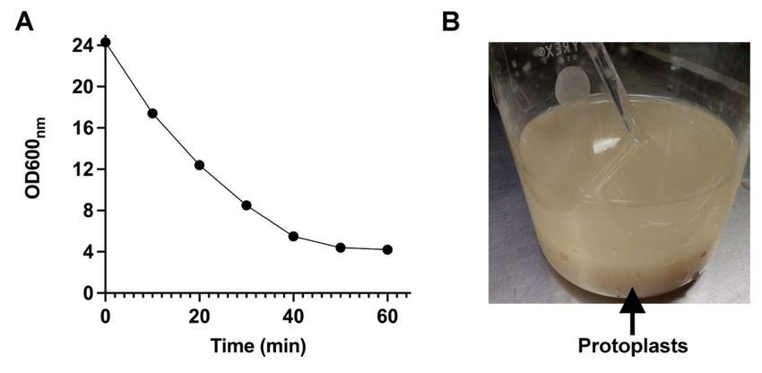
7. To check the formation of protoplasts, shake the cell suspension every 10 min, withdraw 20 µL
of the suspension, and mix with 980 µL of distilled water. Measure the optical density at 600 nm
(Figure 1A). Due to the osmotic shock, cells without a cell wall burst, and their content are
released to the medium. This results in a decrease in the optical density of the suspension.
When the optical density reaches its minimal value, the formation of protoplasts is complete.
Note: From this step, all procedures should be carried out at 4°C.
8. Centrifuge the suspension at 3,800 × g at 4°C for 5 min to recover the protoplasts. Decant the
supernatant.
9. Add 12.5 mL of cold solution B per gram of initial wet weight, to wash the lysing enzymes. With
a Teflon or glass rod, carefully suspend the protoplasts. Do not use the vortex to disperse the
Copyright © 2022 The Authors; exclusive licensee Bio-protocol LLC. 4Please cite this article as: Pardo, J. P. et al. (2022). Isolation of Mitochondria from Ustilago maydis Protoplasts. Bio-protocol 12(01): e4277. DOI:
10.21769/BioProtoc.4277.
Bio-protocol 12(01): e4277.
www.bio-protocol.org/e4277 DOI:10.21769/BioProtoc.4277
protoplasts. Then, centrifuge the suspension for 10 min at 3,800 × g and 4°C. The new pellet
should be compact.
B. Mitochondria isolation
1. Transfer the pellet of protoplasts to a Teflon pestle homogenizer and add cold solution C (40
mL/25-30 g of the initial wet weight of cells) containing the complete cocktail of protease
inhibitors plus PMSF (1 mM final concentration).
2. Attach the Teflon pestle to the electric drill. Connect the drill to the variable autotransformer
(rheostat), select an output intensity of 40, and homogenize for 20 cycles. Avoid the formation
of foam and bubbles.
3. After homogenization, bring the volume to 130 mL with cold solution C (containing protease
inhibitors), transfer the suspension to 50 mL polycarbonate tubes, and eliminate cell debris and
nuclei by centrifuging at 3,800 × g and 4°C for 10 min.
4. Recover the supernatant from the polycarbonate tubes and centrifuge at 17,000 × g and 4°C for
10 min. Carefully decant the supernatant. Then, with the buffer remaining in the tube and with
the help of a soft paintbrush (number 8 or 12, according to the biomass), suspend the
mitochondrial pellet. Keep mitochondria at 4°C for same-day experiments, or at -70°C to be
used later.
5. Determine the protein concentration of your sample by the Lowry method (Lowry et al., 1951).
Prepare a stock solution of 1 mg/mL BSA and construct a standard curve in the range of 5 to
100 µg protein. Dilute the sample tenfold using 0.4% sodium deoxycholate solution, and use 5-
10 µL of the diluted sample for protein determination.
C. Oxygen consumption
To measure the respiratory activity of isolated mitochondria in a thermostated chamber with
magnetic stirring, use a Clark-type oxygen electrode coupled to a YSI5300A biologic oxygen monitor.
1. Bring the thermostated chamber, the solutions D and E, and the distilled water to 30°C.
2. Pour 1.135 mL of distilled water, 65 µL of solution D (final concentration 50 mM), and 100 µL of
solution E (final concentration 20 mM) in the chamber with magnetic stirring. Adjust the signal
to 100%, which corresponds to the dissolved oxygen.
3. Add 5-10 µL of isolated mitochondria; wait until the signal is stabilized and then stimulate oxygen
consumption by adding 10 mM succinate (13 µL from 1 M stock solution), or 0.15 mM NADH
(9.8 µL from 20 mM stock solution).
4. To inhibit respiratory complex IV, add 0.5 mM KCN (2.6 µL from 0.25 M stock solution), or 2 µM
n-octyl gallate (1.3 µL from 2 mM stock solution), to inhibit the alternative oxidase
5. Wash the chamber with distilled water before adding new media.
6. Calculate the specific activity by dividing the oxygen consumption slope by the amount of protein
(mg) or the dry weight (g).
Copyright © 2022 The Authors; exclusive licensee Bio-protocol LLC. 5Please cite this article as: Pardo, J. P. et al. (2022). Isolation of Mitochondria from Ustilago maydis Protoplasts. Bio-protocol 12(01): e4277. DOI:
10.21769/BioProtoc.4277.
Bio-protocol 12(01): e4277.
www.bio-protocol.org/e4277 DOI:10.21769/BioProtoc.4277
D. Mitochondrial membrane potential assay
1. Turn on the Olis Modernized AmincoTM DW-2 spectrophotometer and allow the lamp to warm
up for 30 min. Select the dual-beam option, and adjust the wavelengths of monochromator 1
(MONO1) to 511 nm, and of MONO2 to 533 nm. In the dual-beam mode, the sample is
illuminated with two different wavelengths, one corresponding to the reference wavelength and
the other to the measuring wavelength. The output corresponds to the electronic subtraction of
the absorbance at 511 nm minus the absorbance at 533 nm.
2. Transfer 2.7 mL of solution F into a 3.7 mL glass cuvette containing a magnetic stirrer. Start the
magnetic stirring. Add 2 µM safranine (5.4 µL from 1 mM stock solution) and then the
mitochondrial sample (0.5-1.0 mg protein). Place the cuvette in the spectrophotometer and
close the cover.
3. Adjust the baseline and add, through a small hole made in the chamber cover, the substrate of
your interest (for example, 10 mM of succinate, or 0.15 mM NADH) with a Hamilton syringe.
4. To collapse the membrane potential generated by succinate or any other respiratory substrate,
add 2-10 μM FCCP.
Data analysis
1. Protoplast formation
The formation of protoplasts is followed by the decrease in optical density at 600 nm (OD600nm).
As shown in Figure 1A, after 60 min of incubation in the presence of the lytic enzymes, the
transformation of cells into protoplasts is complete. In addition, Figure 1B shows that the
formation of protoplasts is associated with cell flocculation. Next, respiratory activity and
generation of membrane potential were assayed to evaluate the quality of mitochondria.
Figure 1. Formation of Ustilago maydis protoplast.
During the incubation with lytic enzymes, the OD600nm decreases due to the loss of the yeast cell
wall. (A) In the first 30 min of incubation, 50% of the cells are converted to protoplast. (B)
Because of the degradation of the cell wall, cells begin to form clumps.
2. Mitochondrial respiratory activity
Copyright © 2022 The Authors; exclusive licensee Bio-protocol LLC. 6Please cite this article as: Pardo, J. P. et al. (2022). Isolation of Mitochondria from Ustilago maydis Protoplasts. Bio-protocol 12(01): e4277. DOI:
10.21769/BioProtoc.4277.
Bio-protocol 12(01): e4277.
www.bio-protocol.org/e4277 DOI:10.21769/BioProtoc.4277
U. maydis mitochondria contain the four classic respiratory complexes (I-IV), a glycerol-3-
phosphate dehydrogenase, a cyanide resistant alternative oxidase (AOX), and external and
internal type 2 NADH dehydrogenases (Matuz-Mares et al., 2018; Reyes-Galindo et al., 2019).
To determine the activity of complex I (CI), pyruvate-malate (Pyr-Mal) is added; for complex II
(CII), use succinate; and use TMPD-ascorbate for complex IV (CIV). The activity of the different
complexes can be inhibited by adding rotenone (CI), antimycin A (CIII), or cyanide (CIV) (Juárez
et al., 2004).
The addition of succinate or NADH to the mitochondrial suspension stimulated oxygen
consumption (Figure 2). Respiratory activity was higher with NADH than with succinate,
indicating that the external NADH dehydrogenase is more active than the succinate
dehydrogenase. Because of the presence of the AOX, inhibition of respiration by cyanide was
not complete. However, in the presence of succinate, there was a time-dependent inactivation
of the AOX (Sierra-Campos et al., 2009), probably due to the increased production of H2O2 by
some complexes under this condition. Inhibition of AOX by H2O2 was recently reported
(Yamasaki et al., 2021).
Figure 2. Oxygen consumption by U. maydis mitochondria.
Cells were grown in YPD liquid medium for 24 h at 28°C and collected by centrifugation. Then
mitochondria were isolated as described in the protocol. Oxygen consumption was stimulated
by adding A) succinate (10 mM), or B) NADH (0.15 mM), and the respiratory activity was
inhibited by KCN (1 mM), or n-octyl gallate (2 µM).
3. Mitochondrial membrane potential
Mitochondrial membrane potential (ΔΨ) is the main component of the proton motive force
generated by respiratory complexes I, III, and IV (Figueira et al., 2012). The cationic lipophilic
dye safranine has been used to measure the membrane potential in mitochondria. Upon the
generation of the membrane potential by the addition of respiratory substrates, there is an
accumulation of safranin inside the mitochondrial matrix, which results in dye stacking and
spectral shifts (Akerman and Wikström, 1976). The increase in the 511-533 nm absorbance
differences is related to membrane potential generation, while the decrease in this difference
induced by the protonophore carbonyl cyanide p-trifluoromethoxyphenylhydrazone (FCCP) is
related to the dissipation of ΔΨ.
Copyright © 2022 The Authors; exclusive licensee Bio-protocol LLC. 7Please cite this article as: Pardo, J. P. et al. (2022). Isolation of Mitochondria from Ustilago maydis Protoplasts. Bio-protocol 12(01): e4277. DOI:
10.21769/BioProtoc.4277.
Bio-protocol 12(01): e4277.
www.bio-protocol.org/e4277 DOI:10.21769/BioProtoc.4277
Figure 3A-3C shows the generation of a ΔΨ by different respiratory substrates in mitochondria
obtained from U. maydis protoplasts. Entrance of electrons at the levels of complex I (Pyr-Mal),
complex II (succinate), or the external NADH dehydrogenase induces the rapid formation of a
ΔΨ, which is associated with an increase in the safranine signal (Figure 3). In all cases, the
membrane potential was collapsed by the uncoupler FCCP (Figure 3). Qualitatively, the
membrane potential was the same with the three substrates.
Figure 3. Spectral change of safranine caused by energization of mitochondria.
Mitochondria were energized by adding (A) 10 mM succinate (Succ), (B) 5 mM pyruvate-malate,
or (C) 250 µM NADH. Then potential was abolished with 10 μM FCCP. The system contained:
300 mM sorbitol, 10 mM Hepes pH 7.0, 0.33 EGTA, 0.5 mg (protein) mitochondria, 0.2% BSA,
and 2 μM safranine.
Notes
1. We recommend incubating cells in the water bath for protoplast formation.
2. To obtain high-quality mitochondria preparations, we recommended starting with at least 20 g
of wet weight of cells, and keeping the temperature at 4°C after protoplast formation.
3. We recommend a fresh mitochondrial preparation for oxygen consumption experiments.
4. To achieve inhibition of serine proteases, PMSF should be prepared just before use. Never use
a stored PMSF.
Recipes
1. Solution A
(NH4)2SO4 0.6 M (79.26 g/L)
KH2PO4 20 mM (2.72 g/L)
2. Solution B
Sucrose 0.8 M (273.84 g/L)
Trizma base-HCl (pH 7.0) 10 mM (1.21 g/L)
EDTA 2 mM (0.74 g/L)
KH2PO4 20 mM (2.72 g/L)
BSA 0.3% (3.0 g/L)
Copyright © 2022 The Authors; exclusive licensee Bio-protocol LLC. 8Please cite this article as: Pardo, J. P. et al. (2022). Isolation of Mitochondria from Ustilago maydis Protoplasts. Bio-protocol 12(01): e4277. DOI:
10.21769/BioProtoc.4277.
Bio-protocol 12(01): e4277.
www.bio-protocol.org/e4277 DOI:10.21769/BioProtoc.4277
Adjust pH to 7.0 at room temperature.
3. Solution C
Sucrose 0.4 M (137 g/L)
Trizma base-HCl (pH 7.0) 10 mM (1.21 g/L)
EDTA 2 mM (0.74 g/L)
KH2PO4 20 mM (2.72 g/L)
BSA 0.3% (3.0 g/L)
Adjust pH to 7.0 at room temperature.
4. PMSF
PMSF 1 M (0.0871 g/0.5 mL of DMSO)
5. Solution D
Glucose 1 M (1.80 g/10 mL)
6. Solution E
Tris-HCl (pH 7.0) 250 mM (1.51 g/50 mL)
7. YPD culture medium
Glucose 0.5% (5 g/L)
Select yeast extract 0.5% (5 g/L)
Bacteriological peptone 0.25% (2.5 g/L)
Sterilize at 120°C for 15 min.
For solid YPD, add 2% agar (20 g/L).
8. Solution F
Sorbitol 300 mM (54.65 g/L)
Hepes 20 mM, pH 7.0 (4.76 g/L)
EGTA 0.33 mM (125.51 mg/L)
BSA 0.2% (2 g/L)
9. FCCP solution
FCCP 1 mM (1.27 mg/5 mL)
10. Safranine solution
Safranine 1 mM (3.51 mg/10 mL)
11. Succinate solution
Succinate 1 M (1.3507 g/5 mL)
12. NADH solution
NADH 20 mM (0.07094 g/5 mL)
13. KCN solution
KCN (0.0815 g/5 mL)
14. n-Octyl gallate Solution
n Octyl gallate (0.0028 g/5 mL)
Copyright © 2022 The Authors; exclusive licensee Bio-protocol LLC. 9Please cite this article as: Pardo, J. P. et al. (2022). Isolation of Mitochondria from Ustilago maydis Protoplasts. Bio-protocol 12(01): e4277. DOI:
10.21769/BioProtoc.4277.
Bio-protocol 12(01): e4277.
www.bio-protocol.org/e4277 DOI:10.21769/BioProtoc.4277
Acknowledgments
Author thank to Universidad Nacional Autónoma de México–Programa de Apoyo a Proyectos de
Investigación e Inovación Tecnológica (PAPIIT IV200519 e IA200321) and Consejo Nacional de
Ciencia y Tecnología (CONACYT-254904). Also, we thanks to Oscar Iván Luqueño Bocardo by the
assistance in figures elaboration and Mercedes Esparza Perusquía for technical assistance.
Competing interests
The author declares no competing interest related to this work.
References
1. Akerman, K. E. and Wikström, M. K. (1976). Safranine as a probe of the mitochondrial
membrane potential. FEBS Lett 68(2): 191-197.
2. Brefort, T., Doehlemann, G., Mendoza-Mendoza, A., Reissmann, S., Djamei, A. and Kahmann,
R. (2009). Ustilago maydis as a Pathogen. Annu Rev Phytopathol 47: 423-445.
3. Figueira, T. R., Melo, D. R., Vercesi, A. E. and Castilho, R. F. (2012). Safranine as a fluorescent
probe for the evaluation of mitochondrial membrane potential in isolated organelles and
permeabilized cells. Methods Mol Biol 810: 103-117.
4. Juárez, O., Guerra, G., Martínez, F. and Pardo, J. P. (2004). The mitochondrial respiratory chain
of Ustilago maydis. Biochim Biophys Acta 1658(3): 244-251.
5. Lowry, O. H., Rosebrough, N. J., Farr, A. L. and Randall, R. J. (1951). Protein measurement
with the Folin phenol reagent. J Biol Chem 193(1): 265-275.
6. Matuz-Mares, D., Matus-Ortega, G., Cárdenas-Monroy, C., Romero-Aguilar, L., Villalobos-
Rocha, J. C., Vázquez-Meza, H., Guerra-Sánchez, G., Peña-Díaz, A. and Pardo, J. P. (2018).
Expression of alternative NADH dehydrogenases (NDH-2) in the phytopathogenic fungus
Ustilago maydis. FEBS Open Bio 8(8): 1267-1279.
7. Reyes-Galindo, M., Suarez, R., Esparza-Perusquía, M., de Lira-Sánchez, J., Pardo, J. P.,
Martínez, F. and Flores-Herrera, O. (2019). Mitochondrial respirasome works as a single unit
and the cross-talk between complexes I, III2 and IV stimulates NADH dehydrogenase activity.
Biochim Biophys Acta Bioenerg 1860(8): 618-627.
8. Sierra-Campos, E., Velázquez, I., Matuz-Mares, D., Villavicencio-Queijeiro, A. and Pardo, J. P.
(2009). Functional properties of the Ustilago maydis alternative oxidase under oxidative stress
conditions. Mitochondrion 9(2): 96-102.
9. Sousa, J. S., D’Imprima, E. and Vonck, J. (2018). Mitochondrial Respiratory Chain Complexes.
Subcell Biochem 87: 167-227.
10. Yamasaki, S., Shoji, M., Kayanuma, M., Sladek, V., Inaoka, D. K., Matsuo, Y., Shiba, T., Young,
L., Moore, A. L., Kita, K., et al. (2021). Weak O2 binding and strong H2O2 binding at the non-
Copyright © 2022 The Authors; exclusive licensee Bio-protocol LLC. 10Please cite this article as: Pardo, J. P. et al. (2022). Isolation of Mitochondria from Ustilago maydis Protoplasts. Bio-protocol 12(01): e4277. DOI:
10.21769/BioProtoc.4277.
Bio-protocol 12(01): e4277.
www.bio-protocol.org/e4277 DOI:10.21769/BioProtoc.4277
heme diiron center of trypanosome alternative oxidase. Biochim Biophys Acta Bioenerg 1862(4):
148356.
Copyright © 2022 The Authors; exclusive licensee Bio-protocol LLC. 11You can also read