Lack of Acute Toxicity and Mutagenicity from Recombinant Epinephelus lanceolatus Piscidin Expressed in Pichia pastoris - Mdpi
←
→
Page content transcription
If your browser does not render page correctly, please read the page content below

marine drugs
Article
Lack of Acute Toxicity and Mutagenicity from
Recombinant Epinephelus lanceolatus Piscidin
Expressed in Pichia pastoris
Hsiao-Ching Chen 1,† , Chieh-Yu Pan 2,† , Venugopal Rajanbabu 3 , Yen-Yun Lee 1 , Wei-Ren Tsai 1, *
and Jyh-Yih Chen 4,5, *
 1   Division of Applied Toxicology, Taiwan Agricultural Chemicals and Toxic Substances Research Institute,
     Council of Agriculture, Taichung City 41358, Taiwan; schingc@tactri.gov.tw (H.-C.C.);
     lyy@tactri.gov.tw (Y.-Y.L.)
 2   Department and Graduate Institute of Aquaculture, National Kaohsiung University of Science and
     Technology, Kaohsiung 811, Taiwan; panjade@nkust.edu.tw
 3   Anbil Dharmalingam Agricultural College and Research Institute, Tamil Nadu Agricultural University,
     Tiruchchirapalli, Tamil Nadu 620027, India; vrajanbabu@gmail.com
 4   Marine Research Station, Institute of Cellular and Organismic Biology, Academia Sinica, 23-10 Dahuen Road,
     Jiaushi, Ilan 262, Taiwan
 5   The iEGG and Animal Biotechnology Center, National Chung Hsing University, Taichung 402, Taiwan
 *   Correspondence: sftsai@tactri.gov.tw (W.-R.T.); zoocjy@gate.sinica.edu.tw (J.-Y.C.);
     Tel.: +886-988105706 (W.-R.T.); +886-920802111 (J.-Y.C.)
 †   These authors contributed equally to this work.

 Received: 16 March 2020; Accepted: 8 April 2020; Published: 11 April 2020

 Abstract: The antimicrobial peptide (AMP) piscidin was identified from Epinephelus lanceolatus and
 demonstrated to possess antimicrobial and immune-related functions. Supplementation of feed
 with recombinant Epinephelus lanceolatus piscidin (rEP)-expressing yeast pellets may minimize
 the excessive use of antibiotics and control pathogens in aquaculture or animal husbandry.
 However, before implementing rEP as a supplement, it is necessary to understand whether it
 harbors any toxicity. Since toxicological information on the topic is scarce, the present investigation
 was carried out to test whether rEP exhibits allergenic and/or toxic effects. In an oral acute toxicity test
 (OECD 425), Sprague Dawley (SD) rats were administered rEP dissolved in reverse osmosis water,
 yielding an LD50 > 5000 mg/kg (no observed animal death). The compound was therefore classified
 as non-toxic by oral administration. In an acute respiratory toxicity test (OECD 403), heads and noses
 of SD rats were exposed to liquid aerosol for 4 h (the highest concentration that could be administered
 without causing any animal death), and a lethal concentration (LC50 ) > 0.88 mg/L was obtained.
 The mass medium aerodynamics diameter (MMAD) of rEP aerosol particles was 8.18 µm and mass
 medium aerodynamics diameter (GSD) was 3.04, which meant that 25.90% could enter the airway
 (
Mar. Drugs 2020, 18, 206                                                                             2 of 17

 animals nor mutagenicity. Thus, rEP can be considered safe for use in subsequent research on its
 application as a feed additive for poultry, cattle, or aquatic animals.

 Keywords: Epinephelus lanceolatus piscidin; antimicrobial peptide; recombinant piscidin; toxic effects;
 allergic effects; mutagenetic toxicity

1. Introduction
      The emergence of multidrug-resistant pathogens has necessitated the development of antibiotic
alternatives to control deadly pathogens in humans and animals [1]. According to the World Health
Organization (WHO), an alarming rise in death due to infections with multidrug-resistant pathogens
is expected by the middle of this century [2]. The WHO has defined a category of drug-resistant
pathogens (Enterococcus faecium, Staphylococcus aureus, Klebsiella pneumoniae, Acinetobacter baumannii,
Pseudomonas aeruginosa, and Enterobacter spp.; abbreviated ESKAPE) that represent the most likely to
cause a substantial increase in infectious cases around the globe [3]. In addition, several instances of
polymicrobial infections with necrotizing fasciitis have been reported. In this condition, the microbial
population feeds on the soft tissues in the infected individual, which may be fatal if left untreated for a
sufficient duration [4]. Currently, there is an alarming rise in multidrug-resistant and pan-drug-resistant
microbial species, along with a drying up of the drug discovery pipeline. Together, these developments
have created an emergent need for potential antimicrobial therapeutics [5]. Antimicrobial peptides
(AMPs) can be considered as a promising category of therapeutic agents due to their significant
antimicrobial activity against drug-resistant pathogens [6].
      AMPs are also known as host defense peptides, as they act against invading microbial pathogens [7].
Apart from their utility in innate immune response, anti-cancer and immunomodulatory activities are
also reported for AMPs in various host organisms [8–10]. Defensins [11], cecropins [12], piscidins [13],
and cathelicidins [14] are among the most widely-reported molecules of this class. These molecules are
known to possess a net positive charge as well as amphipathicity. Based on tense structural features,
AMPs electrostatically interact with the anionic bacterial membrane and cause membranolysis [15,16].
Unlike the mammalian membrane, the bacterial surface has a negative charge due to the presence of
anionic lipids like 1-palmitoyl-2-oleoyl-sn-glycero-phosphocholine (POPC) and cardiolopins, as well
as other anionic surface molecules, like lipoteichoic acid and lipopolysaccharide [17]. Despite the fact
that AMPs possess significant therapeutic potential, their utility as drugs has not yet been established.
Potential drawbacks, such as ion sensitivity and susceptibility to enzymatic degradation, can be
overcome by introducing known molecular modifications that retain or enhance therapeutic utility [18].
      In vitro and in vivo studies have been conducted on a vast array of potential AMPs, with the
molecules showing significant activity, negligible toxicity, and lack of long-term allergenicity [8,19,20].
One group of potential therapeutic AMPs are the piscidins, which have been reported to possess
strong antimicrobial activity against a variety of organisms [13]. Generally, AMPs are expressed
by immune cells, i.e., mast cells [13]. The first ever report of AMPs was made about the genus
Morone [13], and the molecules were found to be composed of 22 to 44 amino acids and exhibit an
alpha-helical secondary structure. Since then, many other genera have been reported to express
piscidin homologs. These molecules play important roles in maintaining immunological function in the
host organisms, and they tend to exhibit alpha-helical conformations [21]. Studies have demonstrated
their antimicrobial activity, and minimal cytotoxicity was evident in cellular studies [22,23]. In the
current study, piscidin from Epinephelus lanceolatus was examined. This species is reported to possess
254 putative AMP genes, which may be important for maintaining the health of the fish [24]. We have
described the isolation, subcloning of the expressed vector, and expression of the piscidin isolated
from Epinephelus lanceolatus (rEP) in Pichia pastoris and identified the biological function of rEP in
Gallus domesticus in our previous research [25]. rEP supplementation increased G. g. domesticus weight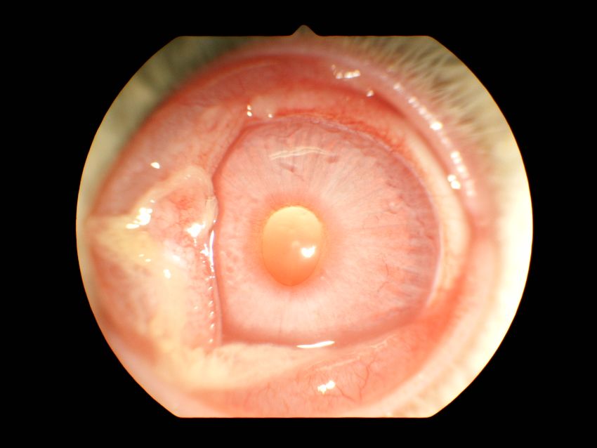
Mar. Drugs 2020, 18, 206                                                                              3 of 17

gain, feed efficiency, and IL-10 and IFN-γ production. Previously, we cloned and characterized five
piscidin-like AMPs (named TP1–TP5) from Oreochromis niloticus [21]. The published results suggest
that piscidins have broad-spectrum antibacterial, antifungal, antiviral, and antiparasitic activities.
From our previous research results, we determined that Epinephelus lanceolatus piscidin (EP) exhibits
high similarity to the highly-active TP3 and TP4 peptides from tilapia [21]. After investigating EP and
other piscidins from fish by MIC methods, we chose the EP peptide as our target gene for expression
in the yeast expression system. The EP experimental results suggested that EP possesses enhanced
antimicrobial activity or growth inhibition activity in Gram-negative and Gram-positive bacteria [25].
      Excessive use of antibiotics in feed supplements is a major concern, as this practice is correlated
with the rise of drug-resistant bacterial species. The repetitive and improper dosing of antibiotics
is one of the main reasons for the steep rise in multidrug-resistant pathogens. Many antibiotics are
available over-the-counter, without stringent and ethical control, and are used by a large percentage
of the population around the world [5]. Therefore, alternatives like AMPs can play a significant role
in overcoming the current deficit of effective antibiotics [7]. AMP-containing feed supplementation
may be a sound strategy to protect domestic animals, which might help prevent the excessive use
of antibiotics [26]. Therefore, the commercial exploitation of AMPs as feed additives requires they
first be examined in a stringent set of toxicological analyses. According to governmental regulatory
bodies, there should be a thorough study of toxicity with model organisms, keeping safety aspects and
health concerns as a priority during drug development [27]. This kind of toxicological analysis can
provide evidence for the absence of toxic analytes/compounds that may be produced by unidentified
modifications [27–30].
      Toxicological analysis is an important part of the drug discovery process, as it helps to ensure
the safe usage of newly-developed potential therapeutics. Tests like oral toxicity, inhalation toxicity,
eye irritation, and skin sensitization are commonly conducted to assess the safety of test molecules [31].
Acute oral toxicity is generally evaluated by oral gavage of rats [32–34]. Similarly, acute inhalation
toxicity studies are performed by exposing rat models to liquid aerosols for a given period of time [32,33].
Another established method of toxicological assessment is to instill the drug into the lower conjunctiva
of the left eye of rabbit models and assess irritation [35]. Skin sensitization studies are also performed,
where the test materials are applied to the skin behind the ears of BALB/c mice, and swelling (if it
occurs) is taken as a measure of sensitization [36–41].
      Toxicological studies on recombinant piscidin (rEP) expressed in Pichia pastoris are limited.
Therefore, it is necessary to perform a panel of basic toxicity tests in accordance with preclinical
guidelines of governmental regulatory bodies. In the present report, we evaluate the toxicity profile of
rEP-expressing yeast pellets. In these tests, we saw that the test candidate did not cause significant oral
toxicity, inhalation toxicity, eye irritation, skin sensitization, bacterial reverse mutation, or chromosomal
effects. Thus, rEP may be considered safe for use in animal fodder as a supplement antibiotic substitute.

2. Results

2.1. Oral Toxicity in Rats
     Oral intake of 5000 mg/kg body weight did not cause any mortality in test rats (Table 1). The body
weight increases of treated rats on day 7 and day 14 were similar to controls (Table 2). No clinical
signs or death of the treated rats was observed within the 14-day observation period (Table 1A). No
significant gross lesions were found in any organs of rats surviving at day 14 of the experiment (Table 1).
The acute oral LD50 for rEP was therefore greater than 5000 mg/kg body weight in female rats.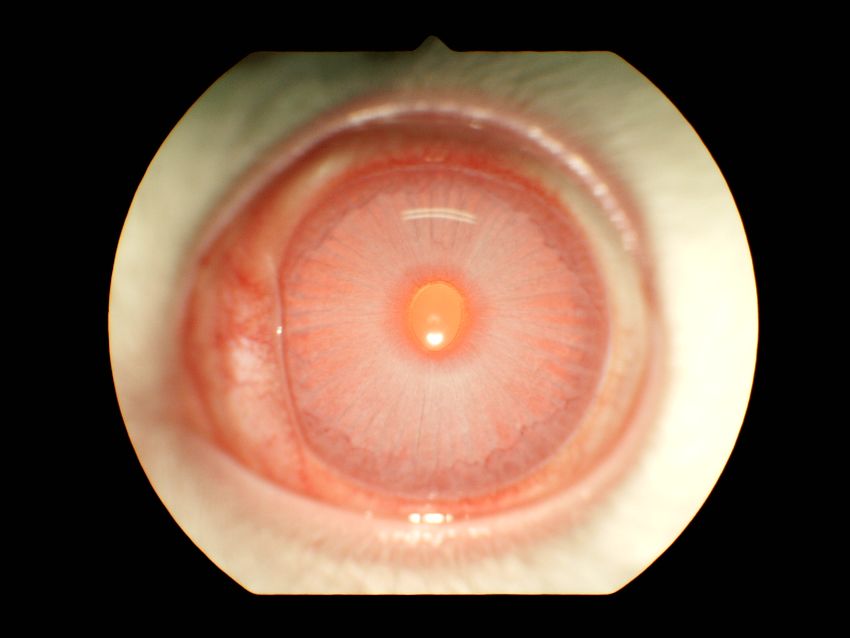
Mar. Drugs 2020, 18, 206                                                                                                         4 of 17

     Table 1. Oral administration of 5000 mg/kg recombinant piscidin (rEP) did not affect examined rats.
     Survival, mortality, clinical signs, and gross pathology after oral dosing of rats.

                                                                                                                       Gross
   Dose Level      Dosing Volume                                                  Observation         Clinical
                                         Survival           Mortality (%)                                            Pathology
    (mg/kg)           (mL/rat)                                                    Period (Day)         Signs
                                                                                                                     Finding *
   5000 (3/3) **           2.4        Survival (3/3) **             0                 14            None (3/3) **   None (3/3) **
      * The gross pathology examination was performed on anus, heart, lung, stomach, liver, pancreas, digestive tract,
      kidney, thymus, and spleen; ** number of animals/total number of examined animals.

     Table 2. Oral administration of 5000 mg/kg recombinant piscidin (rEP) did not affect examined rats.
     Weekly body weights and body weight gain of orally-treated rats.

                                    Body Weight (g)                     Body Weight Gain * (g)        Body Weight Gain ** (%)
      Dose Level
                                     (Mean ± SD)                            (Mean ± SD)                    (Mean ± SD)
       (mg/kg)
                           Day 0        Day 7             Day 14          Day 7         Day 14          Day 7         Day 14
          5000        240.7 ± 3.2     257.6 ± 8.3     272.6 ± 9.9       16.9 ± 5.2     31.9 ± 6.8      7.0 ± 2.1    13.2 ± 2.7
      * Body weight (BW) gain (g) on day N = (day N − day 0) BW; ** body weight (BW) gain (%) on day N = [(day N −
      day 0)/day0] BW × 100%. SD, Standard deviation.

2.2. Inhalation Toxicity in Rats
     rEP was formulated as six liters of liquid aerosol with 34.1% concentration, and Sprague Dawley
(SD) rats were exposed for 4 h. A mean mass medium aerodynamics diameter (MMAD) of 8.18 µM
and geometric standard deviation (GSD) of 3.04 were observed. Comparison with control animals
revealed that these levels are safe and can be inhaled by the test animals without apparent harm.
Some rats treated with rEP aerosol showed piloerection, chromodacryorrhea, hemorrhage of nose,
and tachypnea for a short time, but these reactions disappeared after one day (Table 3). No mortality
was observed, and weight gain of treated male rats was significantly different from blank controls on
the 3rd and 14th day post-treatment (Table 4). Macroscopic lesions in the lungs of necropsy rats 14
days post-treatment had white protrusions and dark red spots (Table S1). Based on these experiments,
the lethal concentration (LC50 ) of aerosol-administered rEP in rats was greater than 0.88 mg/L.

                        Table 3. Clinical signs in rats exposed to 0.88 mg/L liquid aerosol rEP.

                   Time                                                 Clinical Signs *
                                                    Male                                            Female
                   1h                        tachypnea (5/5)                                 tachypnea (5/5)
                                           tachypnea (5/5),                                tachypnea (5/5),
                   2h
                                       abdominal breathing (5/5)                       abdominal breathing (5/5)
                                            piloerection (5/5),
                                                                                           piloerection (5/5),
                                        chromodacryorrhea (4/5),
                   4h                                                                  chromodacryorrhea (5/5),
                                        hemorrhage of nose (1/5),
                                                                                            tachypnea (5/5)
                                             tachypnea (5/5)
               1 to 14 Days                     None (5/5)                                       None (5/5)
      * Number of animals/total number of animals; * observation items: overall normal or not; death; appearance of coat
      skin and mucus; appearance of mouth, eyes, and nose; behavior, movement, and posture; the reflex system; and the
      respiratory and digestive systems.
Mar. Drugs 2020, 18, 206 5 of 17
Table 4. Weight of rats treated with rEP by inhalation. Sprague Dawley (SD) rats were exposed to rEP
liquid aerosol inhalation chamber for 4 h. After 1, 3, 7, and 14 days, weight gain percent was calculated.
Concentration Weight Gain (%) 1
Sex Group
of rEP (mg/L)
Day 1 2 Day 3 2
Blank Control 0 −0.4 ± 2.5 −6.0 ± 0.9
Male
Treated group 0.880 1.6 ± 0.9 −1.7 ± 2.4 *
Blank Control 0 −0.5 ± 1.5 0.1 ± 2.8
Female
Treated group 0.880 0.1 ± 2.1 −1.6 ± 3.9
Blank Control 0 −0.5 ± 1.9 −2.9 ± 3.8
Combined
Treated group 0.880 0.8 ± 1.7 −1.7 ± 3.1
Concentration Weight Gain (%) 1
Sex Group
of rEP (mg/L)
Day 7 2 Day 14 2
Blank Control 0 3.5 ± 2.2 7.8 ± 3.5
Male
Treated group 0.880 0.9 ± 2.2 3.0 ± 2.1 *
Blank Control 0 1.9 ± 3.1 0.5 ± 6.0
Female
Treated group 0.880 3.3 ± 2.2 4.4 ± 5.6
Blank Control 0 2.7 ± 2.6 4.1 ± 6.0
Combined
Treated group 0.880 2.1 ± 2.5 3.7 ± 4.1
1 Data for each sex were derived from 5 treated animals and the combined value represents 10 treated animals;
* indicates significant difference (p < 0.05) from control group by Student’s t test; 2 weight gain (%) on day n = (body
weight of day n – body weight of day 0)/(body weight of day 0) × 100%.
2.3. Eye Irritation in Rabbits
An eye irritation test was conducted using NZW rabbits. No significant changes in body weight
were observed between the time of rEP instillation and the end of the experiment. No obvious major
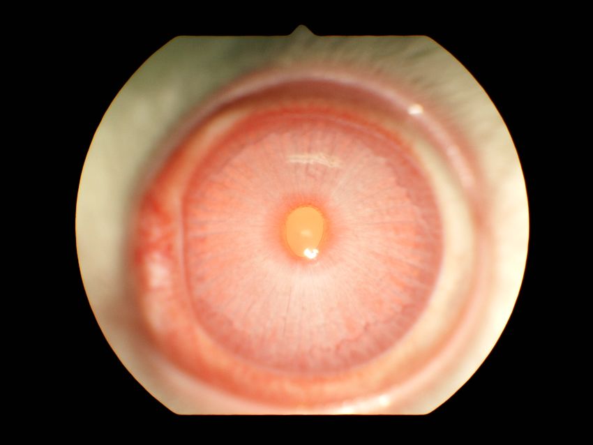
clinical signs (except lacrimation) were observed during the test. After 0.1 g rEP was instilled into
the left eye of each of three rabbits, none of the treated rabbits showed apparent turbidity in the
cornea. The iris was clearly visible, and the cornea still reacted to the light, while the conjunctiva
showed obvious swelling with lids about half-closed, having some discharge and diffuse, crimson color;
individual vessels were not easily discernible. Observations of eye irritation for all three rabbits treated
with rEP were made at 1, 24, 48, and 72 h post-treatment with a hand-held retinal camera. The clinical
appearance of one representative treated eye is showed in Figure 1. After washing with reverse osmosis
water at 24 h post-treatment, the minor signs were gradually lessened and all the signs disappeared by
72 h (Table 5).
Table 5. Ocular score of rabbits treated with rEP. The left eyes of NZW rabbits were instilled with
0.1 g rEP in the lower conjunctiva sac, and the mean irritation score (Supplementary Figure S2) was
calculated at 1, 24, 48, and 72 h post-treatment. N = 3.
Irritant Scoring
Hour
Cornea Iris Conjunctiva
Degree Area Damage Redness Chemosis
1 0 0 1 1.67 2.67
24 0 0 1 1.67 2
48 0 0 0 1 1
72 0 0 0 0 0Mar. Drugs 2020, 18, 206 6 of 17
Mar. Drugs 2020, 18, x 18 of 18
1 hour 24 hours
48 hours 72 hours
Figure
Figure 1. 1. Recovery
Recovery from rEP-mediated
from rEP-mediated mild eye mild eye irritation
irritation after 24 h.after 24 h. Observation
Observation of eyeinirritation
of eye irritation an in
an individual
individual rabbit
rabbit treated withtreated
rEP atwith rEP
1, 24, 48,atand
1, 24,
7248, and 72 h post-treatment.
h post-treatment. Images
Images were were captured
captured with a with
a hand-held
hand-held retinal camera.
retinal camera.
2.4. Skin Sensitization in Mice
Table 5. Ocular score of rabbits treated with rEP. The left eyes of NZW rabbits were instilled with 0.1
g rEP in
Application of the
thelower
peptideconjunctiva
pellet tosac, anddid
skin the not
mean irritation
cause score (Supplementary
significant Figure
changes in body S2) was
weight.
No obvious calculated at 1, 24,
clinical signs, 48, and
except 72 hcoat,
rough post-treatment. N =alopecia,
piloerection, 3. erythema, edema, and emaciation,
were observed during the test (Table 6). The stimulation index Scoring
Irritant (SI) was calculated for treatment groups
(25%, 0.77; 50%, 0.44; and 100%, Hour1.06) and the
Cornea positive-control
Iris group (1.69) (Table 7).
Conjunctiva
Degree Area Damage Redness Chemosis
2.5. Bacterial Reverse Mutation Test
1 0 0 1 1.67 2.67
The results of the main test 24 (including
0 cytotoxicity
0 1test; conducted
1.67 at 5000,
2 1667, 556, 185, 62,
21, and 7 µg/plate ± S9) are shown 48 in Tables
0 8 and
0 9. A confirmatory
0 1 test (conducted
1 at 5000, 2500,
1250, 625, and 313 µg/plate ±72 S9) was 0also conducted,
0 and
0 results0are shown 0in Tables 10 and 11.
No treatment caused a significant elevation in revertants (2- or 3-fold more than negative controls) in
any of the
2.4. five
Skintester strains. in
Sensitization Whether
Mice addition of the S9 liver fraction or not had no apparent impact
on cytotoxicity or precipitate formation. Therefore, we conclude that rEP exhibits a negative response
Application of the peptide pellet to skin did not cause significant changes in body weight. No
in the bacterial reverse mutation test.
obvious clinical signs, except rough coat, piloerection, alopecia, erythema, edema, and emaciation,
Table
were6. Clinical
observedsigns and body
during theweight of mice6).
test (Table after treatment
The of ears with
stimulation indexrEP. Twenty-five
(SI) microliters
was calculated for treatment
of groups
25%, 50%, or 100% rEP solution was applied to the back of Balb/C mice ears for three
(25%, 0.77; 50%, 0.44; and 100%, 1.06) and the positive-control group (1.69) (Table 7). consecutive
days. Sterile water, AOO *, and 2% 2,4-dinitro-1-chlorobenzene (DNCB) were used as controls. Body
weight before treatment and six days after treatment (before sacrifice) are shown.
Groups
Sterile
AOO * 2% DNCB 25% rEP 50% rEP 100% rEP
Water
Time (days) 1 Clinical signs **
Rough coat (5/5),
1 None (5/5) Rough coat (5/5) Piloerection (5/5), None (5/5) Erythema (4/5) Erythema (4/5)
Erythema (5/5)Mar. Drugs 2020, 18, 206 7 of 17
Table 6. Cont.
Groups
Sterile
AOO * 2% DNCB 25% rEP 50% rEP 100% rEP
Water
Rough coat (5/5),
Rough coat (5/5), Piloerection (5/5), Alopecia (3/5),
2 None (5/5) Erythema (3/5) Erythema (5/5)
Erythema (5/5) Erythema (5/5), Erythema (5/5)
Edema (5/5)
Rough coat (5/5),
Rough coat (1/5),
Rough coat (5/5), Piloerection (5/5), Alopecia (3/5),
3 None (5/5) Erythema (5/5) Alopecia (5/5),
Erythema (5/5) Erythema (5/5), Erythema (5/5)
Erythema (5/5)
Edema (5/5)
Rough coat (5/5),
Rough coat (5/5), Piloerection (5/5), Alopecia (5/5), Alopecia (5/5),
4 None (5/5) Erythema (5/5)
Erythema (5/5) Erythema (5/5), Erythema (5/5) Erythema (5/5)
Edema (5/5)
Rough coat (5/5),
Rough coat (1/5), Piloerection (5/5), Alopecia (5/5), Alopecia (5/5),
5 None (5/5) Erythema (5/5)
Erythema (5/5) Erythema (5/5), Erythema (5/5) Erythema (5/5)
Edema (5/5)
rough coat (5/5),
Alopecia (5/5),
piloerection (5/5), Alopecia (5/5),
6 None (5/5) Erythema (5/5) Erythema (2/5) Erythema (5/5),
erythema (5/5), Erythema (2/5)
Emaciation (1/5)
edema (5/5)
Time (treatment) Body weight (g) (mean ± SD)
Before dosing 21.4 ± 1.2 22.0 ± 0.5 21.5 ± 1.0 22.0 ± 0.7 21.7 ± 0.5 21.8 ± 1.3
After dosing
21.4 ± 1.1 22.4 ± 0.9 22.0 ± 0.8 22.5 ± 0.6 21.9 ± 0.6 21.2 ± 1.6
(before sacrifice)
* AOO: acetone: olive oil (4:1 v/v); ** (number of animals/total number of examined animals); 1 day 1 was defined as
the day of treatment.
Table 7. rEP did not cause sensitization of mice in a bromodeoxyuridine (BrDU) assay. Lymph nodes
cells were labeled with BrdU, and the BrdU-labeling index was estimated by measuring absorbance at
370 and 492 nm in an ELISA reader. The stimulation index (SI) value was calculated as the ratio of
control and treatment groups. SI value less than 1.6 means the test item did not cause skin sensitization.
Group Treatment BrdU Labelling Index ÷ Solvent Control BrdU Labelling Index = SI Value
2% DNCB 0.405 ÷ 0.239 = 1.69
25% rEP 0.179 ÷ 0.231 = 0.77
50% rEP 0.102 ÷ 0.231 = 0.44
100% rEP 0.244 ÷ 0.231 = 1.06
Table 8. Revertants of Salmonella typhimurium TA strains treated with rEP peptide (marine peptide) in
the absence of S9 mix (main test including cytotoxicity) (a) .
S9 TA98 TA100 TA1535 TA1537 TA102
Treatment
± Mean ± SD Mean ± SD Mean ± SD Mean ± SD Mean ± SD
BK (b) - 23.0 ± 1.7 119.7 ± 17.7 8.7 ± 1.5 8.0 ± 1.0 200.0 ± 13.1
NC (c) - 24.3 ± 1.5 110.0 ± 9.6 9.7 ± 1.5 8.7 ± 1.2 205.3 ± 16.8
PC (d) - 440.7 ± 40.5 **(18.1) >2000 (e) **(18.2) 1425.3 ± 113.2 **(147.4) >2000 (e) **(230.8) >2000 (e) **(9.7)
Marine
peptide
(µg/plate)
7 - 21.0 ± 1.0(0.9) 118.0 ± 19.3(1.1) 10.7 ± 1.2(1.1) 9.7 ± 2.3(1.1) 205.0 ± 22.6(1.0)
21 - 21.7 ± 3.8(0.9) 120.3 ± 18.6(1.1) 8.7 ± 2.1(0.9) 9.3 ± 1.5(1.1) 193.3 ± 14.6(0.9)Mar. Drugs 2020, 18, 206 8 of 17
Table 8. Cont.
S9 TA98 TA100 TA1535 TA1537 TA102
Treatment
± Mean ± SD Mean ± SD Mean ± SD Mean ± SD Mean ± SD
62 - 24.0 ± 0.0(1.0) 125.0 ± 13.5(1.1) 10.0 ± 2.0(1.0) 9.0 ± 1.7(1.0) 192.3 ± 32.9(0.9)
185 - 26.0 ± 5.0(1.1) 121.3 ± 4.7(1.1) 11.3 ± 3.1(1.2) 10.3 ± 2.3(1.2) 186.7 ± 26.8(0.9)
556 - 24.3 ± 2.1(1.0) 126.7 ± 5.5(1.2) 11.7 ± 1.5(1.2) 11.3 ± 3.1(1.3) 195.0 ± 16.0(0.9)
1667 - 28.7 ± 2.3(1.2) 116.0 ± 19.0(1.1) 12.0 ± 3.6(1.2) 10.0 ± 3.0(1.2) 193.3 ± 9.6(0.9)
5000 - 27.7 ± 1.2(1.1) 135.0 ± 8.2(1.2) 12.0 ± 2.0(1.2) 6.0 ± 1.7(0.7) 196.3 ± 20.0(1.0)
Dose
0.845 ** 0.283 0.602 * 0.115 0.105
response (f)
(a)Data represent means of three replicates. Numbers in parentheses are fold-induction compared with negative
control (NC); (b) blank control (BK); (c) negative control (NC): 100 µL/plate deionized water (DI water); and (d) positive
control (PC): in assay without liver S9: 0.5 µg/plate 4-nitroquinoline-N-oxide (TA98 and TA100), 5 µg/plate sodium
azide (TA1535), 125 µg/plate 9-aminoacridine (TA1537), and 0.5 µg/plate mitomycin C (TA102). ** p < 0.01 compared
to NC (Student’s t test, one-tail); (e) Data were analyzed with 2000 colony numbers/plate; and (f) dose response:
correlation was analyzed for doses without significant cytotoxicity and revertants per plate. * p < 0.05. ** p < 0.0.1.
Table 9. Revertants of Salmonella typhimurium TA strains treated with rEP peptide in the presence of S9
mix (main test including cytotoxicity) (a) .
S9 TA98 TA100 TA1535 TA1537 TA102
Treatment
± Mean ± SD Mean ± SD Mean ± SD Mean ± SD Mean ± SD
BK (b) + 24.0 ± 1.0 126.7 ± 10.0 9.7 ± 0.6 7.3 ± 0.6 227.0 ± 18.1
NC (c) + 26.0 ± 3.6 133.7 ± 12.1 10.7 ± 1.5 9.0 ± 1.7 214.3 ± 4.7
PC (d) + >2000 (e) **(76.9) 1812.7 ± 106.7 **(13.6) 227.7 ± 23.9 **(21.3) 117.0 ± 24.9 **(13.0) 1228.0 ± 200.0 **(5.7)
Marine
peptide
(µg/plate)
7 + 26.3 ± 3.1(1.0) 120.3 ± 17.1(0.9) 11.0 ± 1.7(1.0) 9.3 ± 2.3(1.0) 210.7 ± 12.5(1.0)
21 + 26.7 ± 3.5(1.0) 116.3 ± 20.0(0.9) 10.0 ± 1.0(0.9) 8.7 ± 0.6(1.0) 215.7 ± 18.0(1.0)
62 + 23.3 ± 3.2(0.9) 118.3 ± 21.6(0.9) 9.3 ± 1.5(0.9) 8.3 ± 2.3(0.9) 202.3 ± 9.7(0.9)
185 + 25.7 ± 3.2(1.0) 128.3 ± 18.2(1.0) 11.7 ± 2.5(1.1) 7.7 ± 1.5(0.9) 203.0 ± 24.6(0.9)
556 + 23.0 ± 3.0(0.9) 118.0 ± 9.2(0.9) 10.3 ± 2.5(1.0) 9.3 ± 2.5(1.0) 202.0 ± 24.6(0.9)
1667 + 25.7 ± 3.2(1.0) 115.0 ± 10.4(0.9) 13.7 ± 1.5(1.3) 8.0 ± 2.0(0.9) 211.3 ± 16.3(1.0)
5000 + 25.7 ± 2.9(1.0) 138.3 ± 11.0(1.0) 9.7 ± 1.2(0.9) 7.7 ± 1.5(0.9) 202.3 ± 24.3(0.9)
Dose
0.054 0.222 0.050 0.331 0.215
response (f)
(a)
Data represent means of three replicates. Numbers in parentheses are fold-induction compared with NC; (b) blank
control (BK); (c) negative control (NC): 100 µL/plate deionized water (DI water); and (d) positive control (PC): in assay
with liver S9: 5 µg/plate 2-aminofluorene (TA98), 10 µg/plate 2-aminofluorene (TA100), 5 µg/plate 2-aminoanthracene
(TA 1535), 20 µg/plate 2-aminofluorene (TA1537), and 30 µg/plate danthron (TA102). ** p < 0.01 compared to NC
(Student’s t test, one-tail); (e) Data were analyzed with 2000 colony numbers/plate; and (f) dose response: correlation
was analyzed for doses without significant cytotoxicity and revertants per plate.
Table 10. Revertants of Salmonella typhimurium TA strains treated with rEP peptide in the absence of S9
mix (confirmation test) (a) .
S9 TA98 TA100 TA1535 TA1537 TA102
Treatment
± Mean ± SD Mean ± SD Mean ± SD Mean ± SD Mean ± SD
BK (b) - 23.0 ± 2.6 113.0 ± 14.4 11.0 ± 1.0 9.0 ± 2.6 200.7 ± 9.0
NC (c) - 25.7 ± 3.5 105.7 ± 7.2 13.7 ± 2.5 8.3 ± 0.6 202.0 ± 27.2
PC (d) - 445.0 ± 35.8 **(17.3) >2000 (e) **(18.9) 1488.0±150.7 **(108.9) >2000 (e) **(240.0) >2000 (e) **(9.9)
Marine
peptide
(µg/plate)
313 - 24.7 ± 0.6(1.0) 112.3 ± 4.9(1.1) 12.3 ± 3.1(0.9) 7.7 ± 1.5(0.9) 203.0 ± 8.9(1.0)
625 - 26.0 ± 1.0(1.0) 100.0 ± 7.2(0.9) 9.7 ± 1.2(0.7) 9.3 ± 2.1(1.1) 195.7 ± 8.5(1.0)
1250 - 25.7 ± 6.1(1.0) 112.0 ± 9.5(1.1) 9.0 ± 0.0(0.7) 7.7 ± 0.6(0.9) 197.3 ± 25.9(1.0)
2500 - 26.7 ± 3.1(1.0) 136.7 ± 24.7(1.3) 10.3 ± 2.5(0.8) 11.0 ± 2.6(1.3) 225.7 ± 30.7(1.1)
5000 - 24.7 ± 0.6(1.0) 126.3 ± 28.5(1.2) 10.7 ± 2.1(0.8) 8.3 ± 2.1(1.0) 216.0 ± 30.8(1.1)
Dose
0.015 0.517 0.113 0.114 0.471
response (f)
(a)
Data represent means of three replicates. Numbers in parentheses are fold-induction compared with NC; (b) blank
control (BK); (c) negative control (NC): 100 µL/plate deionized water (DI water); and (d) positive control (PC): in
assay without liver S9: 0.5 µg/plate 4-nitroquinoline-N-oxide (TA98 and TA100), 5 µg/plate sodium azide (TA1535),
125 µg/plate 9-aminoacridine (TA1537), and 0.5 µg/plate mitomycin C (TA102). ** p < 0.01 compared to NC (Student’s
t test, one-tail); (e) Data were analyzed with 2000 colony numbers/plate; and (f) dose response: correlation was
analyzed for doses without significant cytotoxicity and revertants per plate. * p < 0.05.Mar. Drugs 2020, 18, 206 9 of 17
Table 11. Revertants of Salmonella typhimurium TA strains treated with rEP peptide in the presence of
S9 mix (confirmation test) (a) .
S9 TA98 TA100 TA1535 TA1537 TA102
Treatment
± Mean ± SD Mean ± SD Mean ± SD Mean ± SD Mean ± SD
BK (b) + 22.7 ± 2.3 109.0 ± 4.0 10.7 ± 2.1 7.3 ± 1.2 209.7 ± 18.1
NC (c) + 25.3 ± 2.5 105.3 ± 1.5 11.7 ± 3.2 10.3 ± 0.6 208.3 ± 4.7
PC (d) + >2000 (e) **(78.9) 961.3 ± 82.8 **(9.1) 167.0 ± 13.0 **(14.3) 100.7 ± 10.3 **(9.7) 1087.7 ± 160.0 **(5.2)
Marine
peptide
(µg/plate)
313 + 26.3 ± 6.1(1.0) 97.7 ± 2.3(0.9) 8.7 ± 2.1(0.7) 10.3 ± 2.9(1.0) 209.7 ± 7.6(1.0)
625 + 30.0 ± 2.6(1.2) 97.3 ± 11.0(0.9) 13.7 ± 2.1(1.2) 11.3 ± 2.5(1.1) 185.7 ± 20.6(0.9)
1250 + 19.7 ± 3.1(0.8) 102.0 ± 4.0(1.0) 13.3 ± 2.9(1.1) 7.3 ± 0.6(0.7) 187.7 ± 14.2(0.9)
2500 + 23.0 ± 2.0(0.9) 99.3 ± 7.8(0.9) 12.0 ± 2.0(1.0) 8.3 ± 2.1(0.8) 199.0 ± 12.5(1.0)
5000 + 29.0 ± 5.3(1.1) 107.7 ± 14.6(1.0) 12.3 ± 2.5(1.1) 10.7 ± 1.2(1.0) 185.7 ± 15.6(0.9)
Dose
0.004 0.667 0.204 0.048 0.268
response (f)
(a)
Data represent means of three replicates. Numbers in parentheses are fold-induction compared with NC; (b) blank
control (BK); (c) negative control (NC): 100 µL/plate deionized water (DI water); (d) positive control (PC): in assay
with liver S9: 5 µg/plate 2-aminofluorene (TA98), 10 µg/plate 2-aminofluorene (TA100), 5 µg/plate 2-aminoanthracene
(TA 1535), 20 µg/plate 2-aminofluorene (TA1537), and 30 µg/plate danthron (TA102). ** p < 0.01 compared to NC
(Student’s t test, one-tail); (e) Data were analyzed with 2000 colony numbers/plate; and (f) dose response: correlation
was analyzed for doses without significant cytotoxicity and revertants per plate.
2.6. Micronucleus Test in Chinese hamster ovary (CHO)-K1 Cells
No treatments with rEP caused a significant increase in frequency of micronuclei compared to
negative controls (0.5% DI water) in the micronucleus assay. In addition, a dose-response relationship
was not observed (Table 12). The results showed that rEP had no observable effects on chromosomes
(clastogenicity) of CHO-K1 cells.
Table 12. Micronucleus analysis.
Exposure Time (3 + 20) h-S9 (3 + 20) h + S9 (24 + 0) h-S9
Treatment Micronucleus Assay
(µg/mL) Frequency of Micronuclei (%) (a)
BK (b) 4.10 3.25 4.85
NC (c) 3.75 3.85 4.75
125 4.55 3.95 4.60
250 3.95 4.00 3.95
500 3.55 3.25 5.20
1000 3.80 3.15 4.85
2000 3.90 3.75 5.55
PC (d) 9.60 ** 7.20 ** 11.40 **
Dose response (p value) (e) 0.601 0.299 0.134
(a) Frequency of micronuclei (%). Data were analyzed by a chi-squared test with Yate’s correction (GraphPad 5),
** p < 0.01 compared to NC. Micronuclei were scored in at least 2000 cells per treatment (at least 1000 cells per
culture; two cultures per concentration); (b) blank control (BK); (c) negative control (NC): 0.5% DI water; (d) positive
control (PC): 0.25 µg/mg mitomycin C (−S9); 10 µg/mg cyclophosphamide (+S9); and (e) dose response (trend test)
(GraphPad 5).
3. Discussion
The serendipitous discovery of penicillin in the year 1928 was a monumental moment in the field
of antimicrobial therapy [41]. However, the occurrence of resistance was reported as early as 1940,
when an E. coli strain was found to inactivate penicillin by producing penicillinase enzyme [42]. Since
then, numerous antibiotics have been discovered and applied. However, resistance to a large proportion
has developed in microbial populations, resulting an increased global morbidity [5]. Apart from the
clinically-used therapeutics, antibiotics have also been used as growth-promoting agents, for veterinaryMar. Drugs 2020, 18, 206 10 of 17
purposes, in aquaculture maintenance, and for other domestic uses. However, improper usage is
thought to be the most important factor driving the evolution of antibiotic-resistant microbes [43].
The rise in global population has caused a sharp increase in the demand for food supply.
Despite high production levels, a large quantity of food materials must be rejected due to microbial
contamination. One prime reason for this persistent contamination is the recurrent rise of
antibiotic-resistant microbial species. These drug-resistant species have limited the utility of the
current spectrum of antibiotics. Factors like mutations, improper dosing, and non-compliance with
recommendations are commonly responsible for the proliferation of drug-resistant species. Aquaculture
is one example of a food industry that has been severely affected by problems with contamination.
A wide variety of microbial species with acquired resistance to common antibiotics are found in
aquaculture products. Hence, the constant monitoring of systems is crucial, as contamination can
directly affect the general health of the entire culture, humans, and the environment [44]. To counter
these issues, recombinant AMP-containing fodder has been evaluated in aquatic organisms [45].
AMPs are less prone to resistance than conventional antibiotics and they have been reported to kill
multidrug-resistant microbial species [46]. Based on these previous promising results, more research
on the use of AMPs as a fodder supplement in aquaculture is needed. Piscidin is a well-known AMP
that is expressed in the mast cells of fish and exhibits potent antimicrobial activity [1,13]. Moreover,
expression of AMPs, like piscidin, in yeast may serve as a suitable method to introduce the molecules
as fodder supplements. Prior to application, it is necessary to investigate the possible toxicity and
allergenic effects of AMP-expressing yeast pellets.
In this study, the main objective was to assess the toxicological effects of rEP-expressing yeast
pellets before further evaluation of the product as a fodder supplement candidate in aquaculture and
livestock industries. Oral toxicity, inhalation toxicity, eye irritation, and skin sensitization studies were
conducted in vivo, and a bacterial reverse mutation test and micronucleus test in CHO-K1 cells were
also conducted in accordance with OECD recommendations [47].
We performed eye irritation studies because the organ is located externally and it is highly
susceptible to environmental factors. Moreover, the eye is extremely vascularized organ, so any
long-term contact may allow molecules to enter systemic circulation. The eye itself is sensitive and
prone to damage from chemical exposure [48]. We found that NZW rabbits instilled with rEP showed
irritation within 1 h that lasted up to 72 h. Notably rEP treatment did not cause any long-term corneal
opacity, conjunctival redness, or abnormality of the iris, and therefore, it can be considered as safe for
eyes [49]. Apart from the ocular parameters, we also examined the body weight of the test animals.
There was no notable difference in body weights prior to and after the treatment of test animals,
indicating that the eye exposure route does not lead to overt systemic toxicity.
Pesticide poisoning through systemic or inhalation exposures is responsible for around 150,000
deaths annually [50]. Since inhaled agriculture and aquaculture materials, such as pesticides and
herbicides, have been reported to cause harm to humans, it is necessary to assess the inhalation toxicity
of any substance intended for use in aquaculture applications [51]. The inhalation experiments yielded
an LC50 value for rEP that was greater than the maximal tested concentration of 0.88 mg/L, as no
mortality was observed up to 14 days after inhalation exposure. Comparing the inhalation risk of rEP
dust with the liquid aerosol inhalation toxicity study we conducted, the exposure to liquid aerosol
allowed us to generate smaller particle than dust aerosol, and these smaller particles can easily access
deeper regions of the lung. In the inhalation toxicity study, the liquid aerosol had an 8.18 µm MMAD
and 3.04 GSD, meaning that about 25.90% of the aerosol can enter the airway of rats (Mar. Drugs 2020, 18, 206 11 of 17
The oral toxicity of pesticides is often the highest of any exposure route, especially for
acetylcholinesterase inhibitors. Organophosphates, carbamates, and organochlorine are some such
well-known pesticides [50–52]. Therefore, we evaluated the oral toxicity of rEP as a routine component
of the toxicity analysis. In the oral toxicity study, we found that the acute oral LD50 of rEP was greater
than 5 g/kg body weight in female rats. This result shows that even a very high concentration did not
cause notable toxicity in test animals [33]. In addition, no mortality or significant variations in body
weight were observed. Furthermore, clinical appearance and gross pathology were normal. These
results allow us to conclude that the candidate lacks significant oral toxicity.
Murine ear is a common site to test allergenicity. In the hypersensitivity tests, animals were treated
with 25 µL of 25%, 50%, and 100% rEP. After treatment, there was no notable change in the body
weight over the 6 days of observation. Animals treated with the same doses were further examined,
and erythema was observed at 25% rEP. Animals treated with the next two higher doses showed the
symptoms of alopecia and erythema. However, only one individual from the 100% test group showed
emaciation on the 6th day.
Since rEP may be considered for future fodder supplementation studies, a sensitization test is
necessary to evaluate hypersensitivity and allergic reactions [43]. The skin sensitization study showed
that the stimulation index (SI) values for 25%, 50%, and 100% rEP treatment groups were 0.77, 0.44,
and 1.07, respectively, and the proliferation ratio of lymph node cells was less than 1.6 for all groups.
These results indicate that rEP does not cause allergic or sensitization effects in mice. Taken together,
the toxicity test results show that rEP is unlikely to cause significant eye, inhalation, oral, or contact
toxicity, and it can be further evaluated as a fodder supplement in aquaculture.
4. Materials and Methods
4.1. Expression of Recombinant Piscidin in Fermenter Cultures
A yeast expression vector with the DNA sequence code for piscidin inserted after a
methanol-inducible AOX promoter was cloned. After transformation into Pichia pastoris, a single
colony was inoculated into 200 mL buffered glycerol-complex medium (BMGY) with PTM4 medium
for 36 h at 28 ◦ C, 200 rpm. The culture was transferred to a 5 L fermenter unit (Winpact, Major Science,
Taoyuan, Taiwan) containing 3L commercial culture medium ((BMGY) and PTM4 medium) [36].
During fermentation, the temperature was maintained at 30 ◦ C. The pH was adjusted to 6.0 with
14% ammonia and 0.1 N H2 SO4 . The concentration of dissolved oxygen was maintained above 20%.
The fermenter culture was grown for 19 h, until the glycerol was completely consumed, followed
by 50% w/v glycerol feeding for 360 min. Next, a 100% methanol feed was started and maintained
for 24–96 h [37]. The cultures were then centrifuged at 6000 rpm for 30 min, and supernatants
were spray-dried (YC-500, Shanghai Pilotech Instrument & Equipment Co, Ltd., Shanghai, China).
Equal amounts of boiled lysate were separated on 12% polyacrylamide gels and then transferred
to PVDF membranes. The membranes were incubated in blocking solution (0.1 M PBS, 5% nonfat
milk, and 0.2% Tween-20) for 1 h at room temperature (RT) and then incubated in the same solution
with the appropriate primary or secondary antibodies. rEP was detected by an anti-His-tag antibody
(His-Tag Antibody (H-3): sc-8036, Santa Cruz Biotechnology, Inc., 10410 Finnell Street, Dallas, Texas
75220, U.S.A.) at a dilution of 1/6000. The second antibody used was an anti-mouse antibody at a
dilution of 1/8000. Then, the membrane were washed three times with TBST buffer for 300 seconds
each time. The membrane was incubated with chemiluminescent HRP substrate and detected by
an imaging system (UVP, BioSpectrum 500, Analytik Jena US LLC (Formerly UVP LLC), Upland,
CA, USA). Signal intensities were determined by densitometric analysis using the ImageJ program.
Finally, the expression of rEP in fermentation cultures was confirmed by Western blotting, and rEP
was quantified by comparison with known concentrations of synthetic piscidin peptide [25]. The rEP
concentration was 262.9 µg/g (Figure S1).Mar. Drugs 2020, 18, 206 12 of 17
4.2. Toxicology Studies
Pellets of yeast expressing rEP were batch produced in a 100-liter fermenter system and analyzed by
the Taiwan Agricultural Chemicals and Toxic Substances Research Institute (TACTRI), Taichung, Taiwan,
for acute oral toxicity, acute inhalation toxicity, acute eye irritation, skin sensitization, bacterial reverse
mutation testing, or chromosomal effects in CHO-K1 cells. These experimental methods for each
test are detailed below. All of the studies were performed in accordance with the guidelines
from OECD, principles of GLP, and US EPA GLP regulations in the Insecticide, Bactericide, and
Rodenticide Act (FIFRA). The animal use protocol was reviewed and approved (19-TACTRI-IACUC-6,
19-TACTRI-IACUC-15, No.19-TACTRI-IACUC-11, No.19-TACTRI-IACUC-14) by the Institutional
Animal Care and Use Committee (IACUC) of TACTRI. Sprague-Dawley (SD) rats were purchased
from BioLASCO (Taipei, Taiwan, ROC), and the studies were performed on rats under the age of
10–11 weeks (young adult). New Zealand White (NZW) rabbits were purchased from the Livestock
Research Institute, COA, EY (Tainan, Taiwan, ROC), and the study was performed on animals under
the age of 17 weeks (young adult). BALB/c mice were purchased from the National Laboratory Animal
Center (Taipei, Taiwan, ROC), and the studies were performed on animals under 10 weeks of age
(young adult). During the experiments, rats (one to three per cage) or mice (five per cage) were
housed in polycarbonate (PC) cages with stainless covers, and rabbits were housed individually in
stainless steel cages. All animals were housed under controlled environmental conditions, including a
temperature of 22 ± 3 ◦ C, a relative humidity of 55 ± 15%, a ventilation more than 15 air exchanges per
hour with HEPA filtered air, and a 12 h light/dark cycle (light period, 06:00–18:00). Drinking water
and pelleted rodent diet were available ad libitum. After a quarantine period, animals in good health,
based on clinical observation and body weight, were selected for experiments.
4.3. Acute Oral Toxicity Study
The procedures were consistent with OECD test guideline 425 (2008). Ten-week-old female SD
rats were administered a dose of 5000 mg/kg body weight of rEP by gavage and observed for clinical
signs, weight gain, mortality, and gross lesions for 14 days.
4.4. Acute Inhalation Toxicity Study
The procedures were consistent with OECD test guideline 403 (2009). A pretest was performed
with a head-nose-only exposure system using rEP liquid aerosol for 4 h on a group of four SD rats
with equal numbers of males and females; no death was observed in the pretest. Five male and five
female SD rats at 11 weeks of age were used for the main inhalation toxicity test. The rEP powder was
autoclaved before administration. The temperature, relative humidity, and concentration of liquid
aerosol in the inhalation chamber were analyzed. Before and after weights of filter paper or metal
collection sheets in the cascade impactor were analyzed for particle size distribution. After exposure
the rats were observed for 14 days and clinical signs were recorded, in addition to body weight and
survival rate. After 14 days, all rats were sacrificed and necropsies were performed.
4.5. Acute Eye Irritation Study
The procedures were consistent with OECD test guideline 405 (2017). Acute eye irritation was
assessed at TACTRI on the eye of NZW rabbits. rEP (0.1 g) was instilled into the lower conjunctiva
sac of the animal [39]. Seventeen-week-old nulliparous and non-pregnant animals were acclimatized
for one week and used in the study. The left eyes of three rabbits were used for the test, and one day
prior to testing, both eyes were examined to ensure that there was no preexisting corneal damage.
In accordance with OECD guidelines, no reference was used in this study. An aliquot of 0.1 g rEP
was placed into the lower conjunctiva sac of the left eye in each treatment rabbit. Proparacaine
hydrochloride (0.5%) and buprenorphine (0.01 mg/kg) were applied as topical anesthetics and systemic
analgesics. Examination of the eyes was performed at 1, 24, 48, and 72 h after instillation. FluoresceinMar. Drugs 2020, 18, 206 13 of 17
dye (2% Fluorescein sodium, Merck KGaA, Darmstadt, Germany) and a hand-held retinal camera
(GENESIS, Kowa, Tokyo, Japan) were used to confirm corneal ulceration in treated eyes. If any signs of
irritation were noted at 72 h, extra observations were made at 7, 14, and 21 days. All signs of irritation
were noted to thoroughly evaluate the irritation of the cornea (degree/area of opacity), iris (damage
value), and conjunctiva (redness and chemosis) (Figure S2). Cornea visibility and reactivity to light,
swelling of lids, visibility of eye vessels, and discharge were also examined to assess eye irritation.
Day of treatment was day 1, and routine clinical signs (increased motor activity, rough coat, reduced
motor activity, piloerection, flaccid, tachypnea, stiffness, dyspnea, tremor, nostril discharge, ataxia,
diarrhea, paralysis, fasciculation, exophthalmos, paleness, lacrimation, erythema, salivation, and
edema) were recorded on a daily basis [40].
4.6. Skin Sensitization Study (Local Lymph Node Assay)
The procedures were consistent with OECD test guideline 442B (2018). The test method had the
advantages of being quantitative, having a short test period (about 1 week), and reducing the pain
and use of animals (more than 50% compared with OECD 406). LLNA- bromodeoxyuridine (BrdU)
was used to test the potential of skin sensitization of rEP, and to analyze whether the test compound
stimulated an allergic reaction [53]. Twenty-five microliters of 25%, 50%, and 100% rEP solution
was applied to the back of Balb/C mice ears for three consecutive days. Six days post-treatment,
the mice were sacrificed and proliferation of lymph node cells was analyzed by bromodeoxyuridine
(BrdU) incorporation into cells using an ELISA read by measuring absorbance at wavelengths 370 and
492 nm [41]. The stimulation index (SI) was derived by dividing the mean BrdU labelling index/mouse
within each treatment group and positive control group by the mean BrdU labelling index for the
solvent control group. When the proliferation ratio of lymph node cells reached 1.6 times or above (SI
≥ 1.6), the test item was considered to have a potential positive sensitization effect.
4.7. Bacterial Reverse Mutation Test
The procedures were consistent with OECD test guideline 471 (1997) and USEPA OCSPP (formerly
OPPTS) harmonized test guidelines 870.5100 (1998) without any modification. Mutagenicity of rEP
(262.9 µg/g) was examined in the bacterial reverse mutation test with Salmonella typhimurium strains
TA98, TA100, TA1535, TA1537, and TA102 by the plate incorporation method. Rat liver S9 enzymes were
used to mimic a mammalian metabolic activation system. rEP, a light yellow powder, was dissolved in
deionized (DI) water to make working solutions. The assay was performed with two independent
replications (main test and confirmatory test) using indicated concentrations and included blank,
negative, and positive controls in triplicate.
4.8. Micronucleus Test in CHO-K1 Cell
The procedures were consistent with OECD test guideline 487 (2016). The rEP (concentration
262.9 µg/g) was evaluated by cytokinesis-block micronucleus (CBMN) assay with Chinese hamster
ovary (CHO-K1) cells. Rat liver S9 enzymes were used to mimic a mammalian metabolic activation
system of biotransformation. Concentrations of rEP tested in the micronucleus assay were selected
based on the results of the cytotoxicity range finder and solubility test. The concentrations selected for
evaluation were 125, 250, 500, 1000, and 2000 µg/mL for short-term treatment (3 + 20) hours with and
without S9 mix and long-term treatment (24 + 0) hours without S9 mix.
5. Conclusions
Aquaculture systems are water bodies that provide a rich environment for the growth of
microorganisms. Of late, many microbes have acquired resistance to a wide spectrum of commercial
antibiotics. Hence, the rise of such super bugs has resulted in major problems for food production.
Infections in food production are not only detrimental to environmental health, but also cause huge
losses in the global economy.Mar. Drugs 2020, 18, 206 14 of 17
The current study was performed to evaluate the safety of a promising formulation for piscidin,
which can be used as a fodder supplement in aquaculture. The peptide has shown bactericidal
activity [13], and here we assessed a routine panel of toxicological parameters, as per OECD guidelines.
The peptide piscidin showed negligible toxicity in all the tests. Hence, this formulation may be
considered for further development as a potential supplement for aquaculture fodder.
Supplementary Materials: The following are available online at http://www.mdpi.com/1660-3397/18/4/206/s1.
Figure S1: Quantitative analysis of synthesized peptides and rEP was accomplished by Western blotting.
The detailed experimental methods are described in Materials and Methods (Section 4.1). The upper left panel
shows the EP synthesis (50, 40, and 20 µg/mL) used to calculate the concentrations and standard concentrations.
The 50 indicates that we loaded 50 ng of synthetic peptide in the well. After Western blotting, the membrane
was incubated with chemiluminescent HRP substrate and detected by an imaging system (UVP, BioSpectrum
500, Analytik Jena US LLC (Formerly UVP LLC), Upland CA, USA). Signal intensities were determined by
densitometric analysis using the ImageJ program. The upper right panel shows the calculated rEP amount from
the methanol induction for 72 h. The bottom left panel shows the quantitation of rEP in the Western blotting
experiment. M is the protein marker; P is the synthetic peptide (40 µg/mL) (in the well containing 40 ng of
synthetic peptide); 0 indicates not treated with methanol; 3 indicates treatment with methanol for 72 h; and V is
the expression vector only without any EP gene insertion as a control, Figure S2: Criteria for the evaluation of
eye irritation. Lower conjunctiva sac of the left eye in NZW rabbits were treated with 0.1 g of rEP and irritancy
description scores were recorded 1, 24, 48, and 72 hours after treatment. (A1) Degree of opacity; (A2) area of
opacity in cornea; (B) iris damage value; (C1) redness; and (C2) chemosis in conjunctiva. Table S1: Macroscopic
findings in treated rats at 14 days after inhalation of liquid aerosol rEP. Sprague Dawley (SD) rats were exposed
to of 0.88 mg/L rEP liquid aerosol in an inhalation chamber for 4 h. After 14 days, the rats were examined for
macroscopic abnormalities.
Author Contributions: Conceptualization, W.-R.T. and H.-C.C.; methodology, H.-C.C. and Y.-Y.L.; validation,
H.-C.C. and Y.-Y.L.; formal analysis, H.-C.C. and Y.-Y.L.; investigation, H.-C.C. and Y.-Y.L.; resources, J.-Y.C.; data
curation, H.-C.C. and Y.-Y.L.; writing—original draft preparation, C.-Y.P. and V.R.; writing—review and editing,
W.-R.T.; visualization, W.-R.T. and H.-C.C.; supervision, W.-R.T.; project administration, C.-Y.P. and J.-Y.C.; funding
acquisition, J.-Y.C. and W.-R.T. All authors have read and agreed to the published version of the manuscript.
Funding: Research funding was from the Council of Agriculture, Executive Yuan (Taiwan/R.O.C.) from
January 2018 to December 2020 (Antimicrobial Peptides as Feed Additives for Fowls, 107AS-22.1.4-LI-L1,
1072101011805-220104L1) to J.Y. Chen. This work was financially supported by the iEGG and Animal Biotechnology
Center from The Feature Areas Research Center Program within the framework of the Higher Education Sprout
Project by the Ministry of Education (MOE-108-S-0023-A) in Taiwan.
Acknowledgments: We thank Marcus Calkins (ICOB) for language editing. We are thankful to Han-Ning Huang
for providing rEP calculation results and pictures for Supplementary Figure S1. We appreciate that Han-Ning
Huang sent rEP to the Division of Applied Toxicology, Taiwan Agricultural Chemicals and Toxic Substances
Research Institute, Council of Agriculture. We are thankful that Han-Ning Huang remains in contact and has
discussed the experiments with the Division of Applied Toxicology. We appreciate Tsung-Yu Tsai for preparing
rEP by expressing the recombinant piscidin in fermenter cultures.
Conflicts of Interest: The authors declare no conflict of interest.
References
1. Lin, W.C.; Chang, H.Y.; Chen, J.Y. Electrotransfer of the tilapia piscidin 3 and tilapia piscidin 4 genes into
skeletal muscle enhances the antibacterial and immunomodulatory functions of Oreochromis niloticus.
Fish Shellfish Immunol. 2016, 50, 200–209. [CrossRef]
2. Tangcharoensathien, V.; Sattayawutthipong, W.; Kanjanapimai, S.; Kanpravidth, W.; Browne, R.;
Sommanustweechai, A. Antimicrobial resistance: From global agenda to national strategic plan, Thailand.
Bulletin 2017, 95, 599–603. [CrossRef]
3. Zhen, X.; Lundborg, C.S.; Sun, X.; Hu, X.; Dong, H. Economic burden of antibiotic resistance in ESKAPE
organisms: A systematic review. Antimicrob. Resist. Infect. Control 2019, 8, 137. [CrossRef]
4. Taviloglu, K.; Yanar, H. Necrotizing fasciitis: Strategies for diagnosis and management. World J. Emergency
Surgery 2007, 2, 19. [CrossRef]
5. Ventola, C.L. The antibiotic resistance crisis: Part 1: Causes and threats. P T 2015, 40, 277–283.
6. Mahlapuu, M.; Hakansson, J.; Ringstad, L.; Bjorn, C. Antimicrobial Peptides: An Emerging Category of
Therapeutic Agents. Front. Cell. Infect. Microbiol. 2016, 6, 194. [CrossRef]
7. Mookherjee, N.; Anderson, M.A.; Haagsman, H.P.; Davidson, D.J. Antimicrobial host defence peptides:
Functions and clinical potential. Nat. Rev. Drug Discov. 2020. [CrossRef]Mar. Drugs 2020, 18, 206 15 of 17
8. Wang, Y.D.; Rajanbabu, V.; Chen, J.Y. Transcriptome analysis of medaka following epinecidin-1 and TH1-5
treatment of NNV infection. Fish Shellfish Immunol. 2015, 42, 121–131. [CrossRef]
9. Rajanbabu, V.; Pan, C.Y.; Lee, S.C.; Lin, W.J.; Lin, C.C.; Li, C.L.; Chen, J.Y. Tilapia hepcidin 2-3
peptide modulates lipopolysaccharide-induced cytokines and inhibits tumor necrosis factor-alpha through
cyclooxygenase-2 and phosphodiesterase 4D. J. Biol. Chem. 2010, 285, 30577–30586. [CrossRef]
10. Rajanbabu, V.; Chen, J.Y. Applications of antimicrobial peptides from fish and perspectives for the future.
Peptides 2011, 32, 415–420. [CrossRef]
11. Ganz, T. Defensins: Antimicrobial peptides of innate immunity. Nat. Rev. Immunol. 2003, 3, 710–720.
[CrossRef] [PubMed]
12. Moore, A.J.; Beazley, W.D.; Bibby, M.C.; Devine, D.A. Antimicrobial activity of cecropins. J. Antimicrob.
Chemother. 1996, 37, 1077–1089. [CrossRef] [PubMed]
13. Noga, E.J.; Silphaduang, U. Piscidins: A novel family of peptide antibiotics from fish. Drug News Perspect.
2003, 16, 87–92. [CrossRef] [PubMed]
14. van Harten, R.M.; van Woudenbergh, E.; van Dijk, A.; Haagsman, H.P. Cathelicidins: Immunomodulatory
Antimicrobials. Vaccines (Basel) 2018, 6, 63. [CrossRef] [PubMed]
15. Huang, H.N.; Chan, Y.L.; Wu, C.J.; Chen, J.Y. Tilapia Piscidin 4 (TP4) Stimulates Cell Proliferation and Wound
Closure in MRSA-Infected Wounds in Mice. Mar. Drugs 2015, 13, 2813–2833. [CrossRef] [PubMed]
16. Wimley, W.C. Describing the mechanism of antimicrobial peptide action with the interfacial activity model.
ACS Chem. Biol. 2010, 5, 905–917. [CrossRef]
17. Malanovic, N.; Lohner, K. Antimicrobial Peptides Targeting Gram-Positive Bacteria. Pharmaceuticals (Basel)
2016, 9, 59. [CrossRef]
18. Fosgerau, K.; Hoffmann, T. Peptide therapeutics: Current status and future directions. Drug Discov. Today
2015, 20, 122–128. [CrossRef]
19. Georgieva, V.; Kamolvit, W.; Herthelius, M.; Luthje, P.; Brauner, A.; Chromek, M. Association between
vitamin D, antimicrobial peptides and urinary tract infection in infants and young children. Acta. Paediatr.
2019, 108, 551–556. [CrossRef]
20. Zhang, D.; He, Y.; Ye, Y.; Ma, Y.; Zhang, P.; Zhu, H.; Xu, N.; Liang, S. Little antimicrobial peptides with big
therapeutic roles. Protein Pept Lett. 2019. [CrossRef]
21. Peng, K.C.; Lee, S.H.; Hour, A.L.; Pan, C.Y.; Lee, L.H.; Chen, J.Y. Five Different Piscidins from Nile Tilapia,
Oreochromis niloticus: Analysis of Their Expressions and Biological Functions. PLoS ONE 2012, 7, e50263.
[CrossRef] [PubMed]
22. Huang, X.; Hu, B.; Yang, X.; Gong, L.; Tan, J.; Deng, L. The putative mature peptide of piscidin-1 modulates
global transcriptional profile and proliferation of splenic lymphocytes in orange-spotted grouper (Epinephelus
coioides). Fish Shellfish Immunol. 2019, 86, 1035–1043. [CrossRef] [PubMed]
23. Li, Z.P.; Chen, D.W.; Pan, Y.Q.; Deng, L. Two isoforms of piscidin from Malabar grouper, Epinephelus
malabaricus: Expression and functional characterization. Fish Shellfish Immunol. 2016, 57, 222–235. [CrossRef]
[PubMed]
24. Wang, D.; Chen, X.; Zhang, X.; Li, J.; Yi, Y.; Bian, C.; Shi, Q.; Lin, H.; Li, S.; Zhang, Y.; et al. Whole Genome
Sequencing of the Giant Grouper (Epinephelus lanceolatus) and High-Throughput Screening of Putative
Antimicrobial Peptide Genes. Mar Drugs 2019, 17, 503. [CrossRef]
25. Tai, H.M.; Huang, H.N.; Tsai, T.Y.; You, M.F.; Wu, H.Y.; Rajanbabu, V.; Chang, H.Y.; Pan, C.Y.; Chen, J.Y.
Dietary supplementation of recombinant antimicrobial peptide Epinephelus lanceolatus piscidin improves
growth performance and immune response in Gallus gallus domesticus. PLoS ONE 2020, 15, e0230021.
[CrossRef]
26. Liu, Q.; Yao, S.; Chen, Y.; Gao, S.; Yang, Y.; Deng, J.; Ren, Z.; Shen, L.; Cui, H.; Hu, Y.; et al. Use of antimicrobial
peptides as a feed additive for juvenile goats. Sci. Rep. 2017, 7, 12254. [CrossRef]
27. Arteaga, M.E.; Mancebo, A.; Molier, T.; Gómez, D.; González, C.; Bada, A.M.; González, B.; Rojas, N.M.;
Rodríguez, G. Dermal toxicity, eye and dermal irritation and skin sensitization evaluation of a new formulation
of Bacillus thuringiensis var israelensis SH-14. Regul. Toxicol. Pharmacol. 2014, 68, 147–151. [CrossRef]
28. Meher, S.M.; Bodhankar, S.L.; Arunkumar; Dhuley, J.N.; Khodape, D.J.; Naik, S.R. Toxicity studies of microbial
insecticide Bacillus thuringiensis var. kenyae in rats, rabbits, and fish. Int. J. Toxicol. 2002, 21, 99–105.
[CrossRef]You can also read