Lipid nanoparticles for topical and transdermal application for alopecia treatment - Dissertation
←
→
Page content transcription
If your browser does not render page correctly, please read the page content below
Mestrado Integrado em Bioengenharia

Lipid nanoparticles for topical and transdermal
       application for alopecia treatment

                         Dissertation

             Maria João Bidarra Tavares Gomes

                                                               Supervisor:

                                                      Prof. Dr. Salette Reis

         Faculdade de Farmácia da Universidade do Porto

                           July 2012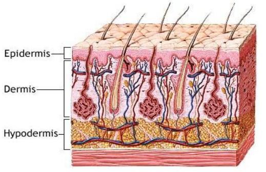
Lipid nanoparticles for topical and transdermal application for alopecia treatment

Approved in public trials by the jury:
President: Alexandre Quintanilha (IBMC/UP)

External examiner: Susana Martins (FF/UP)

Supervisor: Salette Reis (FF/UP)
Lipid nanoparticles for topical and transdermal application for alopecia treatment

Acknowledgments

       This study was carried out in the period between March of 2012 and July of
2012 at the Departamento de Química da Faculdade de Farmácia, under the supervision
of Professor Doctor Salette Reis, at the Universidade do Porto.

        I would like to express my gratitude to my supervisor Prof. Dr. Salette Reis, for
her constant advice, guidance and support, for giving me the opportunity to work on this
interesting topic, and also for the kindness and motivation during all the time.

       I would like to thank Dr. Susana Martins of Departamento de Tecnologia
Farmacêutica da Faculdade de Farmácia da Universidade do Porto, for her help in the
analysis and interpretation of some results.

      I would like to thank Prof. Dr. Amália Jurado and Catarina Grilo from CNC
Coimbra for using the DSC, and also for their help with the DSC experiments.

       I would like to thank Prof. Dr. Marcela Segundo of Departamento de Química da
Faculdade de Farmácia for her help and guidance during HPLC experiments.

       I would like to thank all the people of Departamento de Química da Faculdade
de Farmácia, for the excellent working environment provided, it was a great experience.

        Finally, I want to thank my family and friends, especially my mother for her
support and love, thank you for always being there for me; and my sister that, near or
far, always supported me, thank you for giving me strength in the last days.

                                                                                                          iii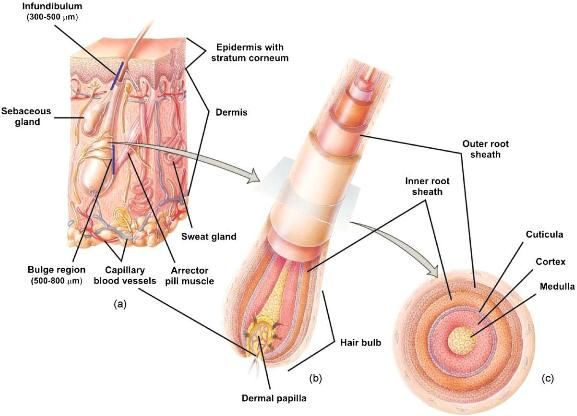
Lipid nanoparticles for topical and transdermal application for alopecia treatment

                                                                               iv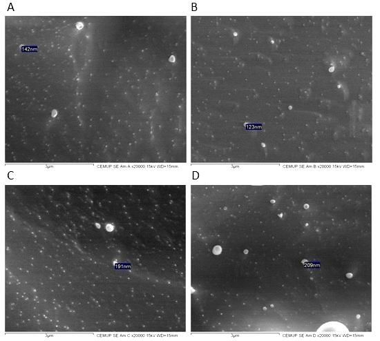
Lipid nanoparticles for topical and transdermal application for alopecia treatment

Abstract

        Alopecia is a dermatological disorder characterized by the reduction of visible
hair. This abnormality affects the hair follicle and causes its shed. There are different
types of alopecia according to its cause, but one of the most common types –
androgenic alopecia – affects up to half of the Caucasian male population by middle
age, and almost all Caucasian men by old age (95%). Besides, it also affects women.
Therefore, and since this disorder also affects psychologically, it is urgent to develop
new drug delivery systems able to improve alopecia therapy. The stratum corneum (SC)
is the main penetration barrier to the access of the majority of the chemicals that come
in contact with the skin. Several nanoparticles (e.g. liposomes and lipid nanoparticles)
have been developed to penetrate the SC. These nanoparticles have proved to be
effective in dermal application of cosmetic and pharmaceutical forms, showing
beneficial properties such as: drug delivery and controlled release; increase in skin
penetration and skin hydration, and an excellent tolerance.

        The present work aims to explore the nanotechnology potential in alopecia
therapy, based on the development and optimization of lipid nanocarriers (NLC) that
were characterized and evaluated regarding the shelf stability. It was also assessed the
suitability of these NLC as carriers for dermal and transdermal delivery of anti-alopecia
drugs by the evaluation of several important parameters.

        Anti-alopecia drugs: minoxidil and finasteride were encapsulated in NLC
prepared by ultrasonication method. NLC for all drugs showed mean particle sizes
bellow (some of them around) 200 nm, as desired to achieve the dermis and the hair
follicles, and zeta potential values around -20 and -30 mV, which indicates a good
physical stability. Over a month of storage little variations in these parameters were
observed, which indicates that all nanoformulations are stable in storage. SEM
measurements showed that all NLC exhibit a spherical shape and a smooth surface
independently of their composition. Differential scanning calorimetry (DSC) studies
allowed the determination of phase transition temperatures. A high loading efficiency
(around 90%) was achieved for finasteride, while only nearly 30% was achieved for
minoxidil NLC over a month. Penetration assays through pig ear skin demonstrated that
NLC loaded with minoxidil and finasteride have low levels of penetration.

       In conclusion, the proposed novel formulation present several good
characteristics which indicate that can be an excellent non-invasive therapy for alopecia.

Key words: nanostructured lipid carriers (NLC), alopecia, anti-alopecia therapy,
minoxidil, finasteride

                                                                                                            v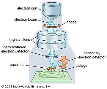
Lipid nanoparticles for topical and transdermal application for alopecia treatment

                                                                               vi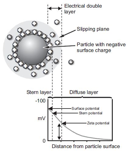
Lipid nanoparticles for topical and transdermal application for alopecia treatment

Contents

Acknowledgments .......................................................................................................... iii
Abstract ........................................................................................................................... v
List of Figures ................................................................................................................ ix
List of Tables .................................................................................................................. xi
1 Aims and Organization of the Dissertation ............................................................... 1
2 Introduction and State of the art ............................................................................... 2
   2.1 Skin ........................................................................................................................ 2
       2.1.1 Histology of the skin ....................................................................................... 2
       2.1.2 Major Skin Functions ...................................................................................... 6
       2.1.3 Skin penetration ............................................................................................... 7
           2.1.3.1 Major routes for drug penetration into the skin with a special focus in the
           transfollicular and transdermal route .................................................................... 9
           2.1.3.2 In vitro evaluation of the release, percutaneous penetration and skin
           retention .............................................................................................................. 13
   2.2 Alopecia and its treatment ................................................................................. 16
       2.2.1 Alopecia aetiology and factors that predispose its appearance ..................... 16
       2.2.2 Drugs used in the treatment of alopecia ........................................................ 18
       2.2.3 Commercial pharmaceutical dosage forms and strategies for alopecia
       treatment ................................................................................................................. 20
   2.3 Nanosystems designed for drug delivery through the transfollicular and
   transdermal route ..................................................................................................... 22
       2.3.1 Lipid nanoparticles: definition and main features ......................................... 23
       2.3.2 Patented lipid nanoparticles applied in dermatology and cosmetic ............... 27
       2.3.3 Nanosystems developed for alopecia treatment: composition and preparation
       methods................................................................................................................... 29
3 Dissertation work plan .............................................................................................. 33
   3.1 Development of lipid nanoparticles (NLC) for alopecia treatment ............... 34
       3.1.1 Optimization of the method of production of lipid nanoparticles ................. 34
           3.1.1.1 Materials and methods ............................................................................ 35
           3.1.1.2 Results and discussion ............................................................................ 41
       3.1.2 Preparation of drug-loaded lipid nanoparticles ............................................. 44
           3.1.2.1 Preparation of minoxidil-loaded lipid nanoparticles .............................. 45

                                                                                                                                  vii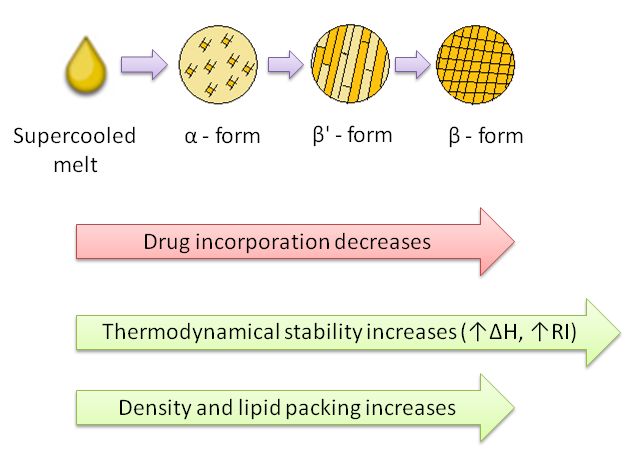
Lipid nanoparticles for topical and transdermal application for alopecia treatment

           3.1.2.2 Preparation of finasteride-loaded lipid nanoparticles ............................. 46
   3.2 Characterization of the lipid nanoparticles ..................................................... 47
       3.2.1 Size ................................................................................................................ 47
           3.2.1.1 Minoxidil ................................................................................................ 47
           3.2.1.2 Finasteride .............................................................................................. 48
       3.2.2 Zeta Potential ................................................................................................. 49
           3.2.2.1 Minoxidil ................................................................................................ 49
           3.2.2.2 Finasteride .............................................................................................. 49
       3.2.3 DSC ............................................................................................................... 51
           3.2.3.1 Characterization of bulk material and drugs........................................... 54
           3.2.3.2 NLC of minoxidil characterization ......................................................... 59
           3.2.3.3 NLC of finasteride characterization ....................................................... 60
           3.2.3.4 Comparison with DLS ............................................................................ 62
       3.2.4 Morphology ................................................................................................... 64
           3.2.4.1 Results and discussion ............................................................................ 65
   3.3 Loading efficiency ............................................................................................... 66
       3.3.1 Minoxidil ....................................................................................................... 66
       3.3.2 Finasteride ..................................................................................................... 68
   3.4 Evaluation of lipid nanoparticles stability ....................................................... 70
       3.4.1 Size ................................................................................................................ 70
           3.4.1.1 Minoxidil ................................................................................................ 70
           3.4.1.2 Finasteride .............................................................................................. 71
       3.4.2 Zeta Potential ................................................................................................. 73
           3.4.2.1 Minoxidil ................................................................................................ 73
           3.4.2.2 Finasteride .............................................................................................. 73
   3.5 Penetration assays .............................................................................................. 75
       3.5.1 Methods ......................................................................................................... 76
       3.5.2 Penetration of minoxidil ................................................................................ 77
       3.5.3 Penetration of finasteride ............................................................................... 78
4 Conclusions and future perspectives ....................................................................... 80
References...................................................................................................................... 82
Appendix I ..................................................................................................................... 87

                                                                                                                                viii
Lipid nanoparticles for topical and transdermal application for alopecia treatment
List of Figures
Figure 1: Schematic cross-sectional representation of skin7. .......................................... 3
Figure 2: Diagram of the (a) structure of the skin and pilosebaceous unit, (b) structure
of the hair follicle, and (c) cross-section of the hair8........................................................ 4
Figure 3: Growth cycle of hair follicle12. ........................................................................ 5
Figure 4: Penetration pathways in the epidermis, through the SC (adapted)5. .............. 10
Figure 5: Chemical structure of finasteride28. ............................................................... 19
Figure 6: Chemical structure of minoxidil28. ................................................................. 19
Figure 7: Models of incorporated compounds in lipid nanoparticles: homogeneous
matrix (A), compound enriched within the shell (B), compound enriched within the core
(C), compounds adhering to the particle surface (D), and clustered compounds adhering
to the particle surface (E)15. ............................................................................................ 26
Figure 8: Chemical structure of cetyl palmitate34. ......................................................... 35
Figure 9: Chemical structure of polysorbate 6037. ......................................................... 36
Figure 10: Schematic representation of lipid nanoparticles preparation method. ......... 37
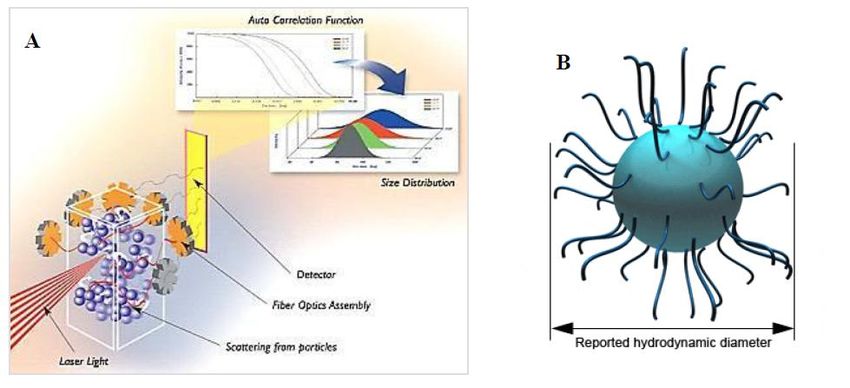
Figure 11: A. Schematic representation of DLS50; B. Hydrodynamic diameter of a
nanoparticle51. ................................................................................................................. 38
Figure 12: Electric potential of a nanoparticle52. ........................................................... 40
Figure 13: Relation between the size and PI for the formulation made – NLC of
minoxidil......................................................................................................................... 41
Figure 14: Relation between the size and PI for different formulations made – NLC of
finasteride – with different times of sonication. ............................................................. 42
Figure 15: Calibration curve of minoxidil, at 230 nm. .................................................. 45
Figure 16: Calibration curve of minoxidil, at 288 nm. .................................................. 45
Figure 17: Calibration curve of finasteride, at 210 nm. ................................................. 46
Figure 18: Mean size distribution and PI with increasing concentrations of minoxidil, of
triplicate samples of minoxidil-loaded NLC. ................................................................. 47
Figure 19: Mean size distribution and PI with increasing concentrations of finasteride,
of triplicate samples of finasteride-loaded NLC............................................................. 48
Figure 20: Mean zeta potential distribution and SD with increasing concentrations of
minoxidil, of triplicate samples of minoxidil-loaded NLC. ........................................... 49
Figure 21: Mean zeta potential distribution and SD with increasing concentrations of
finasteride, of triplicate samples of finasteride-loaded NLC. ......................................... 50
Figure 22: Schematic representation of the polymorphic forms of the lipids in lipid
nanoparticles. Arrows indicate the influence of the polymorphic transitions in: the drug
incorporation; thermodynamic stability and lipid packing. ............................................ 52
Figure 23: DSC thermogram showing the phase transitions to the different polymorphic
forms. .............................................................................................................................. 53
Figure 24: Schematic representation of DSC59.............................................................. 54
Figure 25: DSC melting curve of cetyl palmitate bulk material (second heating). ....... 55
Figure 26: DSC melting curves of bulk mixtures of Cetyl palmitate, Oleic acid and
Polysorbate 60 (Mixture B); and this mixture with minoxidil. ...................................... 56
ixLipid nanoparticles for topical and transdermal application for alopecia treatment
Figure 27: DSC melting curve of Precirol ATO 5 bulk material (second heating). ...... 57
Figure 28: DSC melting curves of bulk mixtures of Precirol ATO 5, Miglyol 812 and
Polysorbate 60 (Mixture A); and this mixture with finasteride. ..................................... 58
Figure 29: DSC melting curves of NLC placebo and NLC loaded with minoxidil. ..... 59
Figure 30: DSC melting curves of NLC placebo and NLC loaded with finasteride. .... 61
Figure 31: Determination of phase transition temperature of NLC of finasteride. ....... 63
Figure 32: Schematic representation of SEM69. ............................................................ 64
Figure 33: SEM images of NLC placebo finasteride (A), 2% finasteride (B), placebo
minoxidil (C) and 2% minoxidil (D). The scale indicated below the images is of 3μm.
Size indicated in image A is 142 nm; in B is 123 nm; in C is 191 nm; in D is 209 nm. 65
Figure 34: Minoxidil loading efficiency into different NLC produced (NLC 2%
minoxidil and NLC 3% minoxidil) with time. ............................................................... 67
Figure 35: Minoxidil loaded mass into different NLC produced (NLC 2% minoxidil
and NLC 3% minoxidil) with time. ................................................................................ 67
Figure 36: Finasteride loading efficiency into different NLC produced (NLC 0,8%
finasteride and NLC 2% finasteride) with time. ............................................................. 68
Figure 37: Finasteride loaded mass into different NLC produced (NLC 0,8% finasteride
and NLC 2% finasteride) with time................................................................................ 69
Figure 38: Mean size distribution and PI of triplicate samples of minoxidil-loaded NLC
with different percentages, measured at different times. ................................................ 71
Figure 39: Mean size distribution and PI of triplicate samples of finasteride-loaded
NLC with different percentages, measured at different times. ....................................... 72
Figure 40: Mean zeta potential distribution of triplicate samples of minoxidil-loaded
NLC with different percentages, at different times. ....................................................... 73
Figure 41: Mean zeta potential distribution of triplicate samples of finasteride-loaded
NLC with different percentages, at different times. ....................................................... 74
Figure 42: Vertical Franz diffusion cell used for penetration assays. ........................... 76
Figure 43: Penetration of minoxidil through the skin over 24 h. The two lines
correspond to duplicate experiments. ............................................................................. 78
xLipid nanoparticles for topical and transdermal application for alopecia treatment
List of Tables
Table 1: Cosmetic products containing NLC currently on the market31. ...................... 28
Table 2: SLN suspension composition13. ....................................................................... 30
Table 3: Commercial solutions compositions (g/100 mL)13. ......................................... 31
Table 4: Drugs solubility in different lipids. The symbol means that the drug is
soluble in the lipid, in that percentage. The symbol means that is not soluble. The -
means that it was not done. ............................................................................................. 34
Table 5: Composition of NLC placebo to produce 5 g of formulation of minoxidil. .... 37
Table 6: Composition of NLC placebo to produce 5 g of formulation of finasteride. .. 37
Table 7: Time of sonication tested for minoxidil formulation (NLC placebo).............. 38
Table 8: Times of sonication for different formulations (NLC placebo of finasteride). 38
Table 9: Composition of the different NLC formulations loaded with minoxidil. ........ 44
Table 10: Composition of the different NLC formulations loaded with finasteride. .... 44
Table 11: Melting point (peak maximum), onset and enthalpy of cetyl palmitate
obtained from the second heating in the DSC analysis. ................................................. 55
Table 12: Melting point (peak maximum), onset and enthalpy of bulk mixture of Cetyl
palmitate, Oleic acid and Polysorbate 60; and this mixture with minoxidil. ................. 56
Table 13: Melting point (peak maximum) and enthalpy of Precirol ATO 5 obtained
from the second heating in the DSC analysis. ................................................................ 57
Table 14: Melting point (peak maximum), onset and enthalpy of bulk mixture of
Precirol ATO 5, Miglyol 812 and Polysorbate 60; and this mixture with finasteride. ... 58
Table 15: Melting point (peak maximum), onset, enthalpy and crystallinity index (RI)
of NLC minoxidil formulations. ..................................................................................... 60
Table 16: Melting point (peak maximum), onset, enthalpy and crystallinity index (RI)
of NLC finasteride formulations. ................................................................................... 62
Table 17: Penetration of finasteride through the skin over 24 h. The two columns
correspond to duplicate experiments. ............................................................................. 79
Table 18: All nanoformulations done with minoxidil entrapped – to select the best
according to its characteristics. Marked in red are the selected ones. ............................ 87
xiLipid nanoparticles for topical and transdermal application for alopecia treatment
xiiLipid nanoparticles for topical and transdermal application for alopecia treatment
1 Aims and Organization of the Dissertation
Lipid nanoparticles have features that make them interest for the pharmaceutical
and cosmetic technology research groups worldwide. This is the main reason why LNs
importance is increasing. Currently, solid lipid nanoparticles (SLN) and nanostructured
lipid carriers (NLC) have been already investigated as carrier systems for many
applications.
This dissertation aims the development of an innovative nanoformulation based
on the combination of two anti-alopecia drugs carried in lipid nanoparticles (NLC). The
formulation developed should be able to deliver the drugs specifically to hair follicles
where they may act. Given the ease of application, and the probably synergic effect
obtained by the combination of two of the most widely used drugs, the formulation
developed is promising.
The first part of this dissertation is organized as follows: in the first sections the
skin histology and physiology are described, as well as the different types of drug
penetration into the skin; also the pharmaceuticals forms and drugs used to treat
alopecia are presented, which are important to understand alopecia aetiology and its
treatment. This introduction of skin and alopecia led to literature review with respect to
the uses of lipid nanoparticles as dermal drug delivery systems. In addition,
characteristics of nanosystems in general and the features of LNs in particular, as topical
delivery systems, were mentioned.
Secondly, it is presented the work developed aiming the production of such
nanosystems with the ability to incorporate and deliver the anti-alopecia drugs into the
dermis and hair follicles. These systems were characterized and investigated with regard
to physical (size, charge, morphology and polymorphic modifications), chemical (drug
loading) properties. The stability of NLC over a month was also assessed. Finally, the
penetration profiles of drugs were also analyzed.
1Lipid nanoparticles for topical and transdermal application for alopecia treatment
2 Introduction and State of the art
2.1 Skin
Skin, the major human organ, is a heterogeneous membrane: lipophilic on its
surface and hydrophilic in its deeper layers1. Skin is very complex due to its histology
and this is translated in multifaceted functions. Those skin roles have an utmost
importance to human survival and welfare. Regarding this and also taking into account
the need to penetrate skin for therapeutic purposes, this field deserves careful attention
in order to progress towards efficient nanomaterials capable of reach and correctly act
on their target while maintaining skin integrity. Consequently, drug penetration routes
into the skin as well as evaluation of drug release, percutaneous penetration and skin
retention are important aspects that need to be studied.
2.1.1 Histology of the skin
Skin has three different layers – epidermis, dermis and hypodermis (Figure 1) –
that have distinct composition and functions. Usually, epidermis and dermis are
considered more important from a penetration perspective2. In epidermis, keratinocytes
are the most abundant cell phenotype and are organized in five different strata (stratum
basale (in contact with dermis), stratum spinosum, stratum granulosum, stratum
lucidum and stratum corneum (SC, in contact with the external environment)), reason
why epidermis is histologically classified as a stratified epithelial layer2. From the
stratum basale to the SC, keratinocytes undergo a progressive modification, varying in
shape and cytoplasm content, that enables epidermis to keep healthy and defensively
competent since this process promotes a continuous regeneration and renewal of its
components. According to this, epidermis could be distinguished in viable epidermis
(VE) and SC where keratinocytes are completely differentiated into anucleated cells
filled with keratin and keratin cross-linked with filaggrin (histidine rich protein,
responsible for filament compaction, that binds keratin fibers3) – called corneocytes2,4.
Those corneocytes (diameter 30 to 50 μm and thickness 0,2-0,8 μm) are disposed along
15 to 20 layers that consist SC and are spaced from each other by a gap of
approximately 75 nm under air-dried conditions. Thus, corneocytes delimit a tortuous
path that is filled with a complex matrix of organized lipid bilayers1,2,5. Summarizing,
SC is the outermost layer of the skin and is comprised of a 10-20 μm thick matrix of
dehydrated and dead corneocytes that are embedded in highly ordered lipid layers,
which serve as a cover5,6. On the other hand, the VE is approximately 100-150 μm thick
and composed of multiple layers of keratinocytes and several other types of cells. The
hypodermis or subcutaneous tissue resides below the dermis and is composed of loose
textured, white, fibrous connective tissue in which fat and elastic fibres are combined6.
2Lipid nanoparticles for topical and transdermal application for alopecia treatment
Regarding hydrophobicity/lipophilicity, SC is lipophilic and contains ≈ 13% of
water, while VE is significantly hydrophilic (> 50%). In the dermis the water content
reaches 70%, favouring hydrophilic drug uptake1.
Figure 1: Schematic cross-sectional representation of skin7.
SC hydration is important to prevent SC mechanical failure and cracking, and
assures preservation of SC enzyme activities that supports the terminal differentiation of
SC layers and a correct self-assembly of intercorneocyte lipids. Therefore, its regulation
(that depends on the proteolysis of corneocyte content, which produces a mixture of
osmotically active amino acids that are able to sequestrate water and to act as
moisturizing factors) is essential as well2. SC microstructure is fundamental to study
skin penetration since it involves hydrophobicity concepts as it refers to supramolecular
organization of intercorneocyte lipids that are assembled in parallel head-head tail-tail
repeating bilayers. As a consequence, repeating hydrophilic and lipophilic regions exists
within those bilayers. Regarding SC spatial organization, it depends on the type and
amount of intercorneocyte lipids2. The most common intercellular lipids (nearly 80%
are apolar) may be classified in four categories: cholesterol and its derivatives,
ceramides, free fatty acids, and triglycerides2,5. Their fluidity is lower on lipid heads in
comparison with tails, and tails region is also known as the transdermal intercellular
apolar (or lipidic) route of skin absorption. Nevertheless, skin may also contain aqueous
pores that contribute to the transdermal intercellular polar (or hydrophilic) route of skin
absorption2 (see section 2.1.3 Skin penetration). Furthermore, SC chemical composition
determines that SC, as a whole, is generally referred as a lipophilic stratum, in contrast
to the higher amount of water in VE which contributes to its hydrophilicity.
Consequently, hydrophobicity change in epidermis is not homogeneous – hydrophilic-
lipophilic gradient. Proving that, it is well known that moderate oil in water (o/w)
partition coefficient of a penetrating molecule is one of the key parameters for a
successful transcutaneous absorption2.
Other important skin components are sweat glands and hair follicles. These
glands are coiled tubular that extend from SC to dermis or hypodermis (2-5 mm in
length) and are involved in thermoregulation and excretion of acids and body wastes.
Hair follicles consist of a hair infundibulum (the part between the skin surface and the
point of the sebaceous gland duct opening to the hair canal) generally supporting one
hair and serving as a route to expel the product of associated sebaceous gland
3Lipid nanoparticles for topical and transdermal application for alopecia treatment
(sebum)2,8. Both components create openings on skin surface, providing breaches that
may be used as potential ports of ingress.
The hair follicle, hair shaft, adjoining arrector pili muscle and associated
sebaceous gland(s) together form an integrated structure recognized as the
pilosebaceous unit (PSU) – a skin appendage (Figure 2). This complex 3-D structure
within the skin possesses a unique biochemistry, metabolism and immunology8,9,10. The
hair shaft is composed of the medulla, the cortex with melanosomes, and the cuticula8.
Figure 2: Diagram of the (a) structure of the skin and pilosebaceous unit, (b) structure of the hair follicle,
and (c) cross-section of the hair8.
The hair follicle, apart from contain many cell types that produce highly
specialized proteins, consists of a hair bulb and shaft enveloped in an inner root sheath,
an outer root sheath, and an outermost acellular basement membrane. The outer root
sheath is a keratinized layer continuous with the epidermis9,10. Hair follicles are also
connected with a network of blood capillaries8. A key function of the PSU involves the
synthesis and release of sebum – a fungistatic and bacteriostatic mixture of short chain
fatty acids produced by sebaceous glands8,10. Those glands (androgen-responsive
glands), connected to the hair follicle by ducts, are outgrowths of epithelial cells9.
There are two types of human hairs - terminal hairs and vellus hairs. Terminal
hairs are those androgen-independent (eyebrows, lashes) and hormone-dependent
(scalp, beard, chest, axilla, pubic region). These hairs are long (> 2 cm), thick (> 0,03
mm), pigmented and usually contain a medullary cavity. These hairs also extend more
than 3 mm into the hypodermis. In contrast, the rest of the body in adults is covered
4Lipid nanoparticles for topical and transdermal application for alopecia treatment
with vellus hairs which are generally short (< 2 cm), thin (< 0,03 mm), unpigmented
and typically extend just 1 mm into the dermis8,10. Skin follicular density varies between
different anatomic regions. On the face and scalp, there are 500-1000 PSU per square
centimeter with each follicular opening exhibiting a diameter of some 50-100 μm. The
area of these orifices may represent 10% of the total surface area of the face and scalp.
In other parts of the body, the follicular openings constitute only about 0,1% of the total
skin area. The sole of the foot, the palm of the hand and the lips do not have hair
follicles8,9,10. Hair shaft diameters have relatively little variation (16-42 μm)8.
In human, the hair grows cyclically (a continuous cycle) with alternating periods
of activity and rest (Figure 3). The cycle has an active growth stage with a rapid hair
matrix cells division and migration upward to form the hair shaft (anagen), with parallel
melanin production11. This phase is followed by a regressive stage with cessation of
mitosis and the lower portion of the hair follicle is largely resorbed through apoptosis
(catagen). Catagen phase conclusion gives rise to a resting follicle (telogen) and finally
a lag phase – resulting telogen hair is a quiescent tissue – during which hair shaft is shed
(exogen). The cycle then returns to anagen and the lower follicle is reformed. Recently
another stage has been described – kenogen – as the lag time between exogen and new
anagen. In human scalp hairs the cycle lasts about 4 years, and nearly 90% of the time is
spent in anagen (catagen and telogen are relatively short)8,9,10,12. This cycle is controlled
by locally active inhibitors. The duration of each growth phase as well as the percentage
of hair in each growth phase differs between vellus and terminal hairs. In addition,
seasonal variations in hair growth are modulated by the endocrine system10. Moreover,
the rate of scalp hair shaft elongation is between 0,3 and 0,4 mm per day8.
Figure 3: Growth cycle of hair follicle12.
5Lipid nanoparticles for topical and transdermal application for alopecia treatment
2.1.2 Major Skin Functions
The skin surface is an ecosystem in equilibrium with precise characteristics.
Skin has many different functions, but the main one is to defend the body from the
external environment – skin provides a natural barrier against exogenous aggressions
and particles penetration13. Therefore, skin confers the capability to prevent the entrance
of chemical and biological agents due to SC structure and composition2. Dermis, which
is a more inner layer than epidermis, has capillary anastomoses capable to carry
nutrients and oxygen to the epidermis, and also clear the dermis from cell metabolic
products and penetrated foreign agents. Otherwise, the outer skin layer epidermis
(majority the SC) acts as a defensive layer and, therefore, influence the ingress and
diffusion of foreign nanometric agents2. Due to that, SC is commonly referred as the
main skin barrier, function that results from the cooperation and interactions between
SC macro and microstructure, supramolecular organization of SC lipidic matrix, and SC
whole composition2. The SC constitutes only 10% of the entire skin but contributes to
over 80% of the cutaneous barrier function5. The SC presents a barrier to most low
molecular weight compounds and prevents intact nanoparticle ingress14. Therefore, as
SC cells, corneocytes represent the first macroscopic physical barrier against the
penetration of foreign agents, also assuring impact resistance. Although, besides those
defensive functions, foreign agents (like therapeutic nanoparticles and microparticles)
can be delivered in diseased skin and to hair follicles openings13.
Besides this, skin also minimizes the effects of UV and IR radiations by
absorbing them (UV), and dissipating associated heat through the regulation of blood
flux, perspiration, and/or sweating (IR). In addition, cellular and molecular barriers are
also important and all molecular or biological agents that manage to overcome the skin
have to face them2. Like that, SC acts as a protective barrier thanks to key enzymatic
reactions, bacterial flora, immune signalling compounds, and preservation of the acidic
pH5. On the one hand, key enzymatic reactions play important roles since they are
necessary for the desquamation, a protective mechanism that contributes to the
elimination of both microorganisms and cells. On the other hand, the resident bacterial
flora in the skin constitutes a complex ecosystem that is very important in skin defence
against potentially pathogenic organisms. Furthermore, the SC is a biosensor that
regulates the responses of the epidermis – SC senses the level of cytokines and growth
factors, which are keys to the inflammatory reaction. Finally, pH in SC is acidic (ranges
between 5 and 6) which is crucial to integrity and cohesion of the SC, and to enzymatic
activities. To maintain the acidic pH, endogenous (such as secretion of sebum) and/or
exogenous (originated outside the epidermis) variables cooperate5. Therefore, skin
protective functions are against external mechanical, chemical, microbial and physical
influences6.
SC structure contributes to its homeostatic function preventing the loss of water
from the epidermis. The existence of a water-resistant skin keeping water in and
exogenous substances out is a key aspect for living on Earth. As the main function of
6Lipid nanoparticles for topical and transdermal application for alopecia treatment
the skin is to protect the inner body and this one is rich in water, the protective function
is also against the dry environment1,5. Therefore, SC allows the maintenance of the body
hydration which is needed to maintain flexibility, among other things. Moreover, water
permeability is a reason why lipid regions are considered to be great barriers of the
skin1,5.
Additionally, the SC provides protection from the outside environment through
its antioxidants – which are capable of protecting against lipid peroxidation and allow
the stabilization of lipid bilayers5. SC is also responsible for the maintenance of body
temperature since it is in charge of isolate the organism from environment, and this
barrier acts also as a reservoir for topically applied substances15.
2.1.3 Skin penetration
The treatment of a local cutaneous dermatologic or pathologic condition could
be made using different means, but there is no doubt that directly applying a
pharmaceutical formulation is easy, convenient, and generally well accepted by patients.
Regarding these reasons, skin has been extensively used for the cutaneous and
percutaneous delivery of therapeutic drugs2. Also because of large surface area and easy
accessibility, skin delivery has potential on application in drug delivery6. Besides this,
local skin targeting is of interest for the pharmaceutical and the cosmetic industry9. A
topically applied substance has distinct possibilities to penetrate into the skin since this
delivery involves, in addition to the transdermal route (through SC), the transfollicular
route through pilosebaceous unit – comprising of hair follicle and sebaceous glands9.
There are many factors that can affect the cutaneous absorption of a molecule
(and consequently a nanoparticle or nanomaterial too), and those may be distinguished
in three different classes: (i) location and skin conditions at the application site; (ii)
physicochemical properties of the penetrating molecule; and (iii) physicochemical
properties of the vehicle dispersing the penetrating molecule2.
The skin integrity variation (by dermatological and other pathological
conditions, damage and trauma...), dimensions of orifices and aqueous pores, and
density of appendages are conditions affecting the absorption of any agent (i). Also
thickness of SC varies in different body regions and pilosebaceous units are unequally
distributed in the body (their density and diameter of hair follicle orifices vary per each
body location). Further, age, skin type and sex hormones influence skin permeability.
Other factors like SC hydrophilic-lipophilic gradient, skin temperature and methods of
application (massaging a formulation on the skin could increase the local temperature)
could favour the penetration of particulate formulations. Therefore, depending on body
site, skin absorption through the transdermal or transfollicular route may be favoured or
limited. Thus, penetration of nanometric agents through hair follicles (transfollicular
route) should be anatomically favoured in the forehead, where they represent 1,28% of
7Lipid nanoparticles for topical and transdermal application for alopecia treatment
skin surface. As well, transdermal route should be favoured in those body locations
where SC is less thick, or has been exfoliated or abraded with cosmetics, sand, or
similar products2,5.
Several properties of the molecule influence its permeation (ii). Some of them
are: solubility and dissolved amount of the penetrating molecule in its vehicle (the
highest solubility of a molecule in its vehicle is correlated with the highest
thermodynamic activity of that formulation, and consequently with the greatest
probability to transdermally deliver that molecule); pKa of the penetrating molecule and
pH of vehicle (only the unionized fraction of a penetrating molecule will be transported
to VE); MW of a penetrating agent (that should be less than 500 Da to significantly
permeate skin); and the diffusion coefficient (D) of the penetrating molecule in its
vehicle and in the skin. Also very important is the o/w partition coefficient of the
penetrating molecule since a very lipophilic molecule will easily partition in SC but will
leave it with difficulty, whereas a hydrophilic molecule will suffer poor penetration.
Additionally, there are some less important features that, however, should not be
underestimated as the potential for binding and metabolism. Particle stability is another
important parameter and it includes particle disassembly, rupture, stability against
chemical reactions (which is capable to modify its superficial properties), and ability to
form micelles after contact with specific structures/components of the skin or to be de-
coated. Therefore, particle stability could influence numerous other properties. Probably
the most significant parameters of the penetrating nanoparticle are the dimension (size),
shape and superficial properties (e.g. charge, polarity) since they influence several
aspects such as the ability to enter the skin, the selection of the penetration route, the
depth of penetration, the coefficient of diffusion in the dispersing vehicle and in the
skin, and the potential to establish interactions with skin components (e.g. charge,
dipole, hydrophobic, and/or hydrogen-bond interactions). “Shape deformability” and
“dimension versus orientation” should be pondered since rigid or deformable shapes
and their orientations may influence nanoparticle/nanomaterial passage across a
structure with a defined porosity. Superficial charges (frequently it is the coating which
is charged), which may interact differently with various components of skin and routes
of penetration, usually prevent nanoparticle/nanomaterial aggregation2,5.
When a formulation is applied on the skin, all its ingredients are too. Thus, all of
them are subjected to skin absorption, although to different extents (iii). As a result, the
type of formulation where agents are dispersed (its physicochemical properties),
possible synergisms/interactions between vehicle-agent-skin, and the application
method will definitely affect the absorption outcomes. It is necessary to take into
account that many different things could happen – water and volatile compounds will
evaporate, non volatile ingredients will be absorbed to different extents, and skin, sweat,
and/or sebum components will diffuse in the applied dose. Therefore, composition and
penetration ability of formulation components (besides the agent) should not be
underestimated – potential enhancing effects could be provided by the ingredients of
applied formulations2.
8Lipid nanoparticles for topical and transdermal application for alopecia treatment
Several techniques and formulations have been developed to successfully
overcome skin barriers2. When penetrating agents are molecules, scientists indicate
small (Lipid nanoparticles for topical and transdermal application for alopecia treatment
Figure 4: Penetration pathways in the epidermis, through the SC (adapted)5.
Although it is generally believed that the intercellular route may dominate
during steady state penetration of compounds, it has been argued that the skin
appendages (hair follicles, pilosebaceous and sweat glands) may offer an alternative
pathway for a diffusing molecule, more significant than previously believed10,17.
Usually, substances smaller than 500 Da, with sufficient oil solubility and high
partition coefficient can be absorbed into the skin. In contrast, larger molecules
(molecular weight higher than 500 Da) are not able to pass the cutaneous barrier5,6.
The worldwide transdermal market, in 2000, was worth US$2 billion and
represented the most successful non-oral systemic drug delivery system. Nevertheless,
transdermal drug delivery is not suited to all drugs and is not justified for all therapies;
however there remains a large number of drugs for which it is desirable but presently
unfeasible4.
The transdermal route of administration avoids hepatic first-pass metabolism and
allows sustained drug release into the systemic circulation1. This route has other
advantages as the skin presents a relatively large and readily accessible surface area (1-2
m2) for absorption4.
Since skin acts as a natural and protective barrier, transdermal drug delivery is a
challenging task for the pharmaceutical scientists. Therefore, in order to increase
therapeutic molecules permeability into/across the skin and expand the range of
transdermally delivered drugs, several methods/strategies have been examined. A drug
penetrates into the skin by a passive diffusion mechanism (according to Fick’s Laws),
depending on its molar mass and physicochemical properties1,4. The simplest
approaches to optimize transdermal drug delivery are passive and involve formulation
manipulation. One of the most important and extensively investigated strategies is the
introduction of reversible structural alterations within the skin (lipidic matrix) by
addition of chemical enhancers4,6. Another main aspect to reduce skin barrier capability
is hydration – using water is the safest method for increasing skin penetration1,4.
10Lipid nanoparticles for topical and transdermal application for alopecia treatment
Moreover, other way to do so is changing lipophilicity of the drug/formulation. Also a
saturated formulation of the drug will provide the maximal flux4.
If molecules are large (molar mass bigger than 500 Da), active mechanisms have
been developed to overcome the barrier and respond to this challenge1,4. Microneedles,
jet injectors, iontophoresis, ultrasound (sonophoresis), electroporation, photomechanical
waves, magnetophoresis, laser radiation and skin abrasions are some of the main
mechanical, physical and active transport techniques available to enhance skin
penetration of various drugs6. In electroporation (electropermeabilization), high voltage
(100-1000 V) electrical impulses are applied during short time intervals (micro- to
millisecond) to create temporary pores on the skin. Such pores provide drug penetration
routes1,4,18. Sonophoresis uses low frequency ultrasonic energy to disrupt lipid packing
in the SC creating aqueous pores, which improve drug delivery1,4. Iontophoresis is an
electrically assisted method where the drug has to be used in an ionic form – it drives
charged species into tissue. By applying an small external electrical field to the skin the
active ingredient will be accelerated and as a result of electromigration and electro-
osmotic forces it will be transported into the skin layers1,4,18. Drug penetration through
damaged regions of the skin depends on the polarity, valence, mobility of the ions and
formulation components4,18.
After the drug reaches into the skin membrane, it must be sufficiently mobile to
diffuse across the SC, which is a complicated diffusion process due to the viscosity of
the lipid matrix. This diffusion is extremely sensitive on molecular size4.
The transfollicular route – generally associated with the lipidic pathway because
of the lipophilicity of sebum that is released by sebaceous glands in the hair follicles –
has been largely ignored because hair follicles constitute only 0,1% of the total skin9.
However, the hair follicle has great potential for skin treatment due to its deep extension
into the dermis and so it could provide much deeper penetration and absorption of
compounds under the skin than seen with the transdermal route. Consequently, there are
doubts about transfollicular route and if it plays an important role on the skin absorption
of nanoparticles. Scientists have distinct opinions about transfollicular route
importance2,10. Still, for certain drug delivery systems, hair follicles are privileged
penetration pathways. They enter faster into them than through the SC, and then offer
the possibility to create high local concentrations of the active compounds within the
follicular duct9. It has been shown that smaller particles accumulate better and deeper in
the hair follicle than larger ones2. Furthermore, it was calculated that the storage time of
the particle based drug delivery systems in the hair follicles was 10 days compared to
short-term storage time in the SC – hair follicles are long term reservoirs6,8.
In some skin diseases, delivery to sweat glands or to the pilosebaceous unit is
essential for the effectiveness of the drug. As an example, there are many diseases of the
hair cycle (androgenic alopecia, hair colour loss, alopecia areata ...) that need effective
therapeutics which would be more successful if they could be specifically targeted to
the hair follicle10,11. Within the hair follicles, different target sites of interest have been
11Lipid nanoparticles for topical and transdermal application for alopecia treatment
defined – the sebaceous gland is of particular interest and is associated with the
aetiology of androgenic alopecia; the midfollicle bulge since it has stem cells
responsible for follicle reconstitution; and follicular papilla and hair matrix cells, as
both have an important role in controlling hair growth (number of matrix cells correlates
with the size of the new hair, while melanin concentrations in the matrix cells modulate
hair pigmentation)8,10,13. In addition, transfollicular route enables the delivery to specific
sites of the hair follicle, the increasing of drug concentration within the pilosebaceous
units, the possibility to reduce the applied dose of a drug and/or the frequency of its
administration, the reduction of hepatic metabolism and drug systemic toxicity. Due to
those reasons, transfollicular administration has been increasingly recognised as
potentially significant in the percutaneous drug delivery paradigm, and therefore is
having more therapeutic interest10. Thus, recent studies have focused on the hair follicle
as a potential pathway for both localized and systemic drug delivery9,10. Therefore,
targeted drug delivery to the pilosebaceous compartment may have several therapeutic
applications for treating numerous hair follicle associated disease states. Those
applications include targeting drugs to the bulge region for gene delivery to facilitate
long-term gene correction of congenital hair disorders or genetic skin disorders; PSU
utilization as reservoirs for localized therapy or as a transport pathway for systemic drug
delivery; and target sebaceous glands to the treatment of acne and androgenetic
alopecia8.
Nevertheless, access to transfollicular route for drug penetration can have some
architectural and physicochemical constraints like the membrane that surrounds the
entire follicle and the keratinous layers of the inner and outer root sheaths that may
physically restrict passage of molecules deep within the follicle. Other barriers include
size selectivity of the follicular openings, the sebum flow into the hair follicle
(assuming that sebum flow has an upward movement) which may impede drug
transport, and the hair growth cycle that also appears to influence pilosebaceous drug
delivery9,10.
Numerous studies have suggested that the enhancement of follicular delivery
may be done by applying certain approaches – such as the use of optimised vehicles
(use a volatile organic solvent like ethanol – lipophilic – in order to dissolve sebum
from the follicular canal); decreasing particle size; or massage following application8,10.
Furthermore, pre-treatment of the skin with cyanoacrylate skin surface stripping (CSSS)
removes the superficial part of the SC and sebum, facilitating penetration8.
A different method has been introduced – skin sandwich model – to estimate the
importance of the transfollicular route8,10,18.
In summary, transfollicular penetration could be used by those agents whose
dimensions are below follicular openings (10-210 μm) and able to disperse themselves
into sweat or sebum (lipophilic). Penetrating agents could easily enter the VE or blood
stream depending on its penetration depth2. In contrast to transdermal route,
transfollicular is favourable for high-molecular weight substances8.
12Lipid nanoparticles for topical and transdermal application for alopecia treatment
2.1.3.2 In vitro evaluation of the release, percutaneous
penetration and skin retention
Deliver an active compound transdermally implies the reduction of barrier
function of skin and the enhancement of permeation of the active compound through
skin. Consequently, several approaches (select an adequate vehicle, chemical
modification of an active gradient, among others) were used to enhance the skin
permeation of the active ingredient19.
As formulations remain stable (a characteristic also important to assess), the
evaluation of the drug release rates, percutaneous absorption/penetration and skin
retention became a crucial step to further validate the possible usefulness of that drug in
those formulations20. In order to assess nanosystems’ drug release, penetration and
retention in skin, in vivo studies could be made. However, when it is not possible to do
them, the equipment and material are not available, or even before their practice, in
vitro tests could be a possibility. Some of those tests are here described.
For this kind of studies, the first step is done in order to identify a suitable
receptor medium for in vitro experiments (to create in vitro conditions which can
replicate the in vivo ones). For this, the saturation solubility of the drug in different
media (solvents) is analyzed (drug content is analysed by HPLC) and the selected
medium should be the one with the highest solubility20.
The release of the drug from topical preparations depends on the
physicochemical properties of the vehicle and the drug employed16. In many studies, in
vitro drug release rates from its formulation is measured through synthetic membranes
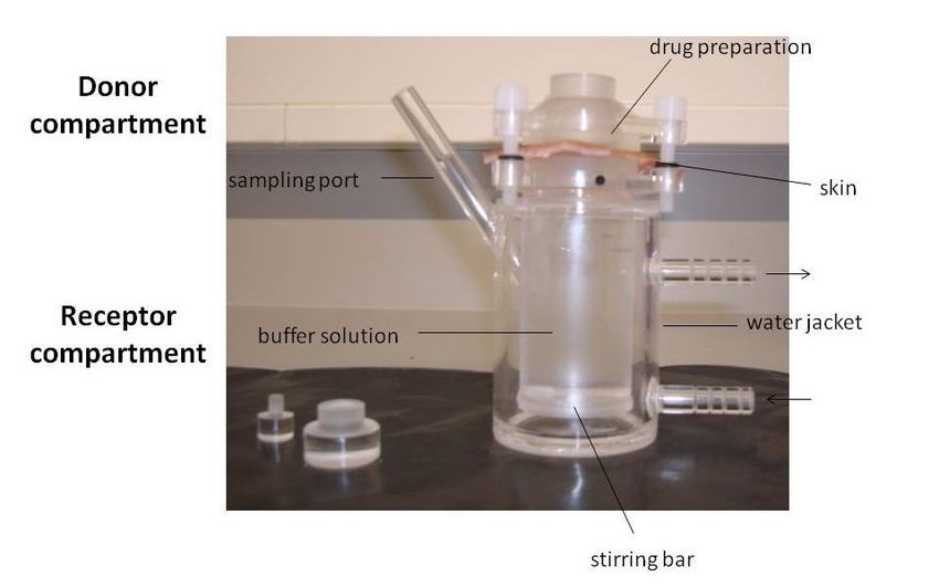
(of nitrocellulose, cellophane, ...) using vertical Franz diffusion cells setup16,20,21.
Synthetic membrane is usually sandwiched between the upper donor compartment and
the lower receptor compartment of Franz diffusion cells; a quantity (≈ 0,5 to 1 g) of the
formulation containing the drug is placed on the surface of the membrane in the donor
compartment, while the receptor compartment is filled with the previous selected
receptor medium, which is in contact with the membrane. During the experiment, the
receptor solution is continuously stirred (at ≈ 100 to 300 rpm) and kept at a proper
temperature (37 ± 1 °C). Samples of this receptor medium are taken from the receptor
compartment, at predetermined time intervals, and therefore the amount of released
drug is analysed by HPLC and its diffusion coefficient (D) is calculated. It is important
to note that D reflects the facility by which molecules move through the membrane and
through the formulation. All measurements are typically performed, at least, in triplicate
and formulations without the drug are used as control16,20,21.
The drug released from nanocarriers (as solid lipid nanoparticles) into the
receptor medium at room temperature could be measured by weighing ≈ 0,5 g samples
of nanocarriers into test tubes containing 10 mL receptor medium. Those tubes are
13You can also read