Synthesis and design of Ag-Fe bimetallic nanoparticles as antimicrobial synergistic combination therapies against clinically relevant pathogens
←
→
Page content transcription
If your browser does not render page correctly, please read the page content below
www.nature.com/scientificreports
OPEN Synthesis and design
of Ag–Fe bimetallic nanoparticles
as antimicrobial synergistic
combination therapies
against clinically relevant
pathogens
A. L. Padilla‑Cruz1,2, J. A. Garza‑Cervantes1,2, X. G. Vasto‑Anzaldo1,2, Gerardo García‑Rivas3,4,
A. León‑Buitimea1,2 & J. R. Morones‑Ramírez1,2*
The inappropriate use of antibiotics and the inadequate control of infections have led to the
emergence of drug-resistant strains. In recent years, metallo-pharmaceutics and metallic
nanoparticles have been proposed as potential alternative antimicrobials due to their broad-spectrum
antimicrobial properties. Moreover, recent findings have shown that combinations of transition metal
compounds can exhibit synergistic antimicrobial properties. Therefore, the synthesis and design
of bimetallic nanoparticles is a field worth exploring to harness the interactions between groups of
metals and organic complex structures found in different microbial targets, towards the development
of more efficient combinatorial antimicrobials composed of synergistic metals. In this study, we
present a green synthesis of Ag–Fe bimetallic nanoparticles using an aqueous extract from the leaves
of Gardenia jasminoides. The characterization of the nanoparticles demonstrated that the synthesis
methodology produces homogenously distributed core–shell Ag–Fe structures with spherical shapes
and average diameter sizes of 13 nm (± 6.3 nm). The Ag–Fe bimetallic nanoparticles showed magnetic
and antimicrobial properties; the latter were evaluated against six different, clinically relevant
multi-drug-resistant microbial strains. The Ag–Fe bimetallic nanoparticles exhibited an antimicrobial
(bactericidal) synergistic effect between the two metals composing the bimetallic nanoparticles
compared to the effects of the mono-metallic nanoparticles against yeast and both Gram-positive
and Gram-negative multidrug-resistant bacteria. Our results provide insight towards the design of
bimetallic nanoparticles, synthesized through green chemistry methodologies, to develop synergistic
combinatorial antimicrobials with possible applications in both industrial processes and the treatment
of infections caused by clinically relevant drug-resistant strains.
The fast spread of drug-resistant infectious diseases poses a global health threat1. If the trend continues at the
current speed, by 2050, it will lead to 10 million people dying every year from drug-resistant infections2. Metal
nanoparticles (NPs) have great potential to be utilized as antimicrobial a gents3. Noble metal NPs, such as silver
(Ag) and gold (Au), have been shown to exhibit strong and sustainable antibacterial action against a wide array
of microorganisms; therefore, they have been utilized in medical devices, food preservatives, dental resin com-
posites, cosmetics, medical device coatings, implants, and medical instruments4. Bimetallic NPs have gained
1
Universidad Autónoma de Nuevo León, UANL, Facultad de Ciencias Químicas, Av. Universidad S/N. CD. Universitaria,
San Nicolás de Los Garza, NL 66455, Mexico. 2Centro de Investigación en Biotecnología y Nanotecnología, Facultad
de Ciencias Químicas, Universidad Autónoma de Nuevo León, Parque de Investigación E Innovación Tecnológica,
Km. 10 autopista al Aeropuerto Internacional Mariano Escobedo, Apodaca, Nuevo León 66629, México. 3Cátedra
de Cardiología Y Medicina Vascular, Escuela de Medicina. Tecnologico de Monterrey, Monterrey, Nuevo León,
Mexico. 4Centro de Investigación Biomédica, Hospital Zambrano‑Hellion, Tecnologico de Monterrey, San Pedro
Garza‑García, Nuevo León, Mexico. *email: jose.moronesrmr@uanl.edu.mx
Scientific Reports | (2021) 11:5351 | https://doi.org/10.1038/s41598-021-84768-8 1
Vol.:(0123456789)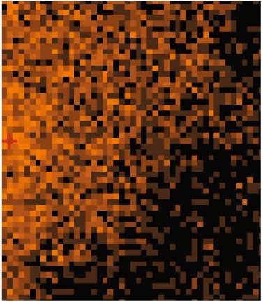
www.nature.com/scientificreports/
specific attention in the last decade due to their optical, electronic, magnetic, and catalytic properties, which, in
most cases, are significantly distinct from their monometallic c ounterparts4. Bimetallic NPs are synthesized by
combining two different metal elements, resulting in various morphologies and structures5.
Green synthesis provides a new scope for NP synthesis since it is an eco-friendly, simple, stable, rapid, and
less-costly method6. In general, the synthesis of bimetallic NPs involves the mixing of two different aqueous metal
solutions with an environmentally friendly reducing agent, such as a plant extract7. The plant phytochemicals,
with antioxidant or reducing properties, are believed to reduce metal ions into metal n anoparticles8. Theoreti-
cally, metal ions with a stronger reduction potential are reduced faster than metal ions with a weaker reduction
potential. Such is the well-known system of the Au–Ag bimetallic NPs, where Au ions are reduced first, forming
the nuclei, while Ag ions are reduced later and adsorbed onto the Au particles, forming a core–shell s tructure9.
Gardenia jasminoides (G. jasminoides), an evergreen tree, is commonly used in traditional Chinese medicine
due to its multiple biological activities (antioxidant properties, hypoglycemic effect, and inhibition of inflamma-
tion)10. It has been reported that G. jasminoides leaves possess alkaloid, flavonoid, saponin, tannin, and phenolic
compounds11. Therefore, extracts from the leaves of G. jasminoides possess a high reducing potential for the
synthesis of metallic nanoparticles such as palladium, iron, and s ilver12–14.
The synthesis of bimetallic nanoparticles using plant extracts is a promising method since it has proved to
be cheaper, safer, simpler, quicker, and easier than conventional m ethods8. The green synthesis of Au–Ag bime-
tallic nanoparticles and their biomedical applications have been extensively reported15–18. On the other hand,
silver-iron (Ag–Fe) bimetallic nanoparticles have several applications in optical, medical, and the remediation
fields19–22. However, there are synthesis methods and important properties and characteristics that still need to
be investigated.
Therefore, in this study we synthesized Ag, Fe, and Ag–Fe bimetallic nanoparticles using G. jasminoides
extract as a reducing agent in the redox synthesis of the nanomaterials. We characterized the nanostructures by
spectroscopic and microscopic analyses and studied their magnetic properties as well as the minimal inhibitory
concentration (MIC) and the minimum bactericidal and fungicidal concentration (MBC and MFC) against
clinically relevant pathogenic strains. The findings and results presented in this work contribute to the advance-
ment of knowledge on synthesis and antimicrobial properties of mono and bimetallic nanoparticles composed
of Ag and Fe. We demonstrated the ability to use green synthesis methods to design and synthesize magnetic
bimetallic nanoparticles composed of Ag and Fe and demonstrated that they exhibit a synergistic antimicrobial
effect. Moreover, the results here described give insight towards the development of novel and more effective
antimicrobial therapies for different applications and in the possible treatment of infections caused by both
sensitive and drug-resistant clinically relevant pathogens.
Results and discussion
Nanoparticles characterization. Nanomaterials, including bimetallic nanoparticles, are of growing
interest in many applications. Bimetallic nanoparticles can exhibit a variety of properties due to the synergistic
effect created when two different metals are combined. This phenomena can enhance their features and proper-
ties and therefore expand or accentuate their applications as antimicrobial agents, drug delivery systems, and as
imaging agents23. Nonetheless, while much research has been performed on monometallic nanoparticles, fewer
studies have explored the use of bimetallic nanoparticles in those fields. We therefore first explored the synthesis
of Ag, Fe, and Ag–Fe bimetallic NPs by monitoring the color changes in the reaction mixture, followed by their
physical–chemical characterization and their antimicrobial properties using various analytical techniques.
For the synthesized Ag-NPs, a wide absorption band can be observed between 380 and 580 nm with a maxi-
mum absorption peak at 425 nm (Fig. 1). This result agrees with previous reports where the peak corresponding
to silver nanoparticles is well documented to be between 390 and 580 nm24. For the case of the Fe-NPs and the
synthesis of Ag–Fe bimetallic nanoparticles, both showed the characteristic UV–Vis spectrum, with a peak at
290 nm and a slight peak at 350 nm, corresponding to metallic iron (Fig. 1). These peaks are related to Fe residues
and the Fe surface plasmons’ collective oscillation. These absorption bands of surface resonance plasmon have
been reported to be present in iron nanoparticles25. As can be seen, a characteristic UV–Vis absorption spectrum
was displayed when the silver and iron nanoparticles were analyzed independently. However, for the bimetal-
lic nanoparticles, an absorption spectrum resembling that of iron nanoparticles was observed, suggesting the
formation of silver-iron core–shell bimetallic nanoparticles. Similar results have been observed in the synthesis
of diverse bimetallic n anoparticles26, 27.
To identify the biomolecules contained in the G. jasminoides leaves extract, responsible for the reduction of
Ag+ and Fe+ ions in the synthesis of nanoparticles, an FT-IR analysis was performed (Fig. 2A). We identified IR
bands centered at 3400, 2920, 1640, 1430, and 1080 cm−1. The band at the 3600–3000 cm-1 region corresponds
to hydroxy groups stretching (O–H). The band at 2950 cm−1 corresponds to the stretching of aliphatic (C–H).
The bands at the 1640 and 1430 cm−1 region can be assigned to C=O and C=C stretching, respectively. Finally,
the 1080 cm-1 band appears due to the stretching of C-O. The FT-IR spectra of the G. jasminoides extract
resembled those previously reported12. After the synthesis of the Ag, Fe, and Ag–Fe bimetallic nanoparticles
with the extract, the absorption peaks appeared weaker than the peaks previous to the reaction synthesis. This
result indicates that the extract coated the nanoparticles, reduced metallic ions, and stabilized the nanoparticles
during the synthesis. According to several authors, these reducing and stabilizing properties can be attributed
to polyphenolic compounds like flavonoids and phenolic acids present in the G. jasminoides extract10, 13, 28. In
addition, the RAMAN spectra in Fig. 2B confirmed the presence of two absorption peaks between 1300 and
1500 cm−1, which are characteristic to aromatic rings related to polyphenolic c ompounds29.
The composition, particle size, and size distribution of the Ag–Fe bimetallic NPs were studied by several
methods. TEM analysis (Fig. 3A) confirmed that the bimetallic nanoparticles displayed a spherical morphology,
Scientific Reports | (2021) 11:5351 | https://doi.org/10.1038/s41598-021-84768-8 2
Vol:.(1234567890)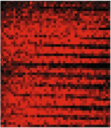
www.nature.com/scientificreports/
4
Ag NPs
Fe NPs
3 Ag/Fe Bimetallic NPs
Absorbance
2
1
0
300 400 500 600 700 800
Wavelength (nm)
Figure 1. UV–visible absorbance spectra of silver (Ag), iron (Fe) nanoparticles, and Ag–Fe bimetallic
nanoparticles synthesized using an aqueous extract from leaves of Gardenia jasminoides.
A Ag NPs Ag/Fe Bimetallic NPs
B
140 Fe NPs G. jasminoides extract 7000
120 6000
Transmitance (A.U.)
100 5000
Intensity
4000
80
3000
60
2000
40
1000
20 1430 1080
3400 2920 1640 C=C C=O 0
OH CH C=O
0 -1000
4000 3500 3000 2500 2000 1500 1000 500 0 500 1000 1500 2000 2500 3000
-1
Wavelength (nm) Raman Shift (cm )
Figure 2. Fourier Transform Infrared (FT-IR) spectra (A) of silver (Ag), iron (Fe) nanoparticles, and Ag-Fe
bimetallic nanoparticles synthesized using an aqueous extract from leaves of Gardenia jasminoides and the
RAMAN spectra of the (B) aqueous extract from leaves of Gardenia jasminoides.
a homogenous distribution with particles that ranged from 3 to 30 nm in diameter and an average diameter size
of 13 nm with an SD + /− 6.13 nm (Fig. 3B and C). The Ag–Fe bimetallic nanoparticles obtained in this study
were smaller than those previously reported, where their average size ranged from 20 to 60 nm30, 31. However,
the differences are related to the reducing and stabilizing agents used during the synthesis p rocess32. The high-
resolution TEM image displayed in Fig. 3B shows clear fringes and morphology that suggests the formation of a
core–shell (Ag-core Fe-shell) nano-arrangement of the bimetallic nanoparticles, as reported on the l iterature30, 33.
The presence of silver and iron as components of the Ag–Fe bimetallic nanoparticles were confirmed through
a dark-field microscopy analysis (Fig. 4A–F). The results showed a clear overlap of the Fe and Ag within the
selected, analyzed field, corroborating the bimetallic nanoparticle formation. In addition, carbon (C) was also
analyzed to detect the element present in the reducing organic extract. Further analysis using energy dispersive
X-Ray (EDX) spectroscopy was carried out to obtain an elemental spectrum in the Ag–Fe bimetallic nanoparticle
sample. Figure 4G shows the EDX spectra of the synthesized Ag–Fe bimetallic NPs, where the binding energies
of metallic silver are observed, at approximately 3 keV, while the peaks around 1, 6, and 7 keV are related to the
binding energies of F e34, 35. The elemental composition of the bimetallic nanoparticles was determined by EDX
and confirmed that the Ag/Fe bimetallic NPs were composed of 58.59% Ag and 41.4% Fe.
Magnetic properties. Magnetic NPs have been reported as non-immunogenic nanomaterials with excel-
lent biocompatibility, suitable for medical applications36. Therefore, the magnetic bimetallic nanoparticles are an
outstanding option for diagnostic (sensing and imaging) and therapeutic applications (thermal treatments and
drug-delivery systems)37. Hence, in the present study, we evaluated the magnetic properties of the Ag–Fe bime-
tallic NPs (Fig. 5). As can be observed, when an external magnetic field is applied to the Ag–Fe bimetallic NPs,
Scientific Reports | (2021) 11:5351 | https://doi.org/10.1038/s41598-021-84768-8 3
Vol.:(0123456789)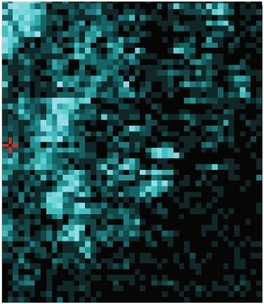
www.nature.com/scientificreports/
A B
50 nm 5 nm
C
35
30
25
Frequency
20
15
10
5
0
0-4.9 5-9.9 10-14.9 15-19.9 20-24.9 25-30
Diameter (nm)
Figure 3. Transmission Electron Microscopy (TEM) characterization of Ag–Fe bimetallic nanoparticles
synthesized using an aqueous extract from leaves of Gardenia jasminoides (A) Low magnitude TEM (B) High-
resolution TEM images and (C) Size distribution histogram of Ag–Fe bimetallic nanoparticles.
they adhered to the magnet, therefore exhibiting magnetic properties. This magnetic property has previously
been described in nanoparticles containing iron30, 31.
Antimicrobial activity. The emerging resistance in bacteria and the high costs of advanced antimicrobial
drugs have encouraged scientists to search for effective, economically viable, and broadly applicable drugs38. In
search of a new therapeutic alternative, we determined the minimal inhibitory concentrations (MIC) of AgNPs,
FeNPs, and Ag–Fe bimetallic NPs against Gram-positive, Gram-negative bacteria, and a yeast using the broth
microdilution method (Table 1).
The present study showed that AgNPs had an antimicrobial effect in concentrations ranging from 15.62 to125
ppm. The MIC values of Staphylococcus aureus (S. aureus), a Gram-positive bacteria, were the same in the ATCC
and the multidrug-resistant strains (125 ppm). In contrast, Pseudomonas aeruginosa (P. aeruginosa), a Gram-
negative bacteria, exhibited lower MIC values (from 15.62 to 62.5 ppm) in both ATCC and multidrug-resistant
strains, compared to the Gram-positive bacteria. Escherichia coli (E. coli; Gram-negative bacteria) and Candida
albicans (C. albicans; yeast), displayed MIC values equal to the Gram-positive bacteria, S. aureus (125 ppm).
These trends in MIC results are in agreement with previous s tudies39–41.
The MIC results of the present study demonstrated that the FeNPs exhibited no antimicrobial activity
(> 250 ppm), at the explored ranges, in all tested strains. As we previously mentioned, the antibacterial activity
of FeNPs has not been thoroughly studied. Most of the studies have evaluated this activity by the well-diffusion
method in Gram-positive and Gram-negative bacteria13, 42, 43. We studied the antimicrobial activity using the
broth microdilution method; therefore, the results are difficult to comparable.
On the other hand, the MIC values of Ag–Fe bimetallic NPs were similar or lower than those obtained with
AgNPs and FeNPs alone. We considered the elemental composition of Ag and Fe in the bimetallic NPs to analyze
their antimicrobial activity (58.59% of Ag and 41.4% of Fe). In general, the MIC values for Ag–Fe bimetallic
NPs in S. aureus (S. aureus ATCC 6538, S. aureus ATCC 29213) were comparable to those obtained with AgNPs
(125 ppm). However, the elemental composition demonstrated that silver and iron concentrations were 73.23 and
51.75 ppm, respectively. Interestingly, the antibacterial effect of Ag–Fe bimetallic NPs was more evident against
Scientific Reports | (2021) 11:5351 | https://doi.org/10.1038/s41598-021-84768-8 4
Vol:.(1234567890)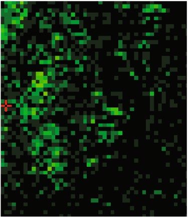
www.nature.com/scientificreports/

                                  A                                Analyzed         B                        C-K   C                  Fe-K
                                                                   Sample

                                                                       100 (nm)

                                  D                                     Fe-L        E                     Ag-K     F                  Ag-L

                                  G                               Ag
                                             2000
                                                    C Mg                            Cu                                      Ag   Ag

                                                        Fe   Si                Fe
                                             1500
                                    Counts

                                             1000

                                              500

                                                0
                                                    0                    5               10             15             20        25
                                                                                              Energy (keV)

                                  Figure 4.  Darkfield image (A)–(F) and energy dispersive X-Ray (EDX) spectra (G) of Ag–Fe bimetallic
                                  nanoparticles synthesized using an aqueous extract from leaves of Gardenia jasminoides.

                                  Figure 5.  Magnetic property of Ag–Fe bimetallic nanoparticles synthesized using an aqueous extract from
                                  leaves of Gardenia jasminoides.

Scientific Reports |   (2021) 11:5351 |                           https://doi.org/10.1038/s41598-021-84768-8                                      5

                                                                                                                                             Vol.:(0123456789)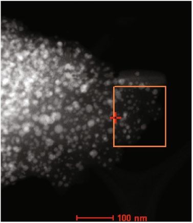
www.nature.com/scientificreports/
MIC Bimetallic MIC MBC or MFC of
NPs AgNPs + FeNPs Bimetallic NPs
Strain MIC AgNPs (ppm) MIC FeNPs (ppm) (Ag / Fe*) (ppm) (Ag / Fe*) (ppm) S value (ppm)
S. aureus
125 > 250 125 (73.23/51.75) 125 (73.23/51.75) 0.53 250
ATCC 6538
S. aureus
125 > 250 125 (73.23/51.75) 125 (73.23/51.75) 0.40 250
ATCC 29,213
S. aureus
125 > 250 250 (146/103.5) 250 (146/103.5) 0.80 500
Multidrug-resistant
P. aeruginosa
62.5 > 250 31.25 (18.3/12.9) 31.25 (18.3/12.9) 0.89 62.5
ATCC 27,853
P. aeruginosa
15.62 > 250 31.25 (18.3/12.9) 31.25 (18.3/12.9) 0.80 62.5
ATCC DMS 50,071
P. aeruginosa
31.25 > 250 15.62 (9.15/6.42) 15.62 (9.15/6.42) 0.90 31.25
Multidrug-resistant
E. coli
125 > 250 125 (73.23/51.75) 62.5 (36.6/25.9) 0.40 250
ATCC 11,229
C. albicans
125 > 250 62.5 (36.6/25.9) 62.5 (36.6/25.9) 0.08 125
Clinical isolate
Table 1. Minimal Inhibitory Concentration (MIC), S value, Minimum Bactericidal Concentration (MBC)
and Minimum Fungicidal Concentration (MFC) of silver nanoparticles (AgNPs), iron nanoparticles (FeNPs),
Ag-Fe bimetallic nanoparticles, and combination of AgNPs + FeNPs, prepared using Gardenia jasminoides
leaves aqueous extract against clinically relevant pathogens. *The elemental composition of Ag and Fe was
considered to analyze nanoparticles’ antimicrobial activity (bimetallic and AgNPs + FeNPs): 58.59% of Ag and
41.4% of Fe. S value: S > 0 Synergistic; S = 0 Additive; S < 0 Antagonistic.
P. aeruginosa. As shown in Table 1, the MIC value in P. aeruginosa ATCC 27853 and ATCC DMS 50071 was
31.25 ppm, while the multidrug-resistant strain was 15.62 ppm. When we analyzed the elemental composition,
it was observed that the concentration of silver and iron was reduced by 3.4 and 19-fold, respectively, compared
to AgNPs and FeNPs MICs values. These data show the potential effects of Ag–Fe bimetallic NPs in multidrug-
resistant bacteria and suggests the importance of the synergistic interactions of these bimetallic NPs, especially
for other Gram-negative bacteria with different antibiotic resistance profiles.
Regarding the antifungal activity of Ag–Fe bimetallic NPs in C. albicans (clinical isolate), we observed a MIC
value of 62.5 ppm (36.6 and 25.9 ppm, Ag and Fe, respectively), which was reduced by 3.4 and 9.6-fold, compared
to AgNPs and FeNPs MICs values. Moreover, it is worth highlighting that the MICs reported in this study were
found to be lower than those previously reported for Ag–Fe bimetallic NPs in S. aureus ATCC, E. coli ATCC, P.
aeruginosa ATCC, and multidrug-resistant P. aeruginosa44, 45.
The synergistic antimicrobial activity of Ag–Fe bimetallic NPs in a complex matrix, such as bacterial cultures,
could follow a non-linear dosage-response behavior. This kind of response is better analyzed when using a syn-
ergy test (such as the Bliss independence model) that takes into account the effect of the antimicrobial agent on
the bacterial culture, rather than tests that assume all types of antimicrobial agents and their combinations have
a linear dosage-respond curve, like the isobologram and the FIC index interpretations46, 47. We therefore evalu-
ated whether the synergistic effect, measured by the Bliss Independence Model, observed in Ag–Fe bimetallic
nanoparticles was conserved or modified after the combination of individually synthesized AgNPs and FeNPs.
The results demonstrate that all of the interactions in the bimetallic nanoparticles were found to be synergistic
(Table 1). Moreover, the combination of AgNPs + FeNPs was also found to be synergistic in the antimicrobial
efficacy against E. coli ATCC 11229. In addition, it is critical to keep in mind that one of the main advantages
of using bimetallic nanoparticles instead of single metal nanoparticles is that incorporating two individual
metals in a single entity makes their synthesis a greener procedure and their delivery a much simpler p rocess15.
Moreover, this process significantly reduces the quantities of hazardous wastes produced during the biosynthesis
of nanoparticles48.
The antimicrobial action of Ag–Fe bimetallic NPs seems to depend on the differences in the cell walls or cell
membrane composition of the microorganism. However, the antimicrobial mechanism of Ag–Fe bimetallic
nanoparticles has not been elucidated yet. Nonetheless, core–shell nanostructures display a more significant
effect than that observed in monometallic n anoparticles44. Porins could act as a transporter channel for the
nanoparticles to and from the cell49. It is also known that silver nanoparticles can anchor to the bacterial cell wall
and penetrate it. Once inside, they contribute to the formation of free radicals generating intracellular oxidative
stress, which leads to cell death50. Recently, it has been described that iron might interact with specific amino
acids (–SH groups of cysteine) present in proteins of the bacteria cell wall. Particularly, cysteine’s thiol side chain
has been identified as the most susceptible to gain electrons from the oxidizing species21. Therefore, we propose
that the antimicrobial mechanism of Ag–Fe bimetallic nanoparticles is carried out in two steps: first, iron, which
is coating the nanoparticles, oxidizes the thiol side chain in cysteine leading to changes in the primary structure
of proteins, alteration in bacterial cell wall permeability and finally cell death. Second, due to the altered perme-
ability of the cell wall, many nanoparticles are uptake by the bacteria. Silver ions are then released from NPs to
the cytoplasm inducing oxidative stress, causing DNA alterations and disruption of membrane morphology.
Scientific Reports | (2021) 11:5351 | https://doi.org/10.1038/s41598-021-84768-8 6
Vol:.(1234567890)www.nature.com/scientificreports/
Thus, silver and iron contribute to disrupting cellular structures, intracellular biological functions, and lead cell
death. Nonetheless, further studies are needed to support this hypothesis.
Next, we determined the minimum bactericidal and fungicidal concentration of Ag–Fe bimetallic NPs to
demonstrate their ability to kill bacteria and yeast (Table 1). A bactericidal agent is defined as a material with a
ratio of MBC to MIC ≤ 4. Antibiotics with a ratio of MBC to MIC > 4 are defined as bacteriostatic agents51. The
bactericidal agent kills bacteria rapidly and reduces bacterial resistance development; hence they are a better
choice for clinicians than bacteriostatic a gents52. Our results showed that the MBC and MFC values were twice
the MIC concentrations; thus, the Ag–Fe bimetallic nanoparticles were bactericidal and fungicidal.
Finally, we conducted a bibliographic search on the evaluation of the biocompatibility of the Ag–Fe bimetallic
nanoparticles. A recent study demonstrated that hybrids silver-iron nanoparticles significantly enhanced the
cytotoxicity of MCF-7 breast cancer cells. Therefore, these nanoparticles could be a potent anticancer drug53.
Another study evaluated magnetite/silver nanoparticles biocompatibility in Vero cells (Kidney epithelial cells).
The results evidenced low cytotoxicity, hemolytic, and platelet aggregation tendency, which confirm a high
biocompatibility54. Moreover, a cytotoxicity study of iron-silver core–shell nanoparticles (FeO/AgNPs) was car-
ried out on Vero cell line, and the results showed that these nanoparticles were biocompatible up to 500 µgml−1
concentration. Hence, even though further studies are required on our specific Ag–Fe bimetallic nanoparticles
system, these studies suggest the biocompatibility of our Ag–Fe bimetallic nanoparticles and hint for their
potential as biocompatible nanomaterials with biological and biomedical applications.
Conclusions
The green synthesis of bimetallic nanoparticles using natural extracts is gaining research attention for environ-
mental and medical applications. In the present study, silver nanoparticles (AgNPs), iron nanoparticles (FeNPs),
Ag–Fe bimetallic nanoparticles, and the combination of AgNPs + FeNPs, were prepared in a redox reaction using
the aqueous G. jasminoides leaves extract as a reducing agent. The evaluation of the antimicrobial activity of
bimetallic NPs showed synergistic bactericidal and fungicidal effects against Gram-positive and Gram-negative
bacteria and yeast. Our results demonstrate the ability to use green synthesis methods to design and synthesize
magnetic bimetallic nanoparticles composed of metals that synergize their antimicrobial effects. Moreover,
this work gives insight into the development of novel and more effective antimicrobial agents with potential
as bactericidal and fungicidal agents, to treat drug-resistant infections, and in other biomedical applications.
Methods
Microbial strains and culture conditions. Strains used in this study include S. aureus ATCC 6538, S.
aureus ATCC 29213, E. coli ATCC 11229, P. aeruginosa ATCC 27853, and P. aeruginosa DSM 50071. Meanwhile,
clinical isolates from a Gram-negative multidrug-resistant strain of P. aeruginosa (kanamycin, chloramphenicol,
and ciprofloxacin-resistant strain), Gram-positive multidrug-resistant strain of S. aureus (kanamycin, chloram-
phenicol, ciprofloxacin, ampicillin-resistant strain), and a yeast, C. albicans, were obtained from the San Vicente
Hospital in Monterrey, Nuevo León, México. All the bacterial assays were performed in Müller-Hinton (MH)
media (BD, Bioxon), and all the assays using C. albicans were performed in Yeast Malt (YM) media (BD, Bioxon).
Gardenia jasminoides extract preparation. Leaves from the plant G. jasminoides were collected in
Monterrey, Mexico. A voucher specimen (number 634) was deposited in ANSM Herbarium (Universidad
Autónoma Agraria Antonio Narro) in Saltillo, Coahuila, México. Leaves were washed using deionized water
to remove dust and were dried at room temperature (22–26 °C) for 24 h before being powdered. An aqueous
extract was prepared as follows: two grams of powdered leaves were mixed with 60 mL of deionized water at
room temperature, stirred for 2 h, and filtered immediately through Whatman filter paper. The aqueous extract
of G. jasminoides was stored at 4 °C until their use in the synthesis of NPs.
Synthesis of monometallic nanoparticles. A total of 12.5 mL of 0.02 M A
gNO3 were mixed with
12.5 mL of G. jasminoides extract and heated for 1 h at 80 °C. The formation of silver nanoparticles was moni-
tored through a change of color of the mixture (from transparent to brownish). The reaction was cooled and
centrifuged at 14,000 rpm for 5 min. The product was then washed with 70% ethanol and lyophilized (FreeZone
6, Labconco, Kansas City, MO, USA) for 24 h to obtain the purified AgNPs. We followed the previous procedure
for the synthesis of the iron nanoparticles; only 12.5 mL of 0.01 M Fe(NO3)3 solution was used instead of the
silver solution, and the color change observed was from transparent to black.
Synthesis of Ag–Fe bimetallic nanoparticles. The synthesis of Ag-Fe bimetallic nanoparticles was per-
formed by adding 12.5 mL of 0.02 M A gNO3 and 12.5 mL of 0.01 M Fe(NO3)3 in a Falcon tube. Then, the tube
was mixed and heated until reaching 60ºC. When this temperature was reached, 25 mL of the filtered G. jasmi-
noides extract was added to the mixture (2:1:1 proportion), kept at constant stir, and heated at 80ºC for 1 h. The
formation of the nanoparticles was monitored and confirmed by a change of color of the mix that changed from
transparent to brown (for silver) or black (for iron). The reaction was cooled at room temperature, centrifuged
at 14,000 rpm for 5 min, and the supernatant was discharged. The product was then washed with 70% ethanol,
left to dry at room temperature for 24 h, and finally lyophilized (FreeZone 6, Labconco, Kansas City, MO, USA)
for another 24 h.
Nanoparticle characterization. Several techniques were used to characterize the synthesized nanopar-
ticles, such as UV–visible (UV–vis) spectra, Fourier-transform infrared (FT-IR) spectroscopy, RAMAN spec-
Scientific Reports | (2021) 11:5351 | https://doi.org/10.1038/s41598-021-84768-8 7
Vol.:(0123456789)www.nature.com/scientificreports/
troscopy, Transmission electron microscopy (TEM), High Resolution (HR)-TEM, dark-field microscopy, and
Energy dispersive X-ray (EDX) analysis. UV–vis absorption spectra of the nanoparticles (1 mg/mL in distilled
water as a dispersive medium) were studied in the 200–600 nm wavelength range using a Multiskan GO Micro-
plate Spectrophotometer (Thermo Fisher Scientific, USA). FT-IR analysis was performed in the 650–4000 cm−1
range in a Shimadzu IRAffinity-1 spectrometer (Shimadzu, Japan) to determine the plant extract components
and the synthesized nanoparticles. The lyophilized NPs and extract (1 mg/mL) were diluted in KBr and analyzed
by the attenuated total reflectance method. To obtain information about the interactions between the nanoparti-
cles and the plant extract, a RAMAN spectrum of the nanoparticles and the extract was measured using a DXR
RAMAN microscope (Thermo Fisher Scientific, USA). The elemental, morphological, and size of the nanopar-
ticles were obtained through a FEI-TITAN 80–300 kV scanning transmission electron microscope. Briefly, the
sample was prepared by depositing and evaporating a drop of NPs solution (1 mg/mL) onto lacey carbon-coated
copper grids. The elements presented in NPs were determined by an energy-dispersive spectrometry analyzer
integrated in the transmission electron microscope. To analyze the particle size and size distribution, we used the
TEM and HR-TEM micrographs to measure de diameter of NPs and then processed them with ImageJ software
developed by the National Institutes of Health.
Magnetic properties. A round-shaped magnet was used to produce an external magnetic field, having
direct contact with the nanoparticles. If the nanoparticles adhered to the magnet, they were considered to exhibit
magnetic properties.
Determination of minimal inhibitory concentration (MIC). The determination of MICs was per-
formed by the microdilution method. Stocks of NPs (mono- and bimetallic) were prepared at a final concentra-
tion of 1000 ppm in LB or YM broth. To perform the MIC test, we used a 96-wells microtiter plaque (Corning,
USA). We added in the top well the necessary volume from the stock solution to reach 500 ppm concentration of
the NPs in a final volume of 200 μL. Then, serial dilutions were performed by taking 100 μL from every very next
well into 100 μL of culture media. The last 100 μL from the last dilution was discarded. The concentration range
was from 250 to 0.97 ppm of each NPs after the bacteria was added. For inoculation, an overnight (ON) culture
was prepared at 37 °C, 150 rpm, and incubated for 20 h, Next, the ON culture was diluted 1:250 in fresh media
and incubated until a critical optical density was reached (OD600nm of 0.2 ± 0.02 for bacteria and an O D600nm of
0.3 ± 0.02 cells/mL for yeast). Finally, a 1:100 dilution was made with fresh media, and 100 μL of this dilution was
added to each test well. Plates were incubated at 37ºC 150 rpm for 20 h, and then, the O D600nm was measured in
a Multiskan GO Microplate Spectrophotometer (Thermo Fisher Scientific, USA). The MIC was defined as the
minimal concentration at which no growth was observed. Each assay was performed in triplicates; growth and
sterility controls were included.
The behavior of the bimetallic NPs against microbial growth was analyzed by the Bliss independence model as
described by Hegreness et al.56, which indicates that synergy can be considered when the effect of the combined
antimicrobial agent (bimetallic NP) is more significant than the predicted of its components (single metal NP).
Therefore, Eq. 1 was used to analyze the synergistic properties of the bimetallic nanoparticles, where the S value;
the difference between the predicted value of individual components x and y (fx0 and f0y, respectively) and the
combined treatment xy (fxy) is denoted in the form:
fx0 f0y fxy
S= − Bliss Independence Model (1)
f00 f00 f00
where, fx0 = treatment with Ag, f0y = treatment with the transition metal, fxy = treatment Ag + metal, f00 = growth
control of a culture that has not been treated with the transition metals. The value of S describes the interaction
between combinatorial treatments as follows: S > 0 Synergistic; S = 0 Additive; S < 0 Antagonistic.
Determination of minimum bactericidal and minimum fungicidal concentration (MBC,
MFC). After obtaining the monometallic and bimetallic nanoparticles MICs, the wells that corresponded
to the twice of the MIC, the MIC, and the growth control of each treatment were selected. Serial dilutions of
selected wells were performed until they reach a final concentration of 1 × 10⁷ cells/mL. Ten microliters of each
dilution were cultured in a Petri dish, incubated at 37ºC for 24 h, and finally, counted the colony-forming units
(CFU) in each treatment.
Received: 22 June 2020; Accepted: 16 February 2021
References
1. World Health Organization [WHO]. Antimicrobial resistance. WHO (2020). Available at: https://www.who.int/news-room/fact-
sheets/detail/antimicrobial-resistance. Accessed: 10th Oct 2020
2. WHO. New report calls for urgent action to avert antimicrobial resistance crisis. Joint News Release 29, (2019).
3. Sánchez-López, E. et al. Metal-based nanoparticles as antimicrobial agents: an overview. Nanomaterials 10, 292 (2020).
4. Arora, N., Thangavelu, K. & Karanikolos, G. N. Bimetallic nanoparticles for antimicrobial applications. Front. Chem. 8, 412 (2020).
5. Belenov, S. V. et al. Phase behavior of Pt–Cu nanoparticles with different architecture upon their thermal treatment. Nanotechnolo-
gies Russ. 12, 147–155 (2017).
6. Behera, A., Mittu, B., Padhi, S., Patra, N. & Singh, J. Bimetallic nanoparticles: Green synthesis, applications, and future perspec-
tives. In Multifunctional Hybrid Nanomaterials for Sustainable Agri-Food and Ecosystems 639–682 (Elsevier, 2020). doi:https://doi.
org/10.1016/B978-0-12-821354-4.00025-X
Scientific Reports | (2021) 11:5351 | https://doi.org/10.1038/s41598-021-84768-8 8
Vol:.(1234567890)www.nature.com/scientificreports/
7. Sharma, G. et al. Novel development of nanoparticles to bimetallic nanoparticles and their composites: a review. J. King Saud
Univ. - Sci. https://doi.org/10.1016/J.JKSUS.2017.06.012 (2017).
8. Kuppusamy, P., Yusoff, M. M., Maniam, G. P. & Govindan, N. Biosynthesis of metallic nanoparticles using plant derivatives and
their new avenues in pharmacological applications: an updated report. Saudi Pharm. J. 24, 473–484 (2016).
9. Mohamad, N. A. N., Jai, J., Arham, N. A. & Hadi, A. A short review on the synthesis of bimetallic nanoparticles using plant extract.
In: 2013 IEEE International Conference on Control System, Computing and Engineering 334–339 (IEEE, 2013). doi:https://doi.
org/10.1109/ICCSCE.2013.6719985
10. Xiao, W., Li, S., Wang, S. & Ho, C.-T. Chemistry and bioactivity of Gardenia jasminoides. J. Food Drug Anal. 25, 43–61 (2017).
11. Kesavan, K., Gnanasekaran, J., Gurunagarajan, S. & Nayagam, A. A. J. microscopic, physicochemical and phytochemical analysis
of gardenia jasminoides (ellis). Int. J. Pharm. Pharm. Sci. 10, 97 (2018).
12. Lü, F., Gao, Y., Huang, J., Sun, D. & Li, Q. Roles of biomolecules in the biosynthesis of silver nanoparticles: case of gardenia jasmi-
noides extract. Chin J. Chem. Eng. 22, 706–712 (2014).
13. Naseem, T. & Farrukh, M. A. Antibacterial activity of green synthesis of iron nanoparticles using Lawsonia inermis and Gardenia
jasminoides leaves extract. J. Chem. 2015, 1–7 (2015).
14. Jia, L., Zhang, Q., Li, Q. & Song, H. The biosynthesis of palladium nanoparticles by antioxidants in Gardenia jasminoides Ellis:
long lifetime nanocatalysts for p-nitrotoluene hydrogenation. Nanotechnology 20, 385601 (2009).
15. Meena Kumari, M., Jacob, J., Philip, D. Green synthesis and applications of Au–Ag bimetallic nanoparticles. Spectrochim. Acta
Part A Mol. Biomol. Spectrosc. 137, 185–192 (2015).
16. Li, J. et al. Biosynthesis of Au, Ag and Au–Ag bimetallic nanoparticles using protein extracts of Deinococcus radiodurans and
evaluation of their cytotoxicity. Int. J. Nanomedicine 13, 1411–1424 (2018).
17. Chavez, K. & Rosas, G. Green synthesis and characterization of Ag@Au core-shell bimetallic nanoparticles using the extract of
hamelia patens plant. Microsc. Microanal. 25, 1102–1103 (2019).
18. Botha, T. L. et al. Cytotoxicity of Ag, Au and Ag–Au bimetallic nanoparticles prepared using golden rod (Solidago canadensis)
plant extract. Sci. Rep. 9, 4169 (2019).
19. Sharma, V. K., Siskova, K. M. & Zboril, R. Magnetic Bimetallic Fe/Ag Nanoparticles: Decontamination and Antimicrobial Agents.
In: ACS Symposium Series 1150, 193–209 (American Chemical Society, 2013).
20. Gallo, A., Bianco, C., Tosco, T., Tiraferri, A. & Sethi, R. Synthesis of eco-compatible bimetallic silver/iron nanoparticles for water
remediation and reactivity assessment on bromophenol blue. J. Clean. Prod. 211, 1367–1374 (2019).
21. Al-Asfar, A., Zaheer, Z. & Aazam, E. S. Eco-friendly green synthesis of Ag@Fe bimetallic nanoparticles: antioxidant, antimicrobial
and photocatalytic degradation of bromothymol blue. J. Photochem. Photobiol. B Biol. 185, 143–152 (2018).
22. Cusimano, M. G. et al. Biogenic iron-silver nanoparticles inhibit bacterial biofilm formation due to Ag+ release as determined by
a novel phycoerythrin-based assay. Appl. Microbiol. Biotechnol. 104, 6325–6336 (2020).
23. Medina-Cruz, D. et al. Bimetallic Nanoparticles for Biomedical Applications: A Review. in Racing for the Surface 397–434 (Springer
International Publishing, 2020). doi:https://doi.org/10.1007/978-3-030-34471-9_16
24. Zhang, X. F., Liu, Z. G., Shen, W. & Gurunathan, S. Silver nanoparticles: Synthesis, characterization, properties, applications, and
therapeutic approaches. Int. J. Mol. Sci. 17, (2016).
25. Klačanová, K. et al. Formation of Fe(0)-nanoparticles via reduction of Fe(II) compounds by amino acids and their subsequent
oxidation to iron oxides. J. Chem. 2013, 1–10 (2013).
26. Fakhri, A., Tahami, S. & Naji, M. Synthesis and characterization of core-shell bimetallic nanoparticles for synergistic antimicrobial
effect studies in combination with doxycycline on burn specific pathogens. J. Photochem. Photobiol. B Biol. 169, 21–26 (2017).
27. Gopinath, K. et al. Green synthesis of silver, gold and silver/gold bimetallic nanoparticles using the Gloriosa superba leaf extract
and their antibacterial and antibiofilm activities. Microb. Pathog. 101, 1–11 (2016).
28. Uddin, R. et al. HPLC-analysis of polyphenolic compounds in gardenia jasminoides and determination of antioxidant activity by
using free radical scavenging assays. Adv. Pharm. Bull. 4, 273–281 (2014).
29. Pierna, J. A. F. et al. Characterization and discrimination of phenolic compounds using Fourier transform Raman spectroscopy
and chemometric tools. https://popups.uliege.be:443/1780-4507 (2018). doi:https://doi.org/10.25518/1780-4507.16270
30. Ruíz-Baltazar, A. Structural characterization of Fe–Ag bimetallic nanoparticles synthesized by chemical reduction. Int. Res. J. Pure
Appl. Chem. 4, 263–269 (2014).
31. Gong, P. et al. Preparation and antibacterial activity of Fe3O4 @Ag nanoparticles. Nanotechnology 18, 285604 (2007).
32. Paszkiewicz, M. et al. Synthesis and characterization of monometallic (Ag, Cu) and bimetallic Ag–Cu particles for antibacterial
and antifungal applications. J. Nanomater. 2016, 1–11 (2016).
33. Carroll, K. J. et al. One-pot aqueous synthesis of Fe and Ag core/shell nanoparticles. Chem. Mater. 22, 6291–6296 (2010).
34. Singh, P., Kim, Y. J., Wang, C., Mathiyalagan, R. & Yang, D. C. The development of a green approach for the biosynthesis of silver
and gold nanoparticles by using Panax ginseng root extract, and their biological applications. Artif. Cells Nanomed. Biotechnol. 44,
1–8 (2015).
35. Prabhu, Y. T., Rao, K. V., Kumari, B. S., Kumar, V. S. S. & Pavani, T. Synthesis of F e3O4 nanoparticles and its antibacterial applica-
tion. Int. Nano Lett. 5, 85–92 (2015).
36. Guo, T. et al. The recent advances of magnetic nanoparticles in medicine. J. Nanomater. 2018, 1–8 (2018).
37. Srinoi, P., Chen, Y.-T., Vittur, V., Marquez, M. & Lee, T. Bimetallic nanoparticles: enhanced magnetic and optical properties for
emerging biological applications. Appl. Sci. 8, 1106 (2018).
38. Aslam, B. et al. Antibiotic resistance: a rundown of a global crisis. Infect. Drug Resist. 11, 1645–1658 (2018).
39. Morones, J. R. et al. The bactericidal effect of silver nanoparticles. Nanotechnology 16, 2346–2353 (2005).
40. Morones-Ramirez, J. R., Winkler, J. A., Spina, C. S. & Collins, J. J. Silver Enhances antibiotic activity against gram-negative bacteria.
Sci. Transl. Med. 5, 190ra81–190ra81 (2013).
41. Jalal, M. et al. Anticandidal activity of biosynthesized silver nanoparticles: effect on growth, cell morphology, and key virulence
attributes of Candida species. Int. J. Nanomed. 14, 4667–4679 (2019).
42. Saqib, S. et al. Synthesis, characterization and use of iron oxide nano particles for antibacterial activity. Microsc. Res. Tech. 82,
415–420 (2019).
43. Vitta, Y., Figueroa, M., Calderon, M. & Ciangherotti, C. Synthesis of iron nanoparticles from aqueous extract of Eucalyptus robusta
Sm and evaluation of antioxidant and antimicrobial activity. Mater. Sci. Energy Technol. 3, 97–103 (2020).
44. Chudasama, B., Vala, A. K., Andhariya, N., Upadhyay, R. V. & Mehta, R. V. Enhanced antibacterial activity of bifunctional F e3O4-Ag
Core-Shell nanostructures. Nano Res 2, 955–965 (2009).
45. Marková, Z. et al. Air stable magnetic bimetallic Fe–Ag nanoparticles for advanced antimicrobial treatment and phosphorus
removal. Environ. Sci. Technol. 47, 5285–5293 (2013).
46. Pillai, S. K., Moellering, R. C. & Eliopoulos, G. M. Antimicrobial combinations. . Antibiot Lab. Med. 5, 365–440 (2005).
47. Garza-Cervantes, J. A. et al. Re-sensitizing ampicillin and kanamycin-resistant E. coli and S. aureus using synergistic metal
micronutrients-antibiotic combinations. Front. Bioeng. Biotechnol. 8, 612 (2020).
48. Roy, N., Gaur, A., Jain, A., Bhattacharya, S. & Rani, V. Green synthesis of silver nanoparticles: an approach to overcome toxicity.
Environ. Toxicol. Pharmacol. https://doi.org/10.1016/j.etap.2013.07.005 (2013).
49. Neal, A. L. What can be inferred from bacterium–nanoparticle interactions about the potential consequences of environmental
exposure to nanoparticles?. Ecotoxicology 17, 362–371 (2008).
Scientific Reports | (2021) 11:5351 | https://doi.org/10.1038/s41598-021-84768-8 9
Vol.:(0123456789)www.nature.com/scientificreports/
50. Prabhu, S. & Poulose, E. K. Silver nanoparticles: mechanism of antimicrobial action, synthesis, medical applications, and toxicity
effects. Int. Nano Lett. 2, 32 (2012).
51. Pankey, G. A. & Sabath, L. D. Clinical relevance of bacteriostatic versus bactericidal mechanisms of action in the treatment of
gram-positive bacterial infections. Clin. Infect. Dis. 38, 864–870 (2004).
52. Finberg, R. W. et al. The importance of bactericidal drugs: future directions in infectious disease. Clin. Infect. Dis. 39, 1314–1320
(2004).
53. de Oliveira Gonçalves, K., Vieira, D. P., Levy, D., Bydlowski, S. P. & Courrol, L. C. Uptake of silver, gold, and hybrids silver-iron,
gold-iron and silver-gold aminolevulinic acid nanoparticles by MCF-7 breast cancer cells. Photodiagnosis Photodyn. Ther. https://
doi.org/10.1016/j.pdpdt.2020.102080 (2020).
54. Ramírez-Acosta, C. M., Cifuentes, J., Cruz, J. C. & Reyes, L. H. Patchy core/shell, magnetite/silver nanoparticles via green and
facile synthesis: routes to assure biocompatibility. Nanomaterials 10, 1857 (2020).
55. Kaur, P., Thakur, R., Malwal, H., Manuja, A. & Chaudhury, A. Biosynthesis of biocompatible and recyclable silver/iron and gold/
iron core-shell nanoparticles for water purification technology. Biocatal. Agric. Biotechnol. 14, 189–197 (2018).
56. Hegreness, M., Shoresh, N., Damian, D., Hartl, D. & Kishony, R. Accelerated evolution of resistance in multidrug environments.
105, 13977–13981 (2008)
Acknowledgements
The authors want to thank to the Universidad Autonoma de Nuevo León and CONACyT for providing financial
support through Paicyt 2016-2017, Paicyt 2019-2020 and Paicyt 2020-2021 Science Grants. CONACyT Grants
for: Basic science grant 221332, Fronteras de la Ciencia grant 1502 and Infraestructura Grant 279957. Ana L
Padilla Cruz for the support from a Beca Nacional de Posgrado from CONACyT and Dr. Angel Leon Buitimea
for the support from a Beca de Posdoctorado Nacional. We also want to thank to the Universidad Autonoma de
Nuevo Leon and the Centro de Investigación en Biotecnología y Nanotecnología for providing the infrastructure
and equipment to perform the experiments.
Author contributions
Conceptualization, A.L.B., and J.R.M.R. Writing-original draft preparation A.L.P.C., J.A.G.C., X.G.V.A., A.L.B.
and J.R.M.R., writing-review, and editing, A.L.P.C., J.A.G.C., G.G.R., A.L.B. and J.R.M.R. Supervision, A.L.B.,
and J.M.R. All authors reviewed the final version of the article and approved the submitted version.
Competing interests
The authors declare no competing interests.
Additional information
Correspondence and requests for materials should be addressed to J.R.M.-R.
Reprints and permissions information is available at www.nature.com/reprints.
Publisher’s note Springer Nature remains neutral with regard to jurisdictional claims in published maps and
institutional affiliations.
Open Access This article is licensed under a Creative Commons Attribution 4.0 International
License, which permits use, sharing, adaptation, distribution and reproduction in any medium or
format, as long as you give appropriate credit to the original author(s) and the source, provide a link to the
Creative Commons licence, and indicate if changes were made. The images or other third party material in this
article are included in the article’s Creative Commons licence, unless indicated otherwise in a credit line to the
material. If material is not included in the article’s Creative Commons licence and your intended use is not
permitted by statutory regulation or exceeds the permitted use, you will need to obtain permission directly from
the copyright holder. To view a copy of this licence, visit http://creativecommons.org/licenses/by/4.0/.
© The Author(s) 2021
Scientific Reports | (2021) 11:5351 | https://doi.org/10.1038/s41598-021-84768-8 10
Vol:.(1234567890)You can also read