MRI and histologically derived neuroanatomical atlas of the Ambystoma mexicanum (axolotl) - Nature
←
→
Page content transcription
If your browser does not render page correctly, please read the page content below
www.nature.com/scientificreports
OPEN MRI‑ and histologically
derived neuroanatomical atlas
of the Ambystoma mexicanum
(axolotl)
Ivan Lazcano1*, Abraham Cisneros‑Mejorado1,2, Luis Concha1, Juan José Ortiz‑Retana1,
Eduardo A. Garza‑Villarreal1* & Aurea Orozco1
Amphibians are an important vertebrate model system to understand anatomy, genetics and
physiology. Importantly, the brain and spinal cord of adult urodels (salamanders) have an incredible
regeneration capacity, contrary to anurans (frogs) and the rest of adult vertebrates. Among these
amphibians, the axolotl (Ambystoma mexicanum) has gained most attention because of the surge
in the understanding of central nervous system (CNS) regeneration and the recent sequencing of its
whole genome. However, a complete comprehension of the brain anatomy is not available. In the
present study we created a magnetic resonance imaging (MRI) atlas of the in vivo neuroanatomy
of the juvenile axolotl brain. This is the first MRI atlas for this species and includes three levels: (1)
82 regions of interest (ROIs) and a version with 64 ROIs; (2) a division of the brain according to the
embryological origin of the neural tube, and (3) left and right hemispheres. Additionally, we localized
the myelin rich regions of the juvenile brain. The atlas, the template that the atlas was derived from,
and a masking file, can be found on Zenodo at https://doi.org/10.5281/zenodo.4595016. This MRI
brain atlas aims to be an important tool for future research of the axolotl brain and that of other
amphibians.
The Mexican axolotl Ambystoma mexicanum, (from the Nahuatl, "water monster") is a mythic native neotenic
salamander that has amazed scientists initially for its peculiar life c ycle1 and later on for its tissue regeneration
capacity2, a trait lost later during vertebrate evolution. Specially interesting is the capacity of the axolotl to
regenerate nervous tissue, which has been described for the pallium3,4, the homologue of the cerebral cortex in
mammals5, as well as for the spinal cord, both of which are able to regenerate after i njury6. Although this capacity
has placed axolotl as an animal model for neural regeneration studies, a thorough comprehension of the nervous
system of this taxon remains obscure. In this context some axolotl brain structures have been well described by
several authors5–7. In general terms, the juvenile-adult brain of axolotl (and other amphibians) is composed by
regions similar to those of other vertebrates: olfactory bulb, telencephalon, diencephalon, mesencephalon, and
rhombencephalon. Histological procedures provide excellent differentiation of tissue types, albeit having limited
sampling of the three-dimensional spatial domain, and the introduction of deformations due to tissue handling.
Magnetic resonance imaging (MRI) is a tool that allows the non-invasive assessment of the neuroanatomy of
virtually any species that fits the scanner bore. MRI has been employed to generate in vivo and ex vivo atlases
of the central nervous system (CNS) from different species, i.e., human-8, rodent-9–11, avian-12,13, reptil14,15 as
well fish16,17. With this in mind, and to further understand the species anatomy and function, we developed a
MRI-based in vivo atlas of the juvenile Ambystoma mexicanum with the advantage of preserving the shape of its
parenchyma, as well as vascular and ventricular systems and correlated this atlas with a histological visualization
of myelin. The relevance of this atlas is that at this moment, an amphibian MRI atlas is not available; therefore,
it will be an invaluable tool for comparative neuroanatomy, biologists, environmentalists or other researchers
interested in this species and other amphibians. Moreover, the importance of axolotl as a model species stresses
the importance of understanding different aspects of the biology of this species brought to danger of extinction
(https://www.iucnredlist.org/species/1095/53947343).
1
Instituto de Neurobiología, Universidad Nacional Autonoma de Mexico (UNAM), Campus Juriquilla, Santiago de
Querétaro, Querétaro, México. 2CONACYT‑Instituto de Neurobiología, Universidad Nacional Autónoma de México
(UNAM), Campus Juriquilla, Santiago de Querétaro, Querétaro, México. *email: ivanlazcano@comunidad.unam.mx;
egarza@comunidad.unam.mx
Scientific Reports | (2021) 11:9850 | https://doi.org/10.1038/s41598-021-89357-3 1
Vol.:(0123456789)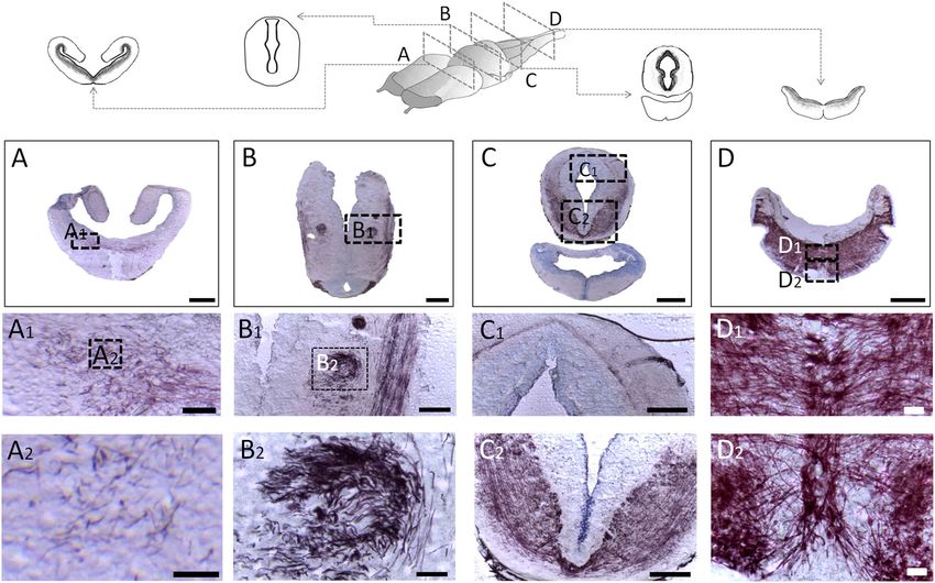
www.nature.com/scientificreports/
Structure Abbreviation Hemisphere Mayor structure Mean volume (mm3) SD Color
Olfactory nerve on19 Left Olfactory bulb 0.161 0.028
Glomerular layer g19 Left Olfactory bulb 0.472 0.077
Mitral cell layer m19 Left Olfactory bulb 0.24 0.031
Granule cell layer gc19 Left Olfactory bulb 0.116 0.018
19
Anterior olfactory nucleus aon Left Olfactory bulb 0.139 0.016
Pallium p20 Left Telencephalon 0.337 0.046
Accessory olfactory bulb aob20 Left Telencephalon 0.442 0.06
4,20
Dorsal pallium dp Left Telencephalon 0.83 0.14
Lateral pallium lp4,20 Left Telencephalon 0.942 0.144
Medial pallium mp4,20 Left Telencephalon 0.732 0.129
4,20,21
Septum s Left Telencephalon 0.668 0.094
Striatum str4,20,21 Left Telencephalon 0.511 0.06
22,23
Medial part of amygdala amc Left Telencephalon 0.068 0.009
Pallial commissure cpa23 Left Telencephalon 0.02 0.004
Anterior commissure ca23 Left Telencephalon 0.025 0.003
23,24
Anterior preoptic nucleus npa Left Telencephalon 0.101 0.01
Lateral/medial forebrain bundle lfb24 Left Telencephalon 0.702 0.083
3
Choroid plexus cp Left Telencephalon 0.171 0.034
Thalamic eminence em th23 Left Telencephalon 0.025 0.005
23
Posterior preoptic nucleus ppn Left Diencephalon 0.068 0.009
Habenula hab21,24 Left Diencephalon 0.041 0.005
21,24
Dorsal thalamus dth Left Diencephalon 0.508 0.052
Ventral thalamus vth21,24 Left Diencephalon 0.193 0.028
21,24
Hypothalamus dorsalis hyth d Left Diencephalon 0.872 0.105
Paraventricular organ pvo21,24 Left Diencephalon 0.052 0.008
21,24
Pars dorsalis hypothalami pdh Left Diencephalon 0.025 0.004
Pars ventralis hypothalami pvh21,24 Left Diencephalon 0.025 0.002
21,24
Subcommissural organ so Left Diencephalon 0.019 0.003
Ependymal cell layer ecl24 Left Diencephalon 0.254 0.232
24
Optic chiasm och Left Mesencephalon 0.023 0.003
Optic tectum to21,24 Left Mesencephalon 0.774 0.118
Tegmentum tgm21,24 Left Mesencephalon 0.482 0.058
21
Nucleus interpeduncularis nip Left Mesencephalon 0.005 0.002
Hypothalamus ventralis hyt v21 Left Mesencephalon 0.193 0.028
25,26
Pituitary hy Left Endocrine 0.086 0.02
Medulla oblongata mo25 Left Rombencephalon 0.36 0.044
25,26
Cerebellum cb Left Rombencephalon 0.157 0.018
Nervous trigeminus V21 Left Rombencephalon 0.078 0.016
21
Nervous lateralis anterior/nervous octavus VIII Left Rombencephalon 0.171 0.028
Gray matter of medulla oblongata gmob25 Left Rombencephalon 0.685 0.08
White matter of medulla oblongata wmob25 Left Rombencephalon 1.403 0.155
19
Olfactory nerve on Right Olfactory bulb 0.193 0.032
Glomerular layer g19 Right Olfactory bulb 0.419 0.068
19
Mitral cell layer m Right Olfactory bulb 0.24 0.039
Granule cell layer gc19 Right Olfactory bulb 0.119 0.017
19
Anterior olfactory nucleus aon Right Olfactory bulb 0.142 0.016
Pallium p20 Right Telencephalon 0.35 0.044
Accessory olfactory bulb aob20 Right Telencephalon 0.413 0.059
Continued
Scientific Reports | (2021) 11:9850 | https://doi.org/10.1038/s41598-021-89357-3 2
Vol:.(1234567890)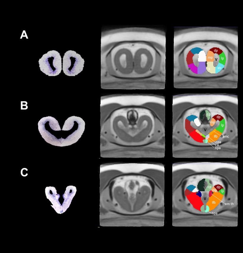
www.nature.com/scientificreports/
Structure Abbreviation Hemisphere Mayor structure Mean volume (mm3) SD Color
Dorsal pallium dp4,20 Right Telencephalon 0.844 0.15
Lateral pallium lp4,20 Right Telencephalon 0.878 0.144
Medial pallium mp4,20 Right Telencephalon 0.718 0.13
4,20,21
Septum s Right Telencephalon 0.675 0.084
Striatum str4,20,21 Right Telencephalon 0.464 0.054
22,23
Medial part of amygdala amc Right Telencephalon 0.07 0.013
Pallial commissure cpa23 Right Telencephalon 0.023 0.003
23
Anterior commissure ca Right Telencephalon 0.027 0.005
Anterior preoptic nucleus npa23,24 Right Telencephalon 0.108 0.015
Lateral/medial forebrain bundle lfb24 Right Telencephalon 0.72 0.093
3
Choroid plexus cp Right Telencephalon 0.142 0.033
Thalamic eminence em th23 Right Telencephalon 0.024 0.004
24
Posterior preoptic nucleus ppn Right Diencephalon 0.07 0.008
Habenula hab21,24 Right Diencephalon 0.04 0.005
21,24
Dorsal thalamus dth Right Diencephalon 0.513 0.053
Ventral thalamus vth21,24 Right Diencephalon 0.174 0.029
21,24
Hypothalamus dorsalis hyth d Right Diencephalon 0.861 0.094
Paraventricular organ pvo21,24 Right Diencephalon 0.052 0.008
Pars dorsalis hypothalami pdh21,24 Right Diencephalon 0.022 0.004
21,24
Pars ventralis hypothalami pvh Right Diencephalon 0.026 0.002
Subcommissural organ so21,24 Right Diencephalon 0.019 0.003
Ependymal cell layer ecl24 Right Diencephalon 0.26 0.031
24
Optic chiasm och Right Mesencephalon 0.021 0.004
Optic tectum to21,24 Right Mesencephalon 0.765 0.132
21,24
Tegmentum tgm Right Mesencephalon 0.467 0.063
Nucleus interpeduncularis nip21 Right Mesencephalon 0.004 0.002
Hypothalamus ventralis hyt v21 Right Mesencephalon 0.551 0.067
25,26
Pituitary hy Right Pituitary 0.09 0.018
Medulla oblongata mo25 Right Rombencephalon 0.363 0.04
25,26
Cerebellum cb Right Rombencephalon 0.153 0.023
Nervous trigeminus V21 Right Rombencephalon 0.07 0.009
Nervous lateralis anterior/nervous octavus VIII21 Right Rombencephalon 0.154 0.022
25
Gray matter of medulla oblongata gmob Right Rombencephalon 0.687 0.076
White matter of medulla oblongata wmob25 Right Rombencephalon 1.349 0.151
Table 1. List of ROIs, abbreviations, references, hemisphere, mayor structure, volume, standard deviation
(SD) and color.
Results and discussion
Although the brain of different vertebrates seems structurally different in morphology, it can be subdivided in
the same way according to the embryonic origin of the neural tube. In particular, the amphibian brain has been
studied in anatomical detail earlier and has been subdivided in olfactory bulb, telencephalon, diencephalon,
mesencephalon, and rhombencephalon18. In the present work, we created an average template from 14 juvenile
axolotls with a final voxel resolution of 0.040 × 0.040 × 0.040 mm. We manually delineated 82 regions of interest
(41 per hemisphere) for the 82-ROI atlas and 64 (32 per hemisphere) regions for the 64-ROI atlas. The average
volume of each region as well as its variability are summarized in Table 1 and Supplementary Table 1. Whole
brain volume per hemisphere (left: 13.4 1.66; right: 13.2 1.65) is shown in Supplementary Fig. 1. We manually
segmented these regions based on previous annotations from axolotl histological studies in which techniques
such as cresyl violet staining, immunohistochemistry or neuronal tracing were used Table 1. At the end of seg-
mentation, we are able to create for the first time a MRI atlas of the axolotl brain (Fig. 1) in which subdivisions of
the main structures present in other vertebrates can be observed (olfactory bulb, telencephalon, etc.). Moreover,
we also segmented the pituitary gland, an endocrine organ which interacts directly and indirectly with the brain.
Scientific Reports | (2021) 11:9850 | https://doi.org/10.1038/s41598-021-89357-3 3
Vol.:(0123456789)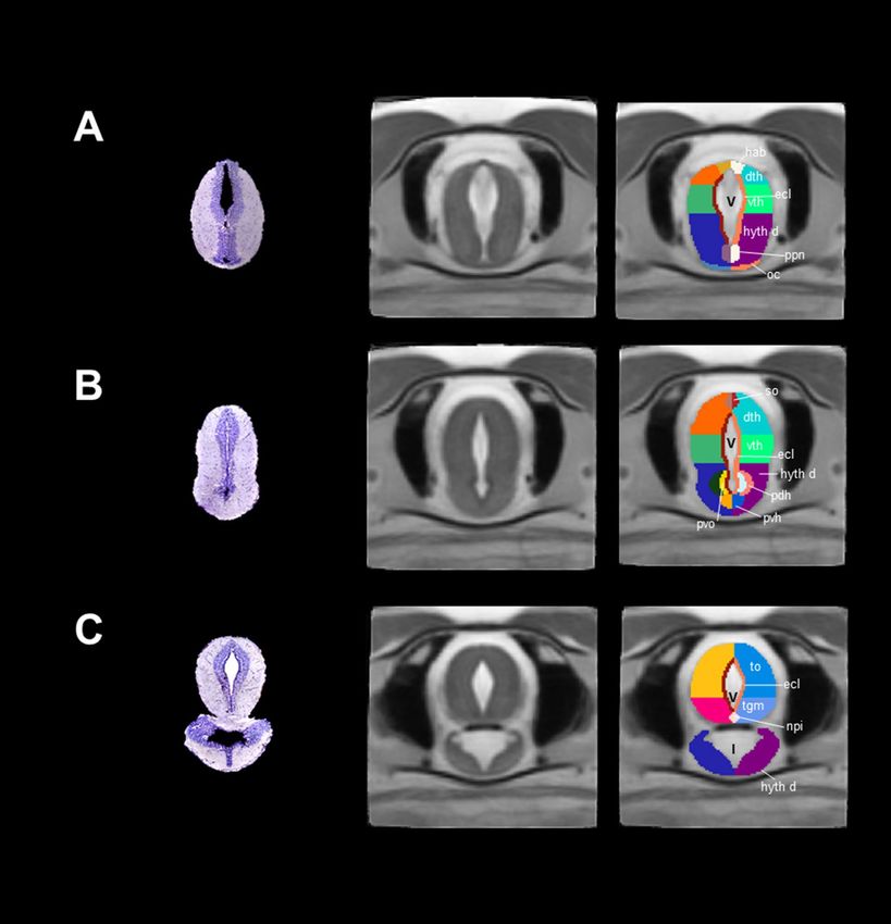
www.nature.com/scientificreports/
Figure 1. 3D reconstruction of the brain of Ambystoma mexicanum employing MRI. (A) Lateral views from the
left and right sides of the complete 3D reconstruction created from our template. (B) Ventral (left) and dorsal
(right) views of the complete 3D reconstruction. Every reconstruction shows each region in a different color
when manual delineation was possible. Yellow arrows denote the pituitary gland.
Olfactory bulb. Employing previous information of the olfactory bulb structures, we are able to distinguish
the olfactory nerve, the glomerular-, mitral- and granular cell layers and the anterior olfactory nucleus (Table 1,
Fig. 2). These structures contribute to the integration of information coming from olfactory neuroreceptors from
the nasal cavity. In mammals, some of the cell structures that contribute to integrate odorant signals are well
documented in terms of morphology, connectivity and function27, but information in amphibians is still scarce.
Telencephalon. This is the biggest brain structure in salamanders; the anterior part of the telencephalon
contains the pallium and subpallium and the lateral ventricles. The pallium and subpallium comprise the left
and right hemispheres. Axolotl pallium is interesting due to its capacity to regenerate after damage4, a trait prob-
ably present in other salamanders7. Because our MRI did not allow enough contrast to differentiate structures
such as the dorsal pallium, lateral pallium, medial pallium and striatum and septum, we decided to further
segment these regions manually using visual inspection from drawings in previous publications (Table 1). The
lateral ventricles (not segmented) were hyperintense in terms of contrast in our T2-weighted MRI images and
are depicted in Fig. 3A. The dorsal, lateral and medial pallium extend to the posterior part of the telencephalon
but present a different morphology as compared with the anterior telencephalon structures. Moreover, at this
brain location, the distance between the lateral ventricles (left and right) is reduced and even located adjacently
at a more posterior region, finally forming the third ventricle (Fig. 3B). We segmented the amygdaloid complex,
the lateral/medial forebrain bundle, the anterior preoptic nucleus, the thalamic eminence, and the pallial and
anterior commissures, according to previous annotations (Fig. 3C). Moreover, the choroid plexus, which secretes
cerebrospinal fluid into the vertebrate brain28 was evident using MRI, contrary to what is observed in histologi-
cal analysis due to the difficulty to preserve this structure.
Diencephalon. At the diencephalon level, we could identify two general structures, thalamus and dorsal
hypothalamus (Fig. 4). We segmented the thalamus into dorsal and ventral thalamus. At the top of the dorsal
thalamus, we identified the habenula (Fig. 4A) and the subcommissural organ (Fig. 4B). The habenula is a struc-
ture present in all vertebrates which participates in the integration of the limbic system, the basal ganglia and the
sensory information29. The subcommissural organ contains secretory ependymal cells located at the roof of the
third ventricle30. The contrast of the latter was hyperintense and centrally located with respect to the thalamus/
hypothalamus. The dorsal hypothalamus includes nuclei that surround the floor of the third ventricle such as
the posterior preoptic nucleus, paraventricular organ, pars dorsalis and pars ventralis hypotallami. All these
regions have been recognized as neurosecretory cells which participate in neuroendocrine systems (Fig. 4A,B)31.
Finally, the optic chiasm was also evident in our MRI images at the floor of the dorsal hypothalamus (Fig. 4A). In
this region, it is well accepted that nerve fibers cross and allow binocular communication between eyes and the
brain32; however, this does not always occur in the vertebrate optic c hiasm33. In Xenopus laevis, for example, fiber
Scientific Reports | (2021) 11:9850 | https://doi.org/10.1038/s41598-021-89357-3 4
Vol:.(1234567890)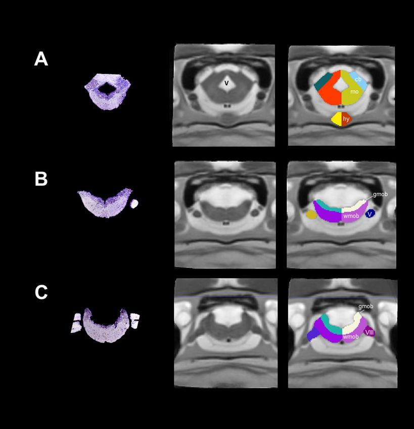
www.nature.com/scientificreports/
Figure 2. Coronal sections and slices of the olfactory bulb of Ambystoma mexicanum. Columns left, central and
right depict the Nissl-staining histological sections; the MRI images and the manual segmentation, respectively
created from our template. (A) left and right single olfactory nerves; (B) the olfactory nerve (on), glomerular
layer (g), and (C) mitral(m) and glomerular cell layers (gc).
cross contralaterally only after metamorphosis34. The type of projections situated in the optic chiasm of neotenic
and metamorphic axolotl is still unknown.
Mesencephalon. This section was subdivided into tectum, tegmentum and ventral hypothalamus. Since
contrast was insufficient to differentiate between tectum and tegmentum, we manually segmented these struc-
tures using previously published drawings (Fig. 4C, Table 1). Optic tectum is the roof of the mesencephalon,
and a big region in the salamander brain; it receives some nerves from the optic chiasm and processes visual
signals in vertebrates35. Tegmentum is located just ventrally from the optic tectum; from this region we were
able to segment the interpeduncularis nucleus, which is known to integrate information for the limbic system36.
At the coronal level, the ventral hypothalamus appears as the lower area of the brain, physically separated from
the optic tectum/tegmentum and surrounding the infundibulum (not segmented). However, the ventral hypo-
thalamus becomes smaller in the left and right sides at posterior levels eventually disappearing, whereas the
infundibulum increases in size. The third ventricle remains in the cerebral midline and the center of the optic
tectum/tegmentum.
Rhombencephalon/pituitary. This is the most posterior area of the brain and contains regions such as the
cerebellum and medulla oblongata. Cerebellum in amphibians is small in size with respect to other v ertebrates37,
Scientific Reports | (2021) 11:9850 | https://doi.org/10.1038/s41598-021-89357-3 5
Vol.:(0123456789)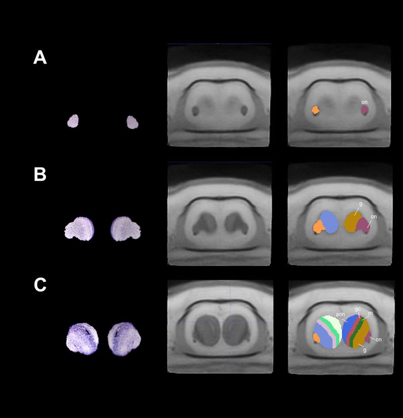
www.nature.com/scientificreports/
Figure 3. Coronal sections and slices of the telencephalon of Ambystoma mexicanum. Columns left, central and
right depict the Nissl-staining histological sections; the MRI images and the manual segmentation, respectively
created from our template. (A) dorsal (dp), lateral (lp) medial pallium (mp), striatum (str) and septum (s);
(B) choroid plexus (cp), medial part of amygdala (amc), pallial(cpa) and anterior commissures (ca), and (C)
thalamic eminence (em th) and anterior preoptic nuclei (npa). V ventricles.
a characteristic inversely correlated to the big genome of these species38. As previously reported, we detected a
small cerebellum in our MRI images (Fig. 5A). Medulla oblongata was identified as a single structure in the ante-
rior rhombencephalon, but we were able to segment gray from white matter in the posterior rhombencephalon,
according to differences in contrast in our MRI images and previous reports (Fig. 5B, Table 1). Moreover, we also
recognized some cranial nerves such as nervous trigeminus (V), nervous lateralis anterior and nervus octavus
(VIII) (Fig. 5B,C). Finally, we identified the pituitary gland, an endocrine organ which releases hormones in
response to some peptides coming from h ypothalamus39,40. In our coronal sections, the pituitary gland appears
at the lower part of the brain as the last visible signal of the ventral hypothalamus, increasing in size as it projects
posteriorly to finally decrease and disappear (Fig. 5A).
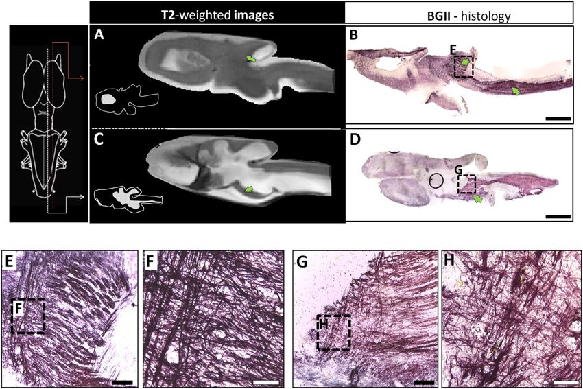
Myelin rich regions in the axolotl CNS. NS41,42 which
Myelin is a characteristic of jawed vertebrate C
allows saltatory propagation of action potentials. Patterns of myelin rich regions are well-documented in
mammals43–45 but in non-mammalian vertebrates, particularly in amphibians, the information is scarce. In an
approach to identify myelin in the axolotl CNS, and due to its contribution to T2-weighted contrast in MRI, we
performed specific myelin staining using the Black Gold II reagent in sagittal and coronal sections (Fig. 6A–D).
Myelin-rich regions were evident in sagittal sections of the medulla oblongata, cerebellum, optic tectum and
Scientific Reports | (2021) 11:9850 | https://doi.org/10.1038/s41598-021-89357-3 6
Vol:.(1234567890)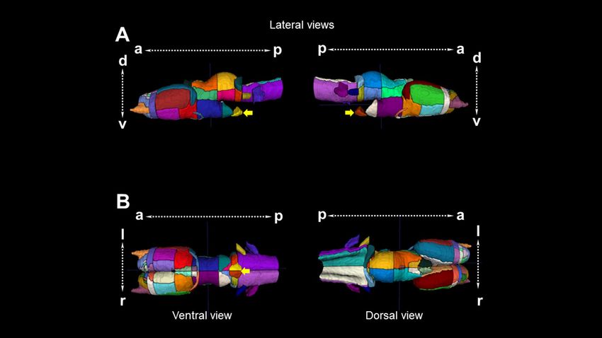
www.nature.com/scientificreports/
Figure 4. Coronal sections and slices of the diencephalon/mesencephalon of Ambystoma mexicanum.
Columns left, central and right depict the Nissl-staining histological sections; the MRI images and the
manual segmentation, respectively created from our template. (A) dorsal (dth) and ventral thalamus (vth),
subcommissural organ (so), optic chiasm (oc), habenula (hab); (B) regions surrounding the floor of the third
ventricle in the hypothalamus dorsalis (hyt d) such as the paraventricular organ (pvo), pars dorsalis (pdh) and
pars ventralis hypotalami (pvh); (C) the mesencephalon, including the optic tectum (to), tegmentum (tgm)and
interpeduncularis nucleus (npi). V ventricle, I infundibulum are indicated.
tegmentum (Fig. 6E–H); however, no myelin staining was observed in the anterior part of the brain, i.e., in the
olfactory bulb and telencephalon (Fig. 6D). These results were confirmed using coronal sections, in which we are
able to detect myelin in the lateral/medial forebrain blundle/amygdaloid complex (Fig. 7A), the ventral thalamus
(Fig. 7B) and the optic tectum/tegmentum (Fig. 7C). Both, sagittal and coronal sections showed that medulla
oblongata is a myelin rich region, particularly in the white matter, confirming the specificity of the myelin stain-
ing method (Fig. 7D). We were not able to detect myelin in the anterior part of the axolotl brain. This discrep-
ancy could have different explanations. In the developing mammal, the myelination process starts in the spinal
cord and gradually covers the posterior part of the CNS postnatally46,47. It is possible that even if the juvenile
stage of the axolotl brain is not fully myelinated, the adult brain could contain myelin in the anterior part of the
brain. Another explanation could be that the axolotl lost the capacity to myelinate axons in the anterior part of
the brain as result of neoteny and/or other unidentified physiological events. In this context, all amphibians go
through dramatic transformations that occur in the transition from a pre- to a post metamorphic stage; however,
axolotl can retain juvenile features throughout adulthood (neoteny/paedomorphism), resulting in adult (repro-
ducing) individuals that maintain juvenile (larval) traits). Lastly, amphibian myelin composition might not be
detected by Black Gold II. Even if this reagent is indeed myelin specific, amphibians have shown to present
Scientific Reports | (2021) 11:9850 | https://doi.org/10.1038/s41598-021-89357-3 7
Vol.:(0123456789)www.nature.com/scientificreports/
Figure 5. Coronal sections and slices of the medulla oblongata and pituitary gland of Ambystoma mexicanum.
Columns left, central and right depict the Nissl-staining histological sections; the MRI images and the manual
segmentation, respectively created from our template. (A) the rhombencephalon, where the cerebellum (cb),
the medulla oblongata (mo) and the pituitary gland (hy) at the lower part of the brain are indicated; (B) white
(wmob) and gray matter from medulla oblongata (gmob) and cranial nerves such as the nervous trigerminus
(V), and (C) nervous lateralis anterior and nervus octavus (VIII).
different compositions of myelin in terms of lipid c ontent48,49. Therefore, it is plausible that axolotl could have a
myelin that is not efficiently stained with Black Gold II in the anterior brain, at least at the stage of development
that was analyzed in the present study. To confirm this last hypothesis, other stages of axolotl development and/
or other methodologies to detect myelin could be tested.
Conclusions
The brain of vertebrates has been studied with different techniques, allowing researchers to visualize different
aspects of its anatomy, according to the methodology employed. MRI of the axolotl brain allowed us to provide
a 3D reconstruction of an amphibian brain and pituitary gland for the first time. In contrast with the results
from histological sections, we were able to visualize native morphology of the brain structures and to transform
this data in a 3D volume. Additionally, this is the first description of axolotl brain myelin distribution. Myelin
rich regions were observed in the posterior, but not the anterior brain, a finding that deserves further attention.
Overall, this work will provide a useful tool to explore new research avenues for the better understanding of this
interesting endemic paedomorphic species, which is an important model in different research areas, including
Scientific Reports | (2021) 11:9850 | https://doi.org/10.1038/s41598-021-89357-3 8
Vol:.(1234567890)www.nature.com/scientificreports/
Figure 6. MRI and BGII techniques in the axolotl brain. (A, C) sagittal sections of in-vivo T2-weighted images
and myelin staining with BGII; (B, D) the approximate myelin localization is indicated in the diagram on the
left. Insets in (A) and (C) show diagrams of the corresponding sagittal plane. In the histological micrographs
of BGII staining, (E) and (G) are amplifications of (B) and (D), while (F) and (H) are amplifications of (E) and
(G), respectively. Bars in (B), (D) indicate 1 mm, in (E), (G) they indicate 100 μm, while in (F, H) they indicate
20 μm.
nervous tissue regeneration, and that is currently listed as critically endangered by the International Union for
Conservation of Nature.
Materials and methods
Animals. Juvenile axolotls were kindly donated by Marco Terrones (Axolkali). All axolotls were maintained
and handled in accordance with protocols approved by the Ethics for Research Committee of the Instituto de
Neurobiología at the Universidad Nacional Autónoma de México (UNAM). The animal experiments were per-
formed following the guidelines for use of live amphibians and reptiles in field and laboratory research of the
American Society of Ichthyologists and Herpetologists. All the experiments complied with the ARRIVE guide-
lines.
Animals of around 3 months after hatching and weighing between 9 and 12 g were kept at our local housing
for at least 20 days prior to imaging at 18 °C in 14/10 light/dark cycles. After habituation, animals were anesthe-
tized by immersion with 0.4% tricain for 10–13 min.
MRI acquisition. MRI was performed on 20 animals at the National Laboratory for MRI, using a 7 T Bruker
Biospec 70/16 scanner and a Helium-cooled two-channel rat-head coil (Bruker Cryoprobe). Once anesthetized,
axolotls were placed in a plastic container and introduced in the scanner (Supplementary Fig. 2). A field map
was first obtained and used for shimming of the main magnetic field. Next, images were acquired using a three-
dimensionally encoded balanced steady-state gradient echo (True FISP) sequence with the following parameters:
TR = 4.4 ms; TE = 2.2 ms; flip angle = 30°; NEX = 3; FOV = 20.48 × 15 × 10.24 mm3 and matrix = 256 × 188 × 128,
yielding isometric voxel dimensions of 0.080 mm; scan time = 5 min 50 s. We also acquired diffusion-weighted
images with a scan time of 11 min 44 s (not reported herein). Total scan time was 17 min 34 s.
MRI template construction. Individual image volumes were visually inspected for quality. Out of n = 20
images, n = 6 were rejected due to poor quality, with a final sample size of n = 14 for template construction.
Images were converted from Bruker format to NIFTI using the software Bruker2nifti50, and then from NIFTI
to MINC using nii2mnc (https://github.com/BIC-MNI/minc-toolkit-v2). Several steps were followed to reach
Scientific Reports | (2021) 11:9850 | https://doi.org/10.1038/s41598-021-89357-3 9
Vol.:(0123456789)www.nature.com/scientificreports/
Figure 7. Myelination in the axolotl. Different coronal sections of the axolotl brain. (A) lateral/medial forebrain
bundle/amygdaloid complex; (B) the dorsal thalamus; (C) the optic tectum/tegmentum, and (D) medulla
oblongata. Below are amplifications for each section. Note the myelination gradient in the anteroposterior
direction. The white matter of medulla oblongata is the structure with the greatest presence of myelin. The bars
in the micrographs indicate 500 µm in (A–D), 100 µm in (A1, B1, C1, C2), and 20 µm in (A2, B2, D1, D2).
the final template construction. STEP1: each image was preprocessed using the following steps and commands;
(1) cleaned header and center image to coordinate 0, 0, 0 (https://github.com/CoBrALab/minctoolkitextras/
blob/master/clean_and_center_minc.pl); (2) reorientation to RAS: 90° rotation of the y axis using volrot; (3)
y axis volume flip using volflip; and (4) N4 bias field correction using an Otsu mask51. STEP 2: we constructed
a first template using the antsMultivariateTemplateConstruction2.sh script (https://github.com/ANTsX/ANTs/
blob/master/Scripts/antsMultivariateTemplateConstruction2.sh)52. STEP 3: an initial brain mask was manually
defined from the first template. STEP 4: fine tuning of the N4 correction and the first template; (1) inverse
registration of the template brain mask to the native space for each subject; (2) reduce field of view to near
brain (crop) using ExtractRegionFromImageByMask; (3) re-run the N4 bias field correction with the improved
individual mask; (4) re-center the images; (5) rotation of the first template to a precise alignment with the axes.
STEP 5: construction of the second template using the raw files and the first template as target. STEP 6: resa-
mpled second template from 0.08 × 0.08 × 0.08 mm to 0.06 × 0.06 × 0.06 mm. STEP 7: construction of the third
template using the second template for warping. STEP 8: resampled final template from 0.06 × 0.06 × 0.06 mm
to final resolution of 0.04 × 0.04 × 0.04 mm. STEP 9: construction of the final template using the third template
for warping. The process is shown in Supplementary Fig. 3. Upsampling of the target templates was applied to
increase model resolution53.
MRI segmentation. Neuroanatomical segmentation was done manually drawing regions of interest (ROIs)
using the ITK-SNAP (version 3.8.0)54 on the final template (0.04 × 0.04 × 0.04 mm). We constructed two atlases:
(1) 82-ROI atlas with sub-segmentation of pallium, telencephalon, tectum and rhombencephalon based on the
available histological anatomic annotations and the nomenclature obtained from the different studies (Table 1),
and (2) 64-ROI atlas without sub-segmentation of the pallium, telencephalon, tectum, and rhombencephalon
(Supplementary Table 1). The ROIs were drawn over our final template while examining all three stereotaxic
planes to reduce inconsistencies in delineation across slices. Labels were hierarchically constructed to simplify
MRI analysis in higher resolutions. There are three levels that describe larger regions: (1) ROI; (2) embryological
origin of the neural tube (olfactory bulb, telencephalon, diencephalon, mesencephalon, endocrine and rhom-
bencephalon), and (3) hemispheres (right and left). Each ROI has a defined abbreviation. The atlas is openly
available through Zenodo (https://doi.org/10.5281/zenodo.4595016).
Histological staining. For histological examination, brains were fixed in PFA (4%) and cryopreserved in
sucrose (30%), then coronal or sagittal (40 μm) cryosections (one juvenile brain for every orientation) were cut
Scientific Reports | (2021) 11:9850 | https://doi.org/10.1038/s41598-021-89357-3 10
Vol:.(1234567890)www.nature.com/scientificreports/
and mounted on electrocharged slides and stored at − 20 °C until staining with the Black Gold II compound
(EMD Millipore Corp., Billerica, MA, USA), using a modified protocol described by55. Briefly, tissue sections
were rehydrated in distilled water for about 2 min at RT. Then, the slides were incubated in 0.3% Black Gold II
solution, dissolved in 0.9% saline vehicle (NaCl), and heated at 60–65 °C for at least 40 min until the staining
was complete. Next, the slides were rinsed for 2 min in Phosphate-Buffered Saline (PBS, RT), transferred to a 1%
sodium thiosulfate solution for 3 min at 60–65 °C, and rinsed two or three times with distilled water. Sections
stained with Black Gold II were counterstained with Nissl. Additional alternate slides were stained only with
the Nissl protocol. For this, slides were transferred to a solution of 0.1–0.2% cresyl violet (blue; EMD Millipore
Corp., Billerica, MA, USA) in 0.1% acetic acid for 5 min and then rinsed two times with distilled water. After, the
tissue was dehydrated using sequential graduated alcohol solutions (50, 70 and 96%) and immersed in xylene for
1 min. The slides were then coverslipped with Entellan mounting medium.
Received: 10 December 2020; Accepted: 12 April 2021
References
1. Voss, S. R. & Shaffer, H. B. Adaptive evolution via a major gene effect: Paedomorphosis in the Mexican axolotl. Proc. Natl. Acad.
Sci. USA 94, 14185–14189 (1997).
2. Satoh, A., Mitogawa, K. & Makanae, A. Nerve roles in blastema induction and pattern formation in limb regeneration. Int. J. Dev.
Biol. 62, 605–612 (2018).
3. Maden, M., Manwell, L. A. & Ormerod, B. K. Proliferation zones in the axolotl brain and regeneration of the telencephalon. Neural
Dev. 8, 1 (2013).
4. Amamoto, R. et al. Adult axolotls can regenerate original neuronal diversity in response to brain injury. Elife 5, e13998 (2016).
5. Medina, L. & Abellán, A. Development and evolution of the pallium. Semin. Cell Dev. Biol. 20, 698–711 (2009).
6. Tazaki, A., Tanaka, E. M. & Fei, J.-F. Salamander spinal cord regeneration: The ultimate positive control in vertebrate spinal cord
regeneration. Dev. Biol. 432, 63–71 (2017).
7. Lust, K. & Tanaka, E. M. A comparative perspective on brain regeneration in amphibians and teleost fish. Dev. Neurobiol. 79,
424–436 (2019).
8. Glasser, M. F. et al. The Human Connectome Project’s neuroimaging approach. Nat. Neurosci. 19, 1175–1187 (2016).
9. Ma, Y. et al. A three-dimensional digital atlas database of the adult C57BL/6J mouse brain by magnetic resonance microscopy.
Neuroscience 135, 1203–1215 (2005).
10. Valdés-Hernández, P. A. et al. An in vivo MRI template set for morphometry, tissue segmentation, and fMRI localization in rats.
Front. Neuroinform. 5, 26 (2011).
11. Wisner, K., Odintsov, B., Brozoski, D. & Brozoski, T. J. Ratat1: A digital rat brain stereotaxic atlas derived from high-resolution
MRI images scanned in three dimensions. Front. Syst. Neurosci. 10, 64 (2016).
12. Kovacević, N. et al. A three-dimensional MRI atlas of the mouse brain with estimates of the average and variability. Cereb. Cortex
15, 639–645 (2005).
13. Vellema, M., Verschueren, J., Van Meir, V. & Van der Linden, A. A customizable 3-dimensional digital atlas of the canary brain in
multiple modalities. Neuroimage 57, 352–361 (2011).
14. Billings, B. K. et al. A three-dimensional digital atlas of the Nile crocodile (Crocodylus niloticus) forebrain. Brain Struct. Funct.
225, 683–703 (2020).
15. Hoops, D. et al. A 3D MRI-based atlas of a lizard brain. J. Compar. Neurol. 526, 2511–2547 (2018).
16. Ullmann, J. F. P., Cowin, G., Kurniawan, N. D. & Collin, S. P. A three-dimensional digital atlas of the zebrafish brain. Neuroimage
51, 76–82 (2010).
17. Simões, J. M., Teles, M. C., Oliveira, R. F., Van der Linden, A. & Verhoye, M. A three-dimensional stereotaxic MRI brain atlas of
the cichlid fish Oreochromis mossambicus. PLoS ONE 7, e44086 (2012).
18. Herrick, C. J. The Brain of the Tiger Salamander, Ambystoma tigrinum. (University of Chicago Press, Chicago, 1948).
19. Wang, H. H., Li, L. Y., Wang, L. W. & Liang, C. C. Morphological and histological studies on the telencephalon of the salamander
Onychodactylus fischeri. Neurosci. Bull. 23(3), 170–174 (2007).
20. Mühlenbrock-Lenter, S., Roth, G. & Laberge, F. Evolution of the Pallium in Amphibians. In Encyclopedia of Neuroscience (eds
Binder, M. D. et al.) (Springer, 2009).
21. Clairambault, P. et al. Organization of the serotoninergic system in the brain of two amphibian species, Ambystoma mexicanum
(Urodela) and Typhlonectes compressicauda (Gymnophiona). Anat. Embryol. 190, 87–99 (1994).
22. Laberge, F., Mühlenbrock-Lenter, S., Grunwald, W. & Roth, G. Evolution of the Amygdala: New insights from studies in Amphib-
ians. Brain Behav. Evol. 67, 177–187 (2006).
23. Krug, L., Wicht, H. & Northcutt, R. G. Afferent and efferent connections of the thalamic eminence in the axolotl Ambystoma
mexicanum. Neurosci. Lett. 149(2), 145–148 (1993).
24. Beltramo, M. et al. Immunolocalization of aromatic L-amino acid decarboxylase, tyrosine hydroxylase, dopamine, and serotonin
in the forebrain of Ambystoma mexicanum. J. Comp. Neurol. 391(2), 227–247 (1998).
25. Dicke, U., Wallstein, M. & Roth, G. 5-HT-like immunoreactivity in the brains of plethodontid and salamandrid salamanders
(Hydromantes italicus, Hydromantes genei, Plethodon jordani, Desmognathus ochrophaeus, Pleurodeles waltl): An immunohisto-
chemical and biocytin double-labelling study. Cell Tissue Res. 287, 513–523 (1997).
26. Bidaud, I. et al. Distribution of the mRNAs encoding the thyrotropin-releasing hormone (TRH) precursor and three TRH receptors
in the brain and pituitary of Xenopus laevis: Effect of background color adaptation on TRH and TRH receptor gene expression. J.
Comp. Neurol. 477(1), 11–28. https://doi.org/10.1002/cne.20235 (2004) (PMID: 15281077).
27. Nagayama, S., Homma, R. & Imamura, F. Neuronal organization of olfactory bulb circuits. Front. Neural Circ. 8, 98 (2014).
28. Bill, B. R. & Korzh, V. Choroid plexus in developmental and evolutionary perspective. Front. Neurosci. 8, 363 (2014).
29. Freudenmacher, L., von Twickel, A. & Walkowiak, W. The habenula as an evolutionary conserved link between basal ganglia,
limbic, and sensory systems—A phylogenetic comparison based on anuran amphibians. J. Comp. Neurol. 528, 705–728 (2020).
30. Meiniel, A. The secretory ependymal cells of the subcommissural organ: Which role in hydrocephalus?. Int. J. Biochem. Cell Biol.
39, 463–468 (2007).
31. Hodos, W. Evolution of the hypothalamus in anamniotes. In Encyclopedia of Neuroscience (eds Binder, M. D., Hirokawa, N. &
Windhorst, U.) 1361–1363 (Springer-Verlag, Berlin, Heidelberg, 2008).
32. Larsson, M. Binocular vision, the optic chiasm, and their associations with vertebrate motor behavior. Front. Ecol. Evol. 3, 89
(2015).
Scientific Reports | (2021) 11:9850 | https://doi.org/10.1038/s41598-021-89357-3 11
Vol.:(0123456789)www.nature.com/scientificreports/
33. Larsson, M. The optic chiasm: A turning point in the evolution of eye/hand coordination. Front. Zool. 10, 41 (2013).
34. Jeffery, G. & Erskine, L. Variations in the architecture and development of the vertebrate optic chiasm. Prog. Retin. Eye Res. 24,
721–753 (2005).
35. Ingle, D. J. Optic tectum. Compar. Neurosci. Neurobiol. https://doi.org/10.1007/978-1-4899-6776-3_40 (1988).
36. Morley, B. J. The interpeduncular nucleus. Int. Rev. Neurobiol. 28, 157–182 (1986).
37. Butts, T., Modrell, M. S., Baker, C. V. H. & Wingate, R. J. T. The evolution of the vertebrate cerebellum: Absence of a proliferative
external granule layer in a non-teleost ray-finned fish. Evol. Dev. 16, 92–100 (2014).
38. Roth, G. & Walkowiak, W. The influence of genome and cell size on brain morphology in amphibians. Cold Spring Harb. Perspect.
Biol. 7, a019075 (2015).
39. Sower, S. A. Breaking dogma on the hypothalamic-pituitary anatomical relations in vertebrates. Endocrinology 156, 3882–3884
(2015).
40. Lazcano, I. et al. Evolution of thyrotropin-releasing factor extracellular communication units. General Compar. Endocrinol. https://
doi.org/10.1016/j.ygcen.2020.113642 (2020).
41. Preston, M. A. & Macklin, W. B. Zebrafish as a model to investigate CNS myelination. Glia 63, 177–193 (2015).
42. Czopka, T. Insights into mechanisms of central nervous system myelination using zebrafish. Glia 64, 333–349 (2016).
43. Ibarrola, N. & Rodríguez-Peña, A. Hypothyroidism coordinately and transiently affects myelin protein gene expression in most
rat brain regions during postnatal development. Brain Res. 752, 285–293 (1997).
44. Barradas, P. C., Vieira, R. S. & De Freitas, M. S. Selective effect of hypothyroidism on expression of myelin markers during develop-
ment. J. Neurosci. Res. 66, 254–261 (2001).
45. Downes, N. & Mullins, P. The development of myelin in the brain of the juvenile rat. Toxicol. Pathol. 42, 913–922 (2014).
46. Jacque, C. M., Collet, A., Raoul, M., Monge, M. & Gumpel, M. Functional maturation of the oligodendrocytes and myelin basic
protein expression in the olfactory bulb of the mouse. Dev. Brain Res. 21, 277–282 (1985).
47. Verity, A. N. & Campagnoni, A. T. Regional expression of myelin protein genes in the developing mouse brain: in situ hybridization
studies. J. Neurosci. Res. 21, 238–248 (1988).
48. Ki, P. F. & Kishimoto, Y. The lipid composition of urodele myelin which lacks hydroxycerebroside and hydroxysulfatide. J. Neuro-
chem. 42, 994–1000 (1984).
49. Ki, P. F., Kishimoto, Y., Stanley, E. F. & Griffin, J. W. Structure and function of urodele myelin lacking alpha-hydroxy fatty acid-
containing galactosphingolipids: Slow nerve conduction and unusual myelin thickness. Brain Res. 345, 19–24 (1985).
50. Ferraris, S. et al. Bruker2nifti: Magnetic resonance images converter from bruker ParaVision to NIfTI format. J. Open Source Softw.
2, 354 (2017).
51. Tustison, N. J. et al. N4ITK: Improved N3 bias correction. IEEE Trans. Med. Imaging 29, 1310–1320 (2010).
52. Avants, B. B. et al. A reproducible evaluation of ANTs similarity metric performance in brain image registration. Neuroimage 54,
2033–2044 (2011).
53. Janke, A. L. & Ullmann, J. F. P. Robust methods to create ex vivo minimum deformation atlases for brain mapping. Methods 73,
18–26 (2015).
54. Yushkevich, P. A. et al. User-guided 3D active contour segmentation of anatomical structures: Significantly improved efficiency
and reliability. Neuroimage 31, 1116–1128 (2006).
55. Schmued, L. et al. Introducing Black-Gold II, a highly soluble gold phosphate complex with several unique advantages for the
histochemical localization of myelin. Brain Res. 1229, 210–217 (2008).
Acknowledgements
The authors would like to thank the technical assistance of M. C. Patricia Villalobos, Guadalupe Yasmín Hernán-
dez Linares, M.C. Leopoldo González Santos, Carlos S. Flores and Dr. Ericka A. de los Ríos. This work received
support from Luis A. Aguilar (Laboratorio Nacional de Visualización Científica Avanzada), and the Laboratorio
Nacional de Imagenología por Resonancia Magnética. We would also like to thank Gabriel A. Devenyi for his
support. This work was supported by a Grant from PAPIIT IN204920.
Author contributions
I.L.: Conceptualization, methodology, investigation, writing—review and editing, formal analysis, resources,
writing—original draft, writing—review and editing, visualization. A.C.M.: Methodology, investigation, writing—
review and editing, visualization. L.C.: Conceptualization, writing—review and editing, supervision. J.J.O.R.:
Methodology, investigation. E.A.G.V.: Conceptualization, methodology, software, formal analysis, resources,
writing—original draft, writing—review and editing, visualization. A.O.: Conceptualization, project adminis-
tration, supervision, funding acquisition, writing—original draft, writing—review and editing, visualization.
Competing interests
The authors declare no competing interests.
Additional information
Supplementary Information The online version contains supplementary material available at https://doi.org/
10.1038/s41598-021-89357-3.
Correspondence and requests for materials should be addressed to I.L. or E.A.G.-V.
Reprints and permissions information is available at www.nature.com/reprints.
Publisher’s note Springer Nature remains neutral with regard to jurisdictional claims in published maps and
institutional affiliations.
Scientific Reports | (2021) 11:9850 | https://doi.org/10.1038/s41598-021-89357-3 12
Vol:.(1234567890)www.nature.com/scientificreports/
Open Access This article is licensed under a Creative Commons Attribution 4.0 International
License, which permits use, sharing, adaptation, distribution and reproduction in any medium or
format, as long as you give appropriate credit to the original author(s) and the source, provide a link to the
Creative Commons licence, and indicate if changes were made. The images or other third party material in this
article are included in the article’s Creative Commons licence, unless indicated otherwise in a credit line to the
material. If material is not included in the article’s Creative Commons licence and your intended use is not
permitted by statutory regulation or exceeds the permitted use, you will need to obtain permission directly from
the copyright holder. To view a copy of this licence, visit http://creativecommons.org/licenses/by/4.0/.
© The Author(s) 2021
Scientific Reports | (2021) 11:9850 | https://doi.org/10.1038/s41598-021-89357-3 13
Vol.:(0123456789)You can also read