The emerging vertebrate model species for neurophysiological studies is Danionella cerebrum, new species (Teleostei: Cyprinidae) - Nature
←
→
Page content transcription
If your browser does not render page correctly, please read the page content below
www.nature.com/scientificreports
OPEN The emerging vertebrate model
species for neurophysiological
studies is Danionella cerebrum, new
species (Teleostei: Cyprinidae)
Ralf Britz1,2*, Kevin W. Conway3,4 & Lukas Rüber5,6
The four described species of Danionella are tiny, transparent fishes that mature at sizes between
10–15 mm, and represent some of the most extreme cases of vertebrate progenesis known to date.
The miniature adult size and larval appearance of Danionella, combined with a diverse behavioral
repertoire linked to sound production by males, have established Danionella as an important model
for neurophysiological studies. The external similarity between the different species of Danionella
has offered an important challenge to taxonomic identification using traditional external characters,
leading to confusion over the identity of the model species. Using combined morphological and
molecular taxonomic approaches, we show here that the most extensively studied species of
Danionella is not D. translucida, but represents an undescribed species, D. cerebrum n. sp. that is
externally almost identical to D. translucida, but differs trenchantly in several internal characters.
Molecular analyses confirm the distinctiveness of D. cerebrum and D. translucida and suggest that the
two species are not even sister taxa. Analysis of the evolution of sexual dimorphisms associated with
the Weberian apparatus reveals significant increases in complexity from the simpler condition found in
D. dracula, to most complex conditions in D. cerebrum, D. mirifica and D. translucida.
Progenesis is an evolutionary process that speeds up gonad development in relation to somatic development, so
that the result is a small organism with larval features yet ripe g onads1,2. Prominent vertebrate examples of this
type of heterochronic change are the species of the cyprinid genus Danionella. So far, four species of the genus
Danionella, maturing at sizes of 10–15 mm in length and including some of the smallest fishes and vertebrates,
have been described from Myanmar and northeastern India3–6. Though progenesis may act at the level of an
individual character or character complex, leading to a mosaic of paedomorphic features in the adult, Danionella
is unusual in that it shows organism–wide progenesis or developmental truncation, a heterochronic change that
leads to tiny adult organisms with an overall larval a ppearance7–10.
It is this larval condition of their adult skeleton, in which the skull roof is missing and the brain is covered only
by skin, that has led various researchers to turn to Danionella as the adult vertebrate model organism to study
neurophysiological questions by deep imaging their brain activity in vivo11–14. Species of the genus Danionella
are further remarkable in possessing a number of unique morphological novelties in males. These include a
redirection of their gut and genital ducts so that both open in between the pelvic fins, a large drumming muscle
that originates from expanded bony flanges on the os suspensorium of their Weberian apparatus and inserts on a
hypertrophied fifth rib, and a conical drumming cartilage that probably works like a drumstick on the anterior
swimbladder chamber4–6,9. This highly complex vocalization apparatus has been identified as a promising organ
system to study n europhysiologically11.
While the organism-wide progenetic nature of Danionella has been the prerequisite for their establishment
as neurophysiological model organisms, their larval appearance has a downside for systematists: few of the
commonly used external character systems are available for study, as colour pattern is highly reduced to a few
1
Senckenberg Natural History Collections Dresden, Museum of Zoology, 01109 Dresden, Germany. 2Research
Associate, Department of Life Sciences, Natural History Museum, London SW75BD, UK. 3Department of Ecology
and Conservation, Biology and Biodiversity Research and Teaching Collections, Texas A&M University, College
Station, College Station, TX 77543, USA. 4Research Associate, Ichthyology, Australian Museum Research Institute,
1 William Street, Sydney, NSW 2010, Australia. 5Naturhistorisches Museum Bern, 3005 Bern, Switzerland. 6Aquatic
Ecology and Evolution, Institute of Ecology and Evolution, University of Bern, 3012 Bern, Switzerland. *email:
ralf.britz@senckenberg.de
Scientific Reports | (2021) 11:18942 | https://doi.org/10.1038/s41598-021-97600-0 1
Vol.:(0123456789)
www.nature.com/scientificreports/
D. cerebrum D. translucida D. mirifica D. dracula D. priapus
Dorsal-fin rays 8–9 8–9 8–10 7–8 9–10
Anal-fin rays 15–18 12–15 17–20 12–14 20–21
Principal caudal-fin rays 9+9 8–9 + 8–9 9+9 7–8 + 7–8 9+9
Dorsal procurrent caudal-fin rays 5–8 5–7 5–6 3–4 7–8
Ventral procurrent caudal-fin rays 5–8 3–7 6–7 2–3 6–8
Pectoral-fin rays 6 6–7 6–7 7 8
Pelvic-fin rays 5 5 5 6 5
Vertebrae 33–35 32–34 36–37 36–37 37–38
Abdominal 13–14 12–14 14–15 15–16 15–16
Caudal 19–21 19–21 21–22 20–22 22–23
Ribs 7–8 7–8 7–9 8 10
Anal-fin pterygiophores 14–17 11–14 16–19 11–13 19–20
Insertion of anal-fin pterygiophores 22–24 19–21 23–25 23–24 27–28
Table 1. Comparative meristic information of the five species of Danionella. Information for D. mirifica, D.
dracula and D. priapus taken from Britz4,5 and Britz et al.6,8. Counts of D. cerebrum (n = 32) based on MTD
39986, MTD 39987 and MTD 39989, those of D. translucida (n = 28) on MTD 39992, MTD 39993.
melanophores, and scales are absent. The four recognized species have thus been distinguished by fin-ray counts
and internal anatomy4–6 and are difficult to tell apart when alive.
With the two species of Danionella that are used in these studies, D. dracula and D. translucida, at the start
of their career as vertebrate models, we began to take a comparative taxonomic and anatomical look at these
fishes. The skeletal anatomy of D. dracula was already the subject of a monographic s tudy9, but information on
D. translucida is so far limited to the scant original description3. When comparing cleared and double stained
specimens of D. translucida from the type locality and from additional localities around the southern tip of the
Rakhine Yoma in Myanmar, we noticed a striking difference in the chondrocranium between individuals from
different localities. This led us to compare in detail the skeletal anatomy of these two forms, and the other species
of Danionella, and to complement our anatomical study with molecular data. Here we show that the organism
that has been introduced as a promising model organism for neurophysiological research is not D. translucida,
but an undescribed species, which we describe here in detail as Danionella cerebrum new species. We discuss
anatomical differences between D. cerebrum new species and the other four species of Danionella and comment
on the anatomical changes in the Weberian apparatus and associated structures during the evolution of this
genus. We conclude by stressing that the study of Danionella presents a promising system to understand major
morphological changes as the five species show anatomical differences far greater even than between species of
only remotely related genera within the subfamily Danioninae.
Results
Taxonomy. Danionella cerebrum new species. Holotype. BMNH 2021.8.30.1, female, 12.6 mm SL, Myan-
mar, Yangon Division, Hmawbi, roadside canal draining into Thandabin Chaung, 17° 06.200′ N 96° 02.890′ E,
Britz et al., 18 Oct 2008.
Paratypes. MTD 39985, 245 specimens, 7.5–12.0 mm SL, same information as holotype. MTD 39986, 20
c&s, 10.5–13.5 mm SL, same information as holotype. ZRC 62210, 36, 7.2–12.4 mm SL, same information as
holotype. NRM 71156, 36, 7.7–10.8 mm SL, same information as holotype. USNM 439009, 36, 7.8–12.0 mm SL,
same information as holotype. BMNH 20.21.6.2.1-200, 200, 7.2–11.8 mm SL, same information as holotype.
MTD 39987, 1, 10.4 mm SL, c&s, Myanmar, Bago Division, Daik U, Daikme Chaung, 17° 48.267′ N 96° 39.826′
E, Britz et al., 19 Oct 2008. MTD 39988, 10–12 mm SL, 4, unnamed stream 14.4 km NE of Daik U, 17.89314° N
96.56361°E, Britz et al., 19 Oct 2008. MTD 39989, 1 c&s, 11.4 mm SL, unnamed stream 14.4 km NE of Daik U,
17.89314° N 96.56361°E, Britz et al., 19 Oct 2008.
Additional material (non type): MTD 39990, 29 specimens, 7.8–11.5 mm SL, aquarium shop in Bago, report-
edly from Thandabin Chaung, Britz et al., 19 Oct 2008. MTD 39991, 8 c&s, 8.5–11.3 mm SL, aquarium shop in
Bago, reportedly from Thandabin Chaung, Britz et al., 19 Oct 2008.
Diagnosis. Danionella cerebrum is distinguished from D. translucida, D. dracula, and D. priapus by the num-
ber of anal-fin rays (15–18 vs. 12–15 in D. translucida, 12–14 in D. dracula, 20–21 in D. priapus, Table 1). It is
further distinguished from D. mirifica, D. dracula, and D. priapus by fewer vertebrae (33–35 vs. 36–38, Table 1),
from D. priapus and D. dracula by fewer pectoral-fin rays (6 vs. 8 in D. priapus and 7 in D. dracula, Table 1),
from D. translucida and D. dracula by the presence of a ventromedially directed cartilage flange from the taenia
marginalis anterior that approaches the trabecula communis (vs. absence, Fig. 2c,d), and from D. dracula by the
presence in the male of bony flanges on the outer arm of the os suspensorium and a connection of these to the
lateral process of vertebra 2 (vs. absence of flanges and of connection to second lateral process), the presence
of a maxillo-mandibular cartilage (vs. absence), the absence of odontoid processes in the male (vs. presence),
more anal-fin pterygiophores (14–17 vs. 11–13, Table 1), more principal caudal fin rays (9 + 9 vs. 8 + 8, Table 1)
Scientific Reports | (2021) 11:18942 | https://doi.org/10.1038/s41598-021-97600-0 2
Vol:.(1234567890)
www.nature.com/scientificreports/

                                  Figure 1.  Danionella cerebrum. (a) male (ca. 10 mm SL) and (b) female (ca. 12 mm SL) in life, not preserved;
                                  note yellowish chromatophores dorsally on head, melanophores scattered in rows on body in both sexes, and
                                  eggs covered by large melanophores in female; (c) MTD 39985, paratype, 10.4 mm SL, male and (d) BMNH
                                  2021.8.30.1, holotype, 12.6 mm SL, female (below), white arrows mark position of vent, which is shifted
                                  anteriorly to the pelvic fins in males; (e) Weberian apparatus in male, MTD 39992, paratype, 11.7 mm SL and
                                  (f) female, MTD 39992, paratype, 11.8 mm SL, in lateral view; the same in male (g) and female (h) in frontal
                                  view; (e) and (g) black arrowhead marks connection between lateral process and outer arm of os suspensorium,
                                  star marks connecting flanges between inner and outer arms of os suspensorium and red arrow marks posterior
                                  extension of inner arm of os suspensorium covering swimbladder dorsally. Abbreviations: cl, claustrum; dc,
                                  drumming cartilage; ios, inner arm of os suspensorium; nc, neural complex; oos, outer arm of os suspensorium; r,
                                  rib; sc, scaphium; sw, swimbladder.

                                                                Range              Average   Standard deviation
                                   Standard length (SL) in mm   10.0–12.6 (12.6)
                                   In % SL
                                   Head length (HL)             20–21.9 (21.4)     21.0      0.6
                                   Predorsal length             66.1–71.4 (69.6)   69.6      1.4
                                   Preanal length               52.5–56.3 (54.5)   54.5      1.3
                                   Body depth at vent           15.4–19.0 (19.0)   17.3      1.1
                                   Caudal peduncle depth         7.7–9.0 (8.7)      8.4      0.4
                                   In % HL
                                   Eye diameter                 28.0–31.8 (29.6)   30.0      1.8
                                   Snout length                 20.0–24.0 (22.2)   22.6      1.4

                                  Table 2.  Morphometric information of 10 specimens (5 males, 5 females) of Danionella cerebrum (BMNH
                                  2021.8.30.1, MTD 39985), information of holotype in parentheses behind range.

                                  and fewer pelvic-fin rays (5 vs. 6, Table 1). Danionella cerebrum can be further distinguished from the similar
                                  syntopically living D. translucida, by the last dorsal-fin ray inserted opposite to the last anal-fin ray (vs. last
                                  dorsal-fin ray inserted posterior to last anal-fin ray, Fig. 2a,b), by the last anal-fin pterygiophore inserted in
                                  front of haemal spine of vertebra 22–24 (vs. 19–21), by the lateral process of the second vertebra blade-like (vs.
                                  axe shaped, Fig. 2g,h), and by the distal tip of the fused inner arms of the ossa suspensoria bifurcated (vs. single,
                                  Fig. 2e,f) and not reaching the middle of the anterior swimbladder (vs. curving around and reaching middle of
                                  anterior swimbladder, Fig. 2e,f).

                                  Description. Maximum known size 13.5 mm SL. General body shape illustrated in Fig. 1a–d. Morphometric
                                  information based on 10 specimens is provided in Table 2. Head and eye are large; mouth supraterminal. Nostrils
                                  well developed. Lateral line canals and pores on head and body absent. Body elongate with a short dorsal fin,

Scientific Reports |   (2021) 11:18942 |                   https://doi.org/10.1038/s41598-021-97600-0                                                3

                                                                                                                                                Vol.:(0123456789)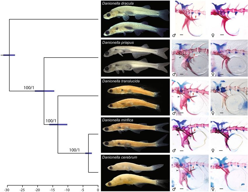
www.nature.com/scientificreports/

                                            Figure 2.  Comparison of skeletal characters of cleared and stained females of Danionella cerebrum, and D.            ▸
                                            translucida. Whole skeleton of D. cerebrum (a), MTD 39986, paratype, 11.5 mm SL and D. translucida (b),
                                            MTD 39992, 13.1 mm SL in lateral view, illustrating differences in relative position of dorsal and anal fins;
                                            vertical lines mark base of anteriormost and posteriormost dorsal-fin ray, respectively, in relation to anal fin.
                                            Neurocranium of D. cerebrum, MTD 39986, paratype,13.5 mm (c) and D. translucida (d), MTD 39992, 11.2 mm
                                            SL, in dorsal view, star marks ventromedial cartilage flange in (c), which is absent in (d). Tip of fused inner
                                            arms of ossa suspensoria in D. cerebrum (e) and D. translucida (f), in ventral view; note bifurcated tip in (e) and
                                            single tip in (f). Vertebrae of Weberian apparatus in D. cerebrum (g) and D. translucida (h), in dorsal view; note
                                            caudally expanded lateral process in (h), margin marked by line of grey dots. Abbreviations: in, intercalarium;
                                            ios, inner arm of os suspensorium; lp, lateral process of second vertebra; oos, outer arm of os suspensorium; r, rib;
                                            sc, scaphium; sw, swimbladder; tr, tripus.

                                            situated opposite to posterior half of long anal fin. Tip of dorsal fin situated posterior to a vertical line through
                                            tip of anal fin. Caudal fin furcate with remnants of larval-fin fold in front of its dorsal and ventral margins. A
                                            remnant of pre-anal larval-fin fold present in adult females, absent in adult males. Anus and genital papilla of
                                            mature males located between pelvic fins, at normal position in front of anal fin in females and in immature
                                            males between pelvic and anal fins. A window (pseudotympanum) present in body musculature at lateral side of
                                            anterior swim bladder chamber, rendering its pigmented surface visible. Scales absent.
                                                Vertebrae totaling 33(7), 34(23), 35(2), abdominal vertebrae 13(15), 14(16) or 15(1); caudal vertebrae 19(4),
                                            20(17) or 21(11). Ribs present on vertebrae 5–11(30) or 5–12(2). Rib on vertebra 5 dimorphic, stout and well
                                            ossified in male, feeble and poorly ossified in female. Dorsal-fin rays 8(31) or 9(1), first two fin rays unbranched
                                            (32) and last unbranched (25) or branched (7). Dorsal-fin pterygiophores 7(31) or 8(1). First dorsal-fin pterygio-
                                            phore inserted behind neural spine of vertebra 18(9), 19(19), 20(3) or 21(1), and last in front of neural spine of
                                            vertebra 22(1), 23(22), 24(8) or 25(1). Anal-fin rays 15(8), 16(15), 17(7) or 18(1) with first two rays unbranched
                                            (32) and last unbranched (26) or branched (6). Number of anal-fin pterygiophores in front of first haemal spine:
                                            0(7), 1(11) or 2(15). Last anal-fin pterygiophore inserted in front of haemal spine of vertebra 22(14), 23(15) or
                                            24(3). Principal caudal-fin rays 9 + 9(32) plus 5(2), 6(8), 7(16) or 8 (6) dorsal and 5(1), 6(18), 7(12) or 8(1) ventral
                                            procurrent rays. Pectoral-fin rays 6(32) and pelvic-fin rays 5(32).
                                                No visible pigmentation in preserved specimens, except a line above anal-fin base in some specimens. In life,
                                            body colourless and largely translucent (Fig. 1a,b), except for a number of melanophores and yellowish coloura-
                                            tion covering dorsal surface of skull. Melanophore pattern including an irregular scattering on top and sides
                                            of head, a row following the posterior margin of shoulder girdle, oblique melanophore rows along neural and
                                            haemal arches and spines in deeper layers of body, a horizontal row along insertion of anal-fin muscles starting
                                            above vent and extending posteriorly along caudal peduncle to anterior ventral procurrent rays, a row of mel-
                                            anophores at base of anal fin itself and along first anal-fin rays, and melanophores marking end of the hypural
                                            plate. Females with eggs, with numerous, large melanophores in lining of abdominal wall.
                                                The cleared and double stained specimens (Fig. 2a,b) revealed that, as in other species of the genus Danionella,
                                            the skull, hyopalatine arch, gill arches, endoskeletal shoulder girdle and pterygiophores are mostly cartilaginous
                                            with only thin perichondral ossifications giving the skeleton an overall larval appearance. The following bones are
                                            absent in D. cerebrum: kinethmoid, preethmoid, vomer, nasal, parietal, intercalar, extrascapular, infraorbitals 2–5,
                                            angular, ectopterygoid, metapterygoid, urohyal, hypobranchials 1–3, posttemporal, postcleithrum, mesocoracoid,
                                            pectoral radials 3–4, pelvic radials 1–3, all supraneurals behind supraneural 3, epineurals, epipleurals, uroneural
                                            2, and scales. Exceptions to this theme of bone loss and reduction in the skull are the heavily ossified and toothed
                                            ceratobranchial 5, which is essential in food processing in conjunction with the well-ossified basioccipital, which
                                            carries the masticatory plate that ceratobranchial 5 works against. The basioccipital along with the equally well-
                                            ossified exoccipital houses the intracranial part of the Weberian apparatus (sinus impar, capsules for lagena
                                            and asteriscus). Especially well-developed are also the Weberian ossicles and the os suspensorium, whose inner
                                            arms are fused in the midline with a bifurcated tip. There is a strong sexual dimorphism in the os suspensorium
                                            with males having the outer and inner arms more massively developed, the inner arms covering the roof of the
                                            swimbladder via posterior processes, the inner and outer arms connected via a broad bony flange and having
                                            the outer arms connected to the transverse processes of the second vertebra by a bony process. In addition males
                                            have a drumming cartilage associated with the fifth rib and swimbladder. Females lack all these modifications.
                                            Also the fifth rib is sexually dimorphic, stout and well ossified in the male and with a ventromedially directed
                                            process near its base, and feeble and poorly ossified in the female and lacking such a process.

                                            Etymology. The species name cerebrum, Latin for brain, a noun in apposition, makes reference to the fact that
                                            this fish with one of the smallest adult brains among vertebrates has become a promising new model species for
                                            neurophysiological studies.

                                            Distribution. Danionella cerebrum is known from a number of streams on the southern and eastern slopes of
                                            the Bago Yoma mountain range (Fig. 3) of Myanmar: Thandabin Chaung and Bala Chaung in Yangon Division,
                                            and from Daikme Chaung (type locality of Danionella translucida) and an unnamed stream northwest of Dai-
                                            kme Chaung in Bago Division.

                                            Habitat. All water bodies in which Danionella cerebrum was found, are turbid low altitude streams (Fig. 3)
                                            with visible flow, surface temperatures of around 30 °C, pH 7.4–7.5 and soft water of 20–100 micro Siemens.
                                            Danionella cerebrum was not found at the surface, but at a depth below ca. 30 cm where the water is significantly

          Scientific Reports |   (2021) 11:18942 |                https://doi.org/10.1038/s41598-021-97600-0                                                      4

Vol:.(1234567890)
www.nature.com/scientificreports/
Scientific Reports | (2021) 11:18942 | https://doi.org/10.1038/s41598-021-97600-0 5
Vol.:(0123456789)www.nature.com/scientificreports/
Figure 3. Raxml tree for the coxI gene of the five species of Danionella (upper left) from different sampling
locations (data set 1) and map (upper right) showing type localities (large circles) and locations of additional
samples (small circles). Note that both species, D. cerebrum and D. translucida, co-occur at each other’s type
locality. Roadside canal at Hmawbi (lower left), type locality of D. cerebrum, and Daikme Chaung (lower right),
type locality of D. translucida, illustrating the typical turbid streams in which these two species occur. Map
created with QGIS version 3.8.3-Zanzibar (http://www.qgis.org).
cooler (ca. 25 °C). The species was abundant at the type locality in Hmawbi on 18 Oct 2008, where it co-occurred
with D. translucida (as evidenced by c&s specimen and DNA sample), which appears to have been less common
(a single c&s D. translucida among 28 D. cerebrum). At Daikme Chaung (Fig. 3), the type locality of D. translu-
cida, D. cerebrum was uncommon (1 out of 27 c&s specimens of Danionella) on 19 Aug 2008, but D. translucida
was abundant.
Molecular results. The 12 individuals of Danionella cerebrum and D. mirifica sequenced for the cytochrome
coxidase subunit I (coxI) gene had unique adenin insertions at positions 95 and 624 of the coxI alignment lead-
ing to frame shifts and premature stop codons as previously reported for the gobioid Parapocryptes serperaster15,
the significance of which is unknown. Interestingly, we were also able to identify two adenin insertions in the
coxI gene in the whole genome sequence of D. cerebrum provided by Kadobianskyi et al.16 (GenBank accession
SRMA00000000 as D. translucida) as well as the expected mt DNA tRNAs flanking the coxI, hundreds of base
pairs up and downstream of the coxI region sequenced in our study, suggesting a genuine mitochondrial origin
of our coxI sequences rather than a nuclear pseudogene (NUMT). For the phylogenetic analyses, however, the
two insertions were excluded. In order to make sure that our phylogenetic analyses were not affected by the
unlikely inclusion of NUMTs, we also conducted phylogenetic analyses based on an additional mitochondrial
marker (16S rRNA) and three nuclear DNA markers (erg2b, rag1, rho). The different loci resulted in the same
Scientific Reports | (2021) 11:18942 | https://doi.org/10.1038/s41598-021-97600-0 6
Vol:.(1234567890)www.nature.com/scientificreports/
Figure 4. Timetree of the five species of Danionella illustrating relationships of D. cerebrum (left), differences in
external appearance of preserved specimens (middle), and sexual dimorphisms in the skeleton of the Weberian
apparatus (right, double column) in cleared and double stained specimens. Preserved specimens (middle) from
top: Danionella dracula, BMNH 2008.1.1.1, male, holotype, BMNH.1.1.2–99, female, paratype, D. priapus,
BMNH 2009.9.9.1, male, holotype, BMNH 2009.9.9.2–37, paratype, female; D. translucida NRM 32235, male
and female paratypes; D. mirifica, USNM 372848, male and female paratypes; D. cerebrum, MTD 39985, male,
paratype, BMNH 2021.8.30.1, female, holotype. Cleared and stained specimens (scale bar 0.1 mm), males, left
column from top: D. dracula, BMNH 2008.1.1.100–119, 16.2 mm; D. priapus, BMNH 2009.9.9.38–43, 16.5 mm;
D. translucida, MTD 39992, 9.8 mm; D. mirifica, USNM 372848, 13.2 mm; D. cerebrum, MTD 39986, 11.7 mm;
black arrowheads mark connection between lateral process and outer arm of os suspensorium, black stars mark
connecting flanges between inner and outer arms of os suspensorium, and red arrows marks posterior extension
of inner arm of os suspensorium covering swimbladder dorsally. Females, right column from top: D. dracula,
BMNH 2008.1.1.100–119, 14.7 mm; D. priapus, BMNH 2009.9.9.38–43, 14.8 mm; D. translucida, MTD 39992,
11.2 mm; D. mirifica, USNM 372848, 13.2 mm, D. cerebrum, MTD 39986, 11.7 mm.
tree topology for the five species of Danionella but showed different support values (Figs. 3, 4, Electronic Sup-
plementary Fig. 2).
Our initial result based on morphological information that the model organism used in Schulze et al.11 is
not Danionella translucida, but a separate species, D. cerebrum, is supported by our molecular analysis. Samples
from three different localities around the southern end of the Bago Yoma mountain range, including the type
locality of D. cerebrum, clustered with samples obtained from the stock kept at Bolton Aquarium, from which
the individuals used in Schulze et al.11, in Penalva et al.12 and Kadobiansky et al.16 originated and with the sample
used in Britz et al.6 and labeled Danionella sp. “South Myanmar” LR1707.
We found that Danionella cerebrum differs significantly from its close congeners and the uncorrected p-dis-
tances in the coxI gene between this species and D. translucida (Electronic Supplementary Table 2), the species
with which it has been previously confused, ranged from 22.2–22.9%. Even between D. cerebrum and its closest
relative D. mirifica, p-distances still range from 10.2–10.9%.
We recovered the following topology among the five species of Danionella independent of the genes analysed
(Fig. 4): (D. dracula,(D. priapus,(D. translucida,(D. mirifica,D. cerebrum)))). The age for the split of D. priapus
from the remaining three taxa was estimated at ~ 17.9 MYA (95% HPD 14.5–20.9) and the split of D. translucida
from D. mirifica + D. cerebrum at ~ 13.3 MYA (95% HPD 10.1–16.1). The sister taxa D. mirifica and D. cerebrum
split at ~ 3.2 MYA (95% HPD 2.0–4.2).
Scientific Reports | (2021) 11:18942 | https://doi.org/10.1038/s41598-021-97600-0 7
Vol.:(0123456789)www.nature.com/scientificreports/
Discussion
Alive or preserved, Danionella cerebrum is difficult to distinguish from D. translucida, because the two look
very similar and the distinguishing count of anal-fin rays is challenging to obtain, as the most posterior fin rays
are very small and often pressed close to the body rendering their number extremely difficult to count. Reliable
anal-fin ray counts, however, can be easily obtained in c&s specimens (see Fig. 2a,b) and distinguish the major-
ity of specimens of both species, with 15–18 rays in D. cerebrum, but 12–14, rarely 15, rays in D. translucida.
The difficulty to distinguish the two species with external characters is contrasted by marked differences in their
internal anatomy. The most obvious one that led us to reinvestigate our samples of Danionella from the Bago
Yoma is the flange of cartilage that extends ventromedially from the taenia marginalis anterior towards the tra-
becula communis (Fig. 2c,d). Its posterior and posteroventral margin is covered by the perichondrally ossified
orbitosphenoid, a bone that is restricted to the taenia marginalis anterior in D. translucida (Fig. 2d). There is
also a difference in the medially fused ossa suspensoria, the tip of which is bifurcated and ends at the anterior
curvature of the anterior swim bladder chamber in D. cerebrum, but is pointed in D. translucida and ends in a
single tip at the middle of the ventral curvature of the swimbladder chamber (Fig. 2e,f). A third clear difference
is in the shape of the lateral process of the second centrum (Fig. 2g,h). This is blade-like in D. cerebrum (Fig. 2g)
but has a bulging axe-shape in D. translucida (Fig. 2h). A less conspicuous though consistent difference is the
presence in D. cerebrum of a small medioventrally directed process at the base of the fifth rib in the male (Fig. 1g),
which potentially plays a role in the production of sound (see below). Such a process is absent in D. translucida.
Like the other species of Danionella, D. cerebrum shows a conspicuous sexual dimorphism putatively related
to the production of sound. In mature males, the os suspensorium is greatly expanded by a lateral bone flange
that bridges the gap between the outer and inner arms and an anterior flange that originates from the anterior
margin of the outer arm. This anterior flange is confluent with an anterior process, which in turn is fused to
the lateral process of the second centrum. This forms a large rigid basket-like structure in front of the anterior
swimbladder chamber. A large drumming muscle originates from these flanges and inserts on the enlarged
fifth rib and covers the large conical drumming cartilage like a cup. This apparatus has been hypothesized to be
related in the production of sounds that aid in intraspecific communication of 60 and 120 Hz and amplitudes
of around 140db with a duration of tens of milliseconds to minutes11. Females do not possess these additional
flanges, the drumming muscle or the drumming cartilage and their fifth rib is smaller and only poorly ossified.
The most striking sexual dimorphism in Danionella is developed in D. dracula, in which mature males have
huge odontoid fangs that resemble teeth in their arrangement and a ppearance6, while in other species of Dan-
ionella including D. cerebrum, the head skeleton and jaws are not sexually dimorphic.
A sexual dimorphism that is developed in all species of Danionella concerns the skeleton of the Weberian
apparatus. Roberts3 illustrated the skeleton of the Weberian apparatus in a male of D. translucida, but he seems
to have been unaware of its dimorphic structure in males and females. This was later clarified by B ritz4 in Dan-
ionella mirifica, and the sexually dimorphic anatomy of the Weberian apparatus was subsequently also described
for D. dracula6,9 and D. priapus5.
A comparison of the os suspensorium and the associated skeletal structures responsible for sound production
shows that there is an increase in complexity of the apparatus and its sexually dimorphic structure during the
evolution of the genus Danionella (Fig. 4). All species of Danionella share the presence of a drumming muscle
and drumming cartilage associated with a sexually dimorphic stouter fifth rib and os suspensorium, all putative
synapomorphies of the genus. However, in Danionella dracula, the sister taxon to all other Danionella6 (Fig. 4),
the apparatus of males is similar to that of females, as the anterior flange on the outer arm, its connection to the
lateral process of the second vertebra, and the flange connecting outer and inner arms are all missing (Fig. 4).
Its main dimorphism is thus the robustness of the different bones, which are wider, better ossified and stouter in
males9. This is true of the fifth rib as well as the os suspensorium, from which the drumming muscle o riginates9.
Males of the Indian Danionella priapus share with D. translucida, D. mirifica, and D. cerebrum the flange
connecting both arms of the os suspensorium, the anterior bone flange on the outer arm and the connecting
process to the lateral process of the second vertebra (Fig. 4), putative synpomorphies of this Indo-Burmese clade.
Interestingly some mature males of D. priapus lack the process connecting it to the lateral process of the second
vertebra and, if present, it is much thinner than in the three Myanmar species of this clade. In contrast all mature
males of D. translucida, D. mirifica, and D. cerebrum show a well-developed connecting process between the os
suspensorium and the lateral process, and their bony flanges on the outer and inner arm are more extensive than
in D. priapus, potentially providing a larger surface for the origin of the drumming muscle.
The striking structural differences in the arrangement of the Weberian apparatus in males of the different
species of Danionella may equate to differences in the sounds produced by males. Unfortunately, sound in Dan-
ionella has been recorded and characterized to date only for a single species, D. cerebrum (as D. translucida11).
We expect additional comparative studies on sound production in the genus Danionella to demonstrate that
species-specific differences in the frequency, amplitude and duration of sounds exist as a result of the differences
in anatomical details of the putative sound producing apparatus that we observed.
The five species of Danionella are textbook examples for the link between miniaturization via developmental
truncation and evolutionary morphological novelty, as proposed by Hanken and W ake2 and discussed in detail
17
for this genus by Rüber et al. and Britz and C onway . Specifically for Danionella, Conway et al.10 have shown
9
that it is the dramatic disparity in heterochronic shifts between different parts of the skeleton that has resulted
in a vertebrate with an open, larval skull roof, but a fully developed Weberian apparatus. Both features render
species of Danionella ideal candidates as neurophysiological model organisms. Unlike the equally tiny cyprinid
species of the genus Boraras with more or less the same skeleton as their larger relatives of the R asborini18,19,
Danionella have a developmentally truncated neurocranium, but a precociously developing Weberian apparatus,
which in the latter not only aids in the perception of sound, but also evolved into a unique apparatus for sound
Scientific Reports | (2021) 11:18942 | https://doi.org/10.1038/s41598-021-97600-0 8
Vol:.(1234567890)www.nature.com/scientificreports/
production. The remarkable combination of features of Danionella sets them apart from other cyprinids of the
same or larger size and provide character combinations that are of interest to various vertebrate researchers.
Whether miniaturization of the body of Danionella and its truncated anatomical condition go hand in hand with
a miniaturization of the genome and loss of hox and other developmental genes, as recently demonstrated for
the equally miniaturized and developmentally truncated danionine Paedocypris20 will need to be demonstrated.
We expect that in the future Danionella cerebrum will become another important vertebrate model organism
beyond its current significance for neurophysiological studies.
Methods
Morphology. Methods, counts and measurements follow Britz et al.9, including the clearing and double
staining (c&s). All sizes are provided as standard length (SL). Studied specimens are deposited in the collec-
tions of the Senckenberg Naturhistorische Sammlungen Dresden (MTD), the Natural History Museum, London
(NHM), the Swedish Museum of Natural History (NRM), Stockholm, and the National Museum of Natural
History (USNM) Smithsonian Institution, Washington DC. Because two species of Danionella co-occur at the
type locality of D. translucida and the ranges in anal-fin counts reported by Roberts3 indicate that the type
series of D. translucida may have included two species, it was necessary to verify the taxonomic identity of the
holotype of D. translucida (NRM 32232). Unfortunately Roberts3 only provided a drawing of the holotype with
little useful information and we had to request and study a photo of the holotype reproduced herein (Electronic
Supplementary Fig. 1). Its close examination confirmed that it has an anal-fin ray count of 13 rays thus matching
the diagnostic character state listed herein for our D. translucida material. Additional material studied: Dan-
ionella translucida: MTD 39992, 27 specimens, 8.5–12.2 mm SL, c&s, Myanmar, Bago Division, Daik U, Daikme
Chaung, 17° 48.267′ N 96° 39.826′ E, Britz et al., 19 Oct 08. MTD 39993, 1 c&s, 9.7 mm SL, aquarium shop in
Bago, reportedly from Thandabin Chaung, Britz et al., 19 Oct 2008. Danionella mirifica, USNM 372849, para-
types, 20 c&s, 10–14 mm SL, Myanmar: Kachin State: hill stream, 8 miles from Kamaing on road to Tanai, Aung
Myint, 20 Feb 2003. Danionella priapus, BMNH 2009.9.9.38–43, 6 c&s, 13.6–15.9 mm SL, India, West Bengal,
Jalpaiguri District, Brahmaputra drainage, Jorai River, a tributary of the Sankosh at Laskarpara, outskirts of
Barobisha town, 26°28′ 52.3″N, 89° 49′ 29.8″E, M. Das, 2 Apr 2008.
Molecular. To provide a phylogenetic framework for Danionella we assembled different data sets with dis-
tinct taxon and/or gene sampling consisting of both newly determined sequences and sequences retrieved from
Genbank (Electronic Supplementary Table 1): Data set 1, 47 Danionella specimens, coxI DNA barcodes (654 bp);
data set 2, 22 Danionella specimens, 16 s rRNA (469 bp); data set 3, 10 Danionella specimens, erg2b (834 bp);
data set 4, 15 Danionella specimens, rag1 (1422 bp); data set 5, 14 Danionella specimens, rho (894 bp). For
all data sets Danio rerio was included as outgroup. DNA extraction, PCR amplification, sequencing, sequence
assembly and alignment were conducted as detailed in Conte-Grand et al.21. PCR amplifications and Sanger
sequencing were conducted with the following primers: coxI, FishF2cox1 and FishR1cox122; 16S rRNA, 16S-AL-
L and 16S-BR-H23; erg2b, E2B-278F and E2B-1117R24; rag1, RAG1R1 and R AG1F125; rhodopsin, RH28F24 and
Rod5R26.
Maximum likelihood analyses for data sets 1–5 were conducted with RAxML v7.3.427 under the − f a setting
and 1000 bootstrap replicates. The optimal partition schemes for the four data sets consisting of protein coding
genes were generated using PartitionFinder 1.0.128 using initial partitions according to codon position. The set-
ting model_selection = BIC and search = greedy was used for the different PartitionFinder runs (models = raxml).
Divergence times for data set 1 were estimated in BEAST v2.6.429. After initial test runs and to ensure con-
vergence and obtain ESS values > 200 we used a strict molecular clock and substitution. Clock and tree models
were linked. The Yule process was used as speciation model. Based on the results of Britz et al.6, who obtained an
age of the most recent common ancestor (MRCA) of Danionella of 29.5 (95% confidence interval 27.4–31.9) we
used a calibration priors for the MRCA of Danionella assuming a normal distribution with a mean of 29.5 and
a Sigma of 1.0. Two separate MCMC chain were run for 200 Million iterations sampling every 20’000 steps and
were combined in L ogCombiner29 using a conservative burnin of 50%. Trees were annotated in T reeAnnotator29
and visualized in FigTree v1.4.4 (http://tree.bio.ed.ac.uk/software/figtree/).
Received: 14 July 2021; Accepted: 26 August 2021
References
1. Gould, S. J. Ontogeny and Phylogeny (Harvard University Press, 1977).
2. Hanken, J. & Wake, D. B. Miniaturization of body size: Organismal consequences and evolutionary significance. Annu. Rev. Ecol.
Evol. Syst. 24, 501–519 (1993).
3. Roberts, T. R. Danionella translucida, a new genus and species of cyprinid fish from Burma, one of the smallest living vertebrates.
Environ. Biol. Fishes 16, 231–241 (1986).
4. Britz, R. Danionella mirifica, a new species of miniature fish from Upper Myanmar (Ostariophysi: Cyprinidae). Ichthyol. Explor.
Freshw. 14, 217–222 (2003).
5. Britz, R. Danionella priapus, a new species of miniature cyprinid fish from West Bengal, India (Teleostei: Cypriniformes: Cyprini-
dae). Zootaxa 2277, 53–60 (2009).
6. Britz, R., Conway, K. W. & Rüber, L. Spectacular morphological novelty in a miniature cyprinid fish, Danionella dracula n. sp.
Proc. R. Soc. B. 276, 2179–2186 (2009).
7. Hanken, J. Adaptation of bone growth to miniaturization of body size. In Bone, Volume 7: Bone Growth (ed. Hall, B. K.) 79–104
(CRC Press, Boca Raton, 1993).
Scientific Reports | (2021) 11:18942 | https://doi.org/10.1038/s41598-021-97600-0 9
Vol.:(0123456789)www.nature.com/scientificreports/
8. Britz, R. & Conway, K. W. Osteology of Paedocypris, a miniature and highly developmentally truncated fish (Teleostei: Ostariophysi:
Cyprinidae). J. Morph. 270, 389–412 (2009).
9. Britz, R. & Conway, K. W. Danionella dracula, an escape from the cypriniform Bauplan via developmental truncation?. J. Morph.
277, 147–166 (2016).
10. Conway, K. W., Kubicek, K. M. & Britz, R. Extreme evolutionary shifts in developmental timing establish the miniature Danionella
as a novel model in the neurosciences. Dev. Dyn. 250, 601–611 (2020).
11. Schulze, L. et al. Transparent Danionella translucida as a genetically tractable vertebrate brain model. Nat. methods 15, 977–983
(2018).
12. Penalva, A. et al. Establishment of the miniature fish species Danionella translucida as a genetically and optically tractable neuro-
science model. bioRxiv, 444026 (2018).
13. Chow, D. M. et al. Deep three-photon imaging of the brain in intact adult zebrafish. Nat. methods 17, 605–608 (2020).
14. Gokul, R. et al. Evolutionary divergence of locomotion in two related vertebrate species. bioRxiv 2021.02.11.430752 (2021).
15. Rahman, M. M., Norén, M., Mollah, A. R. & Kullander, S. O. Building a DNA barcode library for the freshwater fishes of Bangla-
desh. Sci. Rep. 9, 9382 (2019).
16. Kadobianskyi, M., Schulze, L., Schuelke, M. & Judkewitz, B. Hybrid genome assembly and annotation of Danionella translucida.
Sci. Data. 6, 1–7 (2019).
17. Rüber, L., Kottelat, M., Tan, H. H., Ng, P. K. L. & Britz, R. Evolution of miniaturization and the phylogenetic position of Paedocypris,
comprising the world’s smallest vertebrate. BMC Evol. Biol. 7, 38 (2007).
18. Kottelat, M. & Vidthayanon, C. Boraras micros, a new genus and species of minute freshwater fish from Thailand (Teleostei:
Cyprinidae). Ichthyol. Explor. Freshw. 4, 161–176 (1993).
19. Conway, K. W. Monophyly of the genus Boraras (Teleostei: Cyprinidae). Ichthyol. Explor. Freshw. 16, 249–264 (2005).
20. Malmstrøm, M. et al. The most developmentally truncated fishes show extensive Hox gene loss and miniaturized genomes. Genome
Biol. Evol. 10, 1088–1103 (2018).
21. Conte-Grand, C. et al. Barcoding snakeheads (Teleostei, Channidae) revisited: Discovering greater species diversity and resolving
perpetuated taxonomic confusions. PLoS ONE 12, e0184017 (2017).
22. Ward, R., Zemlak, T., Innes, B., Last, P. & Hebert, P. DNA barcoding Australia’s fish species. Philos. Trans. R. Soc. B. 360, 1847–1857
(2005).
23. Palumbi, S. R. et al. The Simple Fool’s Guide to PCR (University of Hawaii, 1991).
24. Chen, W.-J., Miya, M., Saitoh, K. & Mayden, R. L. Phylogenetic utility of two existing and four novel nuclear gene loci in recon-
structing Tree of Life of ray-finned fishes: The order Cypriniformes (Ostariophysi) as a case study. Gene 423, 125–134 (2008).
25. López, J. A., Chen, W. J. & Ortí, G. Esociform phylogeny. Copeia 2004, 449–464 (2004).
26. Sevilla, R. G. et al. Primers and polymerase chain reaction conditions for DNA barcoding teleost fish based on the mitochondrial
cytochrome b and nuclear rhodopsin genes. Mol. Ecol. Notes 7, 730–734 (2007).
27. Stamatakis, A. RAxML version 8: A tool for phylogenetic analysis and post-analysis of large phylogenies. Bioinformatics 30,
1312–1313 (2014).
28. Lanfear, R., Calcott, B., Ho, S. Y. W. & Guindon, S. PartitionFinder: Combined selection of partitioning schemes and substitution
models for phylogenetic analyses. Mol. Biol. Evol. 29, 1695–1701 (2012).
29. Drummond, A. J., Suchard, M. A., Xie, D. & Rambaut, A. Bayesian phylogenetics with BEAUti and the BEAST 1.7. Mol. Biol. Evol.
29, 1969–1973 (2012).
Acknowledgements
We are deeply indebted to the late U Tin Win, his wife Daw Tin Pyone and his son Ye Hein Htet, all Hein
Aquarium, Yangon, Myanmar, for helping RB to organize various collecting trips to Myanmar and providing
additional samples of Danionella. Without their support and continuing enthusiasm, this study would not have
been possible. We thank the Fisheries Department, Yangon, Myanmar, for issuing permits. We are grateful to
Soraya Villalba, Natural History Museum, Bern, for her excellent labwork and Hiranya Sudasinghe Natural His-
tory Museum, Bern, for his expert map creating skills. James Maclaine and Oliver Crimmen, Natural History
Museum, London, helped RB collect Danionella during field work and catalogued specimens. RB thanks Paul
Dixon and Pete Liptrot, Bolton Aquarium, Bolton, U.K., for sharing some of their Danionella cerebrum offspring
and supplying information about their stock. Benjamin Judkewitz and Jörg Henninger, Einstein Center for Neu-
rosciences, Charité Berlin, provided samples of their lab stock of D. cerebrum for barcoding. Andrea Hennyey and
Bo Delling, Swedish Museum of Natural History, Stockholm, provided photos and radiographs of the holotype of
D. translucida, respectively, which were crucial in removing any doubts about the identity of this species. We are
grateful to Sven Kullander and Erik Ahlander, Swedish Museum of Natural History, Stockholm, Lynne Parenti
and Diane Pitassy, National Museum of Natural History, Smithsonian Institution, Washington D.C. and Oliver
Crimmen and James Maclaine Natural History Museum, London, for giving us access to material under their
care. The manuscript benefitted from a critical review by Sven Kullander, George Mattox and Dave Johnson. RB’s
collecting trips to Myanmar were financially supported by the Collection Enhancement Fund of the Department
of Zoology, Natural History Museum, London, and the Simon Birch Memorial Fund of the Fishmongers Com-
pany, London. KWC acknowledges support from Texas A&M Agrilife (HATCH TEX09452). This is publication
1650 of the TAMU BRTC and publication 04 of the TAMU Aquarium Research Facility.
Author contributions
R.B., K.W.C. and L.R. designed the study and all authors contributed to the writing.
Funding
Open Access funding enabled and organized by Projekt DEAL.
Competing interests
The authors declare no competing interests.
Nomenclatural acts
This published work and the nomenclatural acts it contains have been registered in ZooBank, the proposed online
registration system for the International Code of Zoological Nomenclature (ICZN). The ZooBankLSIDs (Life
Scientific Reports | (2021) 11:18942 | https://doi.org/10.1038/s41598-021-97600-0 10
Vol:.(1234567890)www.nature.com/scientificreports/
Science Identifiers) can be resolved and the associated information viewed through any standard web browser
by appending the LSID to the prefix ‘http://zoobank.org/’. The LSIDs for this publication are urn:lsid:zoobank.
org:pub: 65198876-420A-4C63-B953-F84C55F3F1FB (article), and urn:lsid:zoobank.org:act: EB6CE17B-2A2E-
4773-82A4-8EB7A2D40C45 (Danionella cerebrum).
Additional information
Supplementary Information The online version contains supplementary material available at https://doi.org/
10.1038/s41598-021-97600-0.
Correspondence and requests for materials should be addressed to R.B.
Reprints and permissions information is available at www.nature.com/reprints.
Publisher’s note Springer Nature remains neutral with regard to jurisdictional claims in published maps and
institutional affiliations.
Open Access This article is licensed under a Creative Commons Attribution 4.0 International
License, which permits use, sharing, adaptation, distribution and reproduction in any medium or
format, as long as you give appropriate credit to the original author(s) and the source, provide a link to the
Creative Commons licence, and indicate if changes were made. The images or other third party material in this
article are included in the article’s Creative Commons licence, unless indicated otherwise in a credit line to the
material. If material is not included in the article’s Creative Commons licence and your intended use is not
permitted by statutory regulation or exceeds the permitted use, you will need to obtain permission directly from
the copyright holder. To view a copy of this licence, visit http://creativecommons.org/licenses/by/4.0/.
© The Author(s) 2021
Scientific Reports | (2021) 11:18942 | https://doi.org/10.1038/s41598-021-97600-0 11
Vol.:(0123456789)You can also read