Lipogranulomatous oophoritis in a Leach s giant gecko (Rhacodactylus leachianus)
←
→
Page content transcription
If your browser does not render page correctly, please read the page content below

Wiener Tierärztliche Monatsschrift – Veterinary Medicine Austria                             105 (2018)

Clinic for Small Mammals, Reptiles and Birds1, Small Animal Clinic2, and Department of Pathology3, University of
Veterinary Medicine Hannover, Foundation, Germany

Lipogranulomatous oophoritis in a Leach´s giant
gecko (Rhacodactylus leachianus)
S. KOPHAMEL1 , M. LEBENS1, P. GÜNTHER1, P. WOHLSEIN3, C. PUFF3, M. FEHR1,2 and K. MATHES1

                                                                                             received September 18, 2017
                                                                                               accepted January 24, 2018

Keywords: pre-ovulatory follicular            Schlüsselwörter: Präovulatorische
stasis (POFS), reproductive tract,            Follikelstase (POFS), Reproduktions-
cystic ovarian disease, ovariectomy,          trakt, Ovarialzyste, Ovariektomie,
reptile.                                      Reptil.

   Summary                                        Zusammenfassung
   A three-year-old female in-                Lipogranulomatöse Oophoritis
tact     Leach´s     giant     gecko          bei einem Neukaledonischen
(Rhacodactylus leachianus) kept               Riesengecko (Rhacodactylus
for breeding purposes was present-            leachianus)
ed for mass removal of the right
ovary. Six weeks after discharge                 Ein drei Jahre alter, für die       erholte sich der Patient gut.
from the hospital, ultrasound and             Zucht     gehaltener      weiblicher   Kontrolluntersuchungen inklusive
radiography rechecks revealed a               Neukaledonischer       Riesengecko     Ultraschalluntersuchungen bestä-
similar morphological alteration on           (Rhacodactylus leachianus) wur-        tigten dies. Bis zum jetzigen
the left ovary and ovariectomy of             de zur chirurgischen Entfernung        Zeitpunkt, zwei Jahre nach dem
the left ovary was also performed.            des rechten Ovars aufgrund ei-         Eingriff, ist der Gecko weiterhin
In both cases, histopathological              ner Umfangsvermehrung in die-          munter, in guter Körperkondition
examination revealed a lipogran-              sem Bereich vorgestellt. Sechs         und hält sein Gewicht. Nach dem
ulomatous oophoritis with cystic              Wochen nach dem Eingriff wur-          Wissen der Autoren ist eine lipog-
changes that may have developed               de mittels Röntgen- und Ultra-         ranulomatöse, zystische, nicht-in-
due to pre-ovulatory follicular sta-          schalluntersuchung dieselbe Ver-       fektiöse und nicht-neoplastische
sis (POFS) related to inappropriate           änderung im linken Ovar fest-          Oophoritis in dieser Form bisher in
husbandry. No infectious agents or            gestellt.   Daher     wurde     eine   keiner Reptilienspezies beschrie-
neoplastic cells were found. The              Ovariektomie auch des linken           ben worden, obwohl POFS häufig
patient recovered well from both              Ovars durchgeführt. Hauptbefund        in oviparen Echsen auftreten.
surgeries. Check-ups including ul-            der histopathologischen Unter-
trasonographic examination con-               suchung war in beiden Fällen
firmed that the animal was recov-             eine lipogranulomatöse Oophoritis
ering successfully. Currently, two            mit Zystenbildung, die sich wahr-
years post surgery, the gecko re-             scheinlich im Zusammenhang mit
mains bright and in good body con-            einer haltungsbedingten präovula-
dition and is maintaining its weight.         torischen Follikelstase (POFS) ent-
To the best of our knowledge,                 wickelt hat. Es wurden weder infek-
lipogranulomatous, cystic non-                tiöse Erreger noch neoplastische
inflammatory and non-neoplastic               Veränderungen festgestellt. Nach
oophoritis has not been described             beiden erfolgreichen Operationen

* e-mail: sara.kophamel@my.jcu.edu.au

                                                                                                                          73
Wiener Tierärztliche Monatsschrift – Veterinary Medicine Austria

in any species of reptile, although
POFS is often reported in oviparous
lizards.

     Introduction
   The popularity of lizards as pets
is steadily increasing and reproduc-
tive tract diseases in pet reptiles are
being diagnosed more frequently
(RIVERA, 2008; CRUZ CARDONA
et al., 2011; HOCHLEITHNER and
HOLLAND, 2014). Infectious diseas-
es and neoplastic alterations of the
reproductive tract are often asso-
ciated with inflammatory disorders
(SCHILDGER and HÄFELI, 2003;
KNOTEK et al., 2016). However,
                                          Fig. 1: The patient: a female Leach´s giant gecko (Rhacodactylus leachianus). / Der Patient:
there are few reports of ovarian in-
                                          Ein weiblicher Neukaledonischer Riesengecko (Rhacodactylus leachianus).
flammations in geckos (LE SOUEF
et al., 2015). In non-reptilian species
such as humans, granulomatous oophoritis is consid-               is the largest oviparous gecko species worldwide and
ered to be a relatively uncommon and chronic inflam-              belongs to the family Diplodactylidae. It is naturally re-
matory process leading to ovarian tissue destruction.             stricted to the islands of New Caledonia. This case
Histologically, a lipogranulomatous cystic inflammatory           serves to illustrate the clinical signs, diagnostic proce-
process is observed (GUPTA and GUPTA, 2015).                      dures and histopathological findings in an adult female
   The Leach´s giant gecko (Rhacodactylus leachi-                 Rhacodactylus leachianus with ovarian changes con-
anus), also known as the New Caledonian giant gecko,              sistent with a lipogranulomatous oophoritis.

                                                                                               Case description
                                                                                            Medical History

                                                                                               A three-year-old intact female
                                                                                            Leach´s giant gecko with a body
                                                                                            weight of 280 grams was presented
                                                                                            for light tremors as well as lethargy
                                                                                            and inappetence for the preceding
                                                                                            two weeks (Fig. 1). The tremors had
                                                                                            decreased prior to presentation after
                                                                                            nine days’ daily administration of cal-
                                                                                            cium powder with the diet and could
                                                                                            not be observed on initial presenta-
                                                                                            tion at the clinic.
                                                                                               The animal was individually
                                                                                            housed in a 50 x 50 x 90 cm glass
                                                                                            terrarium, on clay and pine bark
                                                                                            substrate. Food items were offered
                                                                                            daily and consisted of Repashy
                                                                                            Superfood´s Crested Gecko Meal
                                                                                            Replacement Powder® and com-
Fig. 2: Dorsoventral radiographic image of the female Leach´s giant gecko (Rhacodactylus
leachianus) upon presentation. A radio-opaque mass is present in the coelomic cavity (ar-
                                                                                            mercially available fruit puree for hu-
row). / Dorsoventrale Röntgenaufnahme des weiblichen Neukaledonischen Riesengeckos          mans (a variety of purees). In addi-
(Rhacodactylus leachianus). Nachweis einer hyperdensen Umfangsvermehrung in der             tion, vitamin supplements containing
Zölomhöhle (Pfeil).                                                                         vitamins A, D3 and B2 were provid-

74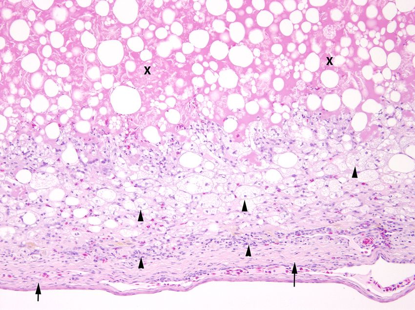
Wiener Tierärztliche Monatsschrift – Veterinary Medicine Austria                                               105 (2018)

ed, as was sepia powder. The enclosure was lit by a                   formed using small vascular clips. The surrounding tis-
fluorescent tube; ultra-violet spectrum lighting was not              sue, as well as the left ovary and salpinx, had no mac-
provided. The humidity was 60 to 70 % and the tem-                    roscopic abnormalities. Other gross findings included
perature in the enclosure ranged from 18 to 27 ºC. The                a firm, yellowish liver with a gritty surface texture and
gecko had no prior medical conditions, had never been                 3 ml brownish coelomic effusion, which was sampled
mated and had never laid eggs.

Physical examination, further dia-
gnosis and surgical intervention

   On physical examination, the an-
imal was bright, alert and in good
body condition but had a firm and dis-
tended coelom. Palpation revealed a
well filled gastrointestinal tract, as
well as a firm, rounded, nonmova-
ble mass in the right caudal coelom.
No other abnormalities were detect-
ed. Radiographs (settings: Bird S/M
60 cm; 25 kV; 4.2 mAs) revealed a
3 x 2 cm caudo-coelomic mass con-
sistent with an alteration of the re-
productive tract (Fig. 2).
   Ultrasonographic findings includ-
ed two altered vitellogenic follicles
of the right ovary (2.3 x 2 cm and
2 x 1.5 cm; Fig. 3). Whereas one fol-
licle was filled with anechogenic flu-
id, the follicular cavity of the other         Fig. 3: Ultrasonographic image of the female Leach´s giant gecko (Rhacodactylus leachianus),
was almost completely occupied by              18 Mhz linear probe, demonstrating the two altered, vitellogenic follicles. / Ultraschallbild
an echogenic, homogeneous mass.                des weiblichen Neukaledonischen Riesengeckos (Rhacodactylus leachianus), 18 Mhz
The examination also revealed an               Linearsonde. Darstellung der zwei veränderten vitellogenen Follikel.
additional homogeneous coelomic
mass with an uneven surface in the
right caudal coelomic cavity, in im-
mediate proximity to the right ovary
and consistent with the radiographic
finding. Ovarian disease in conjunc-
tion with a pre-ovulatory follicular
stasis (POFS) was suspected.
   The animal was hospitalized for
an ovariectomy the next day. The
gecko was intended for breeding
purposes and the owner therefore
requested a unilateral ovariectomy.
The patient was premedicated with
carprofen (4 mg/kg s.c., Rimadyl®)
and pre-anaesthetized with keta-
mine (5 mg/kg i.m., Ketasel-5®) and
diazepam (1 mg/kg i.m., Lipuro®) in
the front legs. Induction and mainte-
nance were achieved by 2–5 % iso-
flurane (Isofluran CP®). The coelio-
tomy revealed a well vascularized              Fig. 4: Coeliotomy in a Leach´s giant gecko (Rhacodactylus leachianus) diagnosed
ovarian mass that did not seem to              with ovarian mass. Right ovary, nodular and well-vascularised mass. / Zöliotomie bei ei-
cause compression of other organs              nem Neukaledonischen Riesengecko (Rhacodactylus leachianus), bei dem eine ovarielle
and was surgically removed (Fig. 4).           Umfangsvermehrung diagnostiziert wurde. Rechtes Ovar, grobknotige und hochgradig ge-
Unilateral ovariectomy was per-                fäßreiche Umfangsvermehrung.

                                                                                                                                           75
Wiener Tierärztliche Monatsschrift – Veterinary Medicine Austria

                                                                           episodes of physical uneasiness were observed during
                                                                           the month after surgery.
                                                                             Six weeks after discharge from the hospital, similar
                                                                           changes were found on the left ovary by means of ul-
                                                                           trasound and radiography. The gecko´s body weight
                                                                           was 273 g. POFS with cystic ovarian changes was
                                                                           suspected on the left ovary. An additional coeliotomy
                                                                           and unilateral ovariectomy of the remaining ovary was
                                                                           therefore proposed. Prior to surgery, and with the ani-
                                                                           mal already sedated, computed tomography (CT) was
                                                                           performed. The images (16 slice CT; layer thickness
                                                                           0.72 mm, pitch 0.64 mm, rotation time 0.5 s at 120 kV
                                                                           and 150 mA/s/slice) revealed two 1.5 x 1 cm, one
                                                                           2.5 x 1.5 cm and one 2.5 x 2.5 cm follicles that were
                                                                           centrally located in the caudal coelom (Fig. 5). The
                                                                           anaesthetic procedure and surgery were conducted in
                                                                           the same manner as the first surgery to remove the left
                                                                           ovary. The patient´s weight after surgery was 239 g.
                                                                           Postoperative care followed the same procedure as
                                                                           for the first operation and recovery was complete and
                                                                           uneventful.

                                                                           Microbiological and histopathological
                                                                           examinations

                                                                             The coelomic effusion was processed by the Institute
                                                                           for Microbiology, University of Veterinary Medicine
Fig. 5: Computed tomography scan image of the female Leach´s giant         Hanover, Germany, following the regular laboratory
gecko (Rhacodactylus leachianus). Coronal image, soft tissue window.       procedure for detecting both aerobic and anaerobic
Four altered layered vittelogenic follicles with different densities and
                                                                           bacteria and yeast species. Several in-house culture
sizes are present in the caudal abdomen: two hypoechogenic (thin
                                                                           media were inoculated by fractional smear: chocolate
arrows), one showing hyperechogenic structures (thick arrow) and
one moderately enlarged (arrowhead). / Computertomographische
                                                                           agar and Kimmig’s agar, Columbia and Gassner agar,
Darstellung des weiblichen Neukaledonischen Riesengeckos                   Staph/Strep selective agar and Hamburg test agar for
(Rhacodactylus leachianus). Coronaler Schnitt, Weichteilfenster. Vier      fungal culture. Aerobic culture was performed for 48–
mehrfach geschichtete vitellogene Follikel unterschiedlicher Dichte        72 h at 30 ºC after processing by enrichment bouil-
und Größe im kaudalen Bereich der Zölomhöhle: Zwei hypoechogene            lon (Institute for Microbiology, University of Veterinary
(dünne Pfeile), einer mit hyperechogenen Strukturen (dicker Pfeil) und     Medicine Hanover, Germany). Chocolate agar plates
einer mittelgradig vergrößert (Pfeilspitze).                               were incubated in a microaerophilic atmosphere (5 %
                                                                           CO2). Neither microbiological nor mycological patho-
and submitted for bacterial and fungal culture. The                        gens could be identified in any culture. The removed
coelomic cavity was lavaged with warm, sterile phys-                       ovaries were fixed in 10 % neutral buffered formalin,
iological saline and closed with 5-0 PDS in a simple,                      paraffin embedded, sectioned at 5 μm and stained
continuous everting pattern.                                               with haematoxylin and eosin (HE). Additionally, Ziehl-
  After surgery, the gecko´s weight was reduced to 261 g.                  Neelsen’s, Gram’s and Giemsa stains, PAS-reaction
Postoperative treatment during the one-week hospitali-                     and Grocott´s methanamine silver impregnation were
zation period included daily pain management with car-                     performed.
profen (4 mg/kg s.c., Rimadyl®) and s.c. fluid therapy with                  Histopathology of the right ovary revealed multiple
sterofundin maintenance fluid (10 ml/kg, Sterofundin                       cysts demarcated by connective tissue with severe ac-
ISO®), butaphosphan and vitamin B12 (100 mg/kg and                         cumulation of intracellular hyaline proteins (confirmed
0.05 mg/kg; Catosal 10 %®) and 24 % calcium boro-                          by PAS-reaction) and empty vacuoles consistent with
gluconate solution (200 mg/kg, Kalziumborogluconat                         fat droplets (Fig. 6). No normal ovarian tissue was
Infusionslösung®), as well as a single administration of                   found. Findings in the left ovary included regular imma-
vitamin B (10 mg/kg s.c., Be-Complex®) and vitamin C                       ture follicles with an intact theca interna and a mature
(200 mg/kg s.c., Vitamin C®). In addition, chloromycetin                   follicle with yolk protein accumulation. A cystic mass
(40 mg/kg p.o., Chloramphenicol®) was given for seven                      with lipogranulomatous inflammation was also found,
days. The gecko was released from the clinic with in-                      consisting of macrophage infiltrations with intracellular
structions to continue peroral chloromycetin (40 mg/kg                     hyaline proteins. Cristalloid and solitary empty gap-like
p.o., Chloramphenicol®) for two more weeks. No other                       structures cholesterol were also identified (Fig. 7). No

76
Wiener Tierärztliche Monatsschrift – Veterinary Medicine Austria                                                 105 (2018)

infectious agents or neoplastic cells
were identified microscopically in ei-
ther ovary.
  Rechecks of the patient (one and
four weeks, four months and one
and two years later) have not re-
vealed any anomalies.

Source of supplies:
5-0 PDS: Ethicon, Norderstedt, Germany;
Brilliance CT 16 slice: DS, Philips GMBH,
Germany; Butaphosphan and Vitamin B12:
Bayer, Leverkusen, Germany; Calcium boro-
gluconate solution: WDT, Garbsen, Germany,
Carprofen: Pfizer, Berlin, Germany, Chloro-
mycetin: Zoetis-Pfizer, Berlin, Germany.;
Diazepam: Braun, Aschaffenburg, Germany,
Gassner agar: Oxoid Deutschland GmbH,
Wesel, Germany; Isoflurane: Henry Schein
VET,     Hamburg,     Germany;       Ketamine:
Selectavet, Weyarn-Holzolling, Germany;
Maintenance fluid sterofundin: Braun,
Aschaffenburg,     Germany;        Radiography      Fig. 6: Histopathology of the right ovary of a Leach´s giant gecko (Rhacodactylus leachi-
device: Gierth HF 400 A, Gierth X-Ray Int.,         anus). Segment of an ovarian cyst demarcated by connective tissue (arrowheads) with severe
Riesa, Germany; Repashy Superfood´s                 accumulation of intracellular proteins and empty vacuoles consistent with fat droplets (X).
Crested Gecko Meal Replacement Powder:              HE, 100x. / Histopathologische Untersuchung des rechten Ovars eines Neukaledonischen
Repashy      Ventures,     Inc.,    Oceanside,      Riesengeckos (Rhacodactylus leachianus). Segment einer ovariellen Zyste mit bindegewe-
California; Ultrasound device: Vivid 7              biger Demarkation (Pfeilspitzen) und hochgradige Akkumulation von Proteinen und leeren
Dimension; GE Healthcare, Munich; 18 Mhz            Vakuolen übereinstimmend mit Fetttröpfchen (X). HE, 100x.
linear probe; Vascular clips: Vitaltec Surgical,
Plymouth, Massachusetts, USA; Vitamin B:
CP-Pharma, Burgdorf, Germany; Vitamin C:
Rebopharm, Bocholt, Germany.

    Discussion
   This clinical case displayed a
pathological change that has not
previously been reported in this form
in reptiles and adds knowledge of re-
productive disorders in Leach´s gi-
ant geckos.
   Reproductive disorders in rep-
tiles may be related to inadequate
husbandry and/or dietary condi-
tions, as the reproductive cycle in
reptiles is mediated by factors such
as the quantity and composition of
diet (with disorders often linked to
an unvaried, high-caloric diet); am-
bient temperature; lighting condi-
tions and humidity; the provision                   Fig. 7: Histopathology of the left ovary of a Leach´s giant gecko (Rhacodactylus leachianus).
                                                    Segment of an ovarian cyst demarcated by connective tissue (arrows). Lipogranulomatous
of a nesting site; and cage mates
                                                    inflammation consisting of infiltration of macrophages with intracellular hayline proteins (ar-
(SYKES, 2010). As imbalances in
                                                    rowheads) as well as empty vacuoles consistent with fat droplets. and severe extracellu-
the reproductive cycle occur, POFS                  lar accumualtion of lipoproteins (X). HE, 400x. / Histopathologische Darstellung des linken
or post-ovulatory egg stasis (POES)                 Ovars eines Neukaledonischen Riesengeckos (Rhacodactylus leachianus). Segment einer
may develop (CAMPBELL, 2009b).                      ovariellen Zyste mit bindegewebiger Demarkation (Pfeile). Lipogranulomatöse Entzündung
Post-ovulatory egg stasis is con-                   mit Infiltration von Makrophagen, die neben hyaline Proteine (Pfeilspitzen) auch Fetttröpfchen
sidered to be frequent in gekkoidae                 (optisch leere Vakuolen, X) gespeichert haben. Extrazellulär hochgradige Akkumulation von
(HOCHLEITHNER and HOLLAND,                          Lipoproteinen (X). HE, 400x .

                                                                                                                                                  77
Wiener Tierärztliche Monatsschrift – Veterinary Medicine Austria 2014). Clinical signs of reproductive disorders may in- as a side effect of the inflammation of the reproductive clude anorexia, apathy, coelomic distension and/or tract (GARDNER and BARROWS, 2010; CAMPBELL, dyspnoea due to the effect of the space-occupying pro- 2015). Due to the owner´s wish not to perform blood cess (STACY et al., 2004; BARTEN, 2006). analysis, and as no liver biopsy was taken, it was not As in the present case, diagnostic imaging proce- possible to confirm the diagnosis of hepatic lipido- dures can assist in the diagnosis of pre-ovulatory fol- sis and any inflammatory process and monocytosis, licular stasis, e.g. in determining whether the follicles which is often associated with granulomatous altera- already create a calcified outer shell, which allows tions (CAMPBELL, 2009a; DI GIROLAMO and MANS, the diagnosis of pre-ovulatory stasis to be eliminated 2016), via haematological assessment. (HERNÁNDEZ-DIVERS, 2006; GUMPENBERGER, Based on evidence from birds, cystic ovarian chang- 2017). The techniques are also useful for detecting tu- es may be either congenital or acquired (SCHMIDT, moural growth within the coelomic cavity (JACOBSON, REAVILL and PHALEN, 2015). Acquired changes in 2007). reptiles may develop due to neoplastic growth (STACY POFS is more commonly observed in captive rep- et al., 2004; CRUZ CARDONA et al., 2011) or, as tiles (BACKUES and RAMSAY, 1994). The alteration shown in birds, secondary to a hormonal disruption is defined by retained follicles due to the lack of ovula- (BOWLES, 2002; BOWLES, 2006; HADLEY, 2010). tion, although vitellogenesis has taken place (SYKES, Hormone analysis was not taken into consideration 2010). The condition may result in the resorption of fol- due to the lack of reference data for the species. licles or coelomitis if the follicles rupture or ectopic ovu- Previously reported granulomatous ovarian changes lation occurs (RIVERA, 2008; CRUZ CARDONA et al., in reptiles were mainly caused by specific pathogens. 2011). Ovariectomy is indicated in cases of POFS in A malignant granulosa cell tumour has been observed lizards that show anorexia and lethargic or abnormal in a green iguana (Iguana iguana) (CRUZ CARDONA behaviour (GIBBONS and TELL, 2009). The procedure et al., 2011) and an ovarian papillary cystadenocarci- is useful for preventing coelomitis and is recommend- nomas (STACY et al., 2004) in the same species. In ed by several authors (OROSZ et al., 1992; FUNK, both cases, cystic alterations were also found. The lit- 2002; SCHILDGER and HÄFELI, 2003). For these rea- erature also includes descriptions of fungi or mycobac- sons, and especially owing to the marked changes in teria as the causal agents of granulomatous inflamma- the reproductive tract, surgical therapy was proposed tions (SOLDATI et al., 2004; MITCHELL, 2012). In the in this case. current case, neither the microbiological nor the his- Despite the use of the diagnostic methods described topathological analysis (HE, Ziehl-Neelsen´s, Gram´s, above, it can be difficult to determine whether a female Giemsa and Grocott´s stains, PAS-reaction) identified is undergoing a physiological or a pathological repro- any infectious agents, suggesting that mycobacterio- ductive cycle, as captive lizards may mature at differ- sis, and particularly a mycotic infection, were unlikely ent ages (DE NARDO, 2006). Female Leach´s giant to be the cause of the granulomatous oophoritis. geckos reach sexual maturity by 2.5 to 4 years and/or The literature also contains scattered reports of by 82 g to 140 g (smaller insular and larger mainland reptile oophoritis. In one case, a Duvaucel´s gecko populations, respectively) and in most of their natural (Hoplodactylus duvaucelii) was documented with in- range they usually lay a single clutch, while in captivity fectious oophoritis due to Salmonella enterica spp. they normally lay three clutches per breeding season (LE SOUEF et al., 2015). A chronic oophoritis as- (DE VOSJOLI, FAST and REPASHY, 2003). Animals sociated with a necrotic ovary in a veiled chamele- in captivity may mature at an earlier age, largely be- on (Chamaeleo calyptratus) has also been described cause the husbandry methods are designed to maxi- (KNOTEK, 2014), as has a post-ovulatory stasis as- mize growth for early breeding. sociated with an oophoritis in a Hermann´s tortoise The fact that this animal´s weight was high despite (Testudo hermanni) (HERNÁNDEZ-DIVERS, 2004). a decreased appetite and the finding of a firm, yellow- Non-neoplastic proliferative ovarian alterations, e.g. ish liver suggests that an underlying fatty liver disease ovarian hyperplasia or oophoritis, are usually associ- may have been present, which may have led to the ac- ated with a follicular rupture or an inflammatory pro- cumulation of hyaline proteins and to the reproductive cess, although evidence from birds suggests that ovar- disorder (MARTÍNEZ SILVESTRE, 2013). The fruit pu- ian atresia or atrophy may also be involved (HADLEY, ree provided may not have satisfied the dietary require- 2010). The cause of the granulomatous oophoritis in ments of the species and could have played a part in this case remains speculative, as there was no associ- the development of adiposity. Vitamin E deficiency can ated infection. Our hypothesis is that it may have been also lead to a disorder of lipid metabolism in reptiles caused due to the follicular degeneration. This altera- (DIERENFELD, 1989). However, because of the gap tion may be attributable primarily to the POFS. In addi- in data on the vitamin E need of the species, it is not tion, the apparent heavy weight of the gecko, hyporex- possible to assess the influence of a putative Vitamin ia and the appearance of the liver indicates fatty liver E deficiency in the present case. A coelomic effusion, disease, which in turn may have been caused by an in- as observed in this patient, can be present in reptiles adequate high-caloric and unbalanced diet. 78
Wiener Tierärztliche Monatsschrift – Veterinary Medicine Austria                                              105 (2018)

  This is the first report of POFS and lipogranuloma-                    Conclusion
tous oophoritis in a Leach´s giant gecko, and, to the                      We show that the patient´s history and diet should be
best of the authors´ knowledge, the first report of a                    carefully examined. Diagnostic imaging procedures are
non-infectious and non-neoplastic lipogranulomatous                      useful in formulating a preliminary diagnosis; howev-
and cystic oophoritis in a reptile. The case expands the                 er, a definitive diagnosis is only possible after histolog-
current knowledge of reproductive disorders in reptiles                  ical examination of ovarian tissue. Non-infectious and
and highlights the importance both of a comprehensive                    non-neoplastic ovarian changes should be included in
study of the husbandry and of regular examination of                     the list of differential diagnoses for reproductive disor-
females to enable an early diagnosis and increase the                    ders in reptiles.
chances of a successful reproductive outcome.

   Fazit für die Praxis:
   Dieser Fallbericht zeigt, dass ovarielle Veränderungen im Zusammenhang mit Haltungsbedingungen kli-
   nisch behutsam untersucht werden sollten. Eine vorläufige Diagnose ist durch Bildgebungsverfahren mög-
   lich, die definitive Diagnose ist jedoch erst durch eine histologische Untersuchung zu bestimmen. Unsere
   Befunde stellen dar, dass nicht-infektiöse und nicht-neoplastische ovarielle Veränderungen in die Liste der
   Differentialdiagnosen für Reproduktionsstörungen bei Reptilien ebenfalls aufgenommen werden sollten.

   References
BACKUES, K.A., RAMSAY, E.C. (1994): Ovariectomy for treatment              guide to their selection and care. Advanced Visions, Inc., Vista,
  of follicular stasis in lizards. J Zoo Wildlife Med 25, 111–116.         California, 296 pp.
BARTEN, S. (2006): Lizards. In: MADER, D.R. (Ed.): Reptile medi-         FUNK, R.S. (2002): Lizard reproductive medicine and surgery. Vet
  cine and surgery. 2nd ed., Saunders Elsevier, St Louis, Missouri,        Clin North Am-Exot Anim Pract 5, 579–613.
  683–695.                                                               GARDNER, B.R., BARROWS, M.G. (2010): Yolk coelomitis in a
BOWLES, H.L. (2002): Reproductive diseases of pet bird species.            white-throated monitor lizard (Varanus albigularis). J S Afr Vet
  Vet Clin N Am-Exot Anim Pract 5, 489–506.                                Assoc 81, 121–122.
BOWLES, K.L. (2006): Evaluating and treating the reproductive sys-       GIBBONS, P.M., TELL, L.A. (2009): Problem solving in reptile prac-
  tem. In: HARRISON, G.J., LIGHTFOOT, T. (Eds.): Clinical Avian            tice. J Exot Pet Med 18, 202–212.
  Medicine. Vol. 2. Spix Publishing, Florida, 519–539.                   GUPTA, N., GUPTA, C. (2015): Xanthogranulomatous oophor-
CAMPBELL, T.W. (2009a): Clinical chemistry of reptiles. In: THRALL,        itis-masquerading as ovarian neoplasm: report of two cases.
  M.A., BAKER, D.C., CAMPBELL, T.W. (Eds.): Veterinary he-                 Annals of Pathology and Laboratory Medicine 2, 24–27.
  matology and clinical chemistry. Lippincott Williams & Wilkins,        GUMPENBERGER, M. (2017): Diagnostic imaging of reproductive
  Philadephia, 493–498.                                                    tract disorders in reptiles. Vet Clin North Am-Exot Anim Pract 20,
CAMPBELL, T.W. (2009b): Clinical Pathology. In: MADER, D.R.,               327–343.
  HERNÁNDEZ-DIVERS, S.J. (Eds.): Current therapy in reptile              HADLEY, T.L. (2010): Management of common psittacine reproduc-
  medicine and surgery. 2nd ed., Saunders Elsevier, St. Louis,             tive disorders in clinical practice. Vet Clin North Am-Exot Anim
  Missouri, 70–92.                                                         Pract 13, 429–438.
CAMPBELL, T.W. (2015): Effusions. In: CAMPBELL, T.W. (Ed.):              HERNÁNDEZ-DIVERS, S.J. (2004): Surgery: principles and tech-
  Exotic animal hematology and cytology. 4th ed., Wiley Blackwell,         niques. In: GIRLING, S.J., RAITI, P., (Eds.): BSAVA Manual of
  Iowa, Pennsylvania, 309–321.                                             Reptiles. 2nd ed., British Small Animal Veterinary Association,
CRUZ CARDONA, J.A., CONLEY, K.J., WELLEHAN, J.F.X., FARINA,                Gloucester, UK, 147–167.
  L.L., ORIGGI, F.C. WAMSLEY, H.L. (2011): Incomplete ovari-             HERNÁNDEZ-DIVERS, S.J. (2006): Surgery. In: MADER, D.R.
  osalpingectomy and subsequent malignant granulosa cell tumor             (Ed.): Reptile Medicine and Surgery. 2nd ed., Saunders Elsevier,
  in a female green iguana (Iguana iguana). JAVMA 239, 237–242.            St. Louis, Missouri, 625.
DE NARDO, D. (2006): Dystocias. In: MADER, D.R. (Ed.): Reptile           HOCHLEITHNER, C., HOLLAND, M. (2014): Ultrasonography. In:
  Medicine and Surgery. 2nd ed., Saunders Elsevier, St Louis,              MADER, D.R., HERNÁNDEZ-DIVERS, S.J. (Eds.): Current ther-
  Missouri, 787–792.                                                       apy in reptile medicine and surgery. 2nd ed. Saunders Elsevier, St.
DIERENFELD, E.S. (1989): Vitamin E deficiency in zoo reptiles,             Louis, Missouri, 119–122.
  birds and ungulates. J Zoo Wildl Med 20, 3–11.                         JACOBSON, E.R. (2007): Overview of reptile biology, anatomy, and
DI GIROLAMO, N., MANS, C. (2016): Reptile soft tissue surgery. Vet         histology. In: JACOBSON, E.R. (Ed.): Infectious diseases and pa-
  Clin North Am-Exot Anim Pract 19, 97–131.                                thology of reptiles. 1st ed. CRC Press, Florida, 1–130.
DE VOSJOLI, P., FAST, F., REPASHY, A. (2003): In: VOSJOLI,
  P., FAST, F., REPASHY, A. (Eds.): Rhacodactylus, the complete

                                                                                                                                             79
Wiener Tierärztliche Monatsschrift – Veterinary Medicine Austria JOHNSON, J.D. (2004): Urogenital system. In: GIRLING, S.J., SCHMIDT, R.E., REAVILL, D.R., PHALEN, D.N. (2015): Reproductive RAITI, P. (Eds.), BSAVA manual of reptiles. 2nd ed. British Small System. In: SCHMIDT, R.E., REAVILL, D.R., PHALEN, D. (Eds.): Animal Veterinary Association, Gloucester, UK, 261–272. Pathology of pet and aviary birds. 2nd ed., Wiley-Blackwell, Iowa, KNOTEK, Z. (2014): Reproductive strategies in captive female veiled 149–153. chameleons. Proceedings of the UPAV/AAVAC/ARAV Conference, SOLDATI, G., LU, Z.H., VAUGHAN, L., POLKINGHORNE, A., 22nd–24th April 2013, Cairns, Australia, 127–130. ZIMMERMANN, D.R., HUDER, J.B., POSPISCHIL, A. (2004): KNOTEK, Z., CERMAKOVA, E., OLIVERI, M. (2016): Reproductive Detection of Mycobacteria and Chlamydiae in granulomatous Medicine in Lizards. Vet Clin N Am Exot Anim Pract 20, 411–438. inflammation of reptiles: A retrospective study. Vet Pathol 41, LE SOUEF, A.T., BARRY, M., BRUNTON, D.H., JAKOB-HOFF, R., 388–397. JACKSON, B. (2015): Ovariectomy as treatment for ovarian bacte- STACY, B.A., VIDAL, J.D., OSOFSKY, A., TERIO, K., KOSKI, M., rial granulomas in a Duvaucel's gecko (Hoplodactylus duvaucelii). DE COCK, H.E.V. (2004): Ovarian papillary cystadenocarcinomas N Z Vet J 63, 340–344. in a green iguana (Iguana iguana). J Comp Pathol 130, 223–228. MARTÍNEZ SILVESTRE, A. (2013): Hepatic lipidosis in reptiles. STAHL, S.J. (2006): Reptile obstetrics. Proceedings of the 20th North Proceedings of the 8th Southern European Veterinary Conference, American Veterinary Conference, 7th–11th January 2006, Orlando, 17th–19th October 2013, Barcelona, Spain. Florida, 1680–1683. MITCHELL, M.A. (2012): Mycobacterial Infections in reptiles. Vet SYKES, J.M. (2010): Updates and practical approaches to reproduc- Clin N Am-Exot Anim Pract 15, 101–111. tive disorders in reptiles. Veterinary Clinics of North America. Vet OROSZ, S.E., TOAL, R.L., KORENEK, N.L., TEUBNER, V.A. (1992): Clin N Am-Exot Anim Pract 13, 349–373. Follicle aspiration for the treatment of pre-ovulatory egg binding in VITT, L.J., CALDWELL, J.P. (2009): Reproduction and reproductive a green Iguana. J Small Exot Anim Med 1, 161–165. modes. In: VITT, L.J., CALDWELL, J.P. (Eds.): Herpetology: An RIVERA, S. (2008): Health assessment of the reptilian reproductive introductory biology of amphibians and reptiles. 3rd ed. Academic tract. J Exotic Pet Med 17, 259–266. Press, Elsevier, Massachusetts, 114. SCHILDGER, B.J., HÄFELI, W. (2003): Chirurgische Therapie der Dystokie bei Reptilien. Tierärztl Praxis K 31, 41–48. 80
You can also read