Looks can be deceiving: contrasting temperature characteristics of two morphologically similar kelp species co-occurring in the Arctic - De Gruyter
←
→
Page content transcription
If your browser does not render page correctly, please read the page content below
Botanica Marina 2021; 64(3): 163–175

Research article

Kiara Franke*, Daniel Liesner, Svenja Heesch and Inka Bartsch

Looks can be deceiving: contrasting temperature
characteristics of two morphologically similar
kelp species co-occurring in the Arctic
https://doi.org/10.1515/bot-2021-0014                                    behaved similarly to cold-temperate populations. Our data
Received February 11, 2021; accepted April 16, 2021;                     suggest that a future increase in seawater temperatures may
published online May 13, 2021                                            hamper the success of H. nigripes and favour L. digitata in
                                                                         Arctic environments.
Abstract: Two morphologically similar digitate kelp spe-
cies, Laminaria digitata and Hedophyllum nigripes, co-occur              Keywords: C:N-ratio; gametogenesis; growth rate; PAM
along a shallow sublittoral depth gradient in the Arctic but,            fluorometry; sporophyte.
in contrast to L. digitata, very few ecophysiological data exist
for H. nigripes. We investigated growth, survival, photosyn-
thetic characteristics and carbon:nitrogen ratios of juvenile
sporophytes, and recruitment and survival of gametophytes                1 Introduction
in genetically verified Arctic isolates of both species along
temperature gradients (0–25 °C) over 14 days. Laminaria                  Two digitate kelp species, Laminaria digitata (Hudson)
digitata gametophytes survived 23–24 °C, while sporophytes               J.V. Lamouroux and Hedophyllum nigripes (J. Agardh)
survived 21–22 °C. Hedophyllum nigripes had lower temper-                Starko, S.C. Lindstrom et Martone are similar in their
ature affinities. Gametophytes survived 19–21 °C, while                   external morphology and they are part of marine forests in
sporophytes survived 18 °C. Male gametophytes were more                  the shallow sublittoral of the Arctic (Dankworth et al.
heat-tolerant than female gametophytes in both species.                  2020; Longtin and Saunders 2016; Starko et al. 2019). They
The pronounced cold adaption of H. nigripes compared                     can be distinguished macroscopically by the presence or
to L. digitata also became apparent in different sporophyte              absence of mucilage ducts in their stipes and the season of
growth optima (L. digitata: 15 °C; H. nigripes: 10 °C) and               fertility (Agardh 1868; Dankworth et al. 2020; Longtin and
gametogenesis optima (L. digitata: 5–15 °C; H. nigripes:                 Saunders 2015). This had already been noted by Agardh
0–10 °C). Higher carbon:nitrogen ratios in H. nigripes suggest           (1868) who first described H. nigripes as Laminaria nig-
an adaptation to nutrient poor Arctic conditions. The overall            ripes from Spitsbergen and compared it to various forms of
temperature performance of H. nigripes possibly restricts the            European Arctic L. digitata.
species to Arctic–Sub-Arctic regions, while Arctic L. digitata                As both species share the same habitat and as their
                                                                         unambiguous identification relies on molecular barcoding,
                                                                         the two species have been partially confused in the Euro-
                                                                         pean Arctic for the past decades (e.g. Fredriksen et al. 2019;
*Corresponding author: Kiara Franke, Alfred Wegener Institute,
Helmholtz Center for Polar and Marine Research, Am Handelshafen 12,      Lund 2014).
27570 Bremerhaven, Germany; and Current Address: Applied Ecology              Meanwhile, it is known that H. nigripes is not present
and Phycology, University of Rostock, Albert-Einstein-Straße 21,         only in the European Arctic, but has a pan-Arctic distri-
18059 Rostock, Germany, E-mail: kiara.franke@uni-rostock.de              bution from the NE-Pacific (Cape Baele, BC, Canada) via the
Daniel Liesner, Alfred Wegener Institute, Helmholtz Center for Polar
                                                                         Canadian Arctic, the Bay of Fundy, USA (∼45°N), and
and Marine Research, Am Handelshafen 12, 27570 Bremerhaven,
Germany; and Department of Algal Development and Evolution, Max          Greenland to Spitsbergen, Norway in the E-Atlantic
Planck Institute for Developmental Biology, Max-Planck-Ring 5, 72076     (Dankworth et al. 2020; Guiry and Guiry 2021; Longtin
Tübingen, Germany                                                        and Saunders 2015; Pedersen 2011; Starko et al. 2019).
Svenja Heesch, Applied Ecology and Phycology, University of Rostock,     Grant et al. (2020) investigated the phylogeography of
Albert-Einstein-Straße 21, 18059 Rostock, Germany
                                                                         H. nigripes, and their genetic studies suggest that the spe-
Inka Bartsch, Alfred Wegener Institute, Helmholtz Center for Polar and
                                                                         cies evolved in the NE-Pacific. The genetic diversity of
Marine Research, Am Handelshafen 12, 27570 Bremerhaven,
Germany. https://orcid.org/0000-0001-7609-2149                           specimens in the Gulf of Alaska indicates the possible

  Open Access. © 2021 Kiara Franke et al., published by De Gruyter.      This work is licensed under the Creative Commons Attribution 4.0
International License.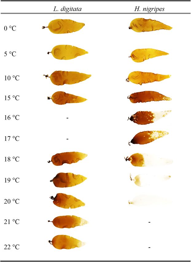
164         K. Franke et al.: Temperature performance of Arctic kelps

existence of northern refugia during ice ages. However, it is           used for the assessment of the current and potential future
uncertain whether N-Atlantic H. nigripes populations                    distribution of both species in a warming Arctic. In
expanded from southern populations, or expanded their                   particular, we quantified gametogenesis, sporophyte
range from northern glacial refugial populations where the              recruitment and growth, optimal quantum yield and car-
species survived (Grant et al. 2020). The exact southern                bon and nitrogen content of sporophytes, as well as sur-
distribution limits of H. nigripes, especially in the                   vival of both life cycle stages along temperature gradients,
N-Atlantic, are unknown. In contrast, L. digitata has a pan-            using several unialgal strains isolated from the European
Atlantic to Arctic distribution, and its southern distribution          Arctic in Kongsfjorden, western Spitsbergen.
limit correlates with the mean 19 °C August sea surface
isotherm (van den Hoek 1982; Müller et al. 2009). The two
species co-occur in the European Arctic and in the                      2 Materials and methods
NW-Atlantic Sub-Arctic (e.g. Bay of Fundy; Dankworth et
al. 2020; Fredriksen et al. 2019; Longtin and Saunders                  2.1 Algal material
2016).
     Both species possess the heteromorphic, dip-                       Clonal vegetative male and female gametophytes were isolated from
lohaplontic life cycle typical for kelps, in which sporo-               four sporophyte individuals each of Laminaria digitata (AWI culture
phytes must recruit from gametophytes (Lüning 1990;                     nos. 3465–3470, 3475, 3476) and Hedophyllum nigripes (AWI culture
Martins et al. 2017; Oppliger et al. 2012). Kelp sporophytes            nos. 3501–3508) collected from Kongsfjorden, Spitsbergen in 2015 and
                                                                        2016, respectively. Vegetative gametophytes were cultivated in sterile
generally have a narrower temperature window for growth
                                                                        half-strength Provasoli enriched seawater (PES, Provasoli 1968; iodide
and reproduction compared to gametophytes (e.g. Bartsch                 enrichment according to Tatewaki 1966, double concentration of Na2
et al. 2008; tom Dieck 1992, 1993, Martins et al. 2017).                glycerophosphate) at either 15 °C (L. digitata) or 10 °C (H. nigripes).
Thermal characteristics of these fitness-related traits reveal           Cultures were maintained at 3 μmol photons m−2 s−1 red light (LED
the physiological temperature performance ranges of life                Mitras lightbar daylight 150 controlled by ProfiLux 3, GHL Advanced
stages and provide basic autecological information. A                   Technology, Kaiserslautern, Germany), in a 16:8 h light:dark (LD)
                                                                        cycle.
recent global overview and a series of global reports during
the last decades showed that warming sea temperatures
have direct and indirect effects on kelp abundance world-               2.2 Genetic confirmation of species identity
wide (Filbee-Dexter et al. 2019) and also in the Arctic
(Bartsch et al. 2016; Krause-Jensen et al. 2020).                       DNA was extracted from approximately 10 mg silica-dried gametophyte
     The temperature performance of L. digitata gameto-                 tissue using the Plant Nucleospin II extraction kit (Macherey-Nagel,
                                                                        Düren) according to the enclosed protocol, with an extended cell lysis
phytes and sporophytes is known for several N-Atlantic
                                                                        step of 1 h. For the amplification of the mitochondrial cytochrome oxi-
populations, showing a broad thermal performance range                  dase 1 gene (COI-5P) and the nuclear-encoded internal transcribed
with evidence for slight local adaptation (e.g. Bolton and              spacer (ITS) 1, polymerase chain reactions (PCRs) of 25 μl contained 8 μl
Lüning 1982; tom Dieck 1992, 1993; Liesner et al. 2020;                 water, 12.5 μl MyTaq™ premix (Bioline), 1 μl of each primer (COI-5P:
Lüning 1980; Martins et al. 2017). However, no eco-                     GazF2 and GazR2, Lane et al. 2007; ITS: AFP2(F) and 5.8S1(R), Peters and
                                                                        Burkhardt 1998; biomers.net) and 2.5 μl DNA extract diluted with water
physiological data currently exist for H. nigripes. In
                                                                        (1:10 or 1:100) prior to amplification. All other methods including the
recent years, it became evident that a digitate kelp species
                                                                        PCR protocol, product purification, sequencing and sequence checks
had considerably increased its biomass at shallow areas in              followed Heesch et al. (2016). Sequences were manually aligned using
Kongsfjorden, western Spitsbergen, as a consequence of                  PhyDE-1 v0.9971 (Müller et al. 2010) and subjected to phylogenetic
ocean warming (Bartsch et al. 2016). A follow-up study                  analyses, with methods given in Heesch et al. (2016, 2020). Strains with
revealed that this increase was most probably due to                    AWI culture nos. 3465–3476 were confirmed to belong to L. digitata
                                                                        (European nucleotide archive [ENA]/GenBank accession nos. for
L. digitata and not H. nigripes (Dankworth et al. 2020).
                                                                        COI-5P: MW626932–MW626942; for ITS: MW616807–MW616813), while
Thus, we hypothesized that, despite sharing a similar                   strains with AWI culture nos. 3501–3508 belonged to H. nigripes (ENA/
habitat and morphology (Dankworth et al. 2020; Longtin                  GenBank accession nos. for COI-5P: MW626943–MW626946; for ITS:
and Saunders 2016), both species possibly do not share                  MW616814–MW616817). In addition, a female strain from Bamfield
similar physiological traits and functions. In order to tackle          (Canada) originally isolated by L. Druehl in 1979 and studied in tom
                                                                        Dieck (1993) under the name of Laminaria bongardiana Postels et
this question, we wanted to compare thermal characteris-
                                                                        Ruprecht (= Laminaria groenlandica sensu Druehl, 1968; original culture
tics of fitness-related traits in two life cycle stages (game-
                                                                        no. 1110) was verified to belong to H. nigripes (AWI culture no. 3275,
tophytes and sporophytes) of both species in laboratory                 ENA/GenBank accession no. for COI-5P: MW626947; for ITS:
experiments. The resulting comprehensive data were also                 MW616818).
K. Franke et al.: Temperature performance of Arctic kelps                     165

2.3 Experiment 1: juvenile sporophytes                                             2.3.5 Carbon and nitrogen contents: Three sporophytes per replicate
                                                                                   were pooled, snap-frozen in liquid nitrogen and preserved at −80 °C at
                                                                                   day 0, 7 and 14. Deep frozen sporophytes were freeze-dried (CHRIST,
2.3.1 Pre-cultivation phase: In both species, gametophyte fertility
                                                                                   ALPHA 1–4 LD plus, Osterode am Harz, Germany) for 24 h, and dry
was induced at 10 °C, 16:8 h LD, at 15 μmol photons m−2 s−1 white LED
                                                                                   weight (DW) was measured. Samples of 2–3 mg were combusted at
light in half-strength PES, following the method of Bartsch (2018).
                                                                                   1000 °C in a CN-Elementar analyser (Euro EA 3000: Euro Vector, Pavia,
Juvenile sporophytes (2 cm long) were transferred to sterile aerated 5 l
                                                                                   Italy) with acetanilide as standard.
glass beakers at 40 μmol photons m−2 s−1. Medium was changed either
every two weeks (gametophyte phase) or weekly (sporophyte phase).
For logistical reasons, experiments had to be performed consecutively              2.4 Experiment 2: gametophytes and reproduction
under otherwise identical conditions. Therefore, H. nigripes sporo-
                                                                                       success
phytes were first grown at 20 μmol photons m−2 s−1 to reduce growth
rates, while the first experiment was conducted with L. digitata. The
former were re-exposed to an irradiance of 40 μmol photons m−2 s−1                 2.4.1 Experimental set-up: Stock gametophyte cultures of H. nigripes
two weeks prior to the experiment, to allow for experimentation under              were acclimatized for two weeks at 15 °C before the start of the
comparable conditions.                                                             experiment. A gametophyte stock solution was prepared separately,
                                                                                   for each species, by pooling even amounts of vegetative male and
                                                                                   female gametophytes, slightly squashing the material in a sterile
2.3.2 Experimental set-up: Sporophytes were subjected to a temper-                 mortar, sieving it through a sterile 100 μm mesh and suspending it in
ature gradient (L. digitata: 0, 5, 10, 15, 18, 19, 20, 21, 22 ± 0.5 °C;            half-strength PES. Because of contamination, AWI culture 3508 (fe-
H. nigripes: 0, 5, 10, 15, 16, 17, 18, 19, 20 ± 0.5 °C) for 14 days (day 0–14),    male of H. nigripes) was excluded from the gametophyte experiments.
followed by a post-cultivation phase (day 14–21) at 10 °C under iden-                    For each species, this stock solution was used to sow a target
tical irradiance conditions. For both species, nine sporophytes per                density of 1000 gametophytes cm−2 into four replicate petri dishes
replicate (n = 5) and temperature treatment were transferred into                  containing 100 ml of half-strength PES. The mean final densities were
aerated glass beakers (1.8 l; half-strength PES). Sporophytes were                 810 ± 178 and 1028 ± 188 gametophytes cm−2 for L. digitata and
acclimatized over six days (day −6 to 0; Supplementary Figure S1),                 H. nigripes, respectively, and were statistically different (t-test,
before being exposed to the temperature gradient above. The last                   p < 0.0005). Gametophytes were acclimatized over six days (day −6 to
temperature step took place at day 0 for all treatments except for the             0, Supplementary Figure S2) at 3 μmol photons m−2 s−1 white LED light,
control (10 °C), in order to start all experimental temperatures at the            16:8 h LD cycle. At day 0, irradiance was increased to 15 μmol pho-
same time (Supplementary Figure S1). All temperatures were main-                   tons m−2 s−1. The last temperature step took place at day 0 for all
tained either in walk-in constant cooling rooms (5–15 °C) or in water              treatments, except for the control (15 °C), in order to start all experi-
baths fitted with thermostats (Huber Variostat CC + Pilot ONE, Peter                mental temperatures at the same time (Supplementary Figure S2;
Huber Kältemaschinen GmbH, Offenburg, Germany). Medium was                         L. digitata: 0, 5, 10, 15, 20, 22, 23, 24, 25 ± 0.5 °C, H. nigripes: 0, 5, 10, 15,
changed every 3–4 days. Thermal performance of sporophytes was                     18, 19, 21, 22 ± 0.5 °C). One replicate of H. nigripes at 15 °C had to be
determined by quantifying growth rates, carbon and nitrogen contents               excluded from analyses due to contamination. After two weeks, the
and optimal quantum yield (FV/FM) (Supplementary Table S1).                        temperature gradient was followed by a post-cultivation phase: Ga-
                                                                                   metophytes exposed to ≤10 °C were kept in the same conditions for
                                                                                   another week (day 14–21) to quantify sporophyte recruitment, while
2.3.3 Growth: Sporophytes were photographed together with a cali-                  gametophytes exposed to ≥15 °C were kept at 15 °C to follow recovery
bration area, and their blade area was determined via image analysis               for two weeks (day 14–28; Supplementary Figure S2). Before post-
(Image J: Rasband, W.S., U. S. National Institutes of Health, Bethesda,            cultivation, 50 ml of the medium was exchanged. After two weeks of
Maryland, USA, 1997–2018) every 3–4 days. Linear relative growth                   post-cultivation, the 15, 24 and 25 °C (L. digitata) and 15, 21 and 22 °C
rates (RGR) were calculated according to the following formula:                    (H. nigripes) treatments received iron-free half-strength PES and were
                                     areafinal −areainitial                        kept at 15 °C under red light to promote vegetative gametophyte
                   RGR[% d−1 ] =
                                          areainitial
                                                              × 100,               growth (Iwai et al. 2015) for long-term post-cultivation (after day 28).
                                               T
                                                                                   Keeping the gametophytes under red light is a standard way for
where T is time (days). In order to discriminate between acclimation of            cultivating gametophytes in a vegetative stage, as blue light included
growth rate over time and overall growth rate, RGR was calculated                  in regular white LED light initiates gametogenesis and reproduction
between consecutive time points and over the 14-day period sepa-                   (Bartsch et al. 2016; Lüning and Dring 1972; Redmond et al. 2014).
rately. For a direct comparison of both species, RGR were normalized               Gametogenesis is easily induced even in vegetative gametophyte
to the respective maximum value per species, which was set as 100%                 cultures that have been kept over decades, under red light conditions
(called: Standardized GR).                                                         (e.g. Martins et al. 2019).

2.3.4 FV/FM: The in vivo chlorophyll a fluorescence of photosystem II               2.4.2 Quantification of gametogenesis: The relative abundance of
of dark-acclimated (5 min) L. digitata and H. nigripes sporophytes was             three ontogenetic stages (vegetative gametophytes (with or without
measured using pulse amplitude modulated (PAM) fluorometry (Mini                    oogonia); gametophytes with released eggs; gametophytes with spo-
PAM, Walz, Effelrich, Germany) as described by Schreiber et al. (1986).            rophytes; Martins et al. 2017) was counted at day 7, 14 and 21 using an
I-PAM images (Imaging PAM, Walz, Effelrich, Germany) were used as a                Olympus CKX41 inverted microscope (Olympus Co., Tokyo, Japan). At
qualitative measure of the overall stress response of sporophytes.                 day 0, approximately 200 female gametophytes per replicate (n = 4)
166          K. Franke et al.: Temperature performance of Arctic kelps

were counted and related to the petri dish area. At each subsequent       14. As the homogeneity of variance was met only partially, the sig-
counting day, ontogenetic stages were quantified in this area.             nificance level was decreased to alpha = 0.01 to reduce the probability
                                                                          of false positive reports.
2.4.3 Temperature tolerance of male and female gametophytes: To
quantify the temperature tolerance of gametophytes, the relative
abundance of living gametophytes of both sexes was recorded at day
7, 14 and 21 and the sex ratio (female/male) was calculated. Gameto-      3 Results
phyte cells were considered alive when no plasmolysis had taken
place and at least one cell per fragment was pigmented.                   3.1 Experiment 1: juvenile sporophytes

2.5 Statistics                                                            3.1.1 Temperature tolerance and growth

Statistical analyses were performed with Statistica 8.0 (Statsoft Inc.,   Standardized GR comparisons revealed tempera-
2007, Minneapolis, USA). Following confirmation of variance homo-         ture × species interactions at 0, 5, 10, 15 and 18 °C (two-way
geneity (Levene’s test, α = 0.05), one- or two-way analysis of variance   ANOVA, F4,40 = 27.3, p < 0.0001; Supplementary Table S2):
(ANOVA) were performed and followed by a Tukey’s post hoc test. For       At 15 and 18 °C, L. digitata grew better than H. nigripes,
data with heterogeneous variance distribution, non-parametric
                                                                          while H. nigripes grew better at 0, 5 and 10 °C compared to
Friedman tests for temperature effects and Kruskal–Wallis tests with
multiple p-value comparison for time effects were applied followed by     L. digitata (Figure 1). In both species, growth responded to
Bonferroni correction.                                                    temperature (two-way ANOVA, F4,40 = 39.3, p < 0.0001;
                                                                          Supplementary Table S2; Standardized GR).
2.6.1 Experiment 1: juvenile sporophytes: Hedophyllum nigripes                 Laminaria digitata sporophytes growth was maximal at
sporophytes exposed to 19 and 20 °C died during the experimental          15 °C (Figure 1; RGR = 9.2 ± 1.4% d−1; data not shown) and
phase and were therefore excluded from statistical analyses. Stan-        higher than in all other temperatures (Tukey’s post hoc test,
dardized growth rate (GR) of both species (0–18 °C) were compared in a
                                                                          p < 0.02). Standardized GR at 0 and ≥20 °C were less than
two-way ANOVA with ‘temperature’ and ‘species’ as fixed factors. For
the interspecies comparison, only the temperature treatments com-
                                                                          20% of the maximum (Figure 1). Moreover, stress was indi-
mon to both species (0–18 °C) were analysed. Standardized GR was          cated by reduced chlorophyll fluorescence at ≥20 °C (I-PAM
analysed in a one-way ANOVA for both species separately after             images, Supplementary Figure S3) and reduced RGR
14 days. RGR over time (experimental phase and recovery) were             (Figure 2) as well as bleached distal areas at ≥21 °C (Figure 3).
grouped by temperatures and analysed in Friedman tests in each            RGR became negative after 14 days at 0 and ≥18 °C (Figure 2);
species separately. FV/FM of both species at day 14 at 0–18 °C was
                                                                          a decrease over time was significant only for 18 °C (Friedman
compared in a two-way ANOVA with ‘temperature’ and ‘species’ as
fixed factors. As variances were heterogeneous, the significance level      test, χ 23,5 = 11.9, p = 0.008) and 19 °C (Friedman test,
was decreased to alpha = 0.01 to reduce the probability of false pos-
itive reports. Due to heterogeneity, the response of FV/FM over time
and temperature was tested separately, applying non-parametric
Friedman tests for both species and for each temperature. The tem-
perature × time interaction for FV/FM of each species was analysed
with Kruskal–Wallis tests. Differences in FV/FM between responses on
day 14 and after recovery were analysed by non-parametric Friedman
tests. Carbon and nitrogen contents and C:N-ratios at day 0 and 14 of
both species were compared in t-tests.

2.6.2 Experiment 2: gametophytes and reproduction success: Ga-
metophytes of L. digitata exposed to 24 and 25 °C, and of H. nigripes
exposed to 21 and 22 °C died during the experimental phase. These
temperatures were therefore excluded from the analyses. Gameto-
phyte density of both species at day 0 was compared in a t-test. Sur-
vival over time was tested in two separate repeated measures (RM)
ANOVAs for each species separately. The recovery of L. digitata was       Figure 1: Standardized growth rates (GR) based on surface area (%)
determined in an RM ANOVA and for H. nigripes in a non-parametric         of Laminaria digitata (white dots) and Hedophyllum nigripes (black
Friedman test separated by temperatures. The change of the sex ratio      dots) sporophytes over two weeks in a temperature gradient (n = 5,
of gametophytes over time was tested in non-parametric Friedman           mean ± SD). Different letters denote significant differences within
tests (L. digitata) or an RM ANOVA (H. nigripes). Sporophyte recruit-     each species (ANOVA with Tukey’s post hoc test: α < 0.05, A–
ment was quantified as percentages of gametophytes bearing sporo-          D = L. digitata; a–c = H. nigripes). Asterisks indicate significant
phytes and was normalized to the respective maximum value per             differences between standardized GR of L. digitata and H. nigripes
species (=100%). Values were analysed in a two-way ANOVA for day          (two-way ANOVA with Tukey’s post hoc test).
K. Franke et al.: Temperature performance of Arctic kelps         167

                                                                                            Figure 2: Relative growth rates (RGR; % d−1) of
                                                                                            Laminaria digitata (top) and Hedophyllum
                                                                                            nigripes (bottom) sporophytes in a
                                                                                            temperature gradient over the experimental
                                                                                            time (14 days; left side of the dotted line) and
                                                                                            recovery at 10 °C (one week; right side of the
                                                                                            dotted line; n = 5, mean ± SD). Each value
                                                                                            denotes the RGR between the indicated time
                                                                                            point and the measuring day before.

χ 23,5 = 10.7, p = 0.01). After one week of recovery at 10 °C, RGR    ANOVA, F4,40 = 14.9, p < 0.0001) and temperature effects
increased only in 0, 5 and 18 °C treatments (Friedman test,           (two-way ANOVA, F4,40 = 13.3, p < 0.0001). The mean FV/FM
χ 21,5 = 5.0, p = 0.025) by up to 6.8% d−1 (0 °C) (Figure 2).         was 0.72 for both species at day 0. In L. digitata sporo-
       Hedophyllum nigripes sporophytes had a growth                  phytes, FV/FM was maximal at 10 and 15 °C with an increase
maximum at 10 °C, which was thereby 5 °C lower than the               of 7% at day 14 (FV/FM = 0.74 ± 0.01; Supplementary Ta-
maximum of L. digitata (Figure 1; RGR = 4.8 ± 0.2% d−1; data          bles S3 and S4, Figure 4). At 21 °C (FV/FM = 0.63 ± 0.05) and
not shown) where growth was higher than in all other tem-             22 °C (FV/FM = 0.34 ± 0.08), FV/FM decreased over time by 15
peratures (Tukey’s post hoc test, p < 0.0002). Standardized           and 54%, respectively (Figure 4, Supplementary Table S4).
GR at 0 °C also did not fall below 20% of the maximum, in             Following the 0 °C treatment, FV/FM of sporophytes
contrast to L. digitata. At temperatures ≥16 °C, however,             increased after one week of recovery at 10 °C (Friedman
standardized GR fell below the 20% level (Figure 1), accom-           test, χ 21,5 = 5, p = 0.02). In sporophytes from the 10, 15, 19
panied by signs of blade bleaching (Figure 3, I-PAM images,           and 20 °C treatments, FV/FM decreased by up to 10% during
Supplementary Figure S3). At 20 °C, RGR became negative               the recovery period (Friedman test, 10 °C: χ 21,5 = 1.8,
already after three days (Figure 2). After one week of recovery       p < 0.05; 15, 19, 20 °C: χ 21,5 = 5, p = 0.03). Sporophytes from
at 10 °C, RGR increased only in 17 °C treatments (1.4% d−1,           21 to 22 °C showed no recovery. Compared to FV/FM of
Friedman test, χ 21,5 = 5, p = 0.03).                                 L. digitata at 0 °C, H. nigripes continuously showed 8%
                                                                      higher mean values at 0 °C than L. digitata (Tukey’s post
                                                                      hoc test, p < 0.02). At 17 and 18 °C, FV/FM in H. nigripes
3.1.2 FV/FM                                                           decreased by 9% (at day 14, FV/FM = 0.63 ± 0.05). After one
                                                                      week of recovery at 10 °C, H. nigripes sporophytes from 0 to
Comparison of FV/FM of both species (0, 5, 10, 15 and 18 °C)          18 °C treatments stayed the same as during the treatment
revealed temperature × species interactions (two-way                  (Figure 4).
168 K. Franke et al.: Temperature performance of Arctic kelps
decreased by 48 and 40% at 0 and 23 °C, respectively
(480 ± 92 and 637 ± 116 gametophytes cm−2, respectively;
Tukey’s post hoc test, p < 0.03). At 25 °C, all gametophytes
died after one week (Figure 5A), and at 24 °C after two
weeks (Figure 5C). After two weeks of post-cultivation at
15 °C, gametophytes from 24 and 25 °C treatments showed
no recovery. However, after seven weeks, 0.2% (1.4 ± 0.8
gametophytes cm−2) of the initial gametophyte number
recovered from the 24 °C treatment.
In H. nigripes only time affected the gametophyte
density in all temperatures (0–19 °C; RM ANOVA,
F2,34 = 35.7, p < 0.0001, Supplementary Table S5, Figure 5).
The gametophyte density (0–19 °C) decreased from initially
1113 ± 201 gametophytes cm−2 to 764 ± 111 gameto-
phytes cm−2 at day 14, which was, however, not significant
(Figure 5). At 21 and 22 °C, all gametophytes died after
14 days. After two weeks of post-cultivation at 15 °C, ga-
metophytes showed no recovery. However, in long-term
culture under red light at 15 °C (one year), 0.08% (0.4 ± 0.03
gametophytes cm−2) of the initial gametophyte number
recovered from the 21 °C treatment.
3.2.2 Sex ratio
All replicates of both species contained approximately two-
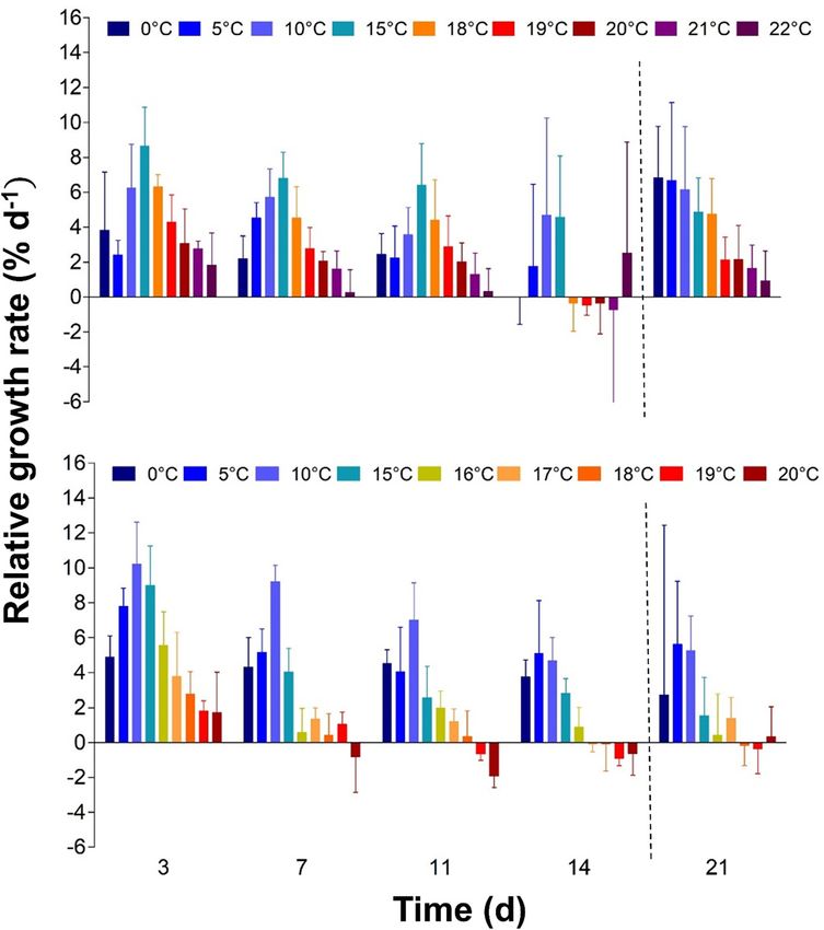
Figure 3: Photographic documentation of Laminaria digitata and
fold fewer female than male gametophytes (ratio: 0.4 ± 0.1,
Hedophyllum nigripes sporophytes exposed to a temperature
gradient after post-cultivation at 10 °C. Images are not to scale.
day 7, data not shown). In both species, the abundance of
Triangular cuts marked individual sporophytes per replicate. females further decreased with increasing temperatures
over time (ratio: 0.24 ± 0.03, day 14, Figure 6). In L. digitata,
sex ratio decreased between day 0 and 14 (Friedman test: 0,
3.1.3 Carbon and nitrogen content
5, 10, 15, 22 °C: χ 22,4 = 6.5. p < 0.04; 20, 21, 23 °C: χ22,4 = 8.0,
Laminaria digitata showed a 10% higher carbon content at p < 0.02). The strongest decrease of 85% (to 0.05 ± 0.01)
day 0 (0–18 °C; t-test, p = 0.05, Table 1) compared to occurred at 23 °C, indicating that many more females than
H. nigripes (t-test, p = 0.007), while the nitrogen content males had died at this temperature. Likewise, in H. nig-
was 100% higher in H. nigripes compared to L. digitata (t- ripes, there was a temperature × time interaction on the sex
test, p < 0.0001) during the whole experiment (Table 1). ratio (RM ANOVA, F5,17 = 37.1, p < 0.0001, Supplementary
Consequently, the C:N-ratio of L. digitata was 40% higher Table S6). The sex ratio was decreasing at 0, 18 and 19 °C
than that of H. nigripes after 14 days (t-test, p < 0.0001, between day 7 and 14 (Tukey’s post hoc test: p < 0.0002),
Table 1). while it kept stable at 5–15 °C.
3.2.3 Development of ontogenetic stages
3.2 Experiment 2: gametophytes and
reproduction success Both species recruited sporophytes between 0 and 15 °C
within 14 days, but not at higher temperatures (Figure 7).
3.2.1 Temperature tolerance and survival There was a temperature × species interaction for sporo-
phyte recruitment (ANOVA, F3,23 = 20.9, p < 0.0001, Sup-
At days 7 and 14, gametophyte density was affected by a plementary Table S7). Laminaria digitata optimally
temperature × time interaction in L. digitata (0–23 °C; RM recruited between 5 and 15 °C (80–90%) and Hedo-
ANOVA, F12,42 = 12, p = 0.01, Supplementary Table S5, phyllum nigripes between 0 and 10 °C (70–80%).
Figure 5). After seven days, the gametophyte density Hedophyllum nigripes recruited more sporophytes at 0 °C
K. Franke et al.: Temperature performance of Arctic kelps 169
Figure 4: Optimal quantum yield (FV/FM) of
Laminaria digitata (top) and Hedophyllum
nigripes (bottom) sporophytes in a
temperature gradient (two weeks; left
graph) and post-cultivation at 10 °C (one
week; right graph). Horizontal lines repre-
sent the median; boxes, the interquartile
range; whiskers, 1.5× of inter-quartile range
(n = 5).
Table : Carbon content (mmol C g− DW), nitrogen content (mmol N g− DW) and carbon:nitrogen (C:N) ratio of Laminaria digitata and
Hedophyllum nigripes at day (T) and day (T) (n = ; mean ± SD).
Carbon Nitrogen C:N-ratio
T T T T T T
L. digitata ± . . ± . . ± . . ± . . ± . . ± .
H. nigripes ± . ± . . ± . . ± . . ± . . ± .
Figure 5: Density of gametophytes of
Laminaria digitata (A, C) and Hedophyllum
nigripes (B, D) at day 7 (A, B) and day 14 (C,
D) in temperature gradients between 0 and
25 °C (L. digitata) and 22 °C (H. nigripes)
(n = 3–4, mean ± SD). Broken horizontal
lines show the mean initial gametophyte
density for each species after the
acclimatization phase (day 0). †All
gametophytes died.170 K. Franke et al.: Temperature performance of Arctic kelps
Figure 6: Sex ratio (female:male) of
gametophytes of Laminaria digitata (left)
and Hedophyllum nigripes (right) after
14 days in temperature gradients between
0 and 25 °C (L. digitata) and 22 °C
(H. nigripes) (n = 4, mean ± SD). Different
letters denote significant differences among
temperatures within each species
(L. digitata: Kruskal–Wallis test with
multiple p-value comparison; H. nigripes:
one-way ANOVA with Tukey’s post hoc test).
Please note that the marked deviation from
an expected initial 50:50 ratio was due to
applied seeding methods. †All gameto-
phytes died.
Figure 7: Effect of temperature on the relative
abundance of different ontogenetic stages
during gametogenesis of Laminaria digitata
(left) and Hedophyllum nigripes (right) in a
temperature gradient after seven days
(above) and 14 days (below; mean of n = 3–4;
SD not shown for clarity). Only the most
developed stage was counted per female
gametophyte. †All gametophytes died.
than L. digitata (Tukey’s post hoc test: p < 0.001), while niches. Upper temperature survival limits for sporophytes
L. digitata recruited more sporophytes at 15 °C than H. nig- and gametophytes as well as optima for sporophyte
ripes (Tukey’s post hoc test: p < 0.004). At 5 and 10 °C, growth, gametophyte survival and sporophyte recruitment
sporophyte recruitment of both species was the same. differed in both kelps by 4–5 °C, with L. digitata showing
While L. digitata showed the first sporophytes at 5–15 °C more temperate characteristics than H. nigripes (Table 2).
after one week (10–40%), only a few sporophytes were For the first time it becomes evident that H. nigripes is a
apparent at 0–10 °C in H. nigripes at the same time (∼5%) species with an Arctic to Sub-Arctic temperature imprint
(Figure 7), showing a delay in recruitment in H. nigripes. (sensu Wiencke et al. 1994). Overall, H. nigripes has a better
performance at 0 °C than L. digitata, and prefers cold
temperatures between 5 and 10 °C for optimum growth and
4 Discussion sporophyte recruitment. Over a two-week exposure,
H. nigripes sporophytes can tolerate a maximum of 18 °C
The present study clearly demonstrates that co-occurring and gametophytes can tolerate 19–21 °C. Thereby, its tem-
Arctic Laminaria digitata and Hedophyllum nigripes from perature performance is comparable to the Arctic endemic
Spitsbergen possess very dissimilar temperature charac- kelp Laminaria solidungula J. Agardh, which shows a
teristics and thereby possibly occupy different ecological similar growth optimum at 5–10 °C, while sporophytesK. Franke et al.: Temperature performance of Arctic kelps 171
Table : Temperature tolerance of Laminaria digitata (Ldig) and Hedophyllum nigripes (Hnig) gametophytes (Game) and sporophytes (Sporo)
determined after two weeks of exposure to the given temperatures and subsequent two-week post-cultivation at °C and long-term culture
under red light at °C (post-cult.).
Species Exp. time Temperature (°C) UST (°C)
Ldig Game One week V S S S V V V V V ––
Two weeks S S S S V V V V –– ––
Post-cult. S S S S V V V V R ––
Sporo One week X X X X X X X X X
Two weeks X X X X X X X X X
Post-cult. X X X X X X X X X
Hnig Game One week S S S V V V V V
Two weeks S S S S V V –– ––
Post-cult. S S S S V V R ––
Sporo One week X X X X X X X X ––
Two weeks X X X X X X X –– ––
Post-cult. X X X X X X X –– ––
UST, upper survival temperature; V, vegetative gametophytes; S, sporophyte recruitment; X, survival; ––, no survival; R, recovery after long term
post-cultivation; Exp. time, exposure time.
survive 16 °C and gametophytes survive 19–20 °C (Bolton In addition to their temperature performance, both
and Lüning 1982; tom Dieck 1992, 1993; Roleda 2016). Arctic species showed major differences in their ability to store
L. digitata has a reduced performance at 0 °C compared to nitrogen under replete nutrient conditions when cultivated
H. nigripes, and sporophyte growth and recruitment are in half-strength PES (274 μmol NO3− l−1; Sarker et al. 2013).
optimal at 10–15 °C. Arctic L. digitata sporophytes survive Interspecific differences in nutrient uptake ability have
21–22 °C and gametophytes survive 23–24 °C for two weeks, also been shown for other Polar seaweeds (Roleda and
which is similar among populations along the latitudinal Hurd 2019). Despite a high concentration of nitrate in the
distribution gradient in the NE-Atlantic (Liesner et al. 2020 medium, L. digitata sporophytes had a C:N-ratio of >20.
and references therein; Schimpf 2021). Therefore, L. dig- This comparatively high C:N-ratio exceeds the mean value
itata is a typical representative of a cold-temperate for temperate and tropical macroalgae and is normally
seaweed species (Wiencke et al. 1994). more indicative of nutrient-poor conditions (Atkinson and
In both kelp species, all replicates showed a strong Smith 1983). In contrast, H. nigripes had approximately
artificial unbalanced sex ratio towards male gametophytes 40% lower C:N-ratios of172 K. Franke et al.: Temperature performance of Arctic kelps
Spitsbergen material, used here, grew with less than 17% of H. nigripes are from Cape Beale and Bamfield, British
the maximum at ≥20 °C. Although Spitsbergen L. digitata Columbia (as Laminaria bongardiana in tom Dieck 1993;
sporophytes looked superficially healthy at 22 °C, even af- Starko et al. 2019). In Bamfield, monthly snapshot mea-
ter two weeks with only a few bleached areas, I-Pam im- surements recorded a minimum SST of 7.8 °C and a
ages revealed that sporophytes were considerably stressed maximum SST of 14.2 °C (2002–2009; Assis et al. 2017;
at ≥19 °C. FV/FM at these temperatures did not recover in Bosch 2020; Tybergbein et al. 2012). As H. nigripes showed
post-cultivation, nor did sporophytes resume growth after highest gametophyte reproduction success at 0–10 °C, this
transfer into optimum temperatures. The Spitsbergen may indicate that the southern distribution limit in the NE-
L. digitata population may thereby belong to an ecotype Pacific is set by temperatures that enable sufficient sporo-
that is slightly more susceptible to high temperatures than phyte recruitment from gametophytes and in addition
southern European populations and supports the existence allow for sufficient sporophyte growth. Following the
of local adaptation towards high temperatures in the spe- phytogeographic distribution concept of van den Hoek
cies (King et al. 2019; Liesner et al. 2020). There is also (1982), locations with winter SST < 10 °C and summer SST
subtle evidence for ecotypic variation in L. digitata game- not surpassing 15 °C would allow for sufficient sporophyte
tophytes, but only at the low temperature range. The upper recruitment and sufficient growth (similarly as around
temperature tolerance of L. digitata gametophytes is 23 °C Bamfield), and thus would provide appropriate tempera-
and was stable in all investigated populations from the ture conditions for this species.
Arctic to the southern distribution limit (tom Dieck 1993; We re-investigated the identity of a ‘L. bongardiana’
Lüning 1980; Schimpf 2021). But North Sea gametophytes culture that had been collected by L. Druehl from Bamfield,
showed a much reduced fertility at 1 °C (tom Dieck 1992), Vancouver Island, Canada in 1979 and was investigated by
while it was relatively high (20%) at 0 °C in our Spitsbergen tom Dieck (1993). Barcode analysis of two genetic makers
material. Similarly, sporophyte recruitment of Spitsbergen (COI-5P and ITS1) revealed its identity as H. nigripes. Also,
gametophytes was slightly better at low temperatures (5 °C) the ‘L. bongardiana’ cultures investigated by tom Dieck
than for North Sea material (Martins et al. 2020). (1992), were most likely H. nigripes, as they were collected at
In contrast to L. digitata, H. nigripes sporophytes grew the same time. Small intraspecific differences in tempera-
with approximately 40% of the maximum rates at 0 °C but ture responses were apparent between the Bamfield and
only with low rates at 16 °C (18%), supporting its affinity for Spitsbergen isolates, with the Bamfield material being
cold Arctic environments. The general cold-adaption of slightly warmer adapted (tom Dieck 1992, this study). The
H. nigripes sporophytes was also apparent in photosyn- upper two-week temperature tolerance of Bamfield game-
thetic parameters. While FV/FM of L. digitata decreased at tophytes was 20–21 °C (tom Dieck 1993) while Spitsbergen
low temperatures ≤5 °C, FV/FM stayed high and stable in isolates survived 19 °C, but recovered with 0.08% from 21 °C
H. nigripes at 0–5 °C but dropped at ≥17 °C, concomitant to treatments in long-term post cultivation. Similarly, juvenile
the reduced growth performance. Most probably an sporophytes from Bamfield showed slightly higher affinities
extended period of 15 °C is already stressful for H. nigripes to warm temperatures than Spitsbergen material. Maximum
sporophytes as photosynthetic inhibition already became growth was at 10 °C with 81 and 75% of the maximum rates at
visible in I-PAM images after two weeks exposure at 15 °C 5 and 15 °C, respectively (tom Dieck 1992), while Spitsbergen
(Supplementary Figure S3). sporophytes also grew maximally at 10 °C but significantly
The southern distribution limit of H. nigripes in the lower at 15 °C than at 5 °C, and the relative growth perfor-
Atlantic is still unknown, but our physiological data may mance at 0 °C was better than in the Bamfield material.
allow assumptions about its potential occurrence along the Although, upper survival temperature of sporophytes from
European coastline. In the western Atlantic, the south- both locations was 18 °C, the Spitsbergen material suffered
ernmost records of H. nigripes are from the Lepreau region considerably at temperatures ≥16 °C (this study: Supple-
in the Bay of Fundy, where monthly snapshot measure- mentary Figure S3; tom Dieck 1992). The differences in
ments recorded a minimum sea surface temperature (SST) temperature adaptation may be a reflection of the slight
of 0.45 °C and a maximum SST of 14.5 °C (2002–2009; Assis genetic differences between investigated H. nigripes pop-
et al. 2017; Bosch 2020; Tybergbein et al. 2012). This may ulations: while COI haplotypes from Svalbard in the NE-
represent a southern growth limit for sporophytes which Atlantic differ by 1–2 mutations from NW-Atlantic material,
drastically reduce their growth at >15 °C over 14 days (this the latter also differs by 1–2 mutations from NE-Pacific ma-
study). In the NE-Pacific, the southernmost records for terial (Grant et al. 2020).K. Franke et al.: Temperature performance of Arctic kelps 173
5 Conclusion Research funding: K.F. thanks the Deutsche For-
schungsgemeinschaft (DFG) for funding in the frame of the
A recent pan-Arctic review compiled the scattered infor- project “Seasonal kelp primary production at a rocky shore
mation on trends in macrophyte abundance during recent site: Integrating physiology and biochemistry into ecological
decades. Most of the 38 sites indicated that macrophyte modelling” (GR5088/2-1). D.L. contributed in the frame of the
abundance, species richness and/or productivity is 2015–2016 BiodivERsA COFUND call for research proposals
increasing in the Arctic (Krause-Jensen et al. 2020) with (program MARFOR), with the national funder DFG (grant no.
medium confidence Intergovernmental panel on climate VA 105/25-1) and S.H. was supported by the DFG in the
change (IPCC) confidence scale, Shapiro et al. 2010). At our framework of the priority programme “Antarctic Research
collection site in Kongsfjorden, especially Laminaria dig- with comparative investigations in Arctic ice areas” SPP 1158.
itata has become very dominant in shallow water during Conflict of interest statement: The authors declare no
recent decades (Bartsch et al. 2016; Dankworth et al. 2020). conflicts of interest regarding this article.
Our data suggest that L. digitata has the long-term poten-
tial of replacing Hedophyllum nigripes, which probably was
the overlooked but prevalent digitate kelp species around References
Spitsbergen (Fredriksen et al. 2019; Lund 2014). Kongsf-
jorden (Spitsbergen) showed a minimum SST of −0.6 °C and Agardh, J.G. (1868). Bidrag till kännedomen af Spetsbergens alger.
Tillägg till föregående afhandling. K. – Sven.
a maximum SST of 3.7 °C between 2002 and 2009 (Assis
Vetenskapsakademiens Handl. 7: 28–49, pl. III.
et al. 2017; Bosch 2020; Tybergbein et al. 2012), and future Assis, J., Tyberghein, L., Bosh, S., Verbruggen, H., Serrão, E.A., and
predictions assume a minimum SST of 1.6 °C and a De Clerck, O. (2017). Bio-ORACLE v2.0: extending marine data
maximum SST of 10.5 °C in 2100 (representative concen- layers for bioclimatic modelling. Global Ecol. Biogeogr. 27:
tration pathway [RCP] 8.5; Assis et al. 2017; Bosch 2020; 277–284.
Atkinson, M.J. and Smith, S.V. (1983). C: N: P ratios of benthic marine
Tybergbein et al. 2012). Although, L. digitata and H. nig-
plants 1. Limnol. Oceanogr. 28: 568–574.
ripes overlap in their thermal range for sporophyte Bartsch, I. (2018). Derivation of clonal stock cultures and
recruitment (0–15 °C), L. digitata showed a better perfor- hybridization of kelps. In: Charrie, B.T., Wichard, T., and
mance in the upper range (5–15 °C). We therefore expect, Reddy, C.R.K. (Eds.). Protocols for macroalgae research. CRC
that this species will be better adapted to future increased Press, Boca Raton, London, New York, pp. 61–78, https://doi.
temperatures than H. nigripes. This hypothesis is supported org/10.1201/b21460-3.
Bartsch, I., Paar, M., Fredriksen, S., Schwanitz, M., Daniel, C., Hop, H.,
by in situ data from the western Atlantic, where H. nigripes
and Wiencke, C. (2016). Changes in kelp forest biomass and
and L. digitata co-occur in the Bay of Fundy, and provide depth distribution in Kongsfjorden, Svalbard, between 1996–
evidence of ongoing competition processes in relation to 1998 and 2012–2014 reflect Arctic warming. Polar Biol. 39:
environmental temperatures. The abundance of H. nigripes 2021–2036.
decreased, and it was replaced by L. digitata after a period Bartsch, I., Wiencke, C., Bischof, K., Buchholz, C.M., Buck, B.H.,
Eggert, A., Feuerpfeil, P., Hanelt, D., Jacobsen, S., Karez, R., et al.
of warm winters surpassing 8 °C (Longtin and Saunders
(2008). The genus Laminaria sensu lato: recent insights and
2016). In times of global warming, and especially a fast- developments. Eur. J. Phycol. 43: 1–86.
warming Arctic, we thus expect H. nigripes to retract to Bolton, J.J. and Lüning, K. (1982). Optimal growth and maximal
more northern locations with unknown consequences for survival temperatures of Atlantic Laminaria species
ecosystem functioning. (Phaeophyta) in culture. Mar. Biol. 66: 89–94.
Bosch, S. (2020). sdmpredictors: Species Distribution Modelling
Predictor Datasets. R package version 0.2.9, Available at:
Acknowledgements: We thank A. Wagner for the isolation https://CRAN.R-project.org/package=sdmpredictors.
Cambridge, M., Breeman, A.M., van Oosterwijk, R., and van den Hoek,
of the stock cultures, and C. Daniel and A. Wagner for their
C. (1984). Temperature responses of some North Atlantic
help during the experimental phase. We thank S. Murawski Cladophora species (Chlorophyceae) in relation to their
(AWI Bremerhaven) for analysing the CN data. This work geographic distribution. Helgol. Meeresunts. 38: 349–363.
was part of the M.Sc. thesis of K.F. at the University of Bre- tom Dieck, I. (1992). North Pacific and North Atlantic digitate
men, Germany which was performed at the Alfred Wegener Laminaria species (Phaeophyta): hybridization experiments and
temperature responses. Phycologia 31: 147–163.
Institute.
tom Dieck, I. (1993). Temperature tolerance and survival in darkness of
Author contributions: All the authors have accepted kelp gametophytes (Laminariales, Phaeophyta): ecological and
responsibility for the entire content of this submitted biogeographical implications. Mar. Ecol. Prog. Ser. 100:
manuscript and approved submission. 253–264.174 K. Franke et al.: Temperature performance of Arctic kelps
Dankworth, M., Heinrich, S., Fredriksen, S., and Bartsch, I. (2020). (Phaeophyceae) – the conundrum of S. groenlandica. Phycologia
DNA barcoding and mucilage ducts in the stipe reveal the 54: 440–450.
presence of Hedophyllum nigripes (Laminariales, Longtin, C.M. and Saunders, G.W. (2016). The relative contribution of
Phaeophyceae) in Kongsfjorden (Spitsbergen). J. Phycol. 56: Saccharina nigripes (Phaeophyceae) to the Bay of Fundy
1245–1254. Laminariaceae: spatial and temporal variability. Mar. Ecol. Prog.
Druehl, L.D. (1968). Taxonomy and distribution of northeast Pacific Ser. 543: 153–162.
species of Laminaria. Can. J. Bot. 46: 539–547. Lund, L. (2014). Morphological diversity in Laminaria digitata –
Filbee-Dexter, K., Wernberg, T., Fredriksen, S., Norderhaug, K.M., and different species or different Phenotypes? Master’s thesis.
Pedersen, M.F. (2019). Arctic kelp forests: diversity, resilience Trondheim, Norwegian University of Science and Technology.
and future. Global Planet. Change 172: 1–14. Lüning, K. (1980). Critical levels of light and temperature regulating
Fredriksen, S., Karsten, U., Bartsch, I., Woelfel, J., Koblowsky, M., the gametogenesis of three Laminaria species (Phaophyceae).
Schumann, R., Moy, S.R., Steneck, R.S., Wiktor, J.M., Hop, H., J. Phycol. 16: 1–15.
et al. (2019). Biodiversity of benthic macro- and microalgae from Lüning, K. (Ed.) (1990). Seaweeds: their environment, biogeography,
Svalbard with special focus on Kongsfjorden. In: Hop, H. and and ecophysiology. Wiley, New York.
Wiencke, C. (Eds.). The ecosystem of Kongsfjorden, Svalbard, Lüning, K. and Dring, M.J. (1972). Reproduction induced by blue light in
Vol. 2. Springer International Publishing, Cham, pp. 331–371. female gametophytes of Laminaria saccharina. Planta 104: 252–256.
Gordillo, F.J., Aguilera, J., and Jiménez, C. (2006). The response of Martins, N., Pearson, G.A., Gouveia, L., Tavares, A.I., Serrão, E.A., and
nutrient assimilation and biochemical composition of Arctic Bartsch, I. (2019). Hybrid vigour for thermal tolerance in hybrids
seaweeds to a nutrient input in summer. J. Exp. Bot. 57: between the allopatric kelps Laminaria digitata and L. pallida
2661–2671. (Laminariales, Phaeophyceae) with contrasting thermal
Grant, W.S., Lydon, A., and Bringloe, T.T. (2020). Phylogeography of affinities. Eur. J. Phycol. 54: 548–561.
split kelp Hedophyllum nigripes: northern ice-age refugia and Martins, N., Pearson, G.A., Bernard, J., Serrão, E.A., and Bartsch, I.
trans-Arctic dispersal. Polar Biol. 43: 1829–1841. (2020). Thermal traits for reproduction and recruitment differ
Guiry, M.D. and Guiry, G.M. (2021). AlgaeBase: world-wide electronic between Arctic and Atlantic kelp Laminaria digitata. PloS One 15:
publication. National University of Ireland, Galway, Available at: e0235388.
http://www.algaebase.org (Accessed 21 January 2021). Martins, N., Tanttu, H., Pearson, G.A., Serrão, E.A., and Bartsch, I.
Heesch, S., Pažoutová, M., Moniz, M.B.J., and Rindi, F. (2016). (2017). Interactions of daylength, temperature and
Prasiolales (Trebouxiophyceae, Chlorophyta) of the Svalbard nutrients affect thresholds for life stage transitions in the
Archipelago: diversity, biogeography, and description of the new kelp Laminaria digitata (Phaeophyceae). Bot. Mar. 60: 109–121.
genera Prasionella and Prasionema. Eur. J. Phycol. 51: 171–187. Müller, J., Müller, K., Neinhus, C., and Quandt, D. (2010). PhyDE®
Heesch, S., Rindi, F., Guiry, M.D., and Nelson, W.A. (2020). Molecular (Phylogenetic Data Editor), Available at: http://www.phyde.de/
phylogeny and taxonomic reassessment of the genus Cladostephus index.html.
(Sphacelariales, Phaeophyceae). Eur. J. Phycol. 55: 1–18. Müller, R., Laepple, T., Bartsch, I., and Wiencke, C. (2009). Impact of
van den Hoek, C. (1982). The distribution of benthic marine algae in oceanic warming on the distribution of seaweeds in polar and
relation to the temperature regulation of their life histories. Biol. cold-temperate waters. Bot. Mar. 52: 617–638.
J. Linn. Soc. 18: 81–144. Oppliger, L.V., Correa, J.A., Engelen, A.H., Tellier, F., Vieira, V.,
Iwai, H., Fukushima, M., Motomura, T., Kato, T., and Kosugi, C. (2015). Faugeron, S., Valero, M., Gomez, G., and Destombe, C. (2012).
Effect of iron complexes with seawater extractable organic Temperature effects on gametophyte life-history traits and
matter on oogenesis in gametophytes of a brown macroalga geographic distribution of two cryptic kelp species. PloS One 7:
(Saccharina japonica). J. Appl. Phycol. 27: 1583–1591. e39289.
King, N.G., McKeown, N.J., Smale, D.A., Wilcockson, D.C., Hoelters, L., Pedersen, P.M. (2011). Grønlands havalger. Forlaget Epsilon,
Groves, E.A., Stamp, T., and Moore, P.J. (2019). Evidence for Copenhagen, Denmark.
different thermal ecotypes in range centre and trailing edge kelp Peters, A.F. and Burkhardt, E. (1998). Systematic position of the kelp
populations. J. Exp. Mar. Biol. Ecol. 514: 10–17. endophyte Laminarionema elsbetiae (Ectocarpales sensu lato,
Krause-Jensen, D., Archambault, P., Assis, J., Bartsch, I., Bischof, K., Phaeophyceae) inferred from nuclear ribosomal DNA sequences.
Filbee-Dexter, K., Dunton, H., Maximova, O., Ragnarsdóttir, S.B., Phycologia 37: 114–120.
Sejr, M.K., et al. (2020). Imprint of climate change on pan-Arctic Piquet, A.T., van de Poll, W.H., Visser, R.J.W., Wiencke, C., Bolhuis, H.,
marine vegetation. Front. Mar. Sci. 7: 617324. and Buma, A.G.J. (2014). Springtime phytoplankton dynamics in
Lane, C.E., Lindstrom, S., and Saunders, G.W. (2007). A molecular Arctic Krossfjorden and Kongsfjorden (Spitsbergen) as a function
assessment of northeast Pacific Alaria species (Laminariales, of glacier proximity. Biogeosciences 11: 2263.
Phaeophyceae) with reference to the utility of DNA barcoding. Provasoli, L. (1968). Media and prospects for the cultivation of marine
Mol. Phylogenet. Evol. 44: 634–648. algae. In: Cultures and collections of algae. Proceedings of the
Liesner, D., Fouqueau, L., Valero, M., Roleda, M.Y., Pearson, G.A., US-Japan Conference. Japan Society of Plant Physiology, Kyoto.
Bischof, K., Valentin, K., and Bartsch, I. (2020). Heat stress Redmond, S., Green, L., Yarish, C., Kim, J., and Neefus, C. (Eds.) (2014).
responses and population genetics of the kelp Laminaria New England seaweed culture handbook. University of
digitata (Phaeophyceae) across latitudes reveal differentiation Connecticut, Groton.
among North Atlantic populations. Ecol. Evol. 10: 9144–9177. Roleda, M.Y. (2016). Stress physiology and reproductive phenology of
Longtin, C.M. and Saunders, G.W. (2015). On the utility of mucilage Arctic endemic kelp Laminaria solidungula J. Agardh. Polar Biol.
ducts as a taxonomic character in Laminaria and Saccharina 39: 1967–1977.K. Franke et al.: Temperature performance of Arctic kelps 175
Roleda, M.Y. and Hurd, C.L. (2019). Seaweed nutrient physiology:
performance of kelps resulting in this manuscript. Her current main
application of concepts to aquaculture and bioremediation.
interest is on seasonal kelp primary production at a rocky shore site
Phycologia 58: 552–562.
and how global warming is influencing kelp primary production. Her
Sarker, M.Y., Bartsch, I., Olischläger, M., Gutow, L., and Wiencke, C.
research is integrating physiology and biochemistry into ecological
(2013). Combined effects of CO2, temperature, irradiance and
modelling.
time on the physiological performance of Chondrus crispus
(Rhodophyta). Bot. Mar. 56: 63–74.
Daniel Liesner
Schimpf, N. (2021). Local adaptation of North Atlantic Laminaria
Alfred Wegener Institute, Helmholtz Center for
digitata gametophytes along latitudes: effects of lower and
Polar and Marine Research, Am Handelshafen
sublethal temperatures, Bachelor’s thesis. Plymouth, University
12, 27570 Bremerhaven, Germany
of Plymouth.
Department of Algal Development and
Schreiber, U., Schliwa, U., and Bilger, W. (1986). Continuous recording
Evolution, Max Planck Institute for
of photochemical and non-photochemical chlorophyll
Developmental Biology, Max-Planck-Ring 5,
fluorescence quenching with a new type of modulation
72076 Tübingen, Germany
fluorometer. Photosynth. Res. 10: 51–62.
Shapiro, H.T., Diab, R., de Brito Cruz, C.H., Cropper, M.L., Fang, J.,
Daniel Liesner is a postdoctoral fellow at Max Planck Institute for
Fresco, L.O., Manabe, S., Mehta, G., Molina, M., and Williams, P.,
Developmental Biology in Tübingen, Germany. He is researching
et al (2010). Climate change assessments: review of the
transitions in sexual systems of kelp, and is specialized in
processes and procedures of the IPCC. Technical Report,
investigating trait variability and plasticity using ecophysiological
InterAcademy Council, Amsterdam.
and molecular methods.
Starko, S., Gomez, M.S., Darby, H., Demes, K.W., Kawai, H., Yotsukura,
N., Lindstrom, S.C., Keeling, P.J., Graham, S.W., and Martone,
P.T. (2019). A comprehensive kelp phylogeny sheds light on the Svenja Heesch
evolution of an ecosystem. Mol. Phylogenet. Evol. 136: 138–150. Applied Ecology and Phycology, University of
Tatewaki, M. (1966). Formation of a crustaceous sporophyte with Rostock, Albert-Einstein-Straße 21, 18059
unilocular sporangia in Scytosiphon lomentaria. Phycologia 6: Rostock, Germany
62–66.
Tyberghein, L., Verbruggen, H., Pauly, K., Troupin, C., Mineur, F., and
De Clerck, O. (2012). Bio-ORACLE: a global environmental dataset
for marine species distribution modelling. Global Ecol. Biogeogr.
21: 272–281.
Wiencke, C., Bartsch, I., Bischoff, B., Peters, A.F., and Breeman, A.M.
(1994). Temperature requirements and biogeography of Antarctic,
Svenja Heesch is a postdoctoral researcher at the University of
Arctic and amphiequatorial seaweeds. Bot. Mar. 37: 247–260.
Rostock in Germany. Her main interests are systematics and
taxonomy, using genetic data in combination with morphology to
unravel the biodiversity of marine as well as terrestrial macroalgae.
Supplementary Material: The online version of this article offers
supplementary material (https://doi.org/10.1515/bot-2021-0014).
Inka Bartsch
Alfred Wegener Institute, Helmholtz Center for
Polar and Marine Research, Am Handelshafen
12, 27570 Bremerhaven, Germany
Bionotes https://orcid.org/0000-0001-7609-2149
Kiara Franke
Alfred Wegener Institute, Helmholtz Center for
Polar and Marine Research, Am Handelshafen Inka Bartsch is a senior scientist at the Alfred Wegener Institute for
12, 27570 Bremerhaven, Germany Polar and Marine Research, Bremerhaven, Germany. Apart from a wide
Applied Ecology and Phycology, University of interest in seaweed research and European Water Framework
Rostock, Albert-Einstein-Straße 21, 18059 Directive monitoring, her major focus is on understanding the impact
Rostock, Germany of abiotic and biotic factors on the regulation of kelp life cycle stages
kiara.franke@uni-rostock.de and communities, and their ability to cope with climate change.
Kiara Franke is a PhD student at the University of Rostock. She Having shifted from temperate NE-Atlantic locations towards the
obtained a Master’s degree in marine biology from the University of Arctic, she focusses on better resolving the inherent latitudinal
Bremen. During her Master’s degree, she studied the temperature complexity of kelp responses.You can also read