Microbial Communities Present in Hydrothermal Sediments from Deception Island, Antarctica
←
→
Page content transcription
If your browser does not render page correctly, please read the page content below
microorganisms

Article
Microbial Communities Present in Hydrothermal Sediments
from Deception Island, Antarctica
Javier Vicente            , Miguel de Celis, Alejandro Alonso             , Domingo Marquina and Antonio Santos *

                                          Unit of Microbiology, Department of Genetics, Physiology and Microbiology, Biology Faculty,
                                          Complutense University of Madrid, 28040 Madrid, Spain; javievic@ucm.es (J.V.); migueldc@ucm.es (M.d.C.);
                                          raalonso@ucm.es (A.A.); dommarq@ucm.es (D.M.)
                                          * Correspondence: ansantos@ucm.es; Tel.: +34-91-913-944-962

                                          Abstract: Deception Island is a geothermal location in Antarctica that presents active fumaroles,
                                          which confers unique characteristics to this habitat. Several studies about microbial communities
                                          in Antarctica have been carried out, nevertheless, Antarctic microbiota is still partially unknown.
                                          Here we present a multidisciplinary study about sediments obtained by deposition during 4 years in
                                          which several approaches have been considered for their characterization. First, a physicochemical
                                          characterization, using ionic chromatography and mass spectrometry for the determination of most
                                          abundant ions (chloride and sulphate) and elements (mainly silicon), was conducted. In addition,
                                          the total microbial community was studied using a metataxonomical approach, revealing a bacterial
                                          community dominated by Proteobacteria and Thaumarchaeota as the main archaeal genera and a fungal

                                          community mainly composed by Aspergillaceae. Culture-dependent studies showed low microbial
                                   diversity, only achieving the isolation of Bacillus-related species, some of them thermophilic, and
Citation: Vicente, J.; de Celis, M.;      the isolation of common fungi of Aspergillus or Penicillium spp. Furthermore, diatoms were detected
Alonso, A.; Marquina, D.; Santos, A.      in the sediment and characterized attending to their morphological characteristics using scanning
Microbial Communities Present in          electron microscopy. The study reveals a high influence of the physicochemical conditions in the
Hydrothermal Sediments from               microbial populations and their distribution, offering valuable data on the interaction between the
Deception Island, Antarctica.             island and water microbiota.
Microorganisms 2021, 9, 1631.
https://doi.org/10.3390/                  Keywords: hydrothermal sediment; metataxonomic study; microbial populations; sediment charac-
microorganisms9081631
                                          terization

Academic Editor: C.P.D. (Corina)
Brussaard

                                          1. Introduction
Received: 2 July 2021
Accepted: 27 July 2021
                                                Microorganisms are the most versatile and ubiquitous life form on Earth. Microbial
Published: 30 July 2021
                                          populations can dwell and grow in extreme environments such as Antarctica. The Antarctic
                                          continent presents unique characteristics, most of its extension is ice-covered, and in some
Publisher’s Note: MDPI stays neutral
                                          parts, different extreme conditions converge. One of these unique locations is Deception
with regard to jurisdictional claims in
                                          Island, which is an extremely cold location with geothermal wells.
published maps and institutional affil-         Deception Island is a ring-shaped island of 15 km of merged diameter [1]. Mont Pond
iations.                                  (542 m above sea level) is the highest point on the island. Deception Island is near the
                                          Antarctic peninsula, being the most active volcano in the South Shetland Islands and has
                                          been the scene of more than twenty identified eruptions over the past two centuries. The
                                          last documented eruption occurred in 1970 [2]. Bathymetric studies accomplished between
Copyright: © 2021 by the authors.
                                          1949 and 1993 indicated a shoaling rate of 0.5 m per year, attributed to fluvial depositions
Licensee MDPI, Basel, Switzerland.
                                          and pyroclastic input [3]. It is a unique ground settled on the expansion axis on Bransfield
This article is an open access article
                                          rift [2], which divides the Archipelago from the Continent. The origin of the island is
distributed under the terms and           directly related to that rift when the upper part of a volcano integrated within collapsed
conditions of the Creative Commons        during the Cenozoic Era.
Attribution (CC BY) license (https://           The great majority of Antarctic volcanoes do not present any evidence of geological
creativecommons.org/licenses/by/          activity, and only four stand out as hydrothermal habitats in Antarctica. Three of them are
4.0/).                                    in the continent, in Victoria Land, and the fourth is Deception Island [4]. Those Antarctic

Microorganisms 2021, 9, 1631. https://doi.org/10.3390/microorganisms9081631                        https://www.mdpi.com/journal/microorganisms
Microorganisms 2021, 9, 1631 2 of 14
geothermal locations are small areas of relatively hot liquid water in a vast, dry, and cold
territory, composed of mineral soils like lapilli or pyroclastic ash [5]. Those locations are
upon regions that present raised temperatures near hydrothermal vents or cracks related
to geological active volcanoes. In Deception Island, numerous hot soils, hot springs, and
fumaroles evidence volcanic activity. The abundant geothermal activity provides the
appropriate characteristics to the development of a singular environment [6].
Deception Island is one of the locations that has contributed to a greater extent of
the knowledge about hydrothermal ecosystems in Antarctica because, since 1930, it has
accommodated numerous scientific stations due to its accessibility [5].
The whole experimental terms offered by this habitat allow the presence of microbial
communities that are extremely adapted to the ground [7]. The communities are locally
adapted, responding to physiochemical gradients and biotic characteristics of soils [8].
The first study of the Antarctic soil microbiology was carried out by Darling and Siple [9],
that, together with other studies [10], described different microbial species. Bacillus subtilis
and B. megaterium were the first species described in the Antarctic continent. Subsequent
researchers indicated an apparent microbial poverty of Antarctic soils, some of them being
described as sterile [11]. Probably, in those cases in which any viabilization was achieved,
the samples were composed only of viable but non-culturable microorganisms [12].
The 21st century, by applying different molecular approaches, was when the real
microbial abundance and diversity present in Antarctic soils was made known [13]. The
most common bacterial phyla in island-soils are Proteobacteria, Bacteroidetes, and Actinobac-
teria [14,15], which are common to those observed in other regions of the continent, which
communities are mainly composed of Actinobacteria, Proteobacteria, Bacteroidetes, Acidobac-
teria, or Deinococcus-Thermus [8]. As far as fungi are concerned, few studies have been
carried out; some of them [16,17] demonstrated that the community is mainly composed of
Ascomycetes. Nevertheless, despite these studies, the microbial community is still poorly
characterized, representing a huge unknown potential of biotechnological applications.
The uniqueness of this study lies in the characteristics of the samples; the sediment
has not been taken in a specific moment, but by the accumulation over four years. During
this time, the material that seawater currents carry in suspension has been accumulated
in the precipitation column used for sampling. This study aims to gain a deeper insight
into the Polar microbiology, by describing the microbiome of the sediments generated in a
unique location on Deception Island. We analyze the nature of the sediments, the microbial
input on the Antarctic continent, and the settlement of new microbial populations.
2. Materials and Methods
2.1. Sampling
The sample object of study was taken from the hydrothermal precipitation column
number 2 from the HYDRODEC-2000 campaign (Special Action Project ANT1998-1557-
E/HESP from the Spanish Antarctic Program). The column was placed in Deception Island,
South Shetland Islands, Antarctica (60◦ 340 9.300” W–62◦ 580 48.30” S), Antarctic Specially
Protected Area 140 (ASPA 140), in an intermediate point between Kroner Lake and Whalers
Bay coast, and in a shallow coastal zone in which the temperature is between 40 and 60
◦ C according to previous works [1,4]. The sediments in the column were obtained by
deposition from the upper zone of material dragged by sea currents and tides for four
years. After column removal, sediments were stored at −20 ◦ C until laboratory processing.
2.2. Geological Study and Determination of Physicochemical Parameters
2.2.1. Ionic and Atomic Compositional Analysis: Inductively Coupled Plasma Atomic
Emission Spectroscopy (ICP-OES) and Ionic Chromatography (IC)
For the aqueous extract from the sample, five grams of sediment were resuspended
in 50.0 milliliters of MilliQ water and kept at room temperature and constant shaking for
24 h. The mixture was centrifuged, and the supernatant was recovered and filtered by
a 0.45 microns PES filter (Whatman, Maidstone, Kent, UK). MilliQ water was added upMicroorganisms 2021, 9, 1631 3 of 14
to 100 milliliters. An ionic chromatograph with a conductivity detector, 940 Professional
Ionic Chromatograph Vario One (Metrohm, Herisau, Switzerland), and a MetroSep A
Supp 7 (250.0 × 4.0 mm) column were used. Fifty microliters of the sample were used in a
mobile phase of Na2 CO3 and a constant flux of 0.70 mL·min−1 . The sample was analyzed
in triplicate.
For the ICP-OES study, 0.25 g of sediment were resuspended in 5.0 of nitric acid. The
mixture was kept at 100 ◦ C up to 24 h. Therefore, 5.0 milliliters of fluorhydric acid was
added for silicate digestion, and the mixture was kept at 100 ◦ C for 24 h. Finally, perchloric
acid was added and the mixture was maintained on a heating plate until it dried. The
product was carried up to 50 milliliters with MilliQ water, and the final concentration of
nitric acid was 8%. A SPECTRO Arcos spectrometer was used and analyzed in triplicate.
International Reference Material CRM-277 Estuarine Sediment was used as standard
for comparison.
Both analyses were carried out at the Geologic Techniques and Archaeometry Re-
search Assistance Centre of the Geological Sciences Faculty at the Complutense University
of Madrid.
2.2.2. Structural Analysis by X-ray Powder Diffraction (XRD)
The sample was ground on an agate mortar and passed through a 52-micron sieve. A
powder coat was spread on an aluminum slide and placed on a D500 Siemens diffractometer
with a graphite monochromator. The analysis was carried out using a cupper Kα anode
(λ = 1.54 Å). An X-ray generator was set to an acceleration voltage of 40 kV and a filament
emission of 30 mA. The exploration angle was set between 2 and 70◦ of 2-Theta, and the
speed was set at 2◦ per minute. The diffract Plus diffraction program and EVA 9 (Siemens,
Munich, Germany) were used as analysis software. Minerals were identified using the
standard reference diffractograms acquired from the American Society for Testing and
Materials (ASTM) database.
The analysis was carried out at the Mineralogy and Crystallography Department of
the Geological Sciences Faculty at the Complutense University of Madrid.
2.3. Microbial Diversity Analysis
2.3.1. Metataxonomic Study
The sediment sample was analyzed following a 16S and ITS metabarcoding strategy
for determining prokaryotic and fungal populations. The sample was stored at −20 ◦ C
until DNA extraction was performed. DNA extraction was carried out using 0.25 g of the
sample and using a commercial kit (DNeasy Powerlyzer Powersoil Kit, Qiagen, Hilden,
Germany). The V4 region of the 16S rRNA gene was amplified by PCR, using primers
515F (GTGYCAGCMGCCGCGGTAA) and 806R (GGACTACNVGGGTWTCTAAT) and
the ITS1 region using WineSeq custom primers (patent number: Patent WO2017096385).
Libraries were prepared following the two-step PCR Illumina® protocol, applying the
Nextera XT index kit (Illumina Inc. San Diego, CA, USA), as described. Then, these were
subsequently sequenced on Illumina® MiSeq instrument (Illumina® , San Diego, CA, USA)
using 2 × 300 paired-end reads. PCR conditions such as the number of cycles, annealing
temperature, thermocycler, and Master-mix composition were carried out according to
the WineSeq® technology procedures [18]. The resulting fastQ sequences were analyzed
following the DADA2 pipeline implemented in R using default parameters [19]. This
pipeline implements an error correction model, allowing the differentiation of a single
nucleotide [20], giving as a final output an Amplicon Sequence Variants (ASV) table. The
taxonomic assignment was performed using the naïve Bayesian classifier implemented
in DADA2 using Silva (release 132) as a reference database [21] for prokaryotes and the
UNITE reference database for fungi [22] with a bootstrap cut-off of 80%.Microorganisms 2021, 9, 1631 4 of 14
2.3.2. Isolation and Identification of Viable Microorganisms
Different culture broths were used to isolate different taxa of bacteria and fungus:
Tryptone Soy Broth (TSB, Conda–Pronadisa Laboratories); Potato Dextrose Broth, (PDB,
Conda–Pronadisa Laboratories, Madrid, Spain); Yeast Malt Broth, (YMB, Conda–Pronadisa
Laboratories, Madrid, Spain); and Sea Water Yeast Extract (SW); 9K and R2A [23]. Sediment
was resuspended in the culture media in a 1:10 ratio on sterile 100 mL Erlenmeyer flasks
and incubated at 15, 32, and 60 ◦ C with constant shaking (120 rpm).
Every 24 h, up to 96 h, 100 µL aliquots were sampled and spread on the correspondent
solid medium and incubated under the same temperature conditions. Attending to their
morphological characteristics, different colonies were picked and streaked on agar plates.
Axenic cultures were maintained in glycerol (20%) at −80 ◦ C.
DNA extraction was carried out according to the Cenis modified method, using silica
spheres (0.2–0.5 mm diameter) for cellular breaking [24]. Identification was conducted
according to the 16S ribosomal (for bacteria) region and ITS region (for fungi) using the ex-
tracted DNA. Y1 (50 TGGCTCAGGACGAAGCTGGCGGC30 ) and Y2 (50 CCTACTGCTGCCT
CCCGTAGGAGT30 ) were the primers used for amplification of the 16S region. PCR condi-
tions, up to 30 cycles, were: denaturing, 95 ◦ C, 45 s; annealing, 58 ◦ C, 1 min; extension, 72 ◦ C,
45 s. NL1 (50 GCATATCAATAAGCGGAGGAAAAG30 ) and NL4 (50 GGTCCGTGTTTCAAG
ACGG30 ) were the primers used for the amplification of the ITS region. PCR conditions,
up to 30 cycles, were: denaturing, 95 ◦ C, 1 min; annealing, 56 ◦ C, 90 s; extension, 72 ◦ C,
2 min. The PCR reaction mixture was: 25 µL DreamTaq Green DNA polymerase 2x (Fisher
Scientific, Waltham, MA, USA), 2 µL of each primer (50 µM), 2 µL of template DNA, and
19 µL of molecular grade water.
PCR amplicons were visualized on a TAE-agarose 1% gel with GelRed® (2 µL/30 mL)
and then purified using the mi-PCR Purification Kit (Metabion, Planneg, Germany). Frag-
ments were Sanger-sequenced (Macrogen Europe, Amsterdam, Netherlands) using an
ABI3730 XL Sanger technology sequencer. Sequences were compared to GenBank se-
quences using the Basic Local Alignment Search Tool (BLAST) from the National Center
for Biotechnology Information (NCBI).
2.3.3. Phenotypical Characterization of the Bacterial Isolates
Growth of the thermophilic strains was verified at 45 and 75 ◦ C using SW as the cul-
ture media and measured turbidically at 600 nm. Salinity resistance characterization of the
bacterial isolates was determined using Sea Water Yeast Extract agar plates with different
marine salt concentrations (0 to 16% through 2% growths). The extracellular enzymatic
characterization was performed using the Ashby for nitrogen-fixation activity, Pikovskaya
for phosphate-solubilizing activity [25], Aleksandrow for potassium-solubilizing activ-
ity [26], and TSA supplemented with 10 g/L of starch (Sigma-Aldrich, Burlington, MA,
USA) for amylolytic activity or 10 g/L gelatin (Sigma-Aldrich, Burlington, MA, USA) for
protease activity determination. Fifty microliters of a bacterial suspension (0.5 McFarland
scale) was spotted onto the plates. Incubation was carried out at 32 and 60 ◦ C, depending
on the isolation temperature, up to 48 h. Starch-TSA and gelatin-TSA were revealed with
Lugol and Frazier reagents, respectively.
2.3.4. Study of Scanning Electron Microscopy (SEM)
The analysis was carried out at the Geologic Techniques and Archaeometry Research
Assistance Centre of the Geological Sciences Faculty from the Complutense University of
Madrid. The sample was placed on a carbon tape, gold-coated, and visualized using a JEOL
JSM-820 scanning electron microscope (SEM). For morphological identification of diatoms,
SEM digital images were compared with online databases [27–29]. We discriminated
between central and pennal divisions; and, as morphologic differential characteristics, we
used the apical, transversal, and pervalvar ratios, as well as ornamentation characteristics.Microorganisms 2021, 9, 1631 5 of 14
3. Results
3.1. Geological Study and Determination of Physicochemical Parameters
Ionic chromatography (IC) analysis showed that chloride and sulphur are the main
ions in the sample (Table 1), whereas the atomic composition determinations (ICP-OES)
indicated that silicon, aluminum, and iron are the main macroelements, and, regarding the
microelements, strontium, sulphur, and nickel are the commonest (Table 2).
Table 1. Main ions present in the sediment sample determined by ionic chromatography.
Ion Concentration (µg/g)
Cl− 4075 ± 408
SO4 2− 1009 ± 202
Br− 14 ± 1
NO3 − 6±1
F− 0.42 ± 0.04
Table 2. Main elements present in the sample determined by ICP-OES.
Amount
Elements Percentage (%) Trace Elements
(µg/g)
Si 17.49 ± 0.71 Sr 351.64 ± 43.84
Al 8.14 ± 0.35 S 329.27 ± 40.31
Fe 6.34 ± 0.07 Ni 172.00 ± 0.00
Na 3.4 ± 0.00 Zn 87.98 ± 2.83
Ca 2.29 ± 0.17 Cu 55.32 ± 6.36
K 0.77 ± 0.01 Ba 52.21 ± 7.18
P 0.17 ± 0.01 Co 45.99 ± 1.41
Mn 0.11 ± 0.00 Li 18.89 ± 2.82
Mg 0.11 ± 0.03 Mo 17.66 ± 7.76
Ti 0.07 ± 0.00
Using XRD, we could identify two principal minerals in the sediment sample, anorthite
and halite, identified using the American Society for Testing and Materials (ASTM) file
number 00-018-1202 and 01-075-0306, respectively. The sample presented a very low
crystalline structure, conformed by amorph glasses due to the shape; it showed numerous
not well-defined peaks and a central bulge standing out (Appendix A, Figure A1).
3.2. Microbial Diversity Analysis
3.2.1. Metataxonomic Study
The fastQ sequences analyzed in R provided a total of 70.287 good quality reads for
prokaryotes and 86.890 for fungi (NCBI Accession Number PRJNA702109). Considering
prokaryotes, at a phylum level, the most abundant populations detected were Proteobacteria
(more than 50% of the total reads) and Actinobacteria (around 25%); furthermore, some
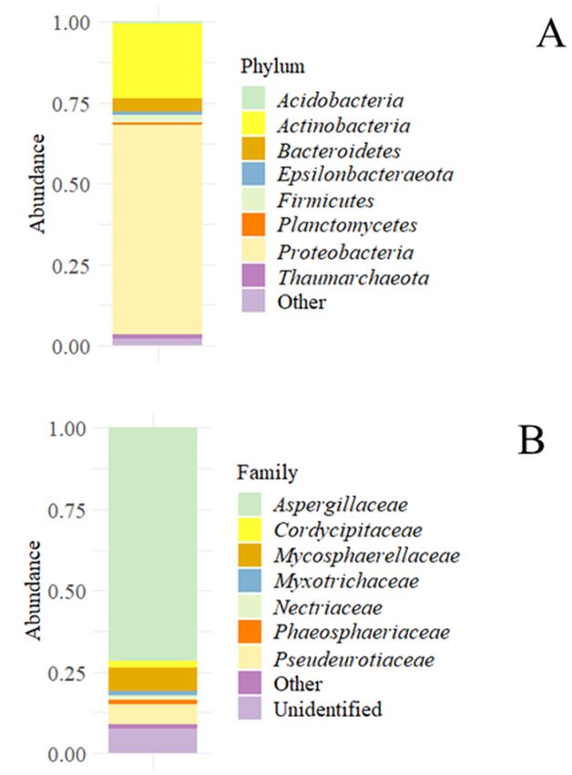
Archaea, as Thaumarchaeota, were also found (Figure 1A). Regarding the fungal popula-
tions found in the sample, they were dominated by almost 75% at the family level by
Aspergillaceae; the remaining the population was formed by Phaeosphaeriaceae, Pseudeuroti-
acee, and other unidentified families (Figure 1B).
3.2.2. Isolation and Identification of Viable Microorganisms
To determine the viability of the meta-taxonomically detected populations, an isolation
study conducted after an enrichment step in different broths was conducted for fungi and
bacteria. No bacteria were isolated when the incubation was conducted at 15 ◦ C; however,
most isolates were isolated at 32 ◦ C, and a few at 60 ◦ C (Table 3). Concerning the bacterial
isolates, a total of 30 strains were isolated, all of them identified as Bacillus-related species.Microorganisms 2021, 9, x FOR PEER REVIEW 6 of 14
some Archaea, as Thaumarchaeota, were also found (Figure 1A.). Regarding the fungal pop-
Microorganisms 2021, 9, 1631 ulations found in the sample, they were dominated by almost 75% at the family level by 6 of 14
Aspergillaceae; the remaining the population was formed by Phaeosphaeriaceae,
Pseudeurotiacee, and other unidentified families (Figure 1B.).
Figure
Figure1. Accumulated microbial
1. Accumulated abundances
microbial in the sediment
abundances sample. (A)
in the sediment Accumulated
sample. bacterial
(A) Accumulated bacterial
and
andarchaeal abundances
archaeal at the
abundances phylum
at the level.
phylum (B) Accumulated
level. fungal
(B) Accumulated abundance
fungal at the at
abundance family
the family level.
level.
Table 3. Isolated bacterial strains in the sediment. Culture media and temperature of isolation.
3.2.2. Isolation and Identification of Viable Microorganisms
Enrichment Salinity NCBI Accesion
Isolates Identification To determine the
Temperature (◦ C)
viability of the meta-taxonomically detected populations, an isola-
Enzymatic Activity
Media Resistance Number
tion study conducted after an enrichment step in different broths was conducted for fungi
DIP-1 Bacillus sp. SW 32 - 8 MZ600240
and bacteria. No bacteria were isolated when the incubation was conducted at 15 °C; how-
DIP-2 Bacillus sp. SW 32 - 8 MZ600241
DIP-3 Bacillus sp. ever, most
SW isolates were isolated
32 at 32 °C, and a few
AMIL, at 60 °C (Table 3).10Concerning the
PROT MZ600242
DIP-4 Bacillus sp. bacterialSW
isolates, a total of3230 strains were isolated,
PHOSall of them identified 8 as Bacillus-re-
MZ600243
DIP-5 Bacillus sp. lated species.
SW 32 - 8 MZ600244
DIP-6 Bacillus sp. SW 32 PROT 16 MZ600245
DIP-7 Bacillus
Table 3.sp. SWstrains in the sediment.
Isolated bacterial 32 Culture NIT,
media PHOS, AMIL, PROTof isolation. 10
and temperature MZ600246
DIP-8 Bacillus sp. SW 32 - 16 MZ600247
DIP-9 Bacillus sp. SW
Enrichment 32
Temperature PHOS Salinity 10
NCBI Accesion MZ600248
Isolates
DIP-10 Identification
Bacillus cereus SW 32 Enzymatic
PHOS,Activity
AMIL, PROT 12 Number MZ600249
Media (°C) Resistance
DIP-11 Bacillus sp. SW 32 PHOS, AMIL, PROT 10 MZ600250
DIP-1 Bacillus sp. SW 32 - 8 MZ600240
DIP-12 Bacillus sp. SW 32 PHOS, AMIL, PROT 16 MZ600251
DIP-2 Bacillus
Bacillussp. SW 32 - 8 MZ600241
DIP-13
DIP-3 Bacillus sp. SWSW 32
32 AMIL,PHOS,
PROTAMIL, PROT 10 10MZ600242 MZ600252
megaterium
DIP-4
DIP-14 Bacillussp.
Bacillus sp. SWSW 32
32 PHOS
PHOS, AMIL, PROT 8 16MZ600243 MZ600253
DIP-15
DIP-5 Bacillus
Bacillussp.
sp. SWSW 32
32 PHOS,
- AMIL, PROT 8 12MZ600244 MZ600254
DIP-16
DIP-6 Bacillus simplex
Bacillus sp. SWSW 32
32 PHOS, AMIL, PROT 16
PROT 10MZ600245 MZ600255
DIP-17 Bacillus sp. SW 32 PHOS, PROT 10 MZ600256
DIP-18 Bacillus sp. SW 32 PHOS, POT, AMIL, PROT 16 MZ600257
Bacillus
DIP-19 SW 32 PHOS, AMIL, PROT 16 MZ600258
megaterium
DIP-20 Bacillus sp. SW 32 PHOS, AMIL, PROT 16 MZ600259
DIP-21 Bacillus mycoides SW 32 PHOS, AMIL, PROT 12 MZ600260
DIP-22 Bacillus simplex SW 32 AMIL 16 MZ600261Microorganisms 2021, 9, 1631 7 of 14
Table 3. Cont.
Enrichment Salinity NCBI Accesion
Isolates Identification Temperature (◦ C) Enzymatic Activity
Media Resistance Number
DIP-23 Bacillus circulans SW 32 PHOS, AMIL, PROT 14 MZ600262
Bacillus
DIP-24 SW 32 PHOS, PROT 16 MZ600263
aryabhattai
DIP-25 Bacillus sp. SW 32 PHOS, AMIL, PROT 14 MZ600264
DIP-26 Bacillus sp. SW 32 PHOS, AMIL, PROT 10 MZ600265
DIP-27 Bacillus sp. SW 32 PHOS, AMIL, PROT 12 MZ600266
Brevibacillus
DIP-28 SW 60 PHOS 12 MZ600267
thermoruber
DIP-29 Geobacillus sp. SW 60 - 10 MZ600268
DIP-30 Bacillus sp. SW 60 PHOS 10 MZ600269
AMIL: amilase; PROT: protease; PHOS: phosphatase; NIT: nitrogen fixation.
Only 12 fungal isolates (Table 4) were obtained after enrichment, most of them belong-
ing to the genus Penicillium, as indicated by molecular identification.
Table 4. Fungal strains isolated in the sediment. Culture media and temperature of isolation.
Enrichment NCBI Accesion
Isolates Identification Temperature (◦ C)
Media Number
DIF-1 Aspergillus sp. PDB 15 MZ602115
Penicillium
DIF-2 YMB 15 MZ602116
chrysogenum
DIF-3 Aspergillus sp. PDB 15 MZ602117
DIF-4 Aspergillus sydowii PDB 15 MZ602118
DIF-5 Penicillium sp. PDB 15 MZ602119
DIF-6 Penicillium sp. YMB 32 MZ602120
DIF-7 Penicillium sp. PDB 32 MZ602121
Penicillium
DIF-8 PDB 32 MZ602122
crustosum
DIF-9 Exophiala sp. PDB 32 MZ602123
Penicillium
DIF-10 PDB 32 MZ602124
chrysogenum
DIF-11 Penicillium sp. PDB 32 MZ602125
Penicillium
DIF-12 YMB 32 MZ602126
crustosum
3.2.3. Phenotypical Characterization of the Bacterial Isolates
The bacterial strains of the present study were diverse according to their phenotypical
characteristics (extracellular enzyme profiles and salinity resistance). All isolates were
able to grow in saline concentrations up to 8% w/v. From the 30 bacterial isolates tested,
25 were able to grow at 10% NaCl, 15 at 12%, 11 at 14%, and 9 at 16%.
The study of a basic panel of metabolic traits of the isolates indicated that only one
bacterial isolate (DIP-7) was able to grow in a nitrogen-free medium, indicating that it
was a free-living nitrogen-fixing strain. Furthermore, only one strain was found to be
able to solubilize potassium (isolate DIP-18). More common among the isolates were the
phosphate solubilizing bacteria (near 73%) which are very important for biogeochemical
cycles, increasing the P availability in soils. Finally, proteolytic (near 67%) and amylolytic
activities (60%) were normal among the isolates.
3.2.4. Study of Scanning Electron Microscopy (SEM)
The microscopic analysis of the sediments revealed that diatoms were present in the
sediment at a high frequency. Up to six different species of diatoms were found according
to the study of their morphological characteristics observed by SEM: Dactyliosolen spp.,
Thalassiosira spp., Coscinodiscus spp., Actynocyclus spp., Odontella spp., and Psammothidium
spp. (Figure 2).activities (60%) were normal among the isolates.
3.2.4. Study of Scanning Electron Microscopy (SEM).
The microscopic analysis of the sediments revealed that diatoms were present in the
sediment at a high frequency. Up to six different species of diatoms were found according
Microorganisms 2021, 9, 1631 to the study of their morphological characteristics observed by SEM: Dactyliosolen spp., 8 of 14
Thalassiosira spp., Coscinodiscus spp., Actynocyclus spp., Odontella spp., and Psammothidium
spp. (Figure 2).
Figure 2. Identified diatoms present in the sediment according to the morphological characteristics
Figure 2. Identified diatoms present in the sediment according to the morphological characteristics
observed by SEM. (A) Dactyliosolen spp., (B) Thalassiosira spp., (C) Coscinodiscus spp., (D) Actynocy-
observed
clus spp., by
(E) SEM. Dactyliosolen
(A) spp.,
Odontella spp., (B) Thalassiosira
(F) Psammothidium spp. spp., (C) Coscinodiscus spp., (D) Actynocyclus
spp., (E) Odontella spp., (F) Psammothidium spp.
4. Discussion
Deception Island is covered by lapilli and pyroclastic ash on most of its surface [30].
The DRX study of the sample revealed two main minerals, anorthite and halite. Anorthite
could be explained by the presence of lapilli since it is formed by basaltic-andesitic and
volcanic glasses. The andesite that conforms lapilli is fundamentally composed by pla-
gioclases, belonging to the albite-anortite series, from the tectosilicates group which their
general formula is (Na, Ca) (Si, Al)3 O8 . Halite, another main component of the sample,
comes from the deposition of marine water.
Recently, some DRX studies of samples coming from Deception Island have been
carried out. Lezcano and colleagues analyzed a soil sample from Cerro Caliente Hill and
determined that the phyllosilicates (montmorillonite, nontronite, and saponite) group
was the most prevalent group of minerals [31]. Nevertheless, regarding the chemical
composition of the sediments, some ions differed in that study compared to our findings:
in the case of soluble SO4 2− , the observed concentration was one thousand times lower
(1.39 ± 1.17 µg/g), and NO3− was ten times less concentrated (0.22 ± 0.15 µg/g). TheMicroorganisms 2021, 9, 1631 9 of 14
chemical analysis of several sediment samples revealed that in Whaler’s Bay some metallic
elements were more concentrated than in other island locations [32]. Among all, Fe was
observed as the most common element (15.814 ± 581 µg/g).
Regarding other kinds of substrates, the chemical composition is more different. The
ionic analysis of melted water [33] revealed quite a different composition if compared to
those obtained in geothermal sediments: the concentration of NO3− rise up to 3 mg/mL,
and other elements, like S (6.76 ppm compared to 329 ppm), on the contrary, were less
concentrated. Some of them, like Mg (0.1 ppm), were in similar concentrations.
Our samples were formed by sediments that air and water transported and then,
were deposited. The chemical analysis carried out in this study showed that the sediment
is mainly composed of materials that conform to the island, probably originating there.
Nevertheless, the ionic composition is slightly different from the sediments obtained in the
island and glacial water. The main difference is in regard to ions such as SO4 2− and NO3− .
In the case of nitrates, our sediments could have been enriched by materials from glaciers.
Due to the presence of the fumaroles, a soil–temperature gradient was established as the
temperature increases, and glaciers melt enriching the seawater. On the contrary, we could
hypothesize that sulfur does not come neither from the island nor from glaciers, in both
cases, it is present in much less concentration. High concentrations of sulphur could be
found due to the presence of fumaroles and volcanic activity [34]. An indicator of volcanic
enrichment is the presence of high concentrations of iron, manganese, and silicon at high
concentrations [35]. In this sample, we cannot affirm that a volcanic enrichment could
have occurred because sulphur and iron concentrations are not in the proper ratio. Silicon
cannot be considered an indicator of volcanic activity in this case either, this element may
come from materials that conform to the island. Either way, geothermal phenomena are
not only the emission of volcanic materials through steam vents or fissures.
We can highlight the presence of amorphous minerals detected by SEM-EDX. These
minerals, composed of sulphur trioxide and aluminum oxide, could be interpreted as parti-
cles of the sediment coming from the volcanic activity that occurred in the last century [2].
Other interesting and abundant elements detected in the optical emission spectroscopy
studies were silicon, aluminum, nickel, sodium, calcium, and strontium, which are the
main elements of basalt and andesite [36].
In conclusion, we can assume that the origin of the materials present in the sample are
sediments that come from the island dragged by water.
Our bacterial meta-taxonomic results are comparable to previously reported re-
sults [36], showing a stable bacterial community in different sediment samples at Whaler’s
Bay over several years. In that study, Proteobacteria, Bacteroidetes, and Actinobacteria were
the main groups. In other analyzed substrates, the results are similar. The microbial
composition of different microbial structures such as biofilms and microbial mats have
been studied. Proteobacteria is the most common phylum (in around 75%) in different
bacterial biofilms isolated at Whaler’s Bay [37], as well as in microbial mats taken from the
island (in around 30%). Bacteroidetes and Acidobacteria (in different ranges depending on the
microbial mat) are also usually present [31]. Martínez-Alonso and colleagues studied the
volcanic endoglacial sediments and described Actinobacteria as the most common phylum
in this ecosystem (30%), followed by Bacteroidetes (27%) and Proteobacteria (15%) among
others [33].
The archaeal phylum Thaumarcheota detected in the meta-taxonomic analysis has
been yet to be described on the island. Lezcano and colleagues described this as the
most common phylum in different microbial mats, representing near 35% of the archaeal
community [30].
To study the viable microbial population present in the sediment sample and their
adaptations to such an environment, an isolation protocol was conducted. It should be
highlighted that the great difference observed between the present microbiota, analyzed
using molecular approaches (meta-taxonomic study), and the viable microbiota, by cul-
tivation on agar plates, is known as the “the great plate count anomaly”. Nevertheless,Microorganisms 2021, 9, 1631 10 of 14
this low percentage of viabilization has been previously reported in similar Antarctic
samples from other studies [6]. Using culture media similar to those previously used in
other studies, both for chemoheterotrophs and chemolithotrophs, the majority of isolates
presented a chemoheterotrophic metabolism [6,14]. All the isolated strains belonged to the
Bacillus-related genera, which are spore-forming species, allowing survival under adverse
conditions. Furthermore, only 7.69% of the species detected by the meta-taxonomic analysis
have been described as spore-forming microorganisms. The absence of lithotrophic isolates
could be explained by these two facts: the lack of endospore-forming species (which affects
the long-term survival of the community), and the nutritional requirements.
Studies carried out during the 20th century in soils with different characteristics,
discovered bacteria that could only be identified up to the genus level and, interestingly,
all of them belonged to the Bacillus genus [11,38]. Later, advances in molecular techniques
for microbial identification and taxonomy made possible the new reidentification of these
isolates, describing new genera such as Alicyclobacillus [39] and Geobacillus [40].
Thermophilic strains of Geobacillus spp. and Brevibacillus thermoruber where previously
described at Deception Island by Muñoz and colleagues in a sample from Fumaroles Bay,
located northwest from our sampling point [41]. Other species isolated in this research,
such as Bacillus cereus and B. megaterium, have been described previously on Deception
Island [42]. More recently, some thermophilic strains have been isolated from the island,
despite sediment temperature from Whaler’s and Fumarole Bays, either from glacier or
fumarole zones [14]. The isolates belonged to Bacillus-related genera such as Geobacillus,
Brevibacillus and Anoxybacillus, and were recovered from samples with environmental
temperatures ranging from 0 to 80 ◦ C and using general culture media, such as TSA or
Marine Agar, such as in our study.
High saline resistance of the isolated strains could be an adaptation to the dehydration
caused under freeze and high salinity conditions [43]. This could be related to wider
resistance to several stresses [44]. Our strains were halotolerant, all of them were able to
grow in up to 8% NaCl (w/v) and some of them up to 16%. Some authors have reported
osmophilic bacterial strains, isolated from the rhizosphere of Antarctic plants, resistant
to NaCl concentrations up to 16% [45]. This generalized osmotolerance is linked with
molecular and physiological adaptations to these environments as indicated by [36] that
analyzed the stress response genes in Whaler’s Bay, describing the osmotic stress genes as
the most common stress genes in a metagenomic analysis.
The production, transformation, decomposition, and/or transport of organic matter is
a central part in the geochemical cycle of bioelements. In that way, microorganisms play
a fundamental role, contributing an efficient set of extracellular enzymes to an optimum
nutrient uptake [45]. In our study, the strains presented different enzymatic profiles, with
few strain-specific activities. Phosphatase, protease, and amylase were the commonest in
our study as was also reported in other studies [46].
The studies regarding the fungal diversity on the island reported the same overall
pattern of dominant fungal populations by isolation and meta-taxonomic approaches.
Concerning the cultivable fungal community, we have detected a low diversity level as
other studies have shown [16,47], with most of the isolates identified as members of the
Aspergillus and Penicillium genera. These genera have been isolated regardless of the soil
temperature, both in hot and cold volcanic soils of the island [16]. Only one isolate belonged
to a different genus, Exophiala, nevertheless, this was usually found in the meta-taxonomic
studies carried out in the same island [17]. Exophiala spp., as well as some Penicillium
spp., have been described to be extremely thermotolerant [48]. In other locations, such
as King George Island (South Shetland Islands), Penicillium spp. has also been frequently
isolated [49].
According to the reported results, more than 1000 fungal species have been described
in morphological or cultivation studies of Antarctic fungi, nevertheless, the use of ‘omic’
approaches is essential in describing the microbial life in each habitat [17]. Several ‘omics’
(metabarcoding or metataxonomic) studies have been realized. In all of them [17,31,50],Microorganisms 2021, 9, 1631 11 of 14
Ascomycetes was the dominant phylum. Our metataxonomic data suggests that the fungal
community is dominated by the Aspergillaceae family (Ascomycetes). The meta-barcoding
analysis of fungal diversity in different areas (impacted and non-impacted locations) of
the island revealed that the main phylum is Ascomycota, up to 44.3% of the ASVs, not
being influenced by the consideration or protection status of the area [17]. In other types of
samples, as well as in soils, the population was dominated by Ascomycota.
In microbial mats, sampled at different temperatures, Ascomycota was the predominant
fungal phyla among all the eukaryotes. At 88 ◦ C, this phylum was the most abundant
(9% of the phyla) as well as at 2 ◦ C (2%); interestingly, at 8 ◦ C it practically disappeared
(abundance less than 0.55%) due to an increase of the eukaryotic-autotroph population [30].
As indicated before, a diatom population was also present in the sediment, and it
was described using the morphological data obtained with SEM. Only the Psammothidium
and Thalassiosira genera, two out of the six diatom genera, have been previously described
on the island, near Kroner Lake [51]. The remaining genera (Dactyliosolen, Coscinodiscus,
Actynnocyclus, and Odontella) have not been previously described on the island, but their
presence on the continent has already been documented. Generally, benthonic diatoms
belong to the pennate group, whereas those in the pelagic habitat belong to central ones [52].
In this study, most of the identified genera were identified as central ones and were probably
diatoms (or their cellular rests) with a pelagic habitat that were dragged to the sampling
point by sea-water streams.
In this study, the different analyses carried out in such a unique sediment sample
provided interesting information about the microbiological and physicochemical influences
between the island and the surrounding sea. The ionic and mineral composition revealed
the confluence between the sea and glacier water, creating a transition zone between ice,
land, and sea. The microbiological determinations provided interesting information about
the community present in that confluence zone. The metataxonomic analysis itself could
only show the quantitative/qualitative structure of the community, but its comparison
to other types of samples could give valuable information about the ecological micro-
structure and interactions between two different environments, defining different roles and
influencing mechanisms. In addition, the viable community inhabiting such an extreme en-
vironment showed different metabolic potentials, not only in their enzymatic activities but
also in their resistance profiles to environmental stresses, promising a high biotechnological
potential capable of being screened for useful industrial enzymes or bioactive compounds.
Author Contributions: Conceptualization, A.A., D.M. and A.S.; methodology, D.M. and A.S.; soft-
ware, M.d.C.; formal analysis, J.V., M.d.C., A.A., D.M. and A.S.; investigation, J.V. and A.A.; writing—
original draft preparation, J.V.; writing—review and editing, A.S. All authors have read and agreed
to the published version of the manuscript.
Funding: This research received no external funding.
Acknowledgments: The authors of this work thankJesús Martínez-Frías (Geosciences Institute,
CSIC-UCM) for his contribution by providing the sample under study. We also thank Ana Martín
(Department of Genetics, Physiology and Microbiology, UCM) for the help provided during the
study of diatoms.
Conflicts of Interest: The authors declare no conflict of interest.ing—original draft preparation, J.V.; writing—review and editing, A.S. All authors have read and
agreed to the published version of the manuscript.
Funding: This research received no external funding.
Acknowledgments: The authors of this work thankJesús Martínez-Frías (Geosciences Institute,
CSIC-UCM) for his contribution by providing the sample under study. We also thank Ana Martín
Microorganisms 2021, 9, 1631 12 of 14
(Department of Genetics, Physiology and Microbiology, UCM) for the help provided during the
study of diatoms.
Conflicts of Interest: The authors declare no conflict of interest.
Appendix A
Appendix A
Figure A1. X-Ray Diffractogram showing the anorthite profile according to ASTM file number 00-018-1202.
References
Figure A1. X-Ray Diffractogram showing the anorthite profile according to ASTM file number 00-018-1202.
1. Marti, J.; Vila, J.; Rey, J. Deception Island (Bransfield Strait, Antarctica): An example of a volcanic caldera developed by extensional
tectonics.
ReferencesGeol. Soc. Lond. Spec. Publ. 1996, 110, 253–265. [CrossRef]
2. Barker,
1. D.H.N.; Austin,
Marti, J.; Vila, J.; Rey,J.A. Rift propagation,
J. Deception detachment
Island (Bransfield Strait,faulting, and
Antarctica): Anassociated
example ofmagmatism in Bransfield
a volcanic caldera developedStrait, Antarctic
by exten-
sionalJ.tectonics.
Peninsula. Geophys.Geol.Res.Soc.
Solid Earth
Lond. Publ.103,
1998,
Spec. 24017–24043.
1996, 110, 253–265,[CrossRef]
doi:10.1144/Gsl.Sp.1996.110.01.20.
3. Cooper,
2. A.P.R.;
Barker, Smellie,
D.H.N.; J.L.;
Austin, J.A.Maylin, J. Evidence
Rift propagation, for shallowing
detachment faulting,and
and uplift from
associated bathymetric
magmatism records Strait,
in Bransfield of Deception
AntarcticIsland,
Antarctica. Antarct.
Peninsula. Sci. 2004,
J. Geophys. Res. 10,
Solid455–461.
Earth 1998, [CrossRef]
103, 24017–24043, doi:10.1029/98jb01117.
4. 3. Cooper,
Herbold, C.W.; A.P.R.; Smellie, J.L.;
McDonald, I.R.;Maylin, J. Evidence
Cary, S.C. for shallowing
Microbial Ecology of andGeothermal
uplift from bathymetric
Habitats in records of Deception
Antarctica. Island, Ant-
In Antarctic Terrestrial
arctica. Antarct.
Microbiology; Springer: Sci.Berlin/Heidelberg,
2004, 10, 455–461, doi:10.1017/s0954102098000558.
Germany, 2014; pp. 181–215. [CrossRef]
5. 4.
Smith, Herbold, C.W.; R.;
K.; Baldwin, McDonald,
Kaufmann, I.R.; R.;
Cary, S.C. Microbial
Sturz, A. EcosystemEcology of Geothermal
studies at DeceptionHabitats in Antarctica.
Island, Antarctica:In Antarctic Terrestrial
An overview. Deep.Mi-Sea Res.
crobiology; Springer: Berlin/Heidelberg, Germany, 2014; pp. 181–215, doi:10.1007/978-3-642-45213-0_10.
Part II Top. Stud. Oceanogr. 2003, 50, 1595–1609. [CrossRef]
6. Marquez, S.L.; Blamey, J.M. Isolation and partial characterization of a new moderate thermophilic Albidovulum sp. SLM16 with
transaminase activity from Deception Island, Antarctica. Biol. Res. 2019, 52, 5. [CrossRef]
7. Pepi, M.; Agnorelli, C.; Bargagli, R. Iron demand by thermophilic and mesophilic bacteria isolated from an antarctic geothermal
soil. Biometals 2005, 18, 529–536. [CrossRef]
8. Bottos, E.M.; Scarrow, J.W.; Archer, S.D.; McDonald, I.R.; Cary, S.C. Bacterial Community Structures of Antarctic Soils. In Antarctic
Terrestrial Microbiology; Springer: Berlin/Heidelberg, Germany, 2014; pp. 9–33.
9. Darling, C.A.; Siple, P.A. Bacteria of Antarctica. J. Bacteriol. 1941, 42, 83. [CrossRef] [PubMed]
10. Flint, E.A.; Stout, J.D. Microbiology of Some Soils from Antarctica. Nature 1960, 188, 767–768. [CrossRef]
11. Horowitz, N.H.; Cameron, R.E.; Hubbard, J.S. Microbiology of the dry valleys of antarctica. Science 1972, 176, 242–245. [CrossRef]
12. Smith, J.J.; Tow, L.A.; Stafford, W.; Cary, C.; Cowan, D.A. Bacterial diversity in three different Antarctic Cold Desert mineral soils.
Microb. Ecol. 2006, 51, 413–421. [CrossRef] [PubMed]
13. Cowan, D.A.; Russell, N.J.; Mamais, A.; Sheppard, D.M. Antarctic Dry Valley mineral soils contain unexpectedly high levels of
microbial biomass. Extremophiles 2002, 6, 431–436. [CrossRef]
14. Bendia, A.G.; Araujo, G.G.; Pulschen, A.A.; Contro, B.; Duarte, R.T.; Rodrigues, F.; Galante, D.; Pellizari, V.H. Surviving in hot
and cold: Psychrophiles and thermophiles from Deception Island volcano, Antarctica. Extremophiles 2018, 22, 917–929. [CrossRef]
[PubMed]Microorganisms 2021, 9, 1631 13 of 14
15. Bendia, A.G.; Signori, C.N.; Franco, D.C.; Duarte, R.T.; Bohannan, B.J.; Pellizari, V.H. A mosaic of geothermal and marine features
shapes microbial community structure on deception Island Volcano, Antarctica. Front. Microbiol. 2018, 9, 899. [CrossRef]
16. Figueredo, H.M.; Gonçalves, V.N.; Godinho, V.M.; Lopes, D.V.; Oliveira, F.S.; Rosa, L.H. Diversity and ecology of cultivable fungi
isolated from the thermal soil gradients in Deception Island, Antarctica. Extremophiles 2020, 24, 219–225. [CrossRef]
17. Rosa, L.H.; da Silva, T.H.; Ogaki, M.B.; Pinto, O.H.B.; Stech, M.; Convey, P.; Carvalho-Silva, M.; Rosa, C.A.; Câmara, P.E. DNA
metabarcoding uncovers fungal diversity in soils of protected and non-protected areas on Deception Island, Antarctica. Sci. Rep.
2020, 10, 21986. [CrossRef]
18. Belda, I.; Zarraonaindia, I.; Perisin, M.; Palacios, A.; Acedo, A. From Vineyard Soil to Wine Fermentation: Microbiome Approxi-
mations to Explain the “terroir”. Concept. Front. Microbiol. 2017, 8, 821. [CrossRef]
19. Callahan, B.J.; Sankaran, K.; Fukuyama, J.A.; McMurdie, P.J.; Holmes, S.P. Bioconductor Workflow for Microbiome Data Analysis:
From raw reads to community analyses. F1000Res 2016, 5, 1492. [CrossRef]
20. Callahan, B.J.; McMurdie, P.J.; Holmes, S.P. Exact sequence variants should replace operational taxonomic units in marker-gene
data analysis. ISME J. 2017, 11, 2639–2643. [CrossRef] [PubMed]
21. Quast, C.; Pruesse, E.; Yilmaz, P.; Gerken, J.; Schweer, T.; Yarza, P.; Peplies, J.; Glockner, F.O. The SILVA ribosomal RNA gene
database project: Improved data processing and web-based tools. Nucleic Acids Res. 2013, 41, D590–D596. [CrossRef]
22. Nilsson, R.H.; Larsson, K.H.; Taylor, A.F.S.; Bengtsson-Palme, J.; Jeppesen, T.S.; Schigel, D.; Kennedy, P.; Picard, K.; Glockner,
F.O.; Tedersoo, L.; et al. The UNITE database for molecular identification of fungi: Handling dark taxa and parallel taxonomic
classifications. Nucleic Acids Res. 2019, 47, D259–D264. [CrossRef] [PubMed]
23. Atlas, R.M. Handbook of Media for Environmental Microbiology; CRC Press: Boca Raton, FL, USA, 2005.
24. Cenis, J. Rapid extraction of fungal DNA for PCR amplification. J. Nucleic Acids Res. 1992, 20, 2380. [CrossRef]
25. Nautiyal, C.S. An efficient microbiological growth medium for screening phosphate solubilizing microorganisms. FEMS Microbiol.
Lett. 1999, 170, 265–270. [CrossRef] [PubMed]
26. Aleksandrov, V.; Blagodyr, R.; Ilev, I. Liberation of phosphoric acid from apatite by silicate bacteria. J. Mikrobiol. Z. 1967, 29,
111–114.
27. Olney, M. MIRACLE. Microfossil Image Recovery and Circulation for Learning and Education. Available online: https://www.
ucl.ac.uk/GeolSci/micropal/index.html (accessed on 29 October 2020).
28. Scott, F.J.; Marchant, H.J. Antarctic Marine Protists; Australian Biological Resources Study and Australian Antarctic Division:
Canberra, Australia, 2005.
29. Spaulding, S.A.; Bishop, I.W.; Edlund, M.B.; Lee, S.; Furey, P.; Jovanovska, E.; Potapova, M. Diatoms of North America. Available
online: https://diatoms.org/ (accessed on 29 October 2020).
30. Rey, J.; Somoza, L.; Martínez-Frías, J. Tectonic, volcanic, and hydrothermal event sequence on Deception Island (Antarctica). J.
Geo-Mar. Lett. 1995, 15, 1–8. [CrossRef]
31. Lezcano, M.A.; Moreno-Paz, M.; Carrizo, D.; Prieto-Ballesteros, O.; Fernandez-Martinez, M.A.; Sanchez-Garcia, L.; Blanco, Y.;
Puente-Sanchez, F.; de Diego-Castilla, G.; Garcia-Villadangos, M.; et al. Biomarker Profiling of Microbial Mats in the Geothermal
Band of Cerro Caliente, Deception Island (Antarctica): Life at the Edge of Heat and cold. Astrobiology 2019, 119, 1490–1504.
[CrossRef]
32. Di Giglio, S.; Agüera, A.; Pernet, P.; M’Zoudi, S.; Angulo-Preckler, C.; Avila, C.; Dubois, P. Effects of ocean acidification on
acid-base physiology, skeleton properties, and metal contamination in two echinoderms from vent sites in Deception Island,
Antarctica. Sci. Total Environ. 2020, 765, 142669. [CrossRef]
33. Martinez-Alonso, E.; Pena-Perez, S.; Serrano, S.; Garcia-Lopez, E.; Alcazar, A.; Cid, C. Taxonomic and functional characterization
of a microbial community from a volcanic englacial ecosystem in Deception Island, Antarctica. Sci. Rep. 2019, 9, 12158. [CrossRef]
[PubMed]
34. Ortiz, R.; Vila, J.; García, A.; Camacho, A.; Diez, J.; Aparicio, A.; Soto, R.; Viramonte, J.; Risso, C.; Menegatt, N. Geophysical
Features of Deception Island. In Recent Progress in Antarctic Earth Science; Terrapub: Tokyo, Japan, 1992; pp. 143–152.
35. Elderfield, H. Effects of volcanism on water chemistry, Deception Island, Antarctica. J. Mar. Geol. 1972, 13, M1–M6. [CrossRef]
36. Wilkinson, J.F.G. Classification and Average Chemical-Compositions of Common Basalts and Andesites. J. Petrol. 1986, 27, 31–62.
[CrossRef]
37. Centurion, V.; Lacerda-Júnior, G.; Duarte, A.; Silva, T.; Silva, L.; Rosa, L.; Oliveira, V. Dynamics of microbial stress responses
driven by abiotic changes along a temporal gradient in Deception Island, Maritime Antarctica. Sci. Total Environ. 2021, 758,
143671. [CrossRef]
38. Nicolaus, B.; Marsiglia, F.; Esposito, E.; Trincone, A.; Lama, L.; Sharp, R.; Di Prisco, G.; Gambacorta, A. Isolation of five strains of
thermophilic eubacteria in Antarctica. J. Polar Biol. 1991, 11, 425–429. [CrossRef]
39. Wisotzkey, J.D.; Jurtshuk, P., Jr.; Fox, G.E.; Deinhard, G.; Poralla, K. Comparative sequence analyses on the 16S rRNA (rDNA) of
Bacillus acidocaldarius, Bacillus acidoterrestris, and Bacillus cycloheptanicus and proposal for creation of a new genus, Alicyclobacillus
gen. nov. Int. J. Syst. Bacteriol. 1992, 42, 263–269. [CrossRef]
40. Coorevits, A.; Dinsdale, A.E.; Halket, G.; Lebbe, L.; De Vos, P.; Van Landschoot, A.; Logan, N.A. Taxonomic revision of the genus
Geobacillus: Emendation of Geobacillus, G. stearothermophilus, G. jurassicus, G. toebii, G. thermodenitrificans and G. thermoglucosidans
(nom. corrig., formerly ‘thermoglucosidasius’); transfer of Bacillus thermantarcticus to the genus as G. thermantarcticus comb. nov.;Microorganisms 2021, 9, 1631 14 of 14
proposal of Caldibacillus debilis gen. nov., comb. nov.; transfer of G. tepidamans to Anoxybacillus as A. tepidamans comb. nov.; and
proposal of Anoxybacillus caldiproteolyticus sp. nov. Int. J. Syst. Evol. Microbiol. 2012, 62, 1470–1485. [CrossRef] [PubMed]
41. Muñoz, P.A.; Flores, P.A.; Boehmwald, F.A.; Blamey, J.M. Thermophilic bacteria present in a sample from Fumarole Bay, Deception
Island. Antarct. Sci. 2011, 23, 549–555. [CrossRef]
42. Llarch, A.; Logan, N.A.; Castellvi, J.; Prieto, M.J.; Guinea, J. Isolation and Characterization of Thermophilic Bacillus spp. from
Geothermal Environments on Deception Island, South Shetland Archipelago. Microb. Ecol. 1997, 34, 58–65. [CrossRef]
43. Thomas, D.; Dieckmann, G. Antarctic sea ice—A habitat for extremophiles. J. Sci. 2002, 295, 641–644. [CrossRef]
44. Yukimura, K.; Nakai, R.; Kohshima, S.; Uetake, J.; Kanda, H.; Naganuma, T. Spore-forming halophilic bacteria isolated from
Arctic terrains: Implications for long-range transportation of microorganisms. J. Polar Sci. 2009, 3, 163–169. [CrossRef]
45. Vasileva-Tonkova, E.; Romanovskaya, V.; Gladka, G.; Gouliamova, D.; Tomova, I.; Stoilova-Disheva, M.; Tashyrev, O. Ecophys-
iological properties of cultivable heterotrophic bacteria and yeasts dominating in phytocenoses of Galindez Island, maritime
Antarctica. World J. Microbiol. Biotechnol. 2014, 30, 1387–1398. [CrossRef]
46. Aislabie, J.M.; Chhour, K.-L.; Saul, D.J.; Miyauchi, S.; Ayton, J.; Paetzold, R.F.; Balks, M.R. Dominant bacteria in soils of Marble
Point and Wright Valley, Victoria Land, Antarctica. Soil Biol. Biochem. 2006, 38, 3041–3056. [CrossRef]
47. Alves, I.M.; Gonçalves, V.N.; Oliveira, F.S.; Schaefer, C.E.; Rosa, C.A.; Rosa, L.H. The diversity, distribution, and pathogenic
potential of cultivable fungi present in rocks from the South Shetlands archipelago, Maritime Antarctica. Extremophiles 2019, 23,
327–336. [CrossRef]
48. Tesei, D.; Marzban, G.; Zakharova, K.; Isola, D.; Selbmann, L.; Sterflinger, K. Alteration of protein patterns in black rock inhabiting
fungi as a response to different temperatures. Fungal Biol. 2012, 116, 932–934. [CrossRef] [PubMed]
49. Wentzel, L.C.P.; Inforsato, F.J.; Montoya, Q.V.; Rossin, B.G.; Nascimento, N.R.; Rodrigues, A.; Sette, L.D. Fungi from Admiralty
Bay (King George Island, Antarctica) Soils and Marine Sediments. Microb. Ecol. 2019, 77, 12–24. [CrossRef]
50. Baeza, M.; Barahona, S.; Alcaíno, J.; Cifuentes, V. Amplicon-metagenomic analysis of fungi from Antarctic terrestrial habitats.
Front. Microbiol. 2017, 8, 2235. [CrossRef] [PubMed]
51. Fermani, P.; Mataloni, G.; Van de Vijver, B. Soil microalgal communities on an antarctic active volcano (Deception Island, South
Shetlands). Polar Biol. 2007, 30, 1381–1393. [CrossRef]
52. Sabater, S. Diatoms. Encyclopedia of Inland Waters; Elsevier: Oxford, UK, 2009.You can also read