Modern Microscopy with the Web of Things: The OpenFlexure Microscope Software Stack
←
→
Page content transcription
If your browser does not render page correctly, please read the page content below
Modern Microscopy with the Web of Things: The OpenFlexure
                                                                   Microscope Software Stack
                                                    Joel T. Collins1 , Joe Knapper1 , Julian Stirling1 , Samuel McDermott2 , Filip Ayazi2 ,
                                                                                  and Richard Bowman1*
arXiv:2101.00933v2 [physics.ins-det] 22 Jan 2021

                                                    1
                                                        Centre for Photonics and Photonic Materials, Department of Physics, University of
                                                                                           Bath, UK.
                                                                     2
                                                                       Cavendish Laboratory, University of Cambridge, UK.
                                                                                   *
                                                                                     r.w.bowman@bath.ac.uk

                                                                                                January 25, 2021

                                                                                                      Abstract
                                                              Automated and computerised control of scientific instrumentation is almost ubiquitous in the
                                                          modern laboratory. Most instrumentation is controlled over decades old communication busses or
                                                          is accessed via proprietary system libraries. This limits which languages and operating systems
                                                          can be used to control instruments, and poses a significant problem when interfacing multiple
                                                          instruments into the same experiment. Here we present the OpenFlexure Microscope software
                                                          stack as an example of how a scientific instrument can be controlled using existing, cross-platform,
                                                          language-independent, industry-supported standards. We split the control code into client and
                                                          server applications interfaced via a web API that conforms to the W3C Web of Things standard.
                                                          This enables simple control of the microscope from multiple languages, provides a modern graph-
                                                          ical control interface, and minimises duplicated code. Network control also makes the software
                                                          stack more robust, allows multiple microscopes to be controlled by one computer, and facilitates
                                                          sharing of equipment between local or remote users. Using a Web of Things approach in research
                                                          laboratories has the potential to solve many of the key challenges of experiment integration, using
                                                          technology that is already well established.

                                                   1      Introduction
                                                   Microscopists are increasingly required to write complicated experiment automation code, in multiple
                                                   languages, to bind together inconsistent interfaces and proprietary software to run their experiments.
                                                   Currently, a wide mix of often proprietary or “legacy” connectors and protocols are commonplace even
                                                   in modern microscopes. This has led to many research labs relying on outdated hardware and software
                                                   because of the requirement for interfaces that are largely obsolete in every other field of technology.
                                                   Common practice is to make use of third-party solutions to retrofit interoperability, often maintained
                                                   predominantly by microscope users rather than manufacturers. Since not all hardware is supported by
                                                   these solutions, time must often be spent developing compatibility layers in the project’s programming
                                                   language of choice to enable even the most basic control.
                                                       In recent years, open web technologies have been widely adopted for controlling domestic hardware,
                                                   in the “Web of Things” (WoT) [1]. These network and web technologies have already addressed many
                                                   of the problems faced by laboratories and have been proven robust, fast, and secure by their long-
                                                   standing ubiquity throughout modern life. Nowadays, support staff deeply familiar with networking
                                                   and web technologies are already in place at most research laboratories and teaching institutions.

                                                                                                           1
While prior work has introduced web technology into laboratories [2, 3, 4, 5, 6], these have lacked the
comprehensive standardisation required for true interoperability. Recently however W3C, the primary
international standards organization for the open web, have moved to standardise the Web of Things,
with solid industry and community support [7].
Here, we introduce the software stack developed for the OpenFlexure Microscope [8, 9], an open-
source, 3D-printed, and fully-automated laboratory microscope. The microscope has been deployed
around the world in a wide range of operating environments, posing unique challenges as it is used
in almost equal measure by novice and expert microscopists. While most commercial microscopes
use direct connections to a single, physical computer, we use a web API (application programming
interface) to enable local and remote control through internet protocol (IP) networks. The software
stack makes use of modern networking technologies and Web of Things standards for microscope control
and data management. The presented architecture for network-connected microscopy has allowed
the OpenFlexure Microscope to be used in a diverse range of settings without re-implementation or
code duplication. Additionally, the extensibility and interoperability has allowed users to develop
additional functionality and entirely new imaging modes without having to re-implement the more
complex instrument control code.
By designing our software stack around the W3C WoT Architecture[7], we have avoided introducing
yet another competing device control system, and paved the way for comprehensive integration with
existing microscopy software solutions such as µManager [10, 11, 12, 13] and Microscope Cockpit [14,
15]. Most modern programming languages have well-developed libraries for handling web requests.
This means that WoT extensions can be developed for various microscopy control platforms to enable
networked interaction with not just the OpenFlexure Microscope, but any device using the W3C WoT
API model. This architecture is not limited to microscopy, and the same approach would work for
most instrumentation. Client applications controlling multiple instruments for larger experiments can
then be written in any modern language that supports web requests.
2 Architecture
2.1 Client–Server Model
The microscope’s software stack includes low-level device control code, logic to integrate the hard-
ware components into a useful instrument, automation for common tasks, a graphical interface for
interactive control, and APIs for scripting automated experiments from various languages. Treating
the microscope as an IoT device naturally splits these functions into client applications (the graphical
interface and scripting APIs) and a server handling the bulk of the logic and hardware control.
This split between the server application and clients has several important advantages. First, it
enables multiple client applications to connect to a microscope simultaneously. This allows, amongst
other things, a graphical interface to display a real-time camera feed, while a script controls sample
manipulation, data acquisition, and analysis. Conversely, a single client can manage multiple micro-
scopes simultaneously. This has allowed clinicians to image sample slides from several microscopes
concurrently, dramatically increasing data acquisition throughput.
Lastly, by separating the more complex server application from comparatively simple client applica-
tions, it becomes significantly easier to write client libraries in a broader set of languages. This means
that microscope users can script experiments without having to re-implement the hardware control
code in their language of choice, interface with a binary library, or learn a new language. It also ensures
consistency between different languages and avoids duplicated effort, as most of the complexity is in
the server application.
2.2 Hardware Architecture
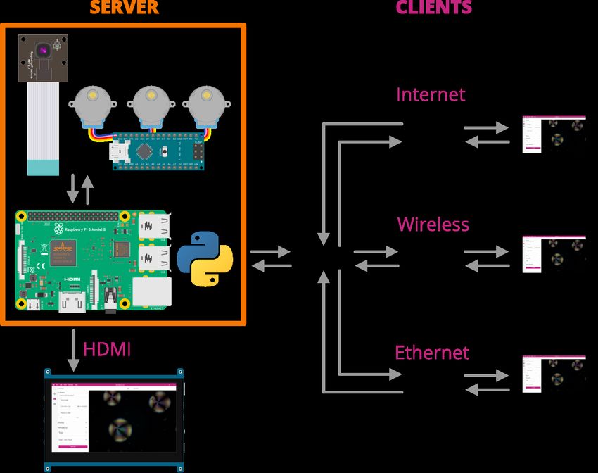
Our server runs on a Raspberry Pi computer embedded in the microscope (Figure 1). This applica-
tion handles communication with the sample translation stage, imaging camera, and any additional
2Figure 1: OpenFlexure Microscope implementation of the client–server architecture. A Raspberry Pi
runs Python code to control physical hardware (camera, Arduino-based motor controller), and set up
an HTTP web server and mDNS discovery records. Users can either control the microscope from the
Raspberry Pi itself using HDMI output and USB peripherals, or connect over any IP network.
hardware, as well as logic for data management and additional functions such as tiled scans and aut-
ofocus. Running the server on an embedded computer ensures the hardware control code is running
in a very well controlled environment. We automatically build and distribute an SD card image with
a correctly configured operating system and our server and client applications pre-installed [16]. This
eliminates the most troublesome aspect of distributing instrument control software, which is correctly
installing and configuring the low-level drivers and libraries on an unknown computer, often clashing
with system-level changes made to support other instruments connected to the same machine.
Client applications can run on the embedded computer, making the microscope a stand-alone
system to which keyboard, monitor, and mouse may be attached. More usually, client applications
will run on other devices connected via a wired or wireless IP network, using the Raspberry Pi’s
ethernet and WiFi interfaces. By using IP networking for instrument control we enable control of
multiple instruments with any external router or switch. Replacing different (often proprietary and
expensive) connectors and adaptors [17] with commodity hardware makes experimental science more
efficient and more accessible to resource-constrained scientists.
The internet protocol itself allows for high-speed, low-latency plug-and play communication. Fully
remote control can be enabled using existing, well-established secure protocols such as SSH forwarding
and VPN connections. Clients can automatically detect the server’s IP address and capabilities via
mDNS (multicast Domain Name System) [18], which is already used extensively by consumer WoT
devices.
3a) READ PROPERTY
CLIENT SERVER
GET REQUEST Read shutter speed
settings/camera/shutter_speed from physical camera.
RESPONSE
17ms Camera returns 17ms
b) WRITE PROPERTY
CLIENT SERVER
PUT REQUEST Write shutter speed
settings/camera/shutter_speed to physical camera.
22ms
RESPONSE
OK Camera returns write successful
Figure 2: Simplified example of an HTTP request flow for reading and writing a “property” resource.
a) Reading a property requires a GET request sent to the URL corresponding to the property to be
read (camera shutter speed). The server then sends a response containing the properties value (17
milliseconds) in the response body. b) Writing a property requires a PUT request sent to the same
URL, containing the new value in the request body (22 milliseconds). The server then sends an “OK”
response confirming the operation’s success.
2.3 Server application and Web API
The server application is written in Python, with much of the back-end code released as a sepa-
rate library (Python-LabThings) [19]. This library uses the “Flask” [20] web application framework,
and includes various utilities to simplify thread-based concurrency, mDNS discovery, hardware syn-
chronisation, and documentation generation. We also make use of the standard scientific python
libraries [21, 22, 23, 24]. Client–server interactions are governed by a web API based W3C Web of
Things interaction model. This model defines properties, actions, and events that map onto HTTP
URLs. The HTTP method then describes the type of operation to be handled. For example, an HTTP
GET request will read the value of a property, whereas an HTTP PUT request will write a new value
to the property (Figure 2).
W3C WoT compatibility is introduced by an automatically generating a description the micro-
scope’s web API functionality in a standardised format [25]. This allows clients to traverse the API
with minimal prior knowledge, aligning closely with the principles of representational state transfer
(REST) [26, 27], an API architectural style widely used across web service and WoT device APIs [27].
The widespread existing use of REST APIs means that users can interact with the OpenFlexure Mi-
croscope using existing standard libraries. We make use of the OpenAPI standard [28] for automatic
generation of interactive documentation [29].
Many long-running tasks, such as acquiring large tile scans, must run in the background without
blocking new API requests. Each request and action is therefore handled by its own thread to allow
concurrency. Access to physical hardware is carefully managed to avoid conflicting instructions by use
of re-entrant locks (Rlocks). In the simplest case, the lock prevents requests from sending instructions
to a device already in use. For example, if the translation stage is currently in the middle of a long
4movement, a request to move elsewhere will be denied until the initial move has completed, releasing
the stage’s lock. Clients can continue to interact with the microscope while an action is running, as
long as they do not require a locked piece of hardware. This, for example, allows the live camera
stream to be monitored during long-running experiments without users having to manually manage
multiple threads.
Background tasks are handled automatically by the server. Whenever an “action” is requested,
the server will start the function in a new thread and immediately send a “created” response back to
the client, including the URL of an “action resource” the client can poll to check its progress or final
return value. In the future, we can improve efficiency by allowing the server to asynchronously push
updates to clients without polling, for example by utilising Server-Sent Events [30].
Most automation and integration can be done using client-side code, but for modifications that are
best done on the server (e.g. adding new hardware, or low-latency integration) we use an extension
system. The main server application handles only the most basic microscope functionality: capturing
images, moving the stage, and managing device settings. All functionality beyond this is provided by
extensions. This allows functionality to be customised on each microscope. Functions deemed broadly
useful have been included and enabled by default in the server application, however all can be disabled
as required. Extensions are written as Python scripts that have direct access to physical components
comprising the microscope (e.g. camera and translation stage) through Python objects. Extensions
can provide HTTP API endpoints and HTML interfaces that are displayed as part of the microscope’s
web app.
2.4 Clients
Our primary client for the OpenFlexure Microscope is a web application included in the microscope’s
internal API server. This application provides a comprehensive graphical interface for the microscope,
including a live stream of the camera, capture functionality, basic data management, and full extension
support. By developing the client as a browser-accessible web application, we are able to support many
different operating systems without any additional code, while simultaneously drawing on the expertise
brought by a large community of existing libraries.
The web application is accompanied by a desktop application (OpenFlexure Connect) handling
device discovery and connection. The application finds and displays discovered microscopes using
mDNS, as well as allowing manual connections and saving a list of commonly accessed microscopes.
Upon connecting, the application finds and loads the microscope’s graphical user interface (Figure 3).
Using a modular interface served by the microscope allows the client to only render user interface
elements for enabled functionality. Server extensions are able to define new graphical interface compo-
nents to be rendered within the main client application. For example, the interface to manage where
images are stored is defined in the relevant extension.
For experiment scripting, we have created a Python client for the microscope [31, 32] that converts
the web API into native Python functions. Extensions functionality is also mapped to Python func-
tions automatically. This enables both scripted experiments as well as interactive sessions using, for
example, iPython/Jupyter notebooks [33, 34]. This lowers the barrier to entry for scripting microscopy
experiments since many students and lab users are familiar with iPython notebooks, especially for data
analysis. The ability to run the graphical client at the same time, e.g. to view the live video feed,
further simplifies setting up and monitoring automated experiments. We have also created a MATLAB
client [35] with similar features. As the scripting clients are lightweight wrappers for the HTTP API,
this involves a minimal amount of duplicated code.
The flexibility of the client–server architecture allows task or experiment specific interfaces to be
created quickly. For example, a client for controlling the microscope with a USB game pad was
developed[36]. This client is useful in remote field applications where a keyboard and mouse are not
practical.
5Figure 3: An example side pane from the OpenFlexure Connect microscope client. Using a web API
for instrument communication allows the client to be written in any programming language. Using
javascript we were able to create a cross-platform, responsive, and modern user interface using general
purpose user interface frameworks.
3 Implementation of microscope functionality
3.1 Live stream
Central to the microscope interface is the integrated live video display. The Raspberry Pi camera
is supported by GPU firmware that provides accelerated JPEG compression of each video frame,
allowing us to serve a real-time Motion JPEG (MJPEG) live stream of the camera. The server starts a
background thread on startup that records JPEG frames from the camera into a buffer. When clients
6connect to the stream, the server will begin sending a multi-part stream of frames taken from that
buffer. A synchronisation event is created for each client thread ensuring that clients never receive the
same frame twice. As a new frame is read from the camera, the event for each client is set, at which
point the response handler for each client will pass the frame onto the client, unset the event, and
then wait for the event to be set again, dropping frames if neccesary to ensure latency is minimised
for clients not able to receive frames at the full rate. This system is based on the work of Miguel
Grinberg [37], and is included in the Python-LabThings library. We use MJPEG in preference to
more sophisticated video formats in order to minimise latency in the stream. The MJPEG format also
makes it easy to extract or drop individual frames, and enables our fast auto-focus algorithm.
3.2 Data collection
The Raspberry Pi camera can capture JPEG images and the raw 8 megapixel Bayer data from the
camera sensor [38, 39]. This raw Bayer data can be used for more advanced analysis, avoiding artefacts
from gamma correction, demosaicing, and compression. The server records metadata about the state
of the microscope at the time of capture (camera settings, stage position, calibration data, and custom
metadata added by the user), stored as a JavaScript Object Notation (JSON) formatted string in
the “UserComment” EXIF field. Captures are stored locally on the Raspberry Pi, either on the SD
card or an available USB storage device, and can be listed (including metadata) and downloaded
through the HTTP API. Separating the actions of capturing images and downloading them avoids the
need to transfer large amounts of data over the network during time-critical phases of an experiment.
The standard graphical client provides an gallery interface to view captured images and view their
metadata.
As previously mentioned, multiple microscopes can be run in parallel to increase data throughput
for a single operator. The greatest time saving can be achieved by setting microscopes to automatically
scan over a large area, building a composite image of hundreds of overlapping fields of view (FOVs).
The server has the option to perform such scans with movements, paths and capture types chosen by
the user. Capture options and metadata are the same as individual captures, and individual images
are saved, with metadata, as the scan runs [9]. Scans run as background tasks, so the microscope’s
video feed and position can be monitored as they run, and scans can be aborted without losing or
corrupting images that are already acquired.
3.3 Auto-focus
Due to the parallelogram-based mechanisms controlling the motion of the OFM translation stage,
changes to the x-y position move the sample over a sphere cap relative to the optics rather than a
plane [8]. This necessitates an auto-focus procedure which can be run reliably at each x-y location
in an automatic scan. As a typical diagnostic scan may require over 100 x-y sites to be imaged, the
software must focus rapidly while still being sufficiently reliable to not invalidate a large scan with any
out of focus images.
A basic auto-focus procedure captures a z-stack of images regularly spaced between points expected
to be far above and below the focal point. At each height, an image is captured and converted to grey-
scale. A Laplacian convolution is applied to the whole image, assigning higher values to areas of
greater spatial brightness variance. These values are raised to the fourth power and summed over the
image to provide a sharpness value. The translation stage is then returned to the z-position with the
highest sharpness. This procedure is based on methods used previously to detect focused areas in an
out of focus image [40], and while highly reliable, typically takes 10–20 seconds to complete, limited
by capturing and image processing time.
A fast auto-focus procedure utilises the MJPEG preview stream as a metric of focus. By disabling
bit rate control, the stream becomes a simple series of independent JPEG images each with identical
compression settings. This JPEG compression uses the discrete cosine transform to describe blocks
of the image [41], where each block is described using a superposition of the fewest discrete cosine
7functions possible, minimising the storage space required. As focused images typically have sharper
feature boundaries, the storage size of an MJPEG frame will peak when the sample is in focus. By
tracking frame size and z-position as the objective moves through the focal point without stopping, the
position of peak sharpness can be identified and returned to. Monitoring the frame size while moving
back to the focus and comparing to the initial data can be used to correct for imperfections such as
backlash. Far from focus, sample features can overflow from one JPEG block to its neighbours. On a
sparse, predominantly dark sample, this can introduce information into blocks which would otherwise
be empty. If this increase in the number of blocks containing some non-zero information outweighs the
reduction of blocks containing a large amount of information, the JPEG size may maximize away from
focus. However, for feature-dense samples the size of a JPEG image can generally be used as a reliable
measure of focus. This auto-focus method has a greater positional resolution than the discrete steps
of the simpler auto-focus as MJPEG frame size can be tracked on-the-fly, reducing the time taken to
less than 5 seconds.
3.4 Automatic Calibration
3.4.1 Lens Shading Table
Due to chief ray angle compensation on the Raspberry Pi camera module’s Sony IMX219 image sensor,
the raw images captured by the microscope suffer from vignetting even when the sample is uniformly
illuminated. However, flat-field correction allows us to recover uniform images in software [42]. We
use a forked version [39] of the “picamera” library [38], to access the lens shading table in the camera’s
GPU-based image processing pipeline, enabling us to correct for vignetting in both captured images
and the real-time preview. A reduction in saturation at the edges of the image remains, but this can
be corrected by post processing at the expense of higher noise in the image [42].
3.4.2 Camera-stage Mapping
It is often convenient to move the microscope stage by a given displacement in pixels on the camera, but
the axes and step size of the translation stage rarely align perfectly. We calibrate this relationship by
moving back and forth along the stage’s x and y axes in turn, analysing the resulting displacement in
the image from the camera [43]. We combine the calibrations into a 2 × 2 affine transformation matrix
that maps stage to camera coordinates. This is a similar approach to µManager’s Pixel Calibrator
plugin [13], but treating the camera’s coordinate system as ground truth rather than the stage. We
avoid hard-coded step sizes by moving the stage in gradually increasing steps, and measure mechanical
backlash by comparing motion in opposite directions. This allows an intuitive click-to-move feature of
the microscope’s graphical interface, and will in the future be used when scanning and tiling images.
The same image analysis used for stage calibration is used as a 2D displacement encoder, allowing
for closed-loop sample translation and closed-loop scanning. This significantly reduces the error in
each individual move, as well as ensuring that errors do not accumulate over the course of a large
scan. Going forward, we will extend this functionality to include simultaneous location and mapping
(SLAM) [44]. This will enable the creation of a map of the sample by comparing predictions based on
commands sent to the motors to observations from the camera. This will enable accurate movements
to features or areas of interest using the camera and estimated motor position.
4 Conclusions
In this manuscript we have demonstrated the benefits of adopting a client–server architecture for in-
strument control, that is built upon standardised web APIs. This architecture allows for local and
remote control of an instrument, allows for multiple devices to safely access an instrument simulta-
neously, or for a device to control multiple instruments. Due to the ubiquity of web requests this
allows experiment to be scripted in almost any modern language on any operating system using only
8standard libraries. We show how this architecture has allowed a highly extensible user interface for the
OpenFlexure Microscope, from simple, cross-platform graphical clients to robust scripting libraries.
We have created a desktop client that provides a graphical interface for the OpenFlexure microscope
suitable for scientific, educational, and clinical use. This can be extended via server plug-ins to allow
the interface to be adapted for new purposes. We enable remote scripting of experiments with our
Python and MATLAB clients, which can run alongside the graphical interface and integrate well with
notebook-based programming.
The core architecture of our software is written as a stand-alone libraries which are not specific to
the microscope itself [19]. This allows other instruments to adopt our standards compliant client–server
architecture. We hope that this will enable greater standardisation of instrument control software built
upon open protocols, and reduce the number of instruments that are tied to specific programming
languages, proprietary software libraries, and legacy communication busses.
5 Acknowledgements
We would like to acknowledge financial support from EPSRC (EP/R013969/1, EP/R011443/1) and
the Royal Society (URF\R1\180153, RGF\EA\181034)
References
[1] Jeretta Horn Nord, Alex Koohang, and Joanna Paliszkiewicz. The Internet of Things: Review
and theoretical framework. Expert Systems with Applications, 133:97–108, nov 2019.
[2] Jeffrey M Perkel. The Internet of Things comes to the lab. Nature, 542(7639):125–126, feb 2017.
[3] J. Zornig, S. Chen, and H. Dinh. RESTlabs: A prototype web 2.0 architecture for Remote Labs. In
2012 9th International Conference on Remote Engineering and Virtual Instrumentation (REV),
pages 1–3. IEEE, jul 2012.
[4] Mohammed Misbah Uddin, Suresh Vakati, and Abul K. M. Azad. Potential of embedded proces-
sors and cloud for remote experimentation. In Michael E. Auer and Dominik May, editors, Cross
Reality and Data Science in Engineering, pages 472–487, Cham, 2021. Springer International
Publishing.
[5] Mohammed Moussa, Abdelhalim Benachenhou, Smail Belghit, Abderrahmane Adda Benattia, and
Abderrahmane Boumehdi. An Implementation of Microservices Based Architecture for Remote
Laboratories. In Michael E Auer and Dominik May, editors, Cross Reality and Data Science in
Engineering, pages 154–161, Cham, 2021. Springer International Publishing.
[6] Christophe Salzmann and Denis Gillet. Smart device paradigm, Standardization for online labs.
In 2013 IEEE Global Engineering Education Conference (EDUCON), pages 1217–1221. IEEE,
mar 2013.
[7] Toru Kawaguchi, Kazuo Kajimoto, Matthias Kovatsch, Michael Lagally, Ryuichi Matsukura, and
Kunihiko Toumura. Web of things (wot) architecture. W3C recommendation, W3C, April 2020.
https://www.w3.org/TR/2020/REC-wot-architecture-20200409/.
[8] James P. Sharkey, Darryl C. W. Foo, Alexandre Kabla, Jeremy J. Baumberg, and Richard W.
Bowman. A one-piece 3D printed flexure translation stage for open-source microscopy. Review of
Scientific Instruments, 87(2):025104, feb 2016.
[9] Joel T Collins, Joe Knapper, Julian Stirling, Joram Mduda, Catherine Mkindi, Valeriana Maya-
gaya, Grace A Mwakajinga, Paul T Nyakyi, Valerian L Sanga, Dave Carbery, Leah White,
9Sara Dale, Zhen Jieh Lim, Jeremy J Baumberg, Pietro Cicuta, Samuel McDermott, Boyko Vo-
denicharski, and Richard Bowman. Robotic microscopy for everyone: the OpenFlexure micro-
scope. Biomedical Optics Express, 11(5):2447, may 2020.
[10] Micro-manager. https://micro-manager.org/wiki/Micro-Manager. (Accessed on 10/06/2020).
[11] Writing plugins for micro-manager. https://micro-manager.org/wiki/Writing_plugins_for_
Micro-Manager. (Accessed on 10/06/2020).
[12] Arthur Edelstein, Nenad Amodaj, Karl Hoover, Ron Vale, and Nico Stuurman. Computer control
of microscopes using µmanager. Current Protocols in Molecular Biology, 92(1):14.20.1–14.20.17,
2010.
[13] Arthur Edelstein, Mark Tsuchida, Nenad Amodaj, Henry Pinkard, Ronald Vale, and Nico Stu-
urman. Advanced methods of microscope control using µmanager software. Journal of Biological
Methods, 1(2):e10, 2014.
[14] Mick A Phillips, David Miguel Susano Pinto, Nicholas Hall, Julio Mateos-Langerak, Richard M
Parton, Danail V Stoychev, Thomas Park, Tiago Susano Pinto, John W Sedat, Martin J Booth,
Ilan Davis, and Ian M Dobbie. Microscope-cockpit: Python-based bespoke microscopy for bio-
medical science. bioRxiv, 2021.
[15] Micronoxford/cockpit: Cockpit is a microscope graphical user interface. https://github.com/
MicronOxford/cockpit. (Accessed on 10/06/2020).
[16] Openflexure / pi-gen · gitlab. https://gitlab.com/openflexure/pi-gen. (Accessed on
21/01/2021).
[17] Shop - ni. https://www.ni.com/en-gb/shop.html. (Accessed on 10/06/2020).
[18] S. Cheshire and M. Krochmal. Multicast dns. RFC 6762, RFC Editor, February 2013. http:
//www.rfc-editor.org/rfc/rfc6762.txt.
[19] labthings/python-labthings: Python implementation of labthings, based on the flask microframe-
work. https://github.com/labthings/python-labthings. (Accessed on 10/08/2020).
[20] pallets/flask: The python micro framework for building web applications. https://github.com/
pallets/flask. (Accessed on 10/08/2020).
[21] Charles R. Harris, K. Jarrod Millman, St’efan J. van der Walt, Ralf Gommers, Pauli Virta-
nen, David Cournapeau, Eric Wieser, Julian Taylor, Sebastian Berg, Nathaniel J. Smith, Robert
Kern, Matti Picus, Stephan Hoyer, Marten H. van Kerkwijk, Matthew Brett, Allan Haldane,
Jaime Fern’andez del R’ıo, Mark Wiebe, Pearu Peterson, Pierre G’erard-Marchant, Kevin Shep-
pard, Tyler Reddy, Warren Weckesser, Hameer Abbasi, Christoph Gohlke, and Travis E. Oliphant.
Array programming with NumPy. Nature, 585(7825):357–362, September 2020.
[22] Pauli Virtanen, Ralf Gommers, Travis E. Oliphant, Matt Haberland, Tyler Reddy, David Cour-
napeau, Evgeni Burovski, Pearu Peterson, Warren Weckesser, Jonathan Bright, Stéfan J. van der
Walt, Matthew Brett, Joshua Wilson, K. Jarrod Millman, Nikolay Mayorov, Andrew R. J. Nel-
son, Eric Jones, Robert Kern, Eric Larson, CJ Carey, İlhan Polat, Yu Feng, Eric W. Moore, Jake
Vand erPlas, Denis Laxalde, Josef Perktold, Robert Cimrman, Ian Henriksen, E. A. Quintero,
Charles R Harris, Anne M. Archibald, Antônio H. Ribeiro, Fabian Pedregosa, Paul van Mulbregt,
and SciPy 1. 0 Contributors. SciPy 1.0: Fundamental Algorithms for Scientific Computing in
Python. Nature Methods, 2020.
[23] G. Bradski. The OpenCV Library. Dr. Dobb’s Journal of Software Tools, 2000.
10[24] J. D. Hunter. Matplotlib: A 2d graphics environment. Computing in Science Engineering, 9(3):90–
95, May 2007.
[25] Victor Charpenay, Michael McCool, Matthias Kovatsch, Sebastian Käbisch, and Takuki
Kamiya. Web of things (wot) thing description. W3C recommendation, W3C, April 2020.
https://www.w3.org/TR/2020/REC-wot-thing-description-20200409/.
[26] Roy Fielding. Architectural Styles and the Design of Network-based Software Architectures. PhD
thesis, University of California, Irvine, 2000.
[27] Leonard Richardson, Mike Amundsen, and Sam Ruby. RESTful Web APIs. O’Reilly Media, Inc.,
2013.
[28] Openapi specification. https://swagger.io/specification/. (Accessed on 10/08/2020).
[29] Rest api documentation tool — swagger ui. https://swagger.io/tools/swagger-ui/. (Ac-
cessed on 10/06/2020).
[30] Ian Hickson. Server-sent events. W3C recommendation, W3C, February 2015.
https://www.w3.org/TR/2015/REC-eventsource-20150203/.
[31] Openflexure / openflexure-microscope-pyclient · gitlab. https://gitlab.com/openflexure/
openflexure-microscope-pyclient. (Accessed on 10/09/2020).
[32] labthings/python-labthings-client: A simple python client for labthings devices. https://github.
com/labthings/python-labthings-client. (Accessed on 10/16/2020).
[33] F. Perez and B. E. Granger. Ipython: A system for interactive scientific computing. Computing
in Science Engineering, 9(3):21–29, May 2007.
[34] Thomas Kluyver, Benjamin Ragan-Kelley, Fernando Pérez, Brian Granger, Matthias Busson-
nier, Jonathan Frederic, Kyle Kelley, Jessica Hamrick, Jason Grout, Sylvain Corlay, Paul Ivanov,
Damián Avila, Safia Abdalla, and Carol Willing. Jupyter notebooks – a publishing format for
reproducible computational workflows. In F. Loizides and B. Schmidt, editors, Positioning and
Power in Academic Publishing: Players, Agents and Agendas, pages 87 – 90. IOS Press, 2016.
[35] Openflexure / openflexure-microscope-matlab-client · gitlab. https://gitlab.com/
openflexure/openflexure-microscope-matlab-client. (Accessed on 21/01/2021).
[36] Openflexure / openflexure-microscope-snesclient · gitlab. https://gitlab.com/openflexure/
openflexure-microscope-snesclient. (Accessed on 10/08/2020).
[37] Miguel Grinberg. Flask video streaming revisited - miguelgrinberg.com. https://blog.
miguelgrinberg.com/post/flask-video-streaming-revisited. (Accessed on 10/06/2020).
[38] waveform80/picamera: A pure python interface to the raspberry pi camera module. https:
//github.com/waveform80/picamera. (Accessed on 10/06/2020).
[39] jtc42/picamerax: A pure python interface for the raspberry pi camera module, with extra features
and fixes. https://github.com/jtc42/picamerax. (Accessed on 10/08/2020).
[40] Xingxing Hao, Hui Zhao, and Jing Liu. Multifocus color image sequence fusion based on mean
shift segmentation. Applied Optics, 54(30):8982, oct 2015.
[41] Gregory K. Wallace. The JPEG still picture compression standard. Communications of the ACM,
34(4):30–44, apr 1991.
[42] Richard W. Bowman, Boyko Vodenicharski, Joel T. Collins, and Julian Stirling. Flat-Field and
Colour Correction for the Raspberry Pi Camera Module. Journal of Open Hardware, 2020.
11[43] Openflexure / microscope-extensions / camera-stage-mapping · gitlab. https://gitlab.com/
openflexure/microscope-extensions/camera-stage-mapping/. (Accessed on 10/06/2020).
[44] Cesar Cadena, Luca Carlone, Henry Carrillo, Yasir Latif, Davide Scaramuzza, Jose Neira, Ian
Reid, and John J. Leonard. Past, Present, and Future of Simultaneous Localization and Mapping:
Toward the Robust-Perception Age. IEEE Transactions on Robotics, 32(6):1309–1332, dec 2016.
12You can also read