Morphological and genetic description of two new species of philometrid nematodes (Philometridae) parasitic in needlefishes (Belonidae) from ...
←
→
Page content transcription
If your browser does not render page correctly, please read the page content below
Institute of Parasitology, Biology Centre CAS
Folia Parasitologica 2021, 68: 008
doi: 10.14411/fp.2021.008                                                                                                                    http://folia.paru.cas.cz

    Research Article

Morphological and genetic description of two new species
of philometrid nematodes (Philometridae) parasitic in
needlefishes (Belonidae) from estuaries of Florida, USA
František Moravec1, Micah D. Bakenhaster2, Seifu Seyoum2 and Michael D. Tringali2

1
    Institute of Parasitology, Biology Centre of the Czech Academy of Sciences, České Budějovice, Czech Republic;
2
    Fish and Wildlife Research Institute, Florida Fish and Wildlife Conservation Commission, St. Petersburg, Florida, USA

Abstract: Two new species of philometrid nematodes (Philometridae) from needlefishes (Belonidae) in Florida are described based on
morphological and genetic characteristics: Philometra aequispiculata sp. n. (males and females) collected from the ovary of Strongy-
lura marina (Walbaum) (type host) and Strongylura notata (Poey), and Philometra notatae sp. n. (females) from the swimbladder of S.
notata. Both species are described and illustrated based on light and scanning electron microscopical examinations. Morphologically,
P. aequispiculata sp. n. differs from all congeners mainly in the unique structure of the distal tip of the gubernaculum, whereas P. no-
tatae sp. n. is mainly characterised by the presence of eight markedly large cephalic papillae of the outer circle in gravid and subgravid
females, the body length of the gravid female (54 mm) and by the absence of caudal projections. Molecular characterisation of the new
species was assessed from phylogenetic analysis of mitochondrial cytochrome c oxidase I (COI) and SSU rRNA small-subunit riboso-
mal RNA (SSU) sequences among closely related philometrids by way of Bayesian inference. Phylogenetic reconstructions based on
COI and SSU sequences show each of the new species comprise discrete ancestor-descendent lineages.
Keywords: Parasitic nematode, Philometra, Barracudia, marine fish, Strongylura, Atlantic Ocean, North America, Tampa Bay,
Apalachicola Bay, integrated taxonomy

    Philometrid nematodes (Philometridae) are widespread                               doma) in the Gulf of Mexico. Naidu and Thakare (1979)
parasites of many freshwater, brackish-water and marine                                reported P. pellucida from the gonad of Xenentodon can-
fishes, in which they usually exhibit a rather high degree of                          cila (Hamilton) (a freshwater fish) in India, but this was an
host specificity (Moravec et al. 2016). In addition to mor-                            apparent misidentification (Moravec et al. 2018).
phological features, each species is characterised by the in-                             Hasegawa et al. (1991) recorded females of Philometra
fection site of its gravid females in the host’s body, which                           sp. from the subcutaneous tissue and anal fin of T. crocodi-
is also important for the identification of philometrids (Mo-                          lus from off Okinawa, Japan. Moravec and Rohde (1992)
ravec 1978, Moravec and Rohde 1992).                                                   established two philometrid species, Philometra kohnae
    In hosts belonging to the beloniform family Belonidae                              Moravec et Rohde, 1992 and Philometra lomi Moravec et
(needlefishes), these parasites were first reported by Linton                          Rohde, 1992, based on available subgravid and gravid fe-
(1907) as Ichthyonema globiceps (Rudolphi, 1819) (= Phi-                               males collected from the subcutaneous tissues of Tylosurus
lometra sp. – see Moravec 2008) from the musculature of                                gavialoides (Castelnau) off Australia. Philometra sp. was
Tylosurus acus (Lacépède) from Bermuda. Rasheed (1965)                                 reported by Petersen et al. (1993) from the musculature of
studied a broken female of Philometra sp. (misidentified                               the same host species (T. crocodilus) from off the Philippi-
as P. pellucida [Jägerskiöld, 1893] – see Moravec 2008),                               nes and by Jacob and Palm (2006) off the southern coast of
55 mm long and with ‘wide oesophageal lumen anterior-                                  Java, Indonesia. It is highly probable that the nematodes
ly’, from ‘Belone liura’ (= apparently Strongylura leiura                              reported by Petersen et al. (1993) and Jacob and Palm
[Bleeker]) from Malabar, southern India, deposited in the                              (2006) were conspecific with Philometroides indonesiensis
British Museum (Nat. Hist.), London in 1935; unfortuna-                                Moravec, Walter et Yuniar, 2012, a species later described
tely, the infection site in the host’s body was not given.                             from T. crocodilus off Indonesia (Moravec et al. 2012).
Nikolaeva and Parukhin (1968) found larvigerous females                                   From the Persian Gulf (off Iraq), Moravec and Ali
of Philometra sp., 75 mm long, in the swimbladder of Tylo-                             (2005) described Philometra strongylurae Moravec et Ali,
surus crocodilus (Péron et Lesueur) (as Strongylura raphi-                             2005 in Strongylura leiura and Strongylura strongylura

Address for correspondence: F. Moravec, Institute of Parasitology, Biology Centre of the Czech Academy of Sciences, Branišovská 31, 370 05 České
Budějovice, Czech Republic. E-mail: moravec@paru.cas.cz ORCID
Zoobank number for article: urn:lsid:zoobank.org:pub:39733EB1-CA92-4558-BE5B-075E1F24E331

           This is an Open Access article distributed under the terms of the Creative Commons Attribution License (http://creativecommons.org/licenses/by/4.0),
                        which permits unrestricted use, distribution, and reproduction in any medium, provided the original work is properly cited.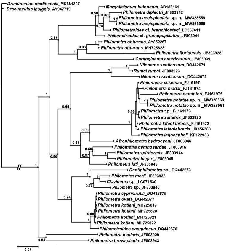
doi: 10.14411/fp.2021.008                                                                Moravec et al.: Two new species of Philometra

(van Hasselt), and Philometra tylosuri Moravec et Ali,                 todes were isolated with fine paintbrushes. A dissecting microsco-
2005 from T. crocodilus, both based on females collected               pe was used to check for male and small female nematodes in
from the hosts’ subcutaneous tissues and musculature. The              gonad rinsing and fine-tipped forceps were used to lift and isolate
authors also mention finding the gravid female of Philome-             any recovered specimens. Only one fish specimen was observed
tra sp. collected allegedly in the stomach of ‘Belone sp.’             during gross observation to host swimbladder-infecting worms
from the Gulf of Beresin, Red Sea, in 1892. Subsequently,              and it was the only case when a swimbladder was also evaluated
Moravec and Justine (2009) described Philometra dentigu-               under magnification to check for male nematodes. The nematode
bernaculata Moravec et Justine, 2009 based on two males                specimens obtained for morphological study were fixed and pre-
found in the oculo-orbit of T. crocodilus off New Caledo-              served in 5% formalin. For light microscopical examination, the
nia. Philometra sp. was listed as a parasite of Strongylura            nematodes were heat-killed with hot (70–80 °C) tap water, then
notata (Poey) (see Moravec and Ali 2005, Moravec 2006)                 fixed and preserved in 5% formalin, while those used for mo-
and Mohamed et al. (2010) reported Philometra sp. from T.              lecular evaluation were placed directly in 95% molecular grade
crocodilus in the Arabian Gulf (off Saudi Arabia). Finally,            ethanol.
Moravec and Bakenhaster (2012) described two subgravid                     For light microscopical examination, the nematodes were
females of Philometra sp. from the ovary of Strongylura                cleared in glycerine. Drawings were made with the aid of a Zei-
marina (Walbaum) off Florida, USA.                                     ss drawing attachment. Specimens used for scanning electron
    Until now, no belonid-infecting philometrid has been               microscopy (SEM) were postfixed in 1% osmium tetroxide (in
molecularly characterised. However, numerous nema-                     phosphate buffer), dehydrated through a graded acetone series,
tode-derived COI and SSU rRNA gene sequences have                      critical-point-dried and sputter-coated with gold; they were exa-
been made available in public databases (Floyd et al 2002,             mined using a JEOL JSM-7401F scanning electron microscope
Holterman et al. 2006); these sequences have been used                 at an accelerating voltage of 4 kV (GB low mode). All measu-
to validate (de Buron et al. 2011, Wang et al. 2015), iden-            rements are in micrometres unless otherwise indicated. The fish
tify (Palesse et al. 2011), and construct phylogenetic trees           nomenclature adopted follows FishBase (Froese and Pauly 2020).
(Holterman et al. 2006, Černotíková et al. 2011) of nema-
tode species.                                                          DNA extraction, PCR amplification and sequencing
    During recent helminthological investigations of some                  Tissue from ethanol-preserved specimens of two of each Phi-
estuarine fishes of Florida, philometrid specimens belonging           lometra aequispiculata sp. n. from Strongylura marina and Phi-
to two undescribed species of Philometra Costa, 1845 were              lometra notatae sp. n. from Strongylura notata was placed in a
collected from sympatric, congeneric hosts, Atlantic need-             biomasher tube (RPI Research Products Mount Prospect, Chica-
lefish (S. marina) and, the redfin needlefish (S. notata). One         go, Illinois). The ethanol was evaporated in an incubator at 37 °C
philometrid species infected gonads of both hosts while the            for one hour and a disposable homogeniser probe was employed
other was found in the swimbladder of only S. notata. New              to grind the tissue in 300 µl of lysis buffer. Genomic DNA was
important data on these nematodes are presented herein.                extracted by using the PureGene DNA isolation kit (Gentra Sys-
                                                                       tems Inc., Minneapolis, Minnesota) and the pellet was rehydra-
MATERIALS AND METHODS                                                  ted in 100 µl of deionised water. The partial cytochrome oxidase
    Fish hosts of the new species were collected with haul seines      subunit 1 (COI) was PCR amplified with the primers COIF, COIR
from June 2017 through April 2019 during routine fisheries inde-       (de Buron et al. 2011), and the SSU rDNA sequence with pri-
pendent population monitoring conducted by the Fish and Wild-          mers PhilonemaF, PhilPCRr, and ameb620f, ameb 620r as inter-
life Research Institute (FWRI) in Tampa and Apalachicola Bays.         nal sequencing primers (Černotíková et al. 2011). PCR reactions
After capture, fish were immediately placed in plastic bags and        had a final volume of 50 µl; each containg 150 ng of the purified
packed in ice. Host specimens collected in Tampa Bay were re-          DNA, 50 µM of dNTP mix, 0.25 µl of 0.1-mg/ml BSA; 0.25 µl
turned within eight hours of capture to FWRI headquarters in St.       of 100 µM of each primer, 10 µl of Taq polymerase buffer (5×),
Petersburg, Florida for laboratory evaluation, while those from        2.5 mM MgCl2 (Promega Corporation, Madison,Wisconsin; final
Apalachicola Bay were shipped by commercial courier overnight          concentration), and 1.25 units of Go Taq DNA polymerase (Pro-
on ice. In many cases, specimens of belonids were returned for         mega Corporation). The reaction profile for COI was as follows:
routine pathological evaluation of grossly visible external abnor-     initial denaturation of 94 °C for 1.3 min, 35 × (94 °C for 40 s,
malities, most frequently jaw deformities characteristic of active     47 °C for 40 s, 72 °C for 45 s), and final extension at 72 °C for
or former infection by females of the copepod Colobomatus goo-         5 min. For SSU rDNA the annealing temperature was, instead,
dingi Cressey et Collette, 1970, and these fish were opportunely       63 °C. Amplified products were gel-purified (Agilent Technolo-
examined for helminths. To increase sample size and gather ad-         gies, Santa Clara, California) and sequenced from both directions
ditional prevalence and intensity data, field crews also returned      by using Big Dye Terminator v 1.1 (Applied Biosystems, Inc.,
grossly normal specimens specifically for parasitological evalua-      Waltham, Massachusetts). Cycle-sequenced products were preci-
tion from September 2017 through April 2018.                           pitated and resuspended in Hi-Di Formamide and visualised on a
    Necropsies of fish included measurement of standard length         3130 Genetic Analyzer (Applied Biosystems, Inc.). Raw sequen-
(SL) and gross observations of external surfaces and visceral or-      ces were aligned and edited by using Sequencher (version 4.0;
gans. Gonads were excised, placed in a petri dish, and cut open        Gene Codes Corporation, Ann Arbor, Michigan) and sequence di-
lengthwise (ovaries) or torn with forceps into pieces (testes), then   vergences were calculated by using MEGA (version 7.0, Tamura
rinsed with physiological saline. Grossly visible female nema-         et al. 2013).

Folia Parasitologica 2021, 68: 008                                                                                            Page 2 of 17
doi: 10.14411/fp.2021.008                                                            Moravec et al.: Two new species of Philometra

Phylogenetic analysis                                                pillae (Figs. 1C, 2A,B). Oesophagus 294 (345) [390–516]
    To evaluate the phylogenetic positions of the new nematode       long, comprising 11% (14%) [15–19%] of body length,
species, 50 sequences were initially extracted from GenBank,         with slight inflation at anterior end measuring 27 × 12 (33
according to the BLAST homology score (Altschul et al. 1990).        × 18) [24–30 × 12–18]; posterior part of muscular oesoph-
After initial alighment of the homologous SSU rDNA sequences         agus overlapped by well-developed oesophageal gland
available in GenBank, the longer sequences of the congeneric and     with large cell nucleus; maximum width of gland 15 (21)
other taxa were trimmed to that of the sequence range of the new     [15–18]. Ventriculus small, 6 long, 9 wide. Nerve ring and
species. Bayesian analysis (BA) (MrBayes, v3.0; Ronquist and         oesophageal nucleus 123 (132) [123–162] and 237 (273)
Huelsenbeck 2003) was used to construct the phylogenetic tre-        [207–396], respectively, from anterior extremity. Excreto-
es. The nst2 and GTR+G+1 models were determined using the            ry pore 177 (192) [171–213] from anterior end.
maximum-likelihood approach and Akaike information criterion            Testis extending anteriorly approximately to level
(AIC) in the program MEGA, were implemented in MRBAYES               of posterior end of oesophagus (Fig. 1D). Posterior end
for COI and SSU rDNA, respectively. Bayesian analysis was            of body blunt, 39 (36) [30–36] wide, with broad caudal
conducted for 10 metropolis-coupled Markov chain Monte Carlo         mound formed by 2 lateral reniform portions well sepa-
generations, with posterior probabilities serched for 106 genera-    rated dorsally (Figs. 1L, 2C,D, 5C,D). Four pairs of small
tions (based on average standard deviation of split frequencies),    adanal papillae present in space between caudal mound
of which the first 2.5 × 10⁵ were considered the burn-in and ex-     and cloacal aperture: 2 pairs of small, markedly elevated
cluded from the analysis. Trees were sampled every 1,000 gene-       papillae situated closely to cloacal aperture and 2 pairs of
rations during the run. The resulting tree file from MrBayes was     very flat, hardly visible papillae located externally to them;
used to construct the phylogenetic tree by using FigTree (http://    pair of small phasmids present dorsolaterally on caudal
tree.bio.ed.ac.uk/software/figtree/). P-distance values between      mound (Figs. 1L, 2D, 5D). Each portion of caudal mound
the putative new species and closely related sequences within the    with 1 subdorsal, minute circular depression (Figs. 1L, 2D,
reconstructions were estimated by using MEGA.                        5D). Spicules needle-like, equally long, with somewhat
    New, supplemental COI sequences from a previously descri-        expanded proximal and sharply pointed distal tips (Figs.
bed species, Philometra diplectri Moravec et Bakenhaster,            1F,G,M, 4C,D, 5C,D); length of spicules 102 (102) [99–
2010, were also obtained by the above methods (GenBank Nos.          111], representing 3.9% (4.1%) [3.6–4.2%] of body length;
MW326919, MW326920, MW326921) and included in the pre-               maximum width of spicules 6 (6) [3–6]. Gubernaculum
sent phylogenetic analysis. The new specimens of P. diplectri        narrow, 69 (63) [60–69] long, with anterior portion some-
were collected during June 2019 from the type host, Diplectrum       what dorsally bent; length of anterior bent part 33 (30) [27–
formosum (Linnaeus), and type locality, northeastern Gulf of Me-     36], comprising 50% (48%) [39–55%] of entire gubernac-
xico as part of an otherwise separate, ongoing study. Because our    ulum length (Figs. 1F,G,M, 4C,D). In lateral view, distal
preliminary molecular evaluations revealed P. diplectri to be a      tip of gubernaculum with large dorsal triangular barb with
near relative of one of the new species, these sequences were con-   smooth terminal part and 4 distinct transverse grooves on
sidered too important to reserve for a future more comprehensive     each side; proper extremity of gubernaculum posterior to
manuscript and were therefore included in the current study.         barb rounded, with 2 deep lateral oblique grooves on each
                                                                     side, united to form 2 anteriorly oriented dorsal serrations;
RESULTS                                                              similar serrations also present in front of barb (Figs. 1J,K,
                                                                     2E,F). Length ratio of gubernaculum and spicules 1 : 1.55
Philometridae Baylis et Daubney, 1926                                (1 : 1.62) [1 : 1.50–1.67]. Spicules and gubernaculum well
                                                                     sclerotised, yellowish, anterior part of gubernaculum and
Philometra aequispiculata sp. n.                      Figs. 1–5      proximal ends of spicules colourless.
                                                                        Gravid female (one incomplete specimen from S. ma-
ZooBank number for species:                                          rina, allotype): Body of fixed specimen brownish; anterior
urn:lsid:zoobank.org:act:BAAAA871-7DB1-41B1-B2CA-3B75DE667C81        part of body tapered, with rounded end. Cuticle smooth.
                                                                     Body of specimen with missing posterior end 100 mm
   Male (two specimens from Strongylura marina; holo-                long, maximum width 1,115. Width of cephalic end 272.
type, measurements of paratype in parentheses. Measure-              Cephalic papillae small, indistinct when laterally viewed
ments of 10 paratypes from Strongylura notata in brack-              (Fig. 1A). Oral aperture almost circular, surrounded by
ets): Body filiform, whitish, 2.64 (2.48) [2.39–2.91] mm             small cephalic papillae arranged in 2 circles and slightly
long, maximum width at middle of body 36 (42) [45–51];               outlined amphids; inner circle of papillae consists of 4 sub-
anterior part of body tapering to anterior extremity, without        median and 2 lateral single papillae, outer circle formed
usual constriction just posterior to cephalic end (Fig. 1H).         by 4 submedian pairs of papillae. Oesophagus including
Maximum width/body length ratio 1 : 73 (1 : 59) [1 : 53–             well-developed anterior bulbous inflation 1.12 mm long;
63]. Cuticle smooth. Cephalic end rounded, 24 (30) [24–              anterior inflation 612 long and 721 wide; maximum width
30] wide. Oral aperture small, circular. Cephalic papillae           of posterior part of oesophagus including gland 150. Oe-
14 in number, arranged in 2 circles; outer circle formed by          sophageal gland well-developed, opening into oesopha-
4 submedian pairs, inner circle consists of 4 submedian and          gus just posterior to nerve ring, with large cell nucleus at
2 lateral papillae (Figs. 1C, 2A,B). Pair of small, slit-like        middle (Fig. 1A). Nerve ring and oesophageal nucleus 367
lateral amphids somewhat posterior to lateral cephalic pa-           and 666, respectively, from anterior extremity. Ventriculus

Folia Parasitologica 2021, 68: 008                                                                                      Page 3 of 17
doi: 10.14411/fp.2021.008 Moravec et al.: Two new species of Philometra Fig. 1. Philometra aequispiculata sp. n. from Strongylura marina (Walbaum), Florida, USA. A – anterior end of gravid female, lateral view; B – cephalic end of subgravid female, apical view; C – cephalic end of male, apical view; D – anterior end of male, lateral view; E – larva; F, G – posterior end of male, lateral and ventral views, respectively; H – cephalic end of male, lateral view; I – posterior end of nongravid female, lateral view; J, K – distal tip of gubernaculum, lateral and subdorsal views, respectively; L – caudal end of male, apical view; M – posterior end of male (another specimen), lateral view. Folia Parasitologica 2021, 68: 008 Page 4 of 17
doi: 10.14411/fp.2021.008                                                                 Moravec et al.: Two new species of Philometra

Fig. 2. Philometra aequispiculata sp. n. from Strongylura marina (Walbaum), Florida, USA; scanning electron micrographs of male.
A, B – cephalic end, sublateral and apical views, respectively (arrows indicate amphids); C, D – caudal end, lateral and apical views,
respectively (arrows indicate phasmids); E, F – distal end of gubernaculum, lateral and subdorsal views, respectively. Abbreviations:
a – pair of cephalic papillae of outer circle; b – submedian cephalic papilla of inner circle; c – lateral cephalic papilla of inner circle;
d – caudal mould; e – cuticular depression; f – cloaca; g – two elevated adanal papillae; o – oral aperture.

Folia Parasitologica 2021, 68: 008                                                                                              Page 5 of 17
doi: 10.14411/fp.2021.008                                                            Moravec et al.: Two new species of Philometra

                                                                    Uterus filled with numerous eggs. Posterior end rounded,
                                                                    299 wide, without caudal projections.
                                                                       Nongravid female (one complete and one incomplete
                                                                    mature specimens without eggs from S. marina; measure-
                                                                    ments of additional three specimens from S. notata in pa-
                                                                    rentheses): Body whitish, that of complete specimen 35.0
                                                                    mm, of incomplete specimen 15.1 (2.34–2.49) mm long,
                                                                    maximum width 286–503 (48–60); maximum width/body
                                                                    length ratio 1 : 114 (1 : 141–152). Width of cephalic end
                                                                    109–163 (30). Oesophagus including anterior bulbous in-
                                                                    flation 925–1,020 (411–480) long, comprising 3% (17–
                                                                    21%) of body length; its anterior inflation 90–114 (30–36)
                                                                    long and 87–111 (18–21) wide (Fig. 4A). Nerve ring and
                                                                    oesophageal nucleus 216–286 (165–189) and 135–598
                                                                    (267–315), respectively, from anterior extremity. Ventricu-
                                                                    lus 15 (9–12) long, 33–81 (24) wide. Intestinal ligament
                                                                    503 (60–75) long (Figs. 1I, 4E). Vulva absent (rudimentary
                                                                    vulva at 1.63–1.73 mm from anterior extremity, i.e. at 69–
                                                                    70% of entire body end; Fig. 4F). Uterus empty. Posterior
                                                                    end rounded, 163 (36–42) wide, without caudal projections
                                                                    (Figs. 1I, 4E).
                                                                    Type host: Atlantic needlefish Strongylura marina (Walbaum)
                                                                       (Belonidae, Beloniformes); 389–508 mm standard length
                                                                       (SL).
                                                                    Other host: Redfin needlefish Strongylura notata (Poey) (Belo-
                                                                       nidae, Beloniformes); 148–379 mm SL.
                                                                    Site of infection: Gonad (ovary and testis).
                                                                    Type locality: Gulf of Mexico, specifically Tampa Bay
                                                                       (27.5474, -82.6333; 27.8287, -82.4420) and Apalachicola Bay
Fig. 3. Philometra aequispiculata sp. n. from Strongylura mari-        (29.6997, -84.7782), Florida, USA (collected 7 November
na (Walbaum), Florida, USA; scanning electron micrographs of
                                                                       2017, and 15 March and 13 June 2018).
subgravid female. A – anterior end of body, subapical view; B
– cephalic end, apical view. Abbreviations: a – pair of cephalic    Prevalence and intensity: Strongylura marina: 26% (5 fish
papillae of outer circle; b – submedian cephalic papilla of inner      infected/19 fish examined); 1–3 (mean 1.4) nematode speci-
circle; c – lateral cephalic papilla of inner circle.                  mens per fish. Strongylura notata: 19% (20/105); 1–4 (mean
                                                                       1.4).
                                                                    Deposition of type specimens: Male holotype and fema-
small, 30 long, 99 wide. Intestine brown. Vulva absent.                le allotype (both mounted on SEM stubs) and 4 paratypes in
Anterior ovary reflexed near anterior body end (Fig. 1A).              the Helminthological Collection of the Institute of Parasitol-
                                                                       ogy, BC CAS, České Budějovice, Czech Republic (Cat. No.
Uterus occupying most space of body, filled with nume-
                                                                       N–1223); 14 paratypes in the Smithsonian National Muse-
rous eggs and larvae (Fig. 1A,E). Larvae (n = 5) 489–495               um of Natural History, Washington, USA (Cat. Nos. USNM
long, maximum width 18; length of oesophagus 156–186                   1640964 and USNM 1640965, respectively).
(32–38% of body length), of sharply pointed tail 138–159            Additional material: Two female specimens taken from go-
(28–32% of body length). Posterior end of female speci-                nads of the type host were used for gene sequencing (GenBank
men absent.                                                            Nos. MW328558, MW328559) and are no longer available for
    Subgravid female (one specimen from S. marina):                    examination.
Body of fixed specimen brownish, 74 mm long, maximum                Etymology: The specific name aequispiculata relates to the fact
width 680; maximum width/body length ratio 1 : 109.                    that spicules of this nematode species are equally long.
Width of cephalic end 177. Cephalic structures (Figs. 1B,
3A,B) essentially identical with those in gravid female.               Remarks. The present data show that nematodes co-
Oesophagus including anterior bulbous inflation 1.05 mm             llected from S. marina and S. notata are morphologically
long, comprising 1.4% of body length; anterior inflation            and biometrically practically identical, they have the same
612 long and 680 wide; maximum width of posterior part              site of infection in the host and localities, their hosts are
of oesophagus including gland 136. Nerve ring and oeso-             congeneric, so that we consider them to be representatives
phageal nucleus 163 and 653, respectively, from anterior            of the same species.
extremity. Intestine brown, straight, ending blindly; poste-           To date, the following six nominal species of philo-
rior end of intestine atrophied, forming ligament 721 long          metrid nematodes are known to parasitise belonid fishes:
attached ventrally to body wall close to posterior extremi-         Philometra dentigubernaculata, P. kohnae, P. lomi, P.
ty. Vulva and anus absent. Ovaries reflexed near body ends.         strongylurae, P. tylosuri and Philometroides indonesien-
                                                                    sis, whereas the previous records of Philometra globiceps

Folia Parasitologica 2021, 68: 008                                                                                       Page 6 of 17
doi: 10.14411/fp.2021.008 Moravec et al.: Two new species of Philometra
Fig. 4. Philometra aequispiculata sp. n. from Strongylura notata (Poey), Florida, USA. A – anterior end of nongravid female, lateral
view; B – cephalic end of male, apical view; C, D – caudal end of male, ventral and lateral views, respectively; E – posterior end of
nongravid female, lateral view; F – vulva of nongravid female, lateral view.
and P. pellucida from these hosts were evidently misiden- portion of the oesophagus (vs bulb much less developed,
tifications (see above). However, in contrast to P. aequispi- not separated); they also differ in the site of infection (sub-
culata sp. n., all these species infect different sites in the cutaneous tissues vs gonad) and their respective hosts have
host (subcutaneous tissues, musculature or oculo-orbits), strongly allopatric ranges (Western Atlantic Ocean vs Indo-
being mostly described solely from available gravid and -West Pacific region).
subgravid females (the male is known only for P. denti- The gravid female of P. lomi has a much longer oe-
gubernaculata). Generally, in addition to morphological sophagus (3.3 mm vs 1.12 mm) provided with a distinct
features, philometrid species are characterised by the site extention of the oesophagus between its anterior bulb and
of infection in the host, particularly that of their gravid fe- the nerve ring (vs without such an extention), whereas the
males (Moravec and Rohde 1992). oesophagus of nongravid females of P. tylosuri is distin-
The new species differs from P. dentigubernaculata cty longer than that of subgravid and gravid females of the
in a distinctly shorter body of the male (2.39–2.91 mm new species (1.63–1.65 mm vs 1.02–1.12 mm), represen-
vs 4.65–4.99 mm), in the spicule length/body length ratio ting 2.3–3.5% (vs 1.4% in a subgravid female) of the body
(3.6–4.2% vs 2.3%), a shorter gubernaculum (60–69 µm vs length, and their caudal end bears a pair of minute caudal
84–90 µm) and mainly in a different structure of the guber- projections (vs caudal projections absent). Philometra lomi
naculum distal tip (with transverse grooves vs without tran- and P. tylosuri are parasites of the host’s subcutaneous tis-
sverse grooves). Moreover, both species have a different sues (latter species also in musculature) (vs in gonad) and
site of infection in the host (gonad vs oculo-orbits) and they they occur in the Indo-Pacific region (vs in North Atlantic
occur in very distant localities (Gulf of Mexico vs South Ocean).
Pacific Ocean). Philometroides indonesiensis mainly differs from
In contrast to P. aequispiculata sp. n., P. kohnae and P. Philometra aequispiculata sp. n. in that the body surface of
strongylurae possess markedly large, dome-shaped cepha- its gravid female bears sparsely distributed small cuticular
lic papillae (vs cephalic papillae very small, hardly visible) bosses (vs cuticular bosses absent). In addition, these spe-
and their anterior oesophageal bulb is conspicuously large, cies utilise hosts belonging to different fish genera (Tylo-
strongly muscular, distinctly separated from the remaining surus Cocco vs Strongylura van Hasselt), they differ in the
Folia Parasitologica 2021, 68: 008 Page 7 of 17doi: 10.14411/fp.2021.008 Moravec et al.: Two new species of Philometra
Fig. 5. Philometra aequispiculata sp. n. from Strongylura notata (Poey), Florida, USA; scanning electron micrographs of male. A, B
– cephalic end, dorsoventral and apical views, respectively (arrows indicate amphids); C, D – caudal end, sublateral and apical views,
respectively (arrows indicate phasmids). Abbreviations: a – pair of cephalic papillae of outer circle; b – submedian cephalic papilla of
inner circle; c – lateral cephalic papilla of inner circle; d – caudal mound; e – cuticular depression; f – cloaca; g – two elevated adanal
papillae; o – oral aperture; s – spicule.
site of infection (musculature vs gonad) and occur in very mm, maximum width 1.80 mm; width of cephalic end
distant localities (Indian Ocean vs North Atlantic Ocean). 394. Maximum width/body length ratio 1 : 30. Oral aper-
Therefore, the present nematodes from the gonads of S. ture circular, surrounded by cephalic papillae arranged in
marina and S. notata are considered to be a new species. 2 circles; inner circle of papillae consists of 4 submedian
Its characteristic feature is the structure of the distal gu- and 2 lateral small single papillae, outer circle formed by 4
bernaculum tip, by which it can be distinguished from all submedian pairs of large papillae; amphids indistinct (Figs.
other Philometra spp. with known males. Philometra aeq- 6C, 7A,C). Oesophagus including well-developed anterior
uispiculata sp. n. is the sixth nominal species of this genus bulbous inflation 2.58 mm long, representing 4.8% of body
described from the Belonidae and the first gonad-infecting length; anterior bulbous inflation 245 long and 299 wide;
species of Philometra from these hosts (needlefishes). maximum width of posterior part of oesophagus including
gland 272. Oesophageal gland well developed, opening
Philometra notatae sp. n. Figs. 6–8 into oesophagus just posterior to nerve ring, provided with
large cell nucleus situated 1.80 mm from anterior end of
ZooBank number for species: body (Fig. 6A). Nerve ring 394 from anterior extremity.
urn:lsid:zoobank.org:act:CCDC5D2D-89EE-4FD4-83F7-1CD078BF162A Ventriculus small, 54 long, 109 wide. Intestine brownish,
very broad, ending blindly; posterior end of intestine atro-
Gravid female (one larvigerous specimen, holotype): phied, forming ligament 748 long attached ventrally to
Body of fixed specimen yellowish; anterior half of body body wall close to posterior extremity (Fig. 6B). Vulva and
broader than posterior one. Anterior part of body tapered, anus absent. Ovaries narrow, making coils near body ends
truncated (Fig. 6A). Cuticle smooth. Length of body 54.1 (Fig. 6A,B). Uterus occupying most space of body, filled
Folia Parasitologica 2021, 68: 008 Page 8 of 17doi: 10.14411/fp.2021.008 Moravec et al.: Two new species of Philometra Fig. 6. Philometra notatae sp. n. from Strongylura notata (Poey), Florida, USA, gravid female. A – anterior end of body, lateral view; B – posterior end of body, lateral view; C – cephalic end, apical view; D – larva from uterus, lateral view; E – shape of caudal end, lateral view; F – cephalic end, lateral view. with numerous eggs and larvae (Fig. 6A). Larvae (n = 5) Posterior end rounded, 245–299 wide, without caudal 390–420 long, maximum width 15–18; length of oesopha- projections. gus 120–129 (31% of body length), of sharply pointed tail Male: Not known. 96–105 (25% of body length) (Fig. 6D). Posterior end of Type host: Redfin needlefish Strongylura notata (Poey) (Belo- female rounded, 272 wide, without caudal projections Fig. nidae, Beloniformes); 381 mm standard length (SL). 6B,E). Site of infection: Swimbladder. Subgravid female (two complete and one incomplete Type locality: Gulf of Mexico, specifically Tampa Bay ovigerous specimens, paratypes): Body of fixed specimens (27.8137, -82.4026), USA (collected 18 April 2019). whitish to yellowish, 46.6–55.8 mm long, maximum width Prevalence and intensity: 0.9% (1 fish infected/105 fish exa- 884–1,564; maximum width/body length ratio 1 : 33–36. mined); 6 nematode specimens. Width of cephalic end 381. Oral aperture circular. Number, Deposition of type specimens: Holotype and paratype size and arrangement of cephalic papillae as in gravid (mounted on SEM stubs) in the Helminthological Collection female (Fig. 7B,D). Oesophagus including anterior bulbous of the Institute of Parasitology, BC CAS, České Budějovice, inflation 2.15–2.18 mm long, comprising 3.9–4.6% of body Czech Republic (Cat. No. N–1224); 3 paratypes in the Smith- length; anterior bulb 177–218 long and 286–299 wide; sonian National Museum of Natural History, Washington, maximum width of posterior part of oesophagus including USA (Cat. No. USNM 1640966). gland 163–258. Nerve ring and oesophageal nucleus 381– Etymology: The specific name notatae is the genitive form of 408 and 1.43 mm, respectively, from anterior extremity. the host’s species name. Intestine brownish, straight, ending blindly; posterior end of intestine atrophied, forming ligament 394–870 long Remarks. The new species P. notatae sp. n. is noted for attached ventrally to body wall close to posterior extremity. the presence of eight conspicuously large cephalic papillae Vulva and anus absent. Uterus filled with numerous eggs. of the external circle in gravid and subgravid females, a Folia Parasitologica 2021, 68: 008 Page 9 of 17
doi: 10.14411/fp.2021.008                                                            Moravec et al.: Two new species of Philometra

Fig. 7. Philometra notatae sp. n. from Strongylura notata (Poey), Florida, USA; scanning electron micrographs of females. A, B –
cephalic end of gravid and subgravid female, respectively, apical views; C – cephalic end of gravid female, sublateral view (arrows
indicate inner submedian papillae); D – cephalic end of subgravid female, dorsal view. Abbreviations: a – submedian cephalic papilla
of outer circle; b – submedian cephalic papilla of inner circle; c – lateral cephalic papilla of inner circle.

feature by which it resembles only seven other congeneric          co (Obiekezie 1986, Obiekezie and Anders 1991, Moravec
species parasitising marine or brackish-water fishes: Philo-       and Rohde 1992, Vidal-Martínez et al. 1995, Moravec et
metra beninensis Obiekezie, 1986 from the subcutaneous             al. 2001, 2006, Moravec and de Buron 2006, Moravec and
tissue of Polydactylus quadrifilis (Cuvier) (Polydactylidae)       Bakenhaster 2010).
in the eastern Central Atlantic (off Nigeria); Philometra              However, in contrast to the new species, the gravid
cynoscionis Moravec, de Buron et Roumillat, 2006 from              female of P. salgadoi is much longer (92–111 mm vs 54
the subcutaneous tissue of Cynoscion nebulosus (Cuvier)            mm) and possesses a pair of caudal projections (vs cau-
(Sciaenidae) in the West Atlantic Ocean (off the USA); P.          dal projections absent). The body of gravid females of all
kohnae from the subcutaneous tissue of Tylosurus gavialo-          other above-mentioned species, except for P. kohnae, is
ides (Castelnau) (Belonidae) in the Pacific Ocean off Aus-         distinctly shorter than that of P. notatae sp. n., not exce-
tralia; Philometra overstreeti Moravec et de Buron, 2006           eding 35 mm, and these also differ in some other features:
from the tissue among teeth of Paralichthys lethostigma            P. beninensis has a shorter oesophagus (1.04 mm vs 2.58
Jordan et Gilbert (Paralichthyidae) in the West Atlantic           mm) and its oesophageal bulb is narrower (110 µm vs 299
Ocean (off the USA); Philometra salgadoi Vidal-Martí-              µm); P. cynoscionis has a shorter oesophagus (1.22–1.78
nez, Aguirre-Macedo et Moravec, 1995 from the occular              mm vs 2.58 mm) and a smaller oesophageal bulb (90–135
cavity of Epinephelus morio (Valenciennes) (Serranidae)            × 114–165 µm vs 245 × 299 µm); P. overstreeti has a shor-
in the Gulf of Mexico; P. strongylurae from the subcutane-         ter oesophagus (1.02–1.33 mm vs 2.58 mm) and a smaller
ous tissue, musculature and gills of Strongylura leiura and        oesophageal bulb (51–68 × 63–87 µm vs 245 × 299 µm);
Strongylura strongylura (Belonidae) in the Persian Gulf            P. strongylurae differs in the maximum width/body len-
(off Iraq); and P. diplectri from the head tissues and the         gth ratio (1 : 17–24 vs 1 : 30) and the relative length of
operculum of Diplectrum formosum in the Gulf of Mexi-              the oesophagus to the body length (9–11% vs 5%); and P.

Folia Parasitologica 2021, 68: 008                                                                                     Page 10 of 17
doi: 10.14411/fp.2021.008 Moravec et al.: Two new species of Philometra
diplectri in a shorter oesophagus (1.07–1.33 mm vs 2.58
Table 1. Number of observed sequence differences between paired taxa represented in this study for COI below the diagonal; p-distances in above the diagonal. For P. carolinensis and P. rubra,
dilpectri aequispiculata
0.132 [0.249] 0.161 [0.259] 0.132 [0.243]
0.197 [0.214] 0.185 [0.205]
sp. n. 400 bp
Philometra Philometra mm) and a smaller oesophageal bulb (48–57 × 111–132
--------
0.188
0.195
0.190
0.166
0.166
0.188
0.129
0.122
0.125
0.149
µm vs 245 × 299 µm). The female of P. kohnae is repor-
ted to be 34–76 mm long, but the length of its oesophagus
represents only 3–4% (vs approximately 5%) of the body
length, the infection site in the host is different (encapsula-
--------
400 bp
0.191
0.226
0.217
0.185
0.178
0.206
0.150
0.148
0.133
60
ted in subcutaneous pouches vs in swimbladder), the host
belongs to a different genus (Tylosurus vs Strongylura) and
their respective host species have strongly allopatric ran-
0.182 [0.255] 0.226 [0.244]
0.179 [0.240] 0.188 [0.210]
0.159 [0.240] 0.180 [0.201]
0.154 [0.228] 0.188 [0.210]
0.188 [0.222] 0.214 [0.234]
0.165 [0.259] 0.199 [0.209]
0.148 [0.247] 0.179 [0.199]
carolinen- rubra 371 bp
Philometra Philometra
ges (South Pacific Ocean endemic to Australia vs western
[392 bp]
--------
73 [82]
69 [79]
0.141
0.184
North Atlantic Ocean estuaries of the Americas and West
Indies).
To date, P. notatae sp. n. is the only nominal species
0.145 [0.220]
0.139 [0.240]
sis 289 bp
of Philometra with gravid females located in the swim-
[331 bp]
--------
37 [81]
45 (84)
37 [79]
bladder of a belonid host. The gravid female of Philometra
sp. reported by Nikolaeva and Parukhin (1968) from the
swimbladder of Tylosurus crocodilus in the Gulf of Mexico
overstreeti paralichthy- cynoscionis
Philometra Philometroides Philometra
(see above) might belong to a different, so far undescribed
--------
39 (78)
359 bp
0.192
0.181
0.173
0.154
0.184
0.192
0.071
0.073
66
48
45
species (considering a different genus of the host), althou-
gh conspecificity of both these forms cannot be excluded.
While no relevance to infection by P. notatae sp. n. is
dis 383 bp
suggested, the type host specimen exhibited several abnor-
--------
42 (78)
65 [75]
0.208
0.201
0.201
0.147
0.185
0.197
0.054
26
57
47
malities that should nevertheless be mentioned. Its jaw had
a lesion grossly consistent with putative histiocytic sarco-
ma previously reported for this species in Florida (Kiryu
et al. 2018) and a tumor-like mass (approximately 40 mm
46 (82)
72 [77]
380 bp
--------
0.212
0.202
0.197
0.162
0.195
0.196
20
25
56
48
diameter) of undetermited nature that caused marked dis-
tension of the fish’s abdomen near the anal fin. Various
other internal abnormalities were noted including a defor-
Philometra
sanguineus
med ovary and an intense infection of the liver and gall-
--------
51 (83)
400 bp
84[94]
0.208
0.205
0.203
0.178
0.199
73
76
69
82
75
-bladder by larval cestodes. Full pathological evaluation of
these lesions remains incomplete.
lagocephali
Philometra
Sequence data
--------
53 (69)
77 [87]
387 bp
0.185
0.192
0.181
0.153
77
71
70
64
69
64
For both P. notatae sp. n. and P. aequispiculata sp.
n., we obtained COI sequences of 400 base pairs (bp)
longer sequence comparisons were available for some taxa [values bracketed].
in length (GenBank accession numbers MW326917,
Philometra
241 bp-w
MW326918 and MW326915, MW326916, respectively);
saltatrix
--------
35 (53)
0.133
0.059
0.033
37
43
39
36
37
34
45
40
for the SSU rDNA, we obtained sequences of 1,671 bp and
1,487 bp long, respectively (GenBank accession numbers
MW328560, MW328561 and MW328558, MW328559).
Philometra
400 bp-w
As expected, there were no stop codons or indels in the
saltatrix
--------
43 (74)
70 [81]
0.133
0.050
70
81
73
77
62
87
76
8
COI sequences. A total of 28 homologous matches of rela-
ted taxa were retained in BLAST searches involving COI
haplotypes for the new species. P-distance values between
Philometra
saltatrix
P. notatae sp. n. and the two most closely related species,
44 (76)
66 [76]
385 bp
--------
0.148
19
14
73
79
75
77
63
87
75
Philometra saltatrix Ramachandran, 1973 and Philomet-
ra lagocephali Moravec et Justine, 2008, were 0.133 and
0.185; between P. aequispiculata sp. n. and its three most
Philometra
notatae sp.
n. 400 bp
closely related species, P. diplectri, P. overstreeti, and
50 (78)
70 [81]
--------
57
53
32
72
83
79
80
69
76
75
Philometroides paralichthydis Moravec et Justine, 2006,
p-distance values ranged from 0.122 to 0.149. These dis-
tances were higher on average than those between pairs of
Philometra notatae sp. n.
Philometra carolinensis
Philometra lagocephali
Philometra cynoscionis
Philometra sanguineus
recognised taxa. Table 1 provides the number of base-pair
Philometra overstreeti
Philometra dilpectri
aequispiculata sp. n.
Philometra saltatrix
Philometra saltatrix
Philometra saltatrix
differences and p-distance values amongst representative
Philometra rubra
taxa based on available COI sequences.
Philometroides
paralichthydis
Philometra
For SSU rDNA, the number of homologous sequences
retained was 38 in BLAST searches; after trimming, the
alignment length was 1846 bp, indicating a substantial in-
Folia Parasitologica 2021, 68: 008 Page 11 of 17doi: 10.14411/fp.2021.008 Moravec et al.: Two new species of Philometra
100% support. It too was reciprocally monophyletic, albeit
with lesser Bayesian support (46%), to a complex of close-
ly related taxa that included P. overstreeti, P. paralichthy-
dis, and P. cynoscionis; P. diplectri was the most recent
common ancestor to this group.
Babesian inference for SSU rDNA also revealed that
both new species comprise discrete evolutionary lineages
(Fig. 10). The clade containing observed haplotypes of P.
notatae sp. n. was monophyletic, having 100% Bayesian
support. In this reconstruction, Philometra nemipteri Luo,
2001 (for which there are no corresponding COI data) was
the closest relative and P. saltatrix was also closely related.
The clade containing observed haplotypes of P. aequispic-
ulata sp. n. was also monophyletic, having 100% support.
In this reconstruction, P. diplectri was the most closest rel-
ative to this new species.
DISCUSSION
As was mentioned in our introduction, Philometra
aequispiculata sp. n. and Philometra notatae sp. n. are so
far the only two known nominal species of philometrids
parasitising needlefishes (Belonidae) in the region of the
Atlantic Ocean. Now it is clear that the female specimens
reported by Moravec and Bakenhaster (2012) as Philome-
tra sp. from the ovary of Strongylura marina in the Gulf
of Mexico off Florida (Charlotte Harbor) were conspecific
with the former species.
Considering the generally high degree of host specifici-
ty of gonad-infecting species of Philometra (see e.g., Mo-
ravec and Manoharan 2014a,b, Moravec et al. 2014, 2016)
and the fact that only males of P. aequispiculata but no
females were found in Strongylura notata, it may be that
Fig. 8. Females of Philometra notatae sp. n. in the swimbladder this host species serves as only the paradefinitive host for
of Strongylura notata (Poey), Florida, USA. A – two specimens this nematode, whereas its true definitive host is S. marina.
(indicated by arrows); B – one specimen (higher magnification). In contrast to the definitive host, the parasite is unable to
reproduce in the paradefinitive host (Odening 1976).
Both newly described philometrid species, P. aequispi-
clusion of indels. The p-distance values between P. notatae culata sp. n. and P. notatae sp. n., were collected from the
sp. n. and its six most closely related species ranged from same host species, S. notata. This is not exceptional that
0.007 to 0.015; whilst in comparisons among these six taxa, more than one sympatric species of philometrids with di-
values ranged from 0.002 to 0.019. Similarly, between P. fferent infection sites occur in the same fish species. For
aequispiculata sp. n. and its four most closely related spe- example, three species of Philometra in different sites are
cies, p-distance values ranged from 0.011 to 0.021; whilst known to occur in the red grouper Epinephelus morio (Va-
in comparisons among these four taxa, values ranged from lenciennes) (Serranidae) in the Gulf of Mexico (Moravec
0.008 to 0.018. Tables 2 and 3 provide the number of base- et al. 2010), whereas two species of Philometra and one of
pair differences and p-distance values amongst representa- Philometroides Yamaguti, 1935 are parasitic in the John’s
tive taxa based on available SSU rDNA sequences. snapper Lutjanus johnii (Bloch) (Lutjanidae) off the north-
ern coast of Australia (Moravec and Barton 2016).
Phylogenetic reconstructions The high level of observed interspecific sequence diver-
Bayesian inference yielded a single topology for COI gence combined with low intraspecific variation confirmed
wherein both putative new species were clustered uniquely that the barcode gene COI represents a useful marker for
in fully supported clades (Fig 9). Haplotypes of P. notatae identification of species Philometra. This gene marker
sp. n. clustered with 99% Bayesian support and were recip- was also helpful in reconstructing some of the phyloge-
rocally monophyletic to a clade containing haplotypes of P. netic relationships amongst maternal lineages, although
saltatrix. However, Bayesian support for the distinguishing deeper branches within the tree generally lacked Bayesi-
node was only 68%. Philometra rubra (Leidy, 1856) re- an support. In contrast to the COI findings, most species
solved as the most recent common (maternal) ancestor to in the SSU rDNA haplotypes were reconstructed within
these two taxa. The clade containing observed haplotypes shallow, weakly supported or non-supported clades. In-
of P. aequispiculata sp. n. was also monophyletic, having deed, sequences for two species, Philometra cyprinirutili
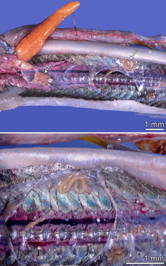
Folia Parasitologica 2021, 68: 008 Page 12 of 17doi: 10.14411/fp.2021.008 Moravec et al.: Two new species of Philometra Fig. 9. Phylogenetic tree inferred from Bayesian analyses of cytochrome oxidase subunit 1 (COI) sequences including those from Philometra aequispiculata sp. n. and P. notatae sp. n. from Strongylura (asterisks) and closely related homologous sequences extracted from GenBank. Numbers at the nodes are Bayesian values for Bayesian posterior probability. (Creplin, 1825) (DQ442675) and Philometra ovata (Zeder, previously noted by Moravec (2006). Despite the limited 1803) (DQ442677), became identical when trimmed to the overall resolution within the SSU rDNA tree, the molecular length of 1684 bp. For these reasons, this segment of the data provided comparatively good support for species-lev- SSU rDNA does not appear to be practical as a general el recognition of P. notatae sp. n. and P. aequispiculata sp. or standard-alone marker for species identification, nor (in n., in terms of sequence divergence and (where applicable) isolation) for the phylogenetic reconstruction of morpholo- Bayesian support for intraspecific haplotypes, relative to gically closely related nematodes. the great majority of taxa represented therein. Moreover, the overal tree contained paraphyletic and Whereas the objective of this work was not to develop polyphyletic patterns within and among genera, suggestive a stand-alone molecular argument for recognition of P. no- of the need for further molecular assessment as has been tatae sp. n. and P. aequispiculata sp. n., the molecular data Folia Parasitologica 2021, 68: 008 Page 13 of 17
doi: 10.14411/fp.2021.008                                                           Moravec et al.: Two new species of Philometra

Fig. 10. Phylogenetic tree inferred from Bayesian analyses of small subunit rRNA (SSU rRNA) sequences including those from Philo-
metra aequispiculata sp. n. and P. notatae sp. n. (asterisks) and closely related homologous sequences extracted from GenBank (note
that Moravec and de Buron [2006] considered Margolisianum bulbosum Blaylock et Overstreet, 1999 to be a genus inquirendum and a
species inquirenda and incertae sedis). Numbers at the nodes are Bayesian values of posterior probability.

are supportive of these respective hypotheses. Under the           Diminishing morphological, biological, and ecological
Evolutionary Species Concept (Wiley 1981), criteria for            factors (e.g., host specificity), discussed elsewhere in this
new species recognition are threefold – the candidate spe-         work, provide supportive evidence for the second and third
cies must possess: 1) a separate and distinct “ancestor-de-        criteria of this recognition concept.
scendant lineage”, 2) its own evolutionary tendencies, and            A related concept, commonly referred to as the Genea-
3) an independent evolutionary fate. As discussed above            logical Species Concept (Baum and Shaw 1995) is further
for both gene markers, the first criterion was robustly sup-       conditioned on the observed state of ‘reciprocal monophy-
ported within the current degree of molecular sampling.            ly’in nuclear gene genealogies. Thus, the genealogical con-

Folia Parasitologica 2021, 68: 008                                                                                    Page 14 of 17
doi: 10.14411/fp.2021.008                                                                                        Moravec et al.: Two new species of Philometra

Table 2. Number of observed sequence differences for SSU rRNA between select pairs of taxa that occupy the monophyletic clade with
Philometra notatae sp. n. below the diagonal; pairwise p-distances are given above the diagonal.

                             Philometra nota- Philometra ne-      Philometra    Philometra sci-   Philometra Philometra lateo- Philometra lago-
                            tae sp. n. 1,671 bp mipteri 1,620 bp madai 1,635 bp aenae 1,664 bp saltatrix 1,648 bp labracis 1,594 bp cephali 1,650 bp
Philometra notatae sp. n.         -------             0.015               0.008                  0.007               0.009          0.009           0.015

Philometra nemipteri               24                 -------             0.012                  0.010               0.013           0.011          0.019

Philometra madai                   13                  20                 -------                0.002               0.004          0.005           0.010

Philometra sciaenae                 11                 16                   4                    -------             0.002          0.002           0.009

Philometra. saltatrix              14                  20                   6                      4                 -------        0.004           0.010

Philometra lateolabracis           15                  17                   8                      4                   6            -------         0.008

Philometra. lagocephali            24                  30                  16                     15                   16            13.5           -------

Table 3. Number of observed sequence differences for SSU rRNA between select pairs of taxa that occupy a monophyletic clade with
(1–5) and sister clade to (6–8) Philometra aequispiculata sp. n. below the diagonal; pairwise p-distances are given above the diagonal.

                     Philometra                      Philometroides Margolisianum Philomteroides Philometra ob-                    Philometra    Caranginema
                    aequispiculata Philometra di- cf. branchioste-    bulbosum    grandipapilla- turans 1,733 bp                   floridensis   americanum
                    sp. n. 1,487 bp plectri 1,734 bp  gi 1,700 bp     1,734 bp     tus 1,754 bp                                     1,734 bp       1,691 bp
Philometra
aequispiculata          -------             0.011               0.013           0.012                  0.021            0.045         0.056         0.051
sp. n.
Philometra              16.5                -------             0.009           0.008                  0.018            0.059         0.073         0.054
diplectri
Philometroides          18.5                 16                 -------         0.012                  0.016            0.054         0.063         0.057
cf. branchiostegi
Margolisianum           17.5                 13                  21             -------                0.017            0.059         0.071         0.052
bulbosum
Philomteroides           31                  32                  28                 29                 -------          0.061         0.069         0.055
grandpapillatus
Philometra               62                  98                  89                 98                  101             -------       0.068         0.055
obturans
Philometra              79.5                 123                 105                120                 117             112.5         -------       0.067
floridensis
Caranginema              71                  91                  94                 87                  93                  90         113          -------
americanum

cept sets the highest bar among all molecular-based appro-                                Czech-BioImaging) and ERDF (No. CZ.02.1.01/0.0/0.0/16_013
                                                                                          /0001775), for their support with obtaining SEM data presented
aches. As is evidenced by the SSU rDNA gene phylogeny,
                                                                                          in this paper, and to Blanka Škoríková of the same Institute for
this bar was met by specimens of P. aequispiculata sp. n.                                 help with the illustrations. This study was partly supported by the
in relation to Philometra diplectri, which was unsurprising                               institutional support of the Institute of Parasitology, BC AS CR
in the absence of representative sequences for Philomet-                                  (60077344). Funding for fish and parasite collection and DNA
ra overstreeti, Philometra paralichthydis, and Philometra                                 sequencing was derived from State of Florida saltwater recrea-
cynoscionis. It remains to be seen if this criterion obtains                              tional fishing license revenues, or the US Department of the In-
upon a more comprehensive molecular analysis with ex-                                     terior (DOI), U.S. Fish and Wildlife Service (USFWS) Wildli-
panded taxonomic sampling.                                                                fe and Sport Fish Restoration program (WSFR) (F16AF00898,
                                                                                          F17AF00793, F16AF00544, F17AF00932, and F18AF00524).
Acknowledgements. We thank our many FWRI colleagues,                                      Statements, conclusions and recommendations are those of the
including Adam Richardson, Clark Gray, Maki Tabuchi, Kristi-                              authors and do not necessarily reflect the views or policies of the
na Deak, Yasunari Kiryu and Eli Bastian, who assisted in fish                             DOI or the USFWS. Any opinions, views, statements, findings,
necropsies. The names of FWRI Fisheries Independent Monito-                               conclusions, and recommendations expressed in this material are
ring field and laboratory biologists who made this study possible                         those of the authors; they do not necessarily reflect, and should
would well exceed reasonably available space here, but we are                             not be interpreted as presenting, the opinions, views, or policies
grateful to each of them. Thanks are also due to the Laboratory                           of the USFWS, WSFR, or the DOI. Nothing contained herein
of Electron Microscopy, Institute of Parasitology, Biology Cen-                           constitutes an endorsement in any respect by any part of the U.S.
tre CAS, institution supported by the MEYS CR (LM2015062                                  Government or the State of Florida.

Folia Parasitologica 2021, 68: 008                                                                                                                Page 15 of 17
doi: 10.14411/fp.2021.008 Moravec et al.: Two new species of Philometra
REFERENCES
Altschul S.F., Gish W., Miller W., Myers E.W., Lipman Moravec F., Bakenhaster M., Fajer-Ávila E.J. 2010: New
D.J. 1990: Basic local alignment search tool. J. Mol. Biol. 215: philometrids (Nematoda, Philometridae) from head tissues
403–410. of two serranid fishes (Epinephelus morio and Mycteroperca
Baum D.A., Shaw K.L. 1995: Genealogical perspectives on the microlepis) off Florida, northern Gulf of Mexico. Acta Parasitol.
species problem. In: P.C. Hoch, A.G. Stephenson (Eds.), Experi- 55: 359–368.
mental and Molecular Approaches to Plant Biosystematics. Mi- Moravec F., Bakenhaster M., Fajer-Ávila E.J. 2014: Three
ssouri Botanical Garden, St. Louis, pp. 289–303. new gonad-infecting species of Philometra (Nematoda: Philo-
de Buron I., France S.G., Connors V.A., Roumillat W.A., metridae) parasitic in Lutjanus spp. (Lutjanidae) in the northern
Tsoi L.C. 2011: Philometrids of the southern flounder Parali- Gulf of Mexico off Florida, USA. Folia Parasitol. 61: 355–369.
chthys lethostigma: a multidimensional approach to determine Moravec F., Barton D.P. 2016: New tissue-dwelling species of
their diversity. J. Parasitol. 97: 466–475. Philometra Costa, 1845 and Philometroides Yamaguti, 1935
Černotíková E., Horák A., Moravec F. 2011: Phylogenetic re- (Nematoda: Philometridae) from marine perciform fishes off the
lationships of some spirurine nematodes (Nematoda: Chroma- northern coast of Australia. Syst. Parasitol. 93: 623–637.
dorea: Rhabditida: Spirurina) parasitic in fishes inferred from Moravec F., de Buron I. 2006: Two species of philometrid ne-
SSU rRNA gene sequences. Folia Parasitol. 58: 135–148. matodes (Nematoda: Philometridae) from the southern flounder
Floyd R., Abebe E., Papert A., Blaxter M. 2002: Molecular Paralichthys lethostigma off South Carolina, USA. Folia Parasi-
barcodes for soil nematode identification. Mol. Ecol. 11: 839– tol. 53: 139–146.
850. Moravec F., de Buron I., Roumillat W.A. 2006: Two new spe-
Froese R., Pauly D. (Eds.) 2020: FishBase. World Wide Web cies of Philometra (Nematoda: Philometridae) parasitic in the
electronic publication, http://www.fishbase.org, version 06/2020. perciform fish Cynoscion nebulosus (Sciaenidae) in the estuaries
Hasegawa H., Williams E.H., Bunkley-Williams L. 1991: of South Carolina, USA. Folia Parasitol. 53: 63–70.
Nematode parasites from marine fishes of Okinawa, Japan. J. Moravec F., Chaabane A., Neifar L., Gey D., Justine J.-L.
Helminthol. Soc. Wash. 58: 186–197. 2016: Descriptions of Philometra aenei n. sp. and P. tunisiensis
Holterman M., van der Wurff A., van den Elsen S., van n. sp. (Nematoda: Philometridae) from Epinephelus spp. off Tu-
Megen H., Bongers T., Holovachov O., Bakker J., Hel- nisia confirm a high degree of host specificity of gonad-infecti-
der J. 2006: Phylum-wide analysis of SSU rDNA reveals deep ng species of Philometra Costa, 1845 in groupers (Serranidae).
phylogenetic relationships among nematodes and accelerated Syst. Parasitol. 93: 115–128.
evolution toward crown clades. Mol. Biol. Evol. 23: 1798–1800. Moravec F., Cutmore S.C., Yong R.Q.-Y. 2018: Redescription
Jacob E., Palm H.W. 2006: Parasites of commercially important of Philometra pellucida (Jägerskiöld, 1893) (Nematoda: Philo-
fish species from the southern Java coast, Indonesia, including metridae) parasitic in the abdominal cavity of the blackspotted
the distribution pattern of trypanorhynch cestodes. Verhandl. puffer Arothron nigropunctatus (Bloch & Schneider) (Teleos-
Gessell. Ichthyol. 5: 163–191. tei: Tetraodontidae) off Australia and Japan. Syst. Parasitol. 95:
Kiryu Y., Landsberg J.H., Bakenhaster M.D., Tyler- 665–671.
-Jedlund A.J., Wilso P.W. 2018: Putative histiocytic sarcoma Moravec F., Justine J.-L. 2009: New data on dracunculoid nema-
in redfin needlefish Strongylura notata (Beloniformes: Beloni- todes from fishes off New Caledonia, including four new species
dae) in Florida USA. Dis. Aquat. Org. 132: 57–78. of Philometra (Philometridae) and Ichthyofilaria (Guyanemi-
Linton E. 1907: Notes on parasites of Bermuda fishes. Proc. U. S. dae). Folia Parasitol. 56: 129–142.
Nat. Mus. 33: 85–126. Moravec F., Manoharan J. 2014a: Gonad-infecting species of
Mohamed A.H., Hassan M.A., Mahmoud M.A. 2010: Infestati- Philometra (Nematoda: Philometridae) from groupers Epine-
on of some marine fish species with red worm Philometra. Arab phelus spp. (Osteichthyes: Serranidae) in the Bay of Bengal, In-
Gulf J. Sci. Res. 28: 137–146. dia. Acta Parasitol. 59: 596–605.
Moravec F. 1978: Redescription of the nematode Philometra ob- Moravec F. Manoharan J. 2014b: Two new gonad-infecting spe-
turans (Prenant, 1886) with a key to the philometrid nematodes cies of Philometra (Nematoda: Philometridae) parasitic in Lutja-
parasitic in European freshwater fishes. Folia Parasitol. 25: 115– nus spp. (Osteichthyes: Lutjanidae) in the Bay of Bengal, India.
124. Parasitol. Res. 113: 3299–3307.
Moravec F. 2006: Dracunculoid and Anguillicoloid Nematodes Moravec F., Rohde K. 1992: Three species of nematodes of the
Parasitic in Vertebrates. Academia, Prague, 634 pp. superfamily Dracunculoidea from Australian fishes. Acta Soc.
Moravec F. 2008: Systematic status of Philometra jordanoi Zool. Bohemoslov. 56: 187–195.
(López-Neyra, 1951) and some other congeneric species pre- Moravec F., Vidal-Martínez V.M., Aguirre-Macedo M.L.,
viously identified as Philometra lateolabracis (Yamaguti, 1935) González-Solís D. 2001: First description of the male and re-
(Nematoda: Philometridae). Folia Parasitol. 55: 159–160. description of the female of Philometra salgadoi Vidal-Martínez
Moravec F., Ali A.H. 2005: Two new species of Philometra (Ne- et al., 1995 (Nematoda: Philometridae) from the ocular cavity
matoda: Philometridae) from needlefishes (Belonidae) in Iraq, of the marine fish Epinephelus morio in Mexico. Parasitol. Res.
with a key to Philometra spp. parasitic in the host’s subcutane- 87: 526–529.
ous tissue, fins and musculature. Folia Parasitol. 52: 267–273. Moravec F., Walter T., Yuniar A.T. 2012: Five new species of
Moravec F., Bakenhaster M. 2010: A new species of Philome- philometrid nematodes (Philometridae) from marine fishes off
tra (Nematoda: Philometridae) from the sand perch Diplectrum Java, Indonesia. Folia Parasitol. 59: 115–130.
formosum (Serranidae) off Florida, northern Gulf of Mexico. J. Naidu T.S.V., Thakare V.K. 1979: Studies on nematode parasites
Parasitol. 96: 987–992. of Belone cancila and Suncus murinus from Naghpur (M. S.),
Moravec F., Bakenhaster M. 2012: New observations on phi- India. Riv. Parassitol. 40: 281–289.
lometrid nematodes (Philometridae) in marine fishes from the Nikolaeva V.M., Parukhin A.M. 1968: [Study of fish helminths
northern Gulf of Mexico and the Indian River Lagoon of Florida in the Gulf of Mexico. In: Investigations of the Central-Ameri-
(USA), with first description of the male of Caranginema ameri- can Seas (Based on Materials of the Soviet-Cuban Marine Expe-
canum. J. Parasitol. 98: 398–403. dition), Part II.] Naukova Dumka, Kiev, pp. 126–149. (In Russian
with Spanish and English summaries.)
Folia Parasitologica 2021, 68: 008 Page 16 of 17You can also read