Morphology of male and female reproductive tract of the ocelot (Leopardus pardalis) - Animal Reproduction
←
→
Page content transcription
If your browser does not render page correctly, please read the page content below
ORIGINAL ARTICLE
Morphology of male and female reproductive
tract of the ocelot (Leopardus pardalis)
Luciana Cristina Machado1* , Jéssica Rodrigues Orlandin1 , Rafael Garcia Karam1 , Felipe Augusto Rós2 ,
Daniele dos Santos Martins1 , Gerlane de Medeiros Costa3 , Carlos Eduardo Ambrósio1
1
Departamento de Medicina Veterinária, Faculdade de Zootecnia e Engenharia de Alimentos, Universidade de São Paulo,
Pirassununga, SP, Brasil
2
Faculdade de Medicina de Ribeirão Preto, Universidade de São Paulo, Ribeirão Preto, SP, Brasil
3
Laboratório de Zoologia e Morfologia Animal, Universidade do Estado do Mato Grosso, Alta Floresta, MT, Brasil
How to cite: Machado LC, Orlandin JR, Karam RG, Rós FA, Martins DS, Costa GM, Ambrósio CE. Morphology of
male and female reproductive tract of the ocelot (Leopardus pardalis). Anim Reprod. 2020;17(2):e20200010.
https://doi.org/10.1590/1984-3143-AR2020-0010
Abstract
The Ocelot (Leopardus pardalis) is the largest species of this genus, despite having broad distribution in
the Americas; it is included in the main list of endangered species. Their conservation is widely studied,
but there is a lack of studies about their morphology. In order to contribute to the knowledge of its
reproductive system, five male and female ocelots were examined macro- and microscopically by
histological techniques. Macroscopic analysis of the male reproductive system revealed presence of
prostate and bulbourethral gland located caudally to the urinary bladder and a penis with small spicules.
Microscopically, the testes were encased by the tunica albuginea and divided it into lobules with
5-10 tubules per lobe. In females, macroscopic analysis demonstrated two ovaries position dorsally in
the sublumbar region and caudal to the kidneys. The bicornuate uterus is composed by uterine horns
(12 to 14 cm in length), which travels from the ovaries in a caudal direction to form a small uterine body
(4 cm in length). The ovary analysis revealed, in longitudinal section, medullary region composed of loose
connective tissue, a stroma rich in blood vessels, and an external parenchymal region surrounded by a
tunica albuginea. The results of the study confirmed the similarity between ocelot's reproductive system
as domestic cat's ones and showing for the first time the complete morphological tool to highlight these
organs and tissue in this male and female endangered wild felid specie. The present study open venue for
other researchers to consider morphological and preservationist features and aimed to help at long-term
conservation of wild felines.
Keywords: morphology, histology, reproductive system, wild felines.
Introduction
The ocelot, Leopardus pardalis (Carnivora: Felidae), is a medium-sized mammal, weighing
8-15 kg, with large legs and a slender body, measuring from 50 cm to 1 m in length, males
being larger than females (Oliveira and Cassaro, 2005). Its activity pattern is typically
nocturnal-crepuscular (Murray and Gardner, 1997; Di Bitetti et al., 2006). Although a good
climber, it is a species with terrestrial habits. Its diet includes large and small mammals
(Ludlow and Sunquist, 1987; Moreno et al., 2006; Bianchi and Mendes, 2007). The most
frequent prey are rats, armadillos, opossums and mice, but it can also feed on anteaters, bats,
deer, hares, iguanas and birds (Emmons, 1987; Ludlow and Sunquist, 1987).
It has a wide distribution from the north of Argentina to the south of Texas (Murray and
Gardner, 1997). This species can occur in environments such as flood plains, coniferous forests,
fields and wet and dry forests (Emmons and Feer, 1997). In Brazil, it occurs in all states with the
*Corresponding author: lucianabiologa@usp.br
Received: February 6, 2020. Accepted: June 9, 2020.
Financial support: None.
Conflicts of interest: The authors have no conflict of interest to declare.
Copyright © The Author(s). This is an Open Access article distributed under the terms of the Creative Commons Attribution License, which
permits unrestricted use, distribution, and reproduction in any medium, provided the original work is properly cited.
Anim Reprod. 2020;17(2):e20200010 | https://doi.org/10.1590/1984-3143-AR2020-0010 1/12
Morphology of reproductive tract of the Leopardus pardalis

                              probable exception of Rio Grande do Sul, occupying the different biomes of the Pantanal,
                              Caatinga, Cerrado, and especially subtropical and tropical forests (Oliveira and Cassaro, 2005).
                              Due to this wide distribution, during the 1960’s and 1970’s, the ocelot was the cat species most
                              exploited by the fur trade (Nowell and Jackson, 1996).
                                  The ocelot currently is included in the Red Book of Brazilian Fauna Threatened by Extinction,
                              elaborated by the IUCN (International Union for Conservation of Nature), where it is classified
                              as of Least Concern - LC (IUCN 3.1) (Caso et al., 2008). However, wild felines such as Leopardus
                              pardalis can be considered important for conservation of the ecosystems they live in due to
                              their abundance and ecological significance (Machado et al., 2016). In Brazil, a Management
                              Plan for the ocelot was adopted by IBAMA in 1994 (Portaria 106 of 12/26/95). This plan is
                              coordinated by the Mata Ciliar Association (ACM - Jundiaí - São Paulo - Brazil), with the support
                              of the Zoological Society of Brazil (SZB) and of IBAMA itself, and its objective is “[…] the
                              structuring of a permanent committee for the preservation of the species, coordination of all
                              relevant activities and establishment of strategies for study, management and protection of
                              the ocelot, seeking resources to program them” (ACM, 1998, p. 44-53). However, this ordinance
                              of IBAMA was revoked in 2004.
                                  Research related to the reproduction biology of wild species, in this case more specifically
                              focusing on morphological and ultrastructural studies, is motivated by the need for a greater
                              understanding of reproductive biology, even of non-threatened species, considering the
                              ecological relevance of the species presented here (Machado et al., 2016). Such studies are of
                              great relevance, as they aim to reproduce in captivity more efficiently, reduce neonatal
                              mortality rates in captivity, as well as the constitution and/or enrichment of animal germplasm
                              banks (Mellen, 1991; Brown et al., 1994; Morato and Barnabé, 1998; Moreira et al., 2001;
                              Kleiman, 2010; Micheletti et al., 2012). Therefore, in view of the countless threats to wildlife, it
                              is essential to have more in-depth knowledge about the reproductive biology of target species,
                              with investments in research and actions aimed at in situ and ex situ conservation.
                                 There are few studies of the ocelot, mainly on its conservation, but there is still a lack of
                              information and data about its morphology with focus in reproductive tools, since structural
                              organization or biobanking (Comizzoli, 2017). Thus, the main objective of this study was to
                              describe the morphology and histology of the organs that compose the male and female
                              reproductive system of Leopardus pardalis. Such information could be useful in the
                              reproductive management of the species as well as in comparative studies of wild cat species.

                              Material and methods
                                  This research was approved by COBEA (Brazilian College of Animal Experimentation) and
                              the University Ethics Committee (CEP - FZEA) Nº 3351210715 and SISBIO Nº. 49271-1. All the
                              animals used in this study were killed in road traffic accidents in Alta Floresta, Mato Grosso,
                              Brazil. A total of five animal’s adults, three males and two females, were assigned to the
                              Laboratório de Zoologia e Morfologia Animal of the Universidade do Estado de Mato Grosso
                              (UNEMAT). Due to the ecological role of great relevance and the difficulty in obtaining corpses
                              of this species, which are rare to be found, we consider that the sample (n = 5) of this study
                              becomes quite representative.
                                 To study the male and female reproductive apparatus of the ocelot, the animals were fixed
                              by perfusion processes, injecting 10% aqueous formaldehyde solution through the external
                              jugular vein, and immersion in the same fixative, where the pieces remained submerged for a
                              minimum period of 48 hours. Subsequently, the animals were placed in the supine position for
                              dissection of the perineal region. After an incision in the Alba line, the skin was retracted and the
                              organs exposed for dissection and photographic recording with a digital camera. For microscopy,
                              small tissue fragments were fixed in 8% buffered paraformaldehyde solution, and then
                              submitted to standard histological procedures for embedding in paraffin. Blocks were cut in 5 µm
                              sections and stained with hematoxylin-eosin (HE) (Banks, 1992; Bacha and Bacha, 2003).

Anim Reprod. 2020;17(2):e20200010                                                                                             2/12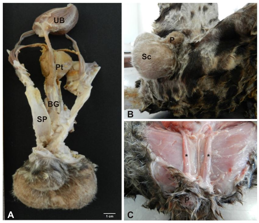
Morphology of reproductive tract of the Leopardus pardalis

                              Results

                              Male reproductive system of the ocelot
                                  The male reproductive system of Leopardus pardalis consists of the following structures:
                              scrotum, penis, testes, vas deferens and epididymis. Also included in this system are the
                              accessory bulbourethral and prostate glands, with absence of vesicular glands
                              (Figures 1 and 2).

                              Figure 1. Photograph of the reproductive tract of a male ocelot. (A) the urinary bladder (UB), prostate (Pt),
                              bulbourethral gland (BG), spermatic funiculus (SP) and penis (P); (B) scrotum (Sc) and penis (P); (C) penis
                              (P) and spermatic funiculus (*).

Anim Reprod. 2020;17(2):e20200010                                                                                                     3/12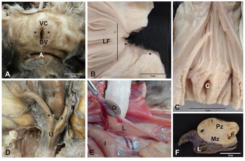
Morphology of reproductive tract of the Leopardus pardalis

                              Figure 2. Photograph of the reproductive tract of a male ocelot. (A) penis (P); (B) cavernous body (CB),
                              deep artery (DA) and urethra (U); (C) spicules (Sp); (D) visceral vaginal tunic (VVT) and parietal vaginal tunic
                              (PVT).

                              Scrotum
                                 The scrotum with a perineal location was positioned as an extension of the skin of the
                              abdominal region in the pelvic region, as a membranous pocket, divided by a median septum,
                              covered by hairs that harbored the testes, epididymides and the vas deferens (Figure 1B).

                              Testis
                                  The paired testes were separated by the scrotal septum. With ovoid shape and rounded
                              contours, they presented a concave border laterally and a straight border positioned medially.
                              They were covered by a fibrous outer membrane of whitish pink color, the visceral vaginal tunic
                              (Figure 2D). Microscopically, the testes were encased by the tunica albuginea, a thick capsule
                              of dense connective tissue, especially at the dorsal surface, which is the mediastinum, through
                              which the fibrous septa that penetrate the testis divided it into lobules. Each lobule was
                              occupied by seminiferous tubules, with 5-10 tubules per lobe (Figures 3A and 3 B).

Anim Reprod. 2020;17(2):e20200010                                                                                                        4/12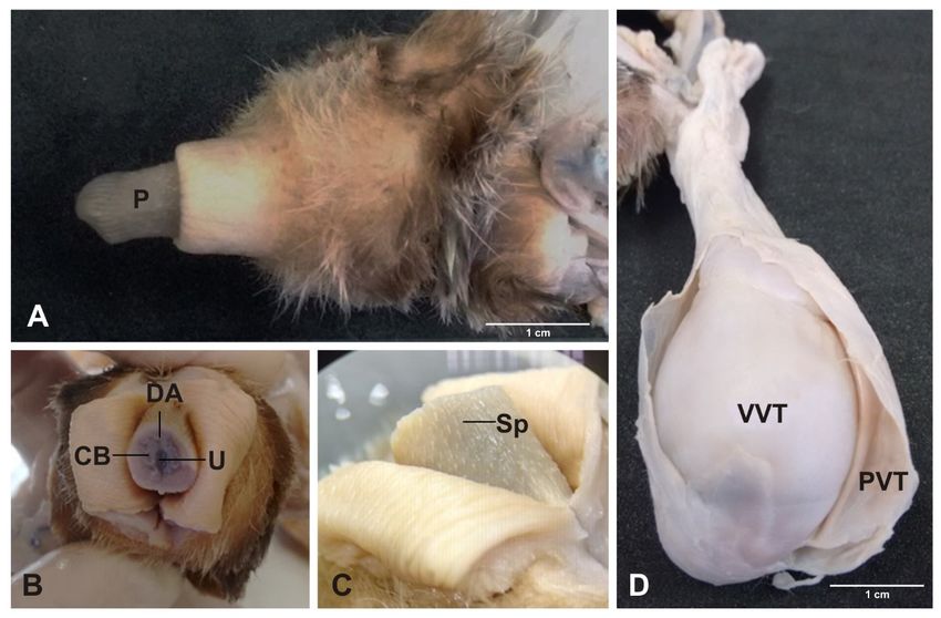
Morphology of reproductive tract of the Leopardus pardalis

                              Figure 3. Photomicrograph of the testis and epididymis of an ocelot. (A) note the Interstitial cell (IC);
                              Seminiferous tubules (ST); (B) Seminiferous epithelium (SE); Sertoli cells (SC); (C and D) Connective tissue
                              (CT); Epididymal duct (ED); Smooth muscle (SM); Basal cells (BC); Spermatozoids (SP). Hematoxylin/eosin
                              technique, bar 20 and 200µm.

                              Spermatic cord
                                 The spermatic cord consisted of the vas deferens, cremaster muscle, visceral lamina of the
                              vaginal tunica and the pampiniform plexus, composed of testicular veins and arteries.
                              Stretching ventrally through the inguinal ring, it was positioned laterally to the penis, reaching
                              up to the testes (Figures 1A and 1B).

                              Epididymis
                                 Covered by a thin serous membrane and located on the medial borders of the testes, were
                              the epididymides, comprising head, body and tail. The most prominent portion, the head of
                              the epididymis, was located in the cranial portion of the gonad, firmly attached to the vaginal
                              tunica. The body extended through the ventral region of the testis, reaching the tail at the
                              caudal end of the testis, from which the vas deferens protruded cranially (Figure 2D).
                              Microscopically the epididymal duct and the basal cells with their nuclei were observed. It was
                              also observed that the epididymis consists of basal cells, loose connective tissue, smooth
                              muscle and a cylindrical pseudo-stratified epithelium (Figures 3C and 3D).

                              Prostate and bulbourethral gland
                                 The prostate was composed of a large, irregularly shaped, compact mass located caudal to
                              the urinary bladder and cranial to the bulbourethral gland. It covered almost the entire pelvic
                              portion of the urethra. The paired bulbourethral glands, with a rounded shape, were located

Anim Reprod. 2020;17(2):e20200010                                                                                                    5/12
Morphology of reproductive tract of the Leopardus pardalis
posterior to the pelvic urethra and caudal to the prostate. In the image, the respective
structures could not be seen (Figure 1A).
Foreskin and penis
The foreskin presented as a thick layer of epithelial tissue, covered by hair, with an orifice
facing the ventral region, through which the urine and the semen are ejected. The penis was
divided into tail, body and glans, and small spicules were observed. In cross section could be
seen the cavernous bodies surrounded by tunica albuginea, erectile tissue and deep artery
(Figures 1B, 1C and 2A).
Female reproductive system of the ocelot
The female reproductive system of Leopardus pardalis consists of the following structures:
ovary, oviducts, uterus, cervix, vagina, clitoris and vulva (Figure 4).
Figure 4. Photograph of female reproductive tract of ocelot. (A) vulvar lips (*), ventral commissure (VC),
dorsal commissure (DC) and anus (A); (B) Figure to observe longitudinal folds (LF) of the cervix and vagina
and vulvar lips (*); (C) External urethral ostio (C); In (D), note intestine (I), uterine horns (*) and uterine
body (U); (E) note the ovary (O), broad ligament of the ovary (L) and intestine (I). In (F), cross section of
ovary. Note the parenchymal zone (Pz), medullar zone (Mz), tertiary follicle (F) and mesosalpinx (L).
Ovary
The paired ovaries were positioned dorsally in the sub lumbar region and caudal to the
kidneys. They had a curved expanded shape with the concave face downwards, similar to a
kidney bean. They were about 1.5 cm in length (Figures 4E and 4F). In longitudinal section, there
was a medullary region composed of loose connective tissue, a stroma rich in blood vessels,
and an external parenchymal region surrounded by a tunica albuginea (Figure 5).
Anim Reprod. 2020;17(2):e20200010 6/12Morphology of reproductive tract of the Leopardus pardalis
Figure 5. Photomicrograph of female reproductive tract of ocelot. Vagina: (A) Lumen (L), Vaginal
epithelium (VE), Muscular layer (ML) and Mucus layer (MU); (B) Lumen (L), Basal cells (BC), Mucus layer
(MU) and Blood Vessel (arrow); (C) Lumen (L), Intermediate cells (IC), Transitional Epithelium (TE) and Blood
Vessel (arrow). Uterus: (D) Lumen (L), Endometrium (E) and Myometrium (M); (E) Lumen (L), Endometrium
(E), Myometrium (M), Stratum basalis (Sb), Stratum functionalis (Sf) and Endometrial gland (arrow);
(F) Lumen (L), Pseudostratified epithelium (PE), Endometrial stroma (St) and Endometrial gland (arrow).
Ovary: (G) Lumen (L), Cortex (C) and Follicle (F); (H) Oocyte (O), Corona radiata (CR), Zona pellucida (ZP),
Antrum (A), Granulosa cells (GC), Cumulus oophofus (CO), Theca interna (Ti), Theca externa (Te) and
Primordial follicle (arrow head); (I) Primary follicle (Pf), Cortical stroma (CS), Oocyte nuclei (ON), Atretic
follicle (arrow). Hematoxylin/eosin technique, bar 20, 100 and 200µm.
Oviduct
The paired oviducts were pinkish-white in color and measured 6 and 10 cm. The uterine
tube is surrounded by a peritoneal tissue derived from the broad ligament of the uterus, called
mesosalpinx, which is subdivided into: the infundibulum of the uterine tube, ampulla and
isthmus (Figure 4D). The uterine tube makes an opening in the internal uterine ostium, being
irrigated by the arteries: ovarian and uterine (Figures 5G, 5H and 5I).
Uterus
The uterus consists of a cervix, a uterine body and two uterine horns (bicornuated).
The uterine body is positioned between the ascending colon and the urinary bladder.
The uterine body was small, about 4 cm in length (Figure 4D). The horns extended to the
ovarian pouch and measured 12 and 14 cm in length. Microscopically the presence of vascular
extract, endometrial glands, glandular epithelium and pseudo-stratified myometrium
epithelium could be shown (Figures 5D, 5E and 5F).
Anim Reprod. 2020;17(2):e20200010 7/12Morphology of reproductive tract of the Leopardus pardalis
Cervix and vagina
Macroscopically, the cervix presented circular longitudinal folds, in annular shape.
Internally, it had a thick wall of smooth muscle. It terminated at the external uterine ostium,
communicating with the vagina, where the longitudinal folds of the mucosa formed
(Figures 4B and 4C). The vagina extended to the external urethral ostium a had a thick wall
composed of smooth musculature with large lips filled by a dense layer of adipose tissue.
Microscopically, the vagina comprised the muscular layer, mucus layer, vaginal epithelium
basal cells, transitional epithelium and intermediate cells (Figures 5A, 5B and 5C).
Vulva and clitoris
The vulva comprised the vulvar lips, which joined together dorsally and ventrally.
The ventral clitoris was the most prominent part of this organ. The dorsal part had more
rounded contours (Figures 4A and 4C).
Discussion
Male reproductive system of the ocelot
A macroscopic description of the male reproductive system of Leopardus pardalis was
previously given by Carneiro et al. (2010), as the penis, scrotum, testis, epididymis, vas deferens
and accessory genital glands such as a bulbourethral gland and prostate are present, but not
the vesicular glands. The latter are also absent in domestic cats (Godinho, 1999; Dyce et al.,
2010) and in wild species as Puma yaguaroundii (Rocha et al., 2017). Leopardus pardalis has
relatively small testes (Sarti, 2006; Carneiro et al., 2010) with the same anatomy as puma (Puma
concolor) (Mansfield and Land, 2002; Guião-Leite, 2002), jaguar (Panthera onca) (Azevedo, 2004)
and Puma yaguaroundii (Rocha et al., 2017). The scrotum is positioned in the perineal region,
lateral to the penis as described by Carneiro et al. (2010) for ocelot and similar to the domestic
cat (Getty, 1987). Microscopically, the albuginea tunica in the examined testes of Leopardus
pardalis is thick and consists of the presence of dense connective tissue moderately modeled,
with considerable amounts of collagen fibers and discrete elastic fibers, with emission of thin
septa into the parenchyma testicular. The findings corroborate the research conducted by
Silva et al. (2009) on domestic felines. As described by Carneiro et al. (2010), in Leopardus
pardalis the vas deferens is a long and thin structure, similar to that of the domestic cat (Getty,
1987). As shown by Junqueira and Carneiro (2015), the body of the epididymis extends through
the ventral region of the testis, reaching the tail at the caudal end of the testis, from which the
vas deferens cranially projects, in the same way as in domestic carnivores. Microscopically, the
epididymal duct and the basal cells with their nuclei were demonstrated. It was also observed
that the epididymis is composed of smooth muscle and a cylindrical pseudo-stratified
epithelium, similar to other mammalian species (Wick and Kress, 2002; Banks, 1992; Junqueira
and Carneiro, 2015). Our findings on the prostate and bulbourethral glands of Leopardus
pardalis complement the findings of Carneiro et al. (2010) for the oncilla (Leopardus tigrinus)
and Puma yaguaroundii (Rocha et al., 2017). Microscopically, it was possible to observe the
internal epithelial layer as well as the prostatic concretion, similar to the findings of Wick and
Kress (2002) and Junqueira and Carneiro (2015).
The penis was divided into three regions: tail, body and glans and the presence of small
spicules was confirmed (Queiroz, 2003; Rocha et al., 2017). Authors relate the function of these
spicules to stimulation of the female reproductive tract to accelerate copulation and enhance
peristalsis to move sperm through the female reproductive tract (Harcourt and Gardiner,
1994). In cross-section it can be observed that the cavernous bodies are surrounded by tunica
albuginea, erectile tissue, deep artery, complementing the findings of Carneiro et al. (2010) for
this species.
Anim Reprod. 2020;17(2):e20200010 8/12Morphology of reproductive tract of the Leopardus pardalis
Female reproductive system of the ocelot
The shape and color of the ovaries was as described generically to the others mammals by
(Derivaux, 1980), Slatter (2007) and Fossum (2008) in domestic felines; Dyce et al. (2010) in
domestic felines and canines and crab-eating fox (Cerdocyon thous) by Machado et al. (2017).
Microscopic analysis of the ovary revealed a medullary region composed of loose connective
tissue, a stroma rich in blood vessels and a parenchyma surrounded by a tunica albuginea,
structures described in domestic cats and dogs by Dyce et al. (2010), Junqueira and Carneiro
(2015) and crab-eating fox by Machado et al. (2017). The oviducts are small with a pink
coloration, conformation and anatomical positioning similar to the findings of Dyce et al. (2010)
in domestic canids and felines and Machado et al. (2017) in crab-eating fox. The importance of
this organ and continuous studies must be highlighted (Andrade et al., 2019).
The uterus of Leopardus pardalis is bicornuate as previously described by Slatter (2007) and
Fossum (2008) and Dyce et al. (2010) in domestic felines. It has characteristics similar to the
uterus of the crab-eating fox (Machado et al., 2017) as well as to the uterus of domestic felines
and canids (Atwood, 1955; Schwarze and Schroder, 1970; Nickel et al., 1979; Getty, 1987; Konig
and Liebich, 2004). The uterine body is small also in agreement with Slatter (2007) and Fossum
(2008) in domestic felines; Dyce et al. (2010) in domestic felines and canids and similar in crab-eating
fox (Machado et al., 2017). Miscroscopically, it is possible to observe the presence of a smooth
muscle, also called the tunica mucosa or myometrium, which is the innermost muscular layer
of the uterus. The presence of uterine glands indicates that the tissue analyzed does not
comprise the region of the uterine body, because it is a region absent from glands, previously
known from domestic canids and felines (Schwarze and Schroder, 1970; Nickel et al., 1979;
Derivaux, 1980; Banks, 1992; Gartner and Hiatt, 1999; Konig and Liebich, 2004; Slatter, 2007;
Fossum, 2008; Dyce et al., 2010; Junqueira and Carneiro, 2015) and the crab-eating fox
(Machado et al., 2017). Macroscopically, the cervix corresponds to the uterine entry, presenting
a musculature thicker than that presented by the uterine body and the vagina, presenting at
the junction vaginal uterus, a knotlike aspect, according to the descriptions performed by
Slatter (2007) and Fossum (2008) in domestic felines. The internal orifice of the cervix is
positioned dorsally and the internal orifice ventrally to the floor of the vagina, in the same way
as described by Slatter (2007), Fossum (2008) and Dyce et al. (2010) in domestic felines.
The vagina extends to the external urethral ostium and has a thick muscular wall. The vulva
comprises vulvar lips and a prominent clitoris. These observations are in agreement with
Slatter (2007) and Fossum (2008) in domestic felines, Dyce et al. (2010) in domestic felines and
canids and Machado et al. (2017) in the crab-eating fox.
Although there are studies on the macroscopic description of the male reproductive system
of Leopardus pardalis, the present study sought to complement previous findings based on the
microscopic description by histological technique (HE). Regarding the morphology of the
female reproductive system of Leopardus pardalis,it is important to note that there are studies
that have not been published so far, which demonstrates the importance of the work
presented here.
The key points and specific findings of this study demonstrated not only the importance of
the morphological description of this species, based on a new macroscopic study, but also
aimed at the ultrastructural description of the reproductive system of males and females of
Leopardus pardalis. It is important to note that the microscopic description by classical
histological technique (Hematoxylin and Eosin), performed in this study is unprecedented, of
great importance and will serve as a complement to the research carried out up to this
moment.
Conclusion
This research emphasizes the importance of the macro- and microscopic description of the
male and female reproductive system of Leopardus pardalis. The information obtained from
the present study made it possible to fill in the gaps that often hinder the reproductive
management of this species and also to complement previous macroscopic studies that have
Anim Reprod. 2020;17(2):e20200010 9/12Morphology of reproductive tract of the Leopardus pardalis
been carried out on the species in question. From the insertion of microscopic studies
accomplished by classical techniques of histology, it was possible to amplify the knowledge,
ultra-structurally. However, we emphasize the importance of further studies on the species in
question (scanning electron microscopy, transmission electron microscopy, for example), with
contributions to the maintenance of biodiversity and preservation of this species.
Acknowledgements
The authors are grateful to Laboratório de Zoologia e Morfologia Animal of the
Universidade do Mato Grosso – UNEMAT (Alta Floresta – Mato Grosso - Brazil), for providing
the animals; the Laboratório de Biologia Molecular do Hemocentro of Ribeirão Preto and
Sandra Navarro Bresciani for construction of figures; the Laboratório de Anatomia Animal
(BDMV/ZMV/FZEA - USP) and PhD. Anthony Michael Carter (Docent Emeritus - University of
Southern Denmark) for technical assistance.
References
ACM: Associação Mata Ciliar. Avaliação das condições veterinárias e de manejo dos pequenos felinos
neotropicais em cativeiro no Estado de São Paulo. Revista de Educação Continuada. 1998;1:44-53.
Andrade GM, del Collado M, Meirelles FV, Silveira JC, Perecin F. Intrafollicular barriers and cellular
interactions during ovarian follicle development. Anim Reprod. 2019;16(3):485-96.
http://dx.doi.org/10.21451/1984-3143-AR2019-0051. PMid:32435292.
Atwood WJ. Comparative anatomy. 2nd ed. St. Louis: The C.V. Mosby Company; 1955. 424 p.
Azevedo MA. Análise morfofuncional do testículo da onça-pintada (Panthera onca) adulta [dissertação].
Viçosa: Universidade Federal de Viçosa; 2004 [cited 2020 Jan 6]. Available from:
http://locus.ufv.br/handle/123456789/5115
Bacha WJ Jr, Bacha LM. Atlas colorido de histologia veterinária. 2nd ed. São Paulo: Roca; 2003 .457 p.
Banks WJ. Histologia veterinária aplicada. 2nd ed. São Paulo: Manole; 1992. 560 p.
Bianchi RC, Mendes SL. Ocelot (Leopardus pardalis) predation on primates in Caratinga Biological Station,
southeast Brazil. Am J Primatol. 2007;69(10):1173-8. http://dx.doi.org/10.1002/ajp.20415.
PMid:17330310.
Brown JL, Wasser SK, Wildt DE, Graham LH. Comparative aspects of steroid hormone metabolism and
ovarian activity in felids, measured noninvasively in feces. Biol Reprod. 1994;51(4):776-86.
http://dx.doi.org/10.1095/biolreprod51.4.776. PMid:7819459.
Carneiro RM, Branco É, Pinheiro LL, Martins DM, Brígida SDS, Araújo EB, Souza ACB, Pereira LC, Lima AR.
Descrição morfológica do sistema reprodutor masculino de jaguatirica (Leopardus pardalis).
Biotemas. 2010;23(4):83-9. http://dx.doi.org/10.5007/2175-7925.2010v23n4p83.
Caso A, Lopez-Gonzalez C, Payan E, Eizirik E, Oliveira TG, Leite-Pitman R, Kelly M, Valderrama C. Panthera
onça. In: International Union for Conservation of Nature and Natural Resources, editor. The IUCN
Red List of Threatened Species, 2008. e: TI5953A5327466. Gland, Switzerland: IUCN; 2008.
http://dx.doi.org./10.2305/IUCN.UK.2008.RLTS.TI5953A5327466.en.
Comizzoli P. Biobanking and fertility preservation for rare and endangered species. Anim Reprod.
2017;14(1):30-3. http://dx.doi.org/10.21451/1984-3143-AR889.
Derivaux J. Reprodução dos animais domésticos, 1. Fisiologia, 2. O macho, inseminação artificial, 3.
patologia. 1st ed. Zaragoza: Editorial Acriba; 1980. p. 3-68.
Di Bitetti MS, Paviolo A, De Angelo C. Density, habitat use and activity patterns of ocelots (Leopardus
pardalis) in the Atlantic Forest of Misiones, Argentina. J Zool. 2006;270(0):153-63.
http://dx.doi.org/10.1111/j.1469-7998.2006.00102.x.
Dyce RM, Sack WO, Wensing CJG. O tratado de anatomia veterinária. 3rd ed. Rio de Janeiro: Elsevier;
2010. 816 p.
Emmons LH. Comparative feeding ecology of felids in a Neotropical Rainforest. Behav Ecol Sociobiol.
1987;20(4):4, 271-83. http://dx.doi.org/10.1007/BF00292180.
Emmons LH, Feer F. Neotropical rainforest mammals: a field guide. 2nd ed. Chicago: University of
Chicago Press; 1997. 307 p.
Anim Reprod. 2020;17(2):e20200010 10/12Morphology of reproductive tract of the Leopardus pardalis
Fossum TW. Cirurgia de pequenos animais. 3rd ed. São Paulo: Elsevier; 2008. Cirurgia dos sistemas
reprodutivo e genital; p. 702-74.
Gartner LP, Hiatt LJ. Tratado de histologia. Rio de Janeiro: Guanabara Koogan; 1999. p. 365-8.
Getty R. Anatomia dos animais domésticos. 5th ed. Rio de janeiro: Guanabara Koogan; 1987. p. 147-53.
Godinho CL. Análise histométrica do testículo e duração da espermatogênese em gatos (Felis domestica)
sexualmente maduros [tese]. Belo Horizonte: Universidade Federal de Minas Gerais; 1999.
Guião-Leite FL. Análise morfológica do testículo e do processo espermatogênico da onça parda (Puma
concolor, Wozencraft, 1993) adulta [dissertação]. Viçosa: Universidade Federal de Viçosa; 2002 [cited
2020 Jan 6]. Available from:
https://www.locus.ufv.br/bitstream/handle/123456789/7910/texto%20completo.pdf?sequence=1&is
Allowed=y
Harcourt AH, Gardiner J. Sexual selection and genital anatomy of male primates. Proc Biol Sci.
1994;255(1342):47-53. http://dx.doi.org/10.1098/rspb.1994.0007. PMid:8153136.
Junqueira LCU, Carneiro J. Histologia básica: texto y atlas. 12nd ed. Barcelona: Editorial Médica
Panamericana; 2015. 556 p.
Kleiman DG. Reproduction. In: Kleiman DG, Thompson KV, Baer CK, editors. Wild mammals in captivity:
principles and techniques for zoo management. Chicago: University of Chicago Press; 2010. p. 377-8.
http://dx.doi.org/10.7208/chicago/9780226440118.001.0001.
König HE, Liebich HJ. Anatomia dos animais domésticos. 2nd ed. Porto Alegre: Artmed; 2004. 400 p.
Ludlow ME, Sunquist ME. Ecology and behavior of ocelots in Venezuela. Natl Geogr Res. 1987;3(4):447-
61.
Machado LC, Oliveira VC, Paraventi MD, Cardoso RNR, Martins DS, Ambrósio CE. Maintenance of
Brazilian biodiversity by germplasm bank. Pesq Vet Bras. 2016;36(1):62-6.
http://dx.doi.org/10.1590/S0100-736X2016000100010.
Machado LC, Roballo KCS, Cury FS, Ambrósio CE. Female reproductive system morphology of crab-eating
fox (Cerdocyon thous) and cryopreservation of genetic material for animal germplasm bank
enrichment. Anat Histol Embryol. 2017;46(6):539-46. http://dx.doi.org/10.1111/ahe.12306.
PMid:28913836.
Mansfield KG, Land D. Cryptorchidism in Florida panthers: prevalence, features, and influence of genetic
restoration. J Wildl Dis. 2002;38(4):693-8. http://dx.doi.org/10.7589/0090-3558-38.4.693.
PMid:12528434.
Mellen JD. Factors influencing reproductive success in small captive exotic felids (Felis spp.): a multiple
regression analysis. Zoo Biol. 1991;10(2):95-110. http://dx.doi.org/10.1002/zoo.1430100202.
Micheletti T, Cubas ZS, Moraes W, Oliveira MJ, Moreira N. Reprodução natural de felídeos selvagens em
cativeiro: dificuldades e orientações. Rev Bras Reprod Anim [serial on the Internet]. 2012 [cited 2020
Jan 6];36(1):39-43. Available from: http://locus.ufv.br/handle/123456789/5115
Morato R, Barnabé R. Biotécnicas de reprodução aplicadas à preservação de felídeos selvagens. Clín Vet
[serial on the Internet]. 1998 [cited 2020 Jan 6];(12):24-6. Available from:
http://www.cbra.org.br/portal/downloads/publicacoes/rbra/v42/n3-4/p141-145%20(RB751).pdf
Moreira N, Monteiro‐Filho E, Moraes W, Swanson W, Graham L, Pasquali O, Gomes M, Morais R, Wildt D,
Brown J. Reproductive steroid hormones and ovarian activity in felids of the Leopardus genus. Zoo
Biol. 2001;20(2):103-16. http://dx.doi.org/10.1002/zoo.1010. PMid:11429781.
Moreno RS, Kays RW, Samudio R Jr. Competitive release in diets of ocelot (Leopardus pardalis) and puma
(Puma concolor) after jaguar (Panthera onca) decline. J Mammal. 2006;87(4):808-16.
http://dx.doi.org/10.1644/05-MAMM-A-360R2.1.
Murray JL, Gardner GL. Leopardus pardalis. Mamm Species. 1997;(548):1-10.
http://dx.doi.org/10.2307/3504082.
Nickel R, Schummer A, Seiferle E. The viscera of the domestic mammals. 2nd ed. Berlin: Verlag Paul
Parey: 1979. p. 351-89. http://dx.doi.org/10.1007/978-1-4757-6814-5.
Nowell K, Jackson P. Wild cats, status survey and conservation action plan [Internet]. Gland, Switzerland:
IUCN/SSC Cat Specialist Group, International Union for Conservation of Nature and Natural
Resources; 1996. 382 p. [cited 2020 Jan 6]. Available from:
https://portals.iucn.org/library/efiles/documents/1996-008.pdf
Oliveira TG, Cassaro K. Guia de campo dos felinos do Brasil. São Paulo: Instituto Pró-
Carnívoros/Fundação Parque Zoológico de São Paulo; 2005. 80 p.
Anim Reprod. 2020;17(2):e20200010 11/12Morphology of reproductive tract of the Leopardus pardalis
Queiroz VS. Estudo do efeito das condições de manipulação do sêmen de jaguatiricas (Leopardus
pardalis: Linnaeus, 1758) sobre a capacitação e a integridade morfológica e funcional dos
espermatozóides [dissertação]. São Paulo: Faculdade de Medicina Veterinária e Zootecnia,
Universidade de São Paulo; 2003. https://doi.org/10.11606/d.10.2003.tde-09062004-140805.
Rocha EF, Santos NTA, Dias RFF, Diniz JARA, Dos Santos JRS, De Menezes DJA. Anatomia macroscópica
dos órgãos reprodutores de Puma yagouaroundi (Geoffroy, 1803) macho. Pubvet. 2017;(11):744-839.
http://dx.doi.org/10.22256/pubvet.v11n8.767-770.
Sarti P. Avaliação morfométrica do testículo e da espermatogênese de jaguatiricas (Leopardus pardalis,
Linnaeus, 1758) adultas [dissertation]. Viçosa: Universidade Federal de Viçosa; 2006.
Schwarze E, Schröder L. Compêndio de anatomia veterinária: sistema visceral. Zaragoza: Acribia; 1970.
p. 277-86. Vol. 2.
Silva CAO, Perri SHV, Koivisto MB, Silva AM, Carvalho RG, Monteiro CMR. Histological and morphometric
evaluation of the testes of cats (Felis catus). Aspectos histológicos e morfométricos dos testículos de
gatos domésticos (Felis catus). Pesq Vet Bras. 2009;29(4):312-6. http://dx.doi.org/10.1590/S0100-
736X2009000400006.
Slatter D. Manual de cirurgia de pequenos animais. 3rd ed. São Paulo: Manole; 2007.
Wick R, Kress A. Ultrastructural changes in the uterine luminal and glandular epithelium during the
oestrous cycle of the marsupial Monodelphis domestica (Grey Short‐Tailed Opossum). Cells Tissues
Organs. 2002;170(2-3):111-31. http://dx.doi.org/10.1159/000046185. PMid:11731700.
Author contributions
LCM: Writing – revision & editing; JRO: Conceptualization, Writing – editing; RGK: Writing – original draft, Writing – editing; FAR: Data curation, Methodology;
DSM: Conceptualization, Formal analysis, Supervision; GMC: Review; CEA: Formal analysis, Writing – review, Supervision.
Anim Reprod. 2020;17(2):e20200010 12/12You can also read