A novel system to collect dual pulse oximetry data for critical congenital heart disease screening research
←
→
Page content transcription
If your browser does not render page correctly, please read the page content below
Journal of Clinical and
        Translational Science
                                                             A novel system to collect dual pulse oximetry
                                                             data for critical congenital heart disease
        www.cambridge.org/cts                                screening research
                                                             Kavish Doshi1, Gregory B. Rehm1, Pranjali Vadlaputi2, Zhengfeng Lai3,
        Research Article                                     Satyan Lakshminrusimha2, Chen-Nee Chuah3 and Heather M. Siefkes2
        Translational Research,                              1
                                                              Department of Computer Science, University of California, Davis, Davis, CA, USA; 2Department of Pediatrics,
        Design and Analysis                                  University of California, Davis, Sacramento, CA, USA and 3Department of Electrical and Computer Engineering,
                                                             University of California, Davis, Davis, CA, USA
        Cite this article: Doshi K, Rehm GB,
        Vadlaputi P, Lai Z, Lakshminrusimha S,
        Chuah C-N, and Siefkes HM. A novel system to             Abstract
        collect dual pulse oximetry data for critical
        congenital heart disease screening research.
                                                                 Introduction: Access to patient medical data is critical to building a real-time data analytic pipe-
        Journal of Clinical and Translational Science 5:         line for improving care providers’ ability to detect, diagnose, and prognosticate diseases. Critical
        e56, 1–7. doi: 10.1017/cts.2020.550                      congenital heart disease (CCHD) is a common group of neonatal life-threatening defects that
                                                                 must be promptly diagnosed to minimize morbidity and mortality. CCHD can be diagnosed
        Received: 2 June 2020
                                                                 both prenatally and postnatally. However, despite current screening practices involving oxygen
        Revised: 28 September 2020
        Accepted: 8 October 2020                                 saturation analysis, timely diagnosis is missed in approximately 900 infants with CCHD annu-
                                                                 ally in the USA and can benefit from increased data processing capabilities. Adding non-
        Keywords:                                                invasive perfusion measurements to oxygen saturation data can improve the timeliness and
        Congenital heart disease screening; pulse                fidelity of CCHD diagnostics. However, real-time monitoring and interpretation of non-
        oximetry
                                                                 invasive perfusion data are currently limited. Methods: To address this challenge, we created
        Address for correspondence:                              a hardware and software architecture utilizing a Pi-top™ for collecting, visualizing, and stor-
        H.M. Siefkes, MD, MSCI, University of California,        ing dual oxygen saturation, perfusion indices, and photoplethysmography data. Data
        Davis Ticon II, 2516 Stockton Blvd, Sacramento,          aggregation in our system is automated and all data files are coded with unique study iden-
        CA 95817, USA. Email: Hsiefkes@ucdavis.edu
                                                                 tifiers to facilitate research purposes. Results: Using this system, we have collected data from
                                                                 190 neonates, 130 presumably without and 60 with congenital heart disease, in total compris-
                                                                 ing 1665 min of information. From these data, we are able to extract non-invasive perfusion
                                                                 features such as perfusion index, radiofemoral delay, and slope of systolic rise or diastolic fall.
                                                                 Conclusion: This data collection and waveform analysis is relatively inexpensive and can be
                                                                 used to enhance future CCHD screening algorithms.

                                                             Introduction
        © The Association for Clinical and Translational     Congenital heart disease is the most common birth defect affecting approximately 0.8% of all
        Science 2020. This is an Open Access article,        births [1]. Critical congenital heart disease (CCHD) accounts for approximately 20% of con-
        distributed under the terms of the Creative
        Commons Attribution-NonCommercial-                   genital heart disease and is life-threatening if not timely diagnosed [1–4]. In fact, prior to uni-
        NoDerivatives licence (http://                       versal oxygen saturation-based CCHD screening, 25% of CCHD were diagnosed after hospital
        creativecommons.org/licenses/by-nc-nd/4.0/),         discharge, with some diagnosed at autopsy [2, 5]. Oxygen saturation screening has since reduced
        which permits non-commercial re-use,                 mortality associated with CCHD and helped with earlier diagnosis, but nearly 900 neonates with
        distribution, and reproduction in any medium,
        provided the original work is unaltered and is
                                                             CCHD remain undiagnosed annually in the USA [6, 7]. Coarctation of the aorta (CoA) is the
        properly cited. The written permission of            most commonly missed CCHD defect despite oxygen saturation screening as it is associated
        Cambridge University Press must be obtained          with poor systemic perfusion without hypoxemia [5, 6, 8]. Late diagnosis of defects such as
        for commercial re-use or in order to create a        CoA can be particularly detrimental. Preliminary analysis of CCHD defects missed by oxygen
        derivative work.                                     saturation screening demonstrates that 18% of newborns with late CoA diagnosis die, some
                                                             before surgery can be done (unpublished data) [9]. More than 50% of deaths due to missed
                                                             CCHD occur before corrective surgery, either at home or shortly after arriving to the
                                                             Emergency Department [4].
                                                                 To improve CCHD detection and minimize false-negative screens, especially of defects
                                                             associated with poor perfusion such as CoA, researchers have investigated the addition of
                                                             non-invasive pulse oximetry measurements such as perfusion index (PIx), radiofemoral pulse
                                                             delay, and other photoplethysmography waveform characteristics to the current oxygen satu-
                                                             ration CCHD screening algorithm [10–14]. However, accessing and interpreting these addi-
                                                             tional pulse oximetry data are not a prevalent practice. In addition, pulse oximetry devices
                                                             currently only store de-identified data, limiting its utility for research. In part due to these lim-
                                                             itations, prior studies evaluating PIx have either included brief (10 s) clinician interpreted and
                                                             manually documented perfusion indices or have utilized retrospective de-identified samplings
                                                             from pulse oximetry devices from routine screening [10–12, 15].
                                                                 To solve this problem, we created an automated real-time data collection and analysis archi-
                                                             tecture that is able to perform non-invasive measurement of perfusion and oxygenation data.

Downloaded from https://www.cambridge.org/core. IP address: 46.4.80.155, on 16 Aug 2021 at 15:12:57, subject to the Cambridge Core terms of use, available at https://www.cambridge.org/core/terms
. https://doi.org/10.1017/cts.2020.550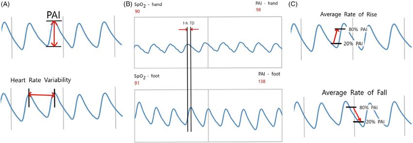
2 Doshi et al.
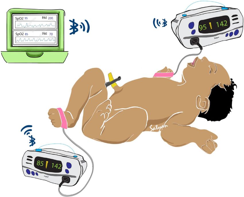
Fig. 1. Illustration of potential uses for this system in post-delivery critical congenital heart disease (CCHD) screening. Two pulse oximeter devices are applied to both a foot and
right hand of a neonate. In our case, we use the Nonin® WristOx2™ 3150 to communicate wirelessly with a central aggregator device. The aggregator can then perform visualization
and analytics on whether the neonate displays risk for CCHD while simultaneously storing the data for later review.
The system is able to wirelessly communicate with pulse oximetry (2) ensure accurate time-stamping of the collected data; and (3)
devices and store data on secure computing systems. The main allow ease of use by non-technical end users in terms of both data
focus of this paper is the novel data collection pipeline that is cus- acquisition hardware and data management software.
tomizable by providers depending on the target clinical outcomes. Defects such as CoA are associated with differences in oxygen
To date, there are no other published research or clinical tools saturation and PIx from the right upper extremity (pre-ductal) and
capable of prospectively collecting abundant high-fidelity CCHD any lower extremity (post-ductal). Most current CCHD screening
screening data. The insights driven by this data collection will methods employ the sequential application of pulse oximetry
be beneficial to developing future algorithmic CCHD screening probe to the right upper extremity followed by a lower extremity
processes and may be applicable to other vascular disease processes with manual documentation and interpretation of data.
that could benefit from non-invasive pulse oximetry perfusion Simultaneous collection of data using dual pulse oximetry
diagnostics (aneurysms, aortic dissections, atherosclerosis, throm- (Fig. 1) enables accurate collection and interpretation of data using
bosis, or vascular graft monitoring). a simple workflow (Fig. 2 and Table 1).
Our general system consists of two independent perfusion and
oxygenation monitors (oximeters) that are attached to the study
Methods subject’s right hand and any foot, and a central aggregator device
The primary focus of our overall study was to explore correlations that is able to communicate with the oximeters and temporarily
between oxygen saturation and perfusion data in neonates with store their data for eventual retrieval. For our oximeter, we use
and without CCHD. The study was registered on ClinicalTrials. the Nonin® WristOx2™ 3150. This device allows simultaneous
gov (NCT04056104). The study was approved by the collection of photoplethysmography and oxygenation and perfu-
Institutional Review Boards at all sites. The parents/guardians of sion data, can externally transmit data using Bluetooth and is rel-
all subjects provided informed consent prior to study enrollment. atively inexpensive. We chose the Pi-top™, an affordable laptop
As part of this study, we developed an integrative software and computer that uses Raspberry Pi microcomputers, as our central
hardware system to collect necessary physiological data. Our sys- aggregator. By developing appropriate software, we were able to
tem needed to: (1) enable continuous and automated data collec- collect oxygenation and perfusion data via Bluetooth through
tion and storage from multiple pulse oximeters simultaneously; the Nonin® devices. The Pi-top is also able to maintain temporal
Downloaded from https://www.cambridge.org/core. IP address: 46.4.80.155, on 16 Aug 2021 at 15:12:57, subject to the Cambridge Core terms of use, available at https://www.cambridge.org/core/terms
. https://doi.org/10.1017/cts.2020.550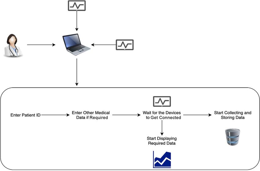
Journal of Clinical and Translational Science                                                                                                                                  3

        Fig. 2. Data collection workflow. Illustration of the workflow of the system. A technician controls the software and attaches the pulse oximeters to the patient. They then enter
        the patient identification number and other medical details. The software will automatically connect to the oximeters via Bluetooth, displays the oximetry and perfusion data in
        real time, and stores the data.

        alignment between the two separate Nonin® devices. This ensured                            congenital heart disease (300 min = CCHD, 100 min =
        that data are accurately time-stamped and synchronized. Time-                              non-critical CHD) and 610 min of oximetry data from newborns
        stamps can also be adjusted to ensure complete de-identification.                          confirmed to be without congenital heart disease. The majority of
        Our entire system is illustrated in Figure 1.                                              patients so far were enrolled at the University of California, Davis,
            Once collected and dated, data files are automatically aggre-                          but four other hospitals (Sutter Medical Center, Sacramento,
        gated and coded with unique study identifiers. The data are then                           University of California, Los Angeles, University of California,
        transferred from the Pi-top using a secure USB drive and uploaded                          San Francisco, and Cohen’s Medical Center, New York) are
        to REDCap through a secure data transfer mode. This architecture                           participating in the study with plans to enroll hundreds of more
        and workflow (Fig. 2) enable ease of data collection and data shar-                        newborns over a total 2-year period.
        ing across different hospitals.                                                                The detailed process of the data collection is as follows: We
                                                                                                   attach two Nonin® WristOx2™ 3150 pulse oximeters to the sub-
                                                                                                   ject, one on the right hand and the other on either foot. Once these
        Results                                                                                    Nonin® devices display valid measurements, and we can ensure
                                                                                                   minimal signal noise resulting from limb movement, we initialize
        Data Collection
                                                                                                   our collection software on the Pi-top™ device. In the software user
        Using this system, we have gathered data from 190 newborns. At                             interface, a unique study identification number is entered. This
        time of enrollment, 130 were presumed to be without congenital                             study identifier is an anonymous token that can only be linked back
        heart disease and 60 were expected to have congenital heart disease.                       to the patient via an encrypted database like REDCap. Next, we
        In total, we have gathered approximately 1665 min of pulse oxime-                          input the fraction of inspired oxygen (FiO2) the patient is receiving.
        try data from these babies. To date, we have confirmed 60 of the                           For study protocols that involve repeat measurements, we included
        newborns enrolled as presumably healthy newborns remain                                    an alphanumeric entry to label specific measurements for later
        healthy. One baby enrolled as a presumably healthy baby before                             identification. The software connects to both pulse oximeters via
        qualifying for the routine CCHD screen, was determined to have                             Bluetooth and starts collecting pulse oximetry, oxygen saturation,
        CCHD based on the first study measurements collected (oxygen                               heart rate, pulse amplitude index (PAI) – synonymous with PIx,
        saturations were in the 60s). The follow-up is still pending for                           and photoplethysmography data. All data are time-stamped so
        the remaining babies enrolled as presumably healthy. Of the babies                         we can maintain temporal accuracy between the two Nonin® devi-
        enrolled presumably with congenital heart disease, we have con-                            ces. During collection, data are also displayed on the Pi-top screen
        firmed the final classification for 48 (31 = CCHD, 13 = non-critical                       in real time, which aids with data quality control (shown in Fig. 3).
        CHD, and 4 = without CHD). In total, this comprises 400 min of                             To further demonstrate our process, we provided stepwise instruc-
        oximetry data from newborns with the final classification of                               tions in an online supplement.

Downloaded from https://www.cambridge.org/core. IP address: 46.4.80.155, on 16 Aug 2021 at 15:12:57, subject to the Cambridge Core terms of use, available at https://www.cambridge.org/core/terms
. https://doi.org/10.1017/cts.2020.550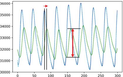
4                                                                                                                                                                Doshi et al.

        Table 1. Raw data fields collected by our system                                          Table 2. Features that can be extracted from the data fields collected

           Data fields        Explanation                                                            Feature
                                                                                                     number            Features
           Patient_ID         Corresponding patient research identification number
                                                                                                     1                 Systolic and diastolic peaks
           Time_Stamp         Time at which the data were recorded
                                                                                                     2                 Perfusion index
           FiO2               Value of fractional inspired oxygen (FiO2) as entered
                              by the clinician                                                       3                 Slope of systolic rise
           Pleth              Value of photoplethysmography as received from the                     4                 Slope of diastolic fall
                              Nonin®
                                                                                                     5                 Area under the curve per pulse
           Measurement        Alphanumeric label assigned to the data to easily dis-
           label              tinguish consecutive repeat measurements                               6                 Delay between systolic maximum points of hand versus
                                                                                                                       foot waveforms
           PAI                Pulse amplitude index as measured by the Nonin®
                                                                                                     7                 Delay between systolic starting points of hand versus
           SpO2               Oxygen saturation (SpO2) as measured by the Nonin®                                       foot waveforms
           Heart_Rate         Heart rate of the patient as measured by the Nonin®                    8                 Oxygen saturation over a defined period of time
                                                                                                     9                 Heart rate

       Data Aggregation
       Management of each Pi-top™, including data aggregation, is per-                            Interestingly, the baby with CoA was trialed off prostaglandin E1
       formed by a technician or research coordinator present on the site.                        therapy to assess the severity of the coarctation and thus the ductus
       During aggregation, a human must manually ensure that data is                              arteriosus was presumably closing and the CoA narrowing during
       correctly aggregated to the database. The technician (1) uploads data                      our data collection. Our measurements occurred approximately
       to the REDCap database, (2) backs up the folders in the                                    10 h and 36 h after the prostaglandin E1 infusion was discontinued.
       Pi-top™ to an external hard drive, and (3) deletes all the files on                        The baby with CoA was monitored in the neonatal intensive care
       the Pi-top™ when the data are stored locally somewhere. Once these                         unit and was asymptomatic. The baby had an echocardiogram at
       steps are complete, uploaded data can be accessed across the                               3 days of age, which noted the ductus arteriosus was closed and
       research network and referenced by the patient identifier.                                 the CoA was minimal. Thus, the baby was discharged home with
                                                                                                  plans to follow-up outpatient with cardiology. The baby then repre-
                                                                                                  sented to care at 7 days of age, and an echocardiogram showed an
       Scalability and Adding New Devices
                                                                                                  increase in the gradient across the CoA prompting readmission and
       Provisioning additional Pi-top™ and pulse oximeter devices enable                          subsequent surgery for CoA repair. Note the foot pulse amplitude
       the collection of multiple patients simultaneously and prevent data                        index (PAI) decreased in this neonate with increasing duration
       collection slowdowns in study progress due to lost or damaged                              off prostaglandin E1 infusion. The average foot PAI decreased from
       equipment. To more rapidly provision devices, we developed an                              96 to 36 during this time period (Fig. 4B and C).
       image of the Pi-top™ with the pre-configured details of our software                           There are some other notable features in this baby with CoA
       that can be copied to the microSD card of the Pi-top™ in a single                          compared to the healthy baby (Fig. 4). Note the closer proximity
       step resulting in a fully functional Pi-top™ without the need to                           of the peaks of the hand and foot (f-h TD) in the healthy baby ver-
       install multiple software components. This workflow substantially                          sus the baby with CoA. Additionally, note how the f-h TD changes
       decreases the number of steps necessary for Pi-top™ deployment                             as more hours off prostaglandin pass and presumably as the ductus
       and may allow non-technical users to assist more experienced per-                          arteriosus closes. However, we are unable to quantify the f-h TD
       sonnel in the provisioning process, which we have successfully done                        accurately at this time. Since the Pi-Top™ can only receive one
       remotely with sites when a software change was needed.                                     Bluetooth data package at a time, there is a Bluetooth transmission
                                                                                                  difference between the hand and foot waveforms that will need to
       Data Collected and Feature Identification                                                  be accounted for in future analyses. Additionally, as a baby’s physi-
                                                                                                  ology changes and the potential for physiological foot peaks occur-
       Table 1 summarizes the data fields collected from the Nonin® devi-                         ring before the hand peaks, this further complicates the analysis.
       ces for each patient enrolled. The data are visualized on the Pi-top                       However, the photoplethysmography segments shown in Fig. 4A
       (Supplemental Figure) for real-time interpretation and then can be                         and B were chosen because the relative hand to foot peak durations
       reconstructed from the logged data for analysis and feature detec-                         were consistent for these measurements. Thus, assuming a similar
       tion on our compute server. From the collected data, we are able to                        Bluetooth transmission across patients, this highlights the poten-
       extract different features stated in Table 2. For example, Fig. 3B and                     tial for a longer f-h TD in patients with CoA. We also captured a
       C illustrates how the delays between systolic peaks of hand and foot                       morphology change to the hand photoplethysmography waveform
       waveforms (Feature #6 in Table 2) and the slopes of the systolic rise                      as the ductus arteriosus closed with the development of a remark-
       and diastolic fall (Feature #3 and #4 in Table 2) can be extracted                         ably notable dicrotic notch (Fig. 4C). This highlighted the need to
       from reconstructed waveforms. Various statistics of these features                         apply smoothing filters to the waveforms so that peaks can more
       (minimum, maximum, mean, standard deviation, and median)                                   easily be identified to ease analysis.
       may be useful for further characterization and development of
       the prediction model for CCHD detection.
                                                                                                  Challenges and Adjustments Made
           To further demonstrate the features collected in our data, we
       included photoplethysmography waveforms from a healthy baby                                We encountered a few challenges, from which others will benefit
       (Fig. 4A) and a baby with critical CoA (Fig. 4B and C).                                    from our experience. First, in regards to data security, we needed an

Downloaded from https://www.cambridge.org/core. IP address: 46.4.80.155, on 16 Aug 2021 at 15:12:57, subject to the Cambridge Core terms of use, available at https://www.cambridge.org/core/terms
. https://doi.org/10.1017/cts.2020.550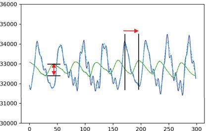
Journal of Clinical and Translational Science                                                                                                                                    5

        Fig. 3. Examples of features that can be extracted from raw waveform. Features that can be extracted from raw waveform include pulse amplitude index (PAI) (Box A), heart rate
        variability (Box A), radiofemoral delay (f-h TD) (Box B), and both the systolic rise and diastolic fall slope of the photoplethysmography waveform (Box C).

               (A)                                                   (B)                                                      (C)

        Fig. 4. Example of pulse oximetry data collected from a healthy newborn and a newborn with critical coarctation of the aorta (CoA). Solid lines are from raw data. Dashed lines
        have a filtered applied to assist with peak identification due to the dicrotic notch interfering with peak identification for the infant with coarctation. (Box A) A normal newborn
        demonstrates minimal time delay between the right hand and foot pulse (f-h TD) and similar pulse amplitude index (PAI) in hand and foot. (Box B and C) A newborn with critical
        CoA shows the foot PAI decrease and the f-h TD change as more time off prostaglandin E1 passes. Additionally, the dicrotic notch appearance in the right hand is notable different
        in the baby with CoA compared to both the healthy newborn and the earlier measurement in the same baby when the ductus arteriosus was presumably more open.

        encrypted USB compatible with Linux operating system and func-                              due to time drift caused by not having the Pi-top™ connected
        tional on an ARM processor (Raspberry Pi). This required using                              to a server for security purposes [16].
        VeraCrypt™ encryption service as opposed to an off-the-shelf USB.
            Additionally, to ensure the cessation of the Bluetooth connec-
        tion, the entire Pi-top™ system has to be shut down. If the                                 Discussion
        Bluetooth connection remains, then the data collection can con-                             We created a system for automated collection of pulse oximetry
        tinue when not intended. This is only problematic if data collection                        data for research related to CCHD screening. This system collects
        has to be restarted. For example, if a technical error is encountered                       and displays real-time data from medical devices while communi-
        mid-data collection, then the system has to be powered down and                             cating wirelessly via Bluetooth. The system does not require access
        restarted, which adds a couple of minutes to the process.                                   to a wireless network making it portable to the regions with little or
            To better identify collected data with specific clinical scenarios,                     no access to the internet and also aids with security. It is
        such as the age of the patient at time of the measurement, we added                         inexpensive and capable of end-to-end automation of data collec-
        an alphanumeric entry that serves as a code specific to the protocol                        tion and storage. The system requires only one clinician or
        time points. We were unable to use the time-stamps on the col-                              coordinator with basic computer skills to monitor the process.
        lected data, as some sites requested we falsify the date and time                           Once it is set up properly, the system can capture oxygen saturation
        on the Pi-top™ to add additional security and completely                                    and perfusion data from the infant in a non-invasive manner, and
        de-identify the data. Even if the time-stamps were not falsified,                           is able to maintain continuous operation for as long as required by
        the time-stamps alone would not be sufficient for identification                            the study circumstances.

Downloaded from https://www.cambridge.org/core. IP address: 46.4.80.155, on 16 Aug 2021 at 15:12:57, subject to the Cambridge Core terms of use, available at https://www.cambridge.org/core/terms
. https://doi.org/10.1017/cts.2020.550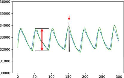
6                                                                                                                                                                Doshi et al.

           Our data collection system is motivated by the need for early                          require additional development efforts. Currently, CCHD screen-
       CCHD detection, and the inability for providers to electronically                          ing capabilities are not offered by this system, but will be part of our
       store pulse oximetry data in a manner conducive to the research                            future work. It is only designed to collect data from neonates who
       and clinical needs. Antenatal echocardiology and postnatal exami-                          have already undergone or will undergo standard of care CCHD
       nation detected only approximately 70% of the patients with                                screening by a qualified provider to generate labeled datasets for
       CCHD leading to the addition of postnatal pulse oximetry to rou-                           research purposes, including the development of advanced
       tine newborn screening [6, 17]. Techniques described in this study                         machine learning-based CCHD detection models. Furthermore,
       address the limitations of the oxygen saturation-based CCHD                                no analytic capabilities for individual data streams such as perfu-
       screen. Researchers have hypothesized that adding non-invasive                             sion are built into the system and our data visualization only dis-
       pulse oximetry measurements such as PIx, radiofemoral pulse                                plays streaming waveform data to the provider. Future work will
       delay (f-h TD), and other photoplethysmography waveform char-                              serve to utilize this streaming waveform data in an analytic capacity
       acteristics to the current diagnostic suite of pulse oximetry mea-                         and possibly even perform CCHD screening interpretation.
       surement will help in more accurate detection of CCHD in                                   However, additional studies and clinical trials will be required to
       patients [10–14]. In a study with a total of 123 normal and 21 new-                        prove its efficacy.
       borns with congenital heart defects, 4 out of 5 critical systemic                             Overall, the system that we created has opened up a new way of
       obstruction subjects passed the regular oxygen saturation-based                            data collection for CCHD related diagnostic information from
       CCHD screen [10].The addition of PIx to the screen resulted in                             neonates. The data collection system and process are mostly auto-
       four of the five newborns failing the combined screen [10]. That                           mated and are capable of being used by non-technical users. It may
       study and prior studies evaluated PIx for a single moment in time                          also serve as a generalized model to be utilized by different
       with an artifact-free waveform for 10 s. In our current study, we                          researchers who are also working to collect streaming waveform
       averaged the pulse amplitude index (PAI), which is PIx multiplied                          information. Using this system we have gathered approximately
       by 100, over 5 min. We captured a patient with CoA and demon-                              1665 min of oximetry data from neonates with and without con-
       strated the foot PAI decrease, as the ductus arteriosus was closing,                       genital heart disease. This information may be invaluable for devel-
       and decreased below thresholds that may be indicative of systemic                          oping future CCHD screening processes and hopefully will serve as
       obstruction such as CoA [11, 18]. Interestingly, we noted this                             the basis for future electronic systems to improve CCHD detection
       decrease in PAI when the echocardiogram estimated only a mild                              and may be adapted for other research endeavors as well.
       CoA and thus it was initially thought the baby may not require sur-
       gery. Therefore, additional non-invasive perfusion measurements                            Supplementary material. To view supplementary material for this article,
       along with the regular oxygen saturation-based screen is a potential                       please visit https://doi.org/10.1017/cts.2020.550.
       technique to not only enhance CCHD detection, but may also help
                                                                                                  Acknowledgments. We are grateful for the support of everyone who helped at
       guide treatment plans when it is already known that a patient has a
                                                                                                  various stages of this project and our study participants. Our engineers: Michael
       CoA, although more research is necessary. Additionally, prior                              Agung, and research coordinators and collaborators: Michele Guillen, Orsolya
       studies have noted prolonged pulse delay and abnormalities in tim-                         Gilicze, Sparsha Govardhan, Alejandra Guzman, Erlinda Manalo, MD, Whitnee
       ing of pre-ductal dichroitic notch in newborns with CoA [13, 19].                          Hogan, MD, Robert Koppel, MD, and Meena Garg, MD.
       Our data captured an identifiable pre-ductal dicrotic notch while                              The project described was supported by the National Center for Advancing
       an infant with CoA was trialed off prostaglandin therapy and the                           Translational Sciences, National Institutes of Health (NIH), through grant
       timing relationship between the hand and foot pulses also                                  number UL1 TR001860 and linked award KL2 TR001859, the Eunice
       appeared to change as well. However, conclusions cannot be drawn                           Kennedy Shriver National Institute of Child Health & Human Development,
       yet on this small sample of patients and additional analyses to sort                       NIH, through grant number 1R21HD099239–01, and the University of
       out Bluetooth transmission versus physiological factors for the                            California, Davis Artificial Intelligence Seed Grant. The content is solely the
                                                                                                  responsibility of the authors and does not necessarily represent the official views
       pulse delay are underway.
                                                                                                  of the NIH.
           Future research into CCHD screening techniques will necessi-
       tate readily accessible medical device data, data standards, and                           Disclosures. The authors do not have conflicts of interest to disclose.
       archival data storage of identifiable data (identifiable at least                          University of California, Davis has intellectual property rights related to inven-
       through unique identification codes) [20]. The use of pulse oxime-                         tions resulting from this work and a provisional patent has been submitted by
       try devices results in huge amounts of data that are yet to be fully                       the university.
       explored for CCHD screening. The current infrastructure limits
       access to this data for research. For example, continuous pulse oxi-
       metry monitoring in the hospital may not be stored long term or be                         References
       readily identifiable, and when it is, researchers have to rely on                           1. Reller MD, Strickland MJ, Riehle-Colarusso T, Mahle WT, Correa A.
       charting to correlate with clinically relevant data such as probe                              Prevalence of congenital heart defects in metropolitan Atlanta, 1998-
       placement [14]. Our pulse oximeter data collection process                                     2005. Journal of Pediatrics 2008; 153(6): 807–813. doi: 10.1016/j.jpeds.
       mitigates many of these barriers and may also help guide future                                2008.05.059
       screenings or research for other diseases that may benefit from                             2. Wren C, Reinhardt Z, Khawaja K. Twenty-year trends in diagnosis of life-
       non-invasive pulse oximetry perfusion diagnostics (aneurysms,                                  threatening neonatal cardiovascular malformations. Archives of Disease in
                                                                                                      Childhood: Fetal & Neonatal Edition 2008; 93(1): F33–F35. doi: 10.1136/
       aortic dissections, atherosclerosis, thrombosis, or vascular graft
                                                                                                      adc.2007.119032
       monitoring).
                                                                                                   3. Koppel RI, et al. Effectiveness of pulse oximetry screening for congenital
           Our system, while providing solutions to the above problems, is                            heart disease in asymptomatic newborns. Pediatrics 2003; 111: 451–455.
       currently limited to the collection of pulse oximetry and perfusion                            doi: 10.1542/peds.111.3.451
       information from a single type of medical device (Nonin®                                    4. Chang R, Gurvitz M, Rodriguez S. Missed diagnosis of critical congenital
       WristOx2™ 3150). Utilizing other devices, and other communica-                                 heart disease. Archives of Pediatrics & Adolescent Medicine 2008; 162(10):
       tion protocols besides Bluetooth is currently unsupported, and will                            969. doi: 10.1001/archpedi.162.10.969

Downloaded from https://www.cambridge.org/core. IP address: 46.4.80.155, on 16 Aug 2021 at 15:12:57, subject to the Cambridge Core terms of use, available at https://www.cambridge.org/core/terms
. https://doi.org/10.1017/cts.2020.550
Journal of Clinical and Translational Science 7
5. Lannering K, Bartos M, Mellander M. Late diagnosis of coarctation 13. Palmeri L, et al. Photoplethysmographic waveform characteristics of
despite prenatal ultrasound and postnatal pulse oximetry. Pediatrics. newborns with coarctation of the aorta. Journal of Perinatology 2017;
2015; 136(2). doi: 10.1542/peds.2015-1155 37(1): 77–80. doi: 10.1038/jp.2016.162
6. Ailes EC, Gilboa SM, Honein MA, Oster ME. Estimated number of infants 14. Sorensen MW, Sadiq I, Clifford GD, Maher KO, Oster ME. Using pulse
detected and missed by critical congenital heart defect screening. Pediatrics oximetry waveforms to detect coarctation of the aorta. BioMedical
2015; 135(6): 1000–1008. doi: 10.1542/peds.2014-3662 Engineering OnLine 2020: 1–12. doi: 10.1186/s12938-020-00775-2
7. Abouk R, Grosse S, Ailes E, Oster M. Association of US state implemen- 15. Jegatheesan P, Nudelman M, Goel K, Song D, Govindaswami B.
tation of newborn screening policies for critical congenital heart disease Perfusion index in healthy newborns during critical congenital heart
with early infant cardiac deaths. JAMA 2017; 318(21): 2111–2118. doi: disease screening at 24 hours: retrospective observational study from
10.1001/jama.2017.17627 the USA. BMJ Open 2017; 7(12): e017580. doi: 10.1136/bmjopen-2017-
8. Peterson C, et al. Late detection of critical congenital heart disease among 017580
US infants: estimation of the potential impact of proposed universal screen- 16. Mills DL. Network time protocol version 4 reference and implementation
ing using pulse oximetry. JAMA Pediatrics 2014; 168(4): 361–370. doi: 10. guide. 2006; (June): 1–90.
1001/jamapediatrics.2013.4779 17. Sebelius K. Secretary of Health & Human Services Letter to the Secretary’s
9. Hogan W. Pulse Oximetry Screening in Practice: Factors Associated with Advisory Committee on Heritable Disorders in Newborns and
Screening Rates and Performance (Unpublished, Direct Communication Children (SACHDNC). (https://www.hrsa.gov/sites/default/files/hrsa/
with Author). advisory-committees/heritable-disorders/reports-recommendations/
10. Siefkes H, et al. Oxygen saturation and perfusion index-based enhanced response-congenital-cyanotic.pdf)
critical congenital heart disease screening. American Journal of 18. Siefkes H, et al. Oxygen saturation and perfusion index-based enhanced
Perinatology 2020; 37(2): 158–165. doi: 10.1055/s-0039-1685445 critical congenital heart disease screening. American Journal of
11. de-Wahl Granelli A, Ostman-Smith I. Noninvasive peripheral perfusion Perinatology 2020; 37(2). doi: 10.1055/s-0039-1685445
index as a possible tool for screening for critical left heart obstruction. 19. Oyonarte M, Dickinson D, Medici D, Hamilton D. Indirect arterial pulse
Acta Paediatrica 2007; 96(10): 1455–1459. doi: 10.1111/j.1651-2227.2007. tracings in children with coarctation of the aorta before and after operation.
00439.x Thorax 1980; 35: 128–132.
12. Schena F, et al. Perfusion index and pulse oximetry screening for congeni- 20. Oster ME, et al. Lessons Learned From Newborn Screening for Critical
tal heart defects. Journal of Pediatrics 2017; 183: 74–79. doi: 10.1016/j.jpeds. Congenital Heart Defects. Pediatrics. 2016; 137(5): e20154573–
2016.12.076 e20154573. doi: 10.1542/peds.2015-4573
Downloaded from https://www.cambridge.org/core. IP address: 46.4.80.155, on 16 Aug 2021 at 15:12:57, subject to the Cambridge Core terms of use, available at https://www.cambridge.org/core/terms
. https://doi.org/10.1017/cts.2020.550You can also read