MYCOTOXIN ANALYSIS: A FOCUS ON RAPID METHODS - Kristine Wolf and Florian J. Schweigert
←
→
Page content transcription
If your browser does not render page correctly, please read the page content below

MYCOTOXIN ANALYSIS:
A FOCUS ON RAPID METHODS
Kristine Wolf and Florian J. Schweigert

                                          With support from   On behalf of

                                                                             A
MYCOTOXIN ANALYSIS:
A FOCUS ON RAPID METHODS
Kristine Wolf and Florian J. Schweigert
Institute of Nutritional Science
University of Potsdam
ISBN 978-99944-72-06-2
© Partnership for Aflatoxin Control in Africa

This document is in the public domain. For non-profit
purposes, users are welcome to download, save, or distribute
this document electronically or in any other format, including
in foreign language translation without written permission.
We do ask that, if you distribute this document, you credit
the Partnership for Aflatoxin Control in Africa (PACA) and
mention the website http://www.aflatoxinpartnership.org/
and not alter the text.

Suggested citation: Kristine Wolf and Florian J. Schweigert
(2018) Mycotoxin Analysis: A Focus on Rapid Methods.
Partnership for Aflatoxin Control in Africa, African Union
Commission, Addis Ababa, Ethiopia.

Disclaimer: Only the authors are responsible for the content
of this book; mention of a proprietary name does not imply
endorsement of the product over others by PACA or the
funders of this work.
FOREWORD
  Mycotoxin contamination of staple and cash crops is a serious developmental challenge
  that calls for urgent actions. African Union Commission has priority attention to
  aflatoxin control because aflatoxins are a real threat to the achievement of major global
  and continental commitments including ending hunger, boosting trade in agricultural
  commodities and services and eradicating poverty. Aflatoxins thwart Africa’s efforts
  at achieving food security, improving nutrition and health outcomes and attaining
  agriculture-led economic growth.

  The outbreaks of acute aflatoxin poisoning that tragically killed hundreds of people in
  Eastern African countries in recent years are a cause for concern. Reports show that
  chronic aflatoxin exposure is attributable to at least one-third of the liver cancer cases in
  Africa, making liver cancer the number one cause of cancer mortality in Africa. Africa’s
  share of the world groundnut trade has dwindled to a mere 4% from high of 77% in the
  1960’s at least partly due to difficulty to meet aflatoxin standards of major importing
  countries. We should also heed the mounting evidence that aflatoxin is associated with
  childhood stunting and with immune-system suppression.

  The challenge is complex and deserves coordinated efforts. The German Federal Ministry
  for Economic Cooperation and Development (BMZ) and the Deutsche Gesellschaft für
  Internationale Zusammenarbeit (GIZ) collaborated with the African Union Commission
  (AUC) to make this publication available. Proper detection is the basis to deal with
  mycotoxins including aflatoxins which are invisible poisons. Considering the limited
  laboratory infrastructure and capacity in most parts of developing world, it is important
  to have rapid, reliable and accessible test methods that can be easily adopted.

  In this regard, it is our belief that this publication provides practical information for
  researchers, food manufacturers, laboratory managers and anyone involved in the
  management of aflatoxins and mycotoxins in general.

  Christel Weller-Molongua,                                Dr. Godfrey Bahiigwa,
  Head of Department,                                      Director,
  Rural Development and Agriculture,                       Department of Rural
  Deutsche Gesellschaft für                                Economy and Agriculture,
  Internationale Zusammenarbeit (GIZ) GmbH                 African Union Commission (AUC)

                                                                                                  III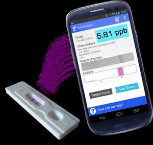
ACKNOWLEDGEMENTS
           The financial support of the Deutsche Gesellschaft für
           Internationale Zusammenarbeit (GIZ) GmbH, Sector Project
           “Sustainable Agriculture” for the desk study is gratefully
           acknowledged. Special thanks go to Mr. Karl Moosman,
           Advisor, Sector Project „Sustainable Agriculture“, Agricultural
           Policy, Food Security, Sustainable Management of Resources
           of GIZ, for making this collaboration possible, for his
           unreserved support, excellent review comments and strong
           desire to see this work reach end users, without which
           this work would not have been materialized. We thank
           Prof. Florian J. Schweigert and Kristine Wolf for their highly
           professional and committed work and for accommodating
           multiple requests. The printing cost of this publication is
           covered by the Partnership for Aflatoxin Control in Africa
           Phase II (PACA II) grant that African Union Commission
           received from the Bill and Melinda Gates Foundation.

IV
CONTENTS
Foreword                                                             III
Acknowledgements                                                     IV
1. Challenges of Mycotoxins and their Control                         1
  1.1 Health impacts of mycotoxins                                    1
  1.2 Prevention and reduction of mycotoxin contamination             2
  1.3 Maximum regulatory limits for major mycotoxins                  3
2. General Steps in the Analysis of Mycotoxins                        7
  2.1 Pre-analysis                                                    8
     2.1.1 Sampling and sample preparation                            8
     2.1.2 Extraction, clean-up and purification                      8
  2.2 Detection and quantification of mycotoxins (analysis step)     11
     1.2    State-of-the-art methods                                 11
     2.2.2 Conventional methods for rapid detection of mycotoxins    12
     2.2.3 Other methods in research use                             22
  2.3 Post-analysis                                                  23
3. Examples for Commercially Available Rapid Analysis Test Systems   25
  3.1 Test kits based on ELISA or LFD                                25
     3.1.1 Charm Sciences Inc.                                       25
     3.1.2 EnviroLogix Inc.                                          27
     3.1.3 Neogen Corporation                                        28
     3.1.4 R-Biopharm AG                                             30
     3.1.5 Romer Labs®                                               33
     3.1.6 VICAM                                                     34
  3.2 Test kits based on fluorescence polarization immunoassays      36
     3.2.1 Aokin AG                                                  36
     3.2.2 Diachemix Inc.                                            37
  3.3 Test kits based on fluorometry                                 38
     3.3.1 ToxiMet Ltd                                               38
4. Comparison between Selected Important Methods                     39
References                                                           41
Helpful links and facts                                              45

                                                                           V
VI
1. CHALLENGES                                              Contaminations with mold and mycotoxins can
                                                           occur pre- and/or post-harvest if conditions are

OF MYCOTOXINS
                                                           poor. Mycotoxins occurrences depend on improper
                                                           conditions with high humidity and temperature
                                                           after harvest and storage. Therefore, mycotoxin

AND THEIR
                                                           contamination is a major concern in tropical regions.
                                                           Because the compounds are chemically stable under
                                                           conditions usually present during food and feed

CONTROL
                                                           processing, they are found in raw materials as well as
                                                           in processed feedstuffs and foodstuffs. Due to their
                                                           stability they are also resistant to high temperature
                                                           and long-term storage. The common occurrences of
                                                           mycotoxins in foodstuffs and feedstuffs pose extensive
                                                           hazard for human and animal health (Hussein and
1.1 HEALTH IMPACTS OF MYCOTOXINS                           Brasel, 2001; Rai, 2012; Wild and Gong, 2010). The
                                                           great variability of mycotoxins in molecular structure
Mycotoxins remain an important global food safety          explains the great variations in clinical symptoms
issue. Many hundreds of natural, toxic secondary fungal    in humans and animals. Mycotoxins toxicity vary
metabolites that are collectively termed as mycotoxins     from one form to another but generally they are
have been identified from fungal cultures. Some of         acutely toxic, mutagenic, carcinogenic, teratogenic
them are observed in foodstuffs and feedstuffs around      estrogenic, and immune suppressants (Table 1).
the world. FAO has estimated that about 25% of global
crops are contaminated by molds and thus affected by       While in the developed world mycotoxin exposure
mycotoxins. The economic losses are estimated to be        has greatly been under control through stringent
billions of dollars (FAO, 2004). Of the many different     regulatory enforcement, the potential health
mycotoxins only a few specific mycotoxins (or groups)      implications of mycotoxins are still considerably high
present considerable food safety concerns. These           in developing countries. Reasons for this are the wide
agriculturally-important mycotoxins are aflatoxins,        spread occurrences of mycotoxins at frequently high
fumonisins, deoxynivalenol (DON), ochratoxin A (OTA)       levels and food consumption patterns that can result in
and zearalenone (ZEA) (Moss, 1991; Steyn, 1995).           large intake of a single cereal such as corn. Additional
Among them aflatoxins have a dominant role in terms        exacerbating factors on health impact are prevalent
of incidence in contaminated material.                     poverty and malnutrition (Shephard, 2008b).

Table 1: Toxic effect of important mycotoxins
 Mycotoxin                   Toxicity
 Aflatoxin                   pulmonary carcinogen, acute aflatoxicosis (Wild and Gong, 2010), liver carcinogen

 Deoxynivalenol (DON)        abdominal stress, increased salivation, malaise, diarrhea, emesis (Pestka, 2005)

 Fumonisin                   toxicosis in swine (Haschek et al., 2001), equine leukoencephalopathy (Marasas et
                             al., 2001)

 Ochratoxin (OTA)            nephrotoxic, hepatotoxic, teratogenic in rats (Abdel-Wahhab MA et al., 2005)

 T-2 and HT-2                nausea, emesis, abdominal pain, diarrhea, dermal necrosis (Omurtag and D.,
                             2001)

 Zearalenone (ZON)           change in the reproductive system of animals such as mice and rats, genotoxic
                             and/or carcinogenic (Food, 2000)

 Source: Rai , 2012

                                                                                                                      1
1.2 PREVENTION AND REDUCTION OF aflatoxin B1 < 0,5 ppb), co-inoculated with highly
toxic strains. Additionally, aflasafe™ (Figure 1) was
MYCOTOXIN CONTAMINATION developed from a partnership of the International
Institute of Tropical Agriculture (IITA) in Nigeria, the
Preventive methods in cultivation and harvest are Agricultural Research Service (ARS) of USDA and the
possible ways to reduce the challenges associated Universities of Bonn (Germany) and Ibadan (Nigeria).
with mycotoxin contamination of crops. In this It includes four native atoxigenic strains adapted to
context biocontrol techniques were investigated to various African countries or agroecosystems. Non-
prevent the spreading of toxigenic fungi in corn. Such germinating sorghum seed is inoculated with the
products are aflaguard® (www.syngenta-us.com) and atoxic strains (Aflasafe™) and thereafter, it should
aflasafe™ (www.aflasafe.com). Basically, atoxigenic be broadcasted to obtain a more even distribution
strains of Aspergillus flavus will be applied to the of the fu ngus across the field. Field tests in Nigeria
field and crowds out the toxigenic strains. Aflaguard and Senegal showed a reduction of aflatoxins in
includes the A. flavus AF36 which does not produce products of maize and groundnuts by 80 - 90%
aflatoxins. A single application reduces the toxigenic (Bandyopadhyay and Cotty, 2013). In relation to
strains in crops by over 80% and leads to a reduction research on aflasafe the “Deutsche Gesellschaft für
of aflatoxins (PJ, 2006). Further studies in the area Internationale Zusammenarbeit” (GIZ) carried out a
of such exclusion techniques exist such as Probst project in Nigeria in which the efficiency of Aflatoxins
et al. (2011) who investigated different isolated biocontrol technology in chili peppers was tested
strains of A. flavus and atoxigenic strains (LOD (GIZ, 2012-2013).
Figure 1: How aflasafe™ works in the field.
Courtesy of Ranajit Bandyopadhyay, IITA (www.iita.org, www.aflasafe.com)
SPORULATION ON MOIST SOIL SOIL COLONIZATION AND DISPLACEMENT
OF TOXIGENIC FUNGI
3-20 days
Insects
Spores
30-33 grains/m2
Wind
BROADCAST
@ 10 kg/ha 2-3 weeks
before flowering AFLASAFE
2Important for the prevention of contaminations
with mycotoxins is the fundamental knowledge in
1.3 MAXIMUM REGULATORY LIMITS
genomics, proteomics and metabolomics among
others - about the fungus and the mycotoxins. This
FOR MAJOR MYCOTOXINS
knowledge enables the development of extensive Ideally, official methods for detection and
prevention methods (Bhatnagar et al., 2008a; quantification of contaminants such as mycotoxins
Bhatnagar et al., 2008b). Genomic studies include in food are set for contaminants for which there are
research in the complete set of genes of an organism. accepted maximum limits (MLs). Different regions
Special methods such as the microarray- or the and countries have set MLs for different mycotoxins
expressed sequence tags (EST) technology could help in food. In Europe limits of 2 ppb (for aflatoxin B1)
to understand the life cycle and the metabolism of the and 4 ppb (for total aflatoxins (B1+B2+G1+G2)) for
fungus which produces certain mycotoxins (Bhatnagar cereals and cereal products (including maize and
et al., 2008b). maize products) for direct human consumption are in
place. Likewise, MLs of 5 ppb for aflatoxin B1 and 10
The study of the whole proteins of a cell as well as ppb for total aflatoxins are set for maize to be sorted
their structure and their functions in the physiological or otherwise processed physically before human
pathways of cells is termed as proteomics. consumption. The European Commission further set
Analytical methods to elucidate the proteom are a method for sampling of cereals and cereals products
the two-dimensional polyacrylamide electrophoresis in view of the prescribed limits. The regulated limits
(2D-PAGE) followed by a protein cleavage and of mycotoxins in the European region are defined
identification by mass spectrometry, particularly the in the regulation of the European Community EG-
matrix-assisted laser desorption-ionisation time-of- VO 1881/2006. Limits for selected mycotoxins are
flight (MALDI-TOF) mass spectrometry. Resistance summarized in Table 2. An abstract of the mycotoxin
associated proteins (RAPs) such as glyoxylase I regulations in food and feed in the United States is
which is a stress related aflatoxin resistance protein shown in Table 3.
(Bhatnagar et al., 2008b), have emerged by these
techniques as promising marker for crop varieties Codex Alimentarius Commission is responsible for
resistant to aflatoxins. setting maximum limits for mycotoxins in Food and
feed at the global level. The Codex Commission has
Metabolomic studies complete the understanding of already adopted MLs for mycotoxins as shown below:
the fungus and the synthesis of mycotoxins during
pre- and postharvest. Metabolomic investigations 1. A maximum level of 10 ppb for total aflatoxins
contain metabolites as the results of specific cellular in treenuts (almonds, hazelnuts, pistachios and
processes in biological organisms. shelled Brazil nuts) “ready to-eat” (CAC, 2017;
IITA, 2015).
With such studies (the field of “omics”) it is possible to
get important information about the contamination 2. ML of 15 ppb for total aflatoxins in peanuts and
of plants and crops with a fungus under special treenuts destined for further processing ” (CAC,
environmental conditions, improving understanding of 2017; IITA, 2015).
their metabolism and the biosynthesis of mycotoxins.
3. ML of 2000 ppb for fumonisins in maize and maize
In long-term orientation the investigations could
flour for direct human consumption (CAC, 2014)
generate knowledge and products to overcome
the contamination with mycotoxins (Bhatnagar et 4. ML of 4000 ppb for fumonisins in maize for further
al., 2008b). processing (CAC, 2014)
35. ML of 2000 ppb for deoxynivalenol in raw cereal aflatoxins (FAO, 2004). In the East Africa region limits
grains (wheat, maize and barley) (CAC, 2015) are set for aflatoxins and fumonisins (EAC Standards
Office, 2015; IITA, 2015). These are 5 ppb for aflatoxins
6. ML of 1000 ppb for deoxynivalenol in flour, B1 and 10 ppb for total aflatoxins and 2000 ppb for
semolina, meal and flakes derived from wheat, fumonisins in maize grain, maize flour, wheat grain,
maize and barley (CAC, 2015) wheat flour, milled rice, macaroni, spaghetti and
vermicelli, durum wheat semolina, finger millet flour,
7. ML of 200 ppb for deoxynivalenol in cereal-based maize gluten, groundnuts (peanuts), sorghum flour,
foods for infants and young children (CAC, 2015) pearl millet/bulbrush flour, dry beans, dry soybeans,
cassava wheat composite flour, composite flour,
Sampling and analysis methods for these MLs are
pearl millet grains, green grams, sorghum grains,
also prescribed by the Codex Commission. Countries
finger millet grains, faba beans, rough (paddy) rice,
in Africa who have set MLs for aflatoxins in food
brown rice, soya protein products and textured soya
prescribe 5 ppb for aflatoxin B1 and 10 ppb for total
protein products
Table 2: Important EU-maximum limits for various mycotoxins
Compound Commodity maximum level [ppb]
B1 total M1
Aflatoxins groundnuts, nuts, dried fruit and processed 2 4 -
products there of intended for direct human
consumption or use as an ingredient in foodstuffs
cereals (including buckwheat) and processed 2 4 -
products there of intended for direct human
consumption or as an ingredient in foodstuffs
rice, including brown rice (intended for direct 2 4 -
human consumption)
milk (raw milk, milk for the manufacturer of milk - - 0.05
based products and heat treated milk)
baby foods and processed cereal, cereal based foods 0.1 - -
for infants and young children
in feed: all feed materials 20 - -
Deoxynivalenol unprocessed cereals (excluding durum wheat, oats 1,250
and maize)
unprocessed maize 1,750
in feed: cereals and cereal products with the 8,000*
exception of maize by-products
4in feed: maize by-products 12,000*
Fumonisins unprocessed maize 4,000
maize and maize based foods intended for direct 1,000
human consumption
in feed: maize and maize based products 60,000*
Ochratoxin A unprocessed cereals 5
dried vine fruit (currants, raisins and sultanas) 10
roasted coffee beans and ground roasted coffee. 5
wine and fruit wine 2
baby foods and processed cereal based foods for 0.5
infants and young children
in feed: cereal and cereal products 250*
T-2/ HT-2 unprocessed cereals
- barley (including malting barley) and maize 200*
cereal grains for direct human consumption
- oats 200*
- maize 100*
breakfast cereals including formed cereal flakes 75*
Zearalenone unprocessed cereals other than maize 100
unprocessed maize 350
in feed: cereal and cereal products with the 2,000*
exception of maize by-products
in feed: maize by-products 3,000*
*guidance level
5Table 3: Important US-Maximum Limits for various mycotoxins
Compound Commodity maximum level [ppb]
B1 total M1
Aflatoxins all food except milk - 20 -
Milk - - 0.5
in feed: corn, corn products, cottonseed meal, and - 20 -
other animal feeds and feed ingredients intended for
dairy animals, for animal species or uses not specified
above, or when the intended use is not known
Deoxynivalenol finished wheat products for consumption by humans 1,000*
in feed: grains and grain by-products destined for 10,000*
ruminating beef and feedlot cattle older than 4
months and for chickens
Fumonisins degermed dry milled corn products (e.g. flaking grits, 2,000*
corn meal, corn flour with fat content of2. GENERAL STEPS
and purification and this is more or less independent
from the method later used for analytical and
quantification such as more instrumental methods
IN THE ANALYSIS
or screening methods (Anfossi et al., 2010; Burger et
al., 2014; Reiter et al., 2009; Shephard, 2008a, 2009;
Whitaker, 2003; Whitaker et al., 2005).
OF MYCOTOXINS Test systems for mycotoxins in food and feed
commodities involve a multiple-step process (Figure
2). Most methods consist of a pre-analytical step in
Fast, inexpensive, portable and reliable determination which the complex sample matrix is homogenized
methods are required for the analysis of mycotoxins and the mycotoxins are solubilized. The samples are
in food and feed – not only for the developing thus extracted, purified and/or enriched. Different
countries but also for farmers and the processing analytical approaches are then used for the detection
industry in developed countries. Techniques should be of individual mycotoxins or mixtures of mycotoxins.
reproducible, sensitive and simple for non-experts. Most In a final post-analysis step results are validated,
methods involve time consuming preparation, clean-up stored or exchanged.
Figure 2: Common sequence of procedures for mycotoxin analysis
matrices
MYCOTOXINS
sampling, extraction, purification, enrichment
PRE-ANALYSIS
HPLC-UV, ELISA, DIPSTICK, BIOSENSORS,
HPLC-FD, FLUORESCENCE INFRARED
LC-MS/MS, GC POLARIZATION SPECTROSCOPY, MIP,
IMMUNOASSAY ELECTRONIC NOSES
DETECTION/ ANALYSIS reference methods rapid methods emerging methods
saving and interpretation of the results, exchange
of data, hard/software help and control
POST-ANALYSIS
72.1 PRE-ANALYSIS Solid products, such as kernels and nuts must
be ground to powder form with a defined size to
enlarge the surface area and make them accessible
2.1.1 Sampling and sample preparation for extraction solvents. The equipment needed in
the process includes mills, grinder, sieves and filters.
The heterogeneous distribution of mycotoxins in cereals, Liquids and paste-like solutions must be gently
nuts, grains and other commodities takes an important stirred before extraction. Subsamples are then
role in the analysis of mycotoxins in food and feed. A taken for extraction and analysis (Beuchat, 1987;
number of wheat kernels can show low contamination International, 2000).
with mycotoxins but another single kernel from the
same unit may show a high contamination. Studies
in corn and peanuts suggest that in one lot only 2.1.2 Extraction, clean-up and purification
0.1% of the kernels are contaminated with e.g.
Based on the fact that the concentrations of mycotoxins
aflatoxin and the concentration of these kernels can
in samples are very low and the determination must
be extremely high. Because of this wide variation
be very sensitive, sample extraction, cleaning and
in concentration range in mycotoxins among a few
purification are needed. Final cleaned-up extracts
contaminated kernels in a lot, variability in replicated
can be concentrated by evaporating the solvents
sampling can be high (Biselli, 2006; Schatzki, 2000;
(e.g. under a nitrogen stream). Techniques in use for
Turner et al., 2009; Whitaker, 2003; Whitaker et al.,
extraction include liquid-liquid extraction and liquid-
2005). Adequate sampling and sample-preparation
solid extraction.
is needed to get a homogenous and representative
sample as basis for precise determination of the level
of contamination of mycotoxins. 2.1.2.1 Liquid-liquid extraction
A tool that provides support in analysing performance Liquid–liquid extraction (LLE) utilizes the solubility
of sampling plans, and determining the most or insolubility of mycotoxins in aqueous phase and
appropriate plan to meet user´s defined objectives, organic phase (hexane, cyclohexane). Non-polar
the Mycotoxin Sampling Tool (V 1.1) is available from substances such as lipids and cholesterol can be
the FAO website http://www.fstools.org/mycotoxins removed by this approach from the sample solution.
Table 4: Strength and weakness of liquid-liquid extraction (LLE).
Strength purification, decrease interfering substances
weakness - time consuming
- extraction effectiveness dependant on the type of matrix or mycotoxin
- loss of sample because of possible adsorption to the glassware
8There are different extraction methods for mycotoxins (Contaminants Bureau) (International, 2000). Briefly,
depending on the type of matrix as well as the in a glass-stoppered flask water, diatomaceous earth
chemical properties of the mycotoxin. High lipophilic and chloroform are added to a powdered sample.
or pigmented samples of food and feed require a After shaking the solution for 30 min it will be filtered
more complex extraction which includes both clean- and 50 ml of an extract will be loaded on a special
up and purification. Based on their chemical property prepared column. After loading the column with
aflatoxins will be extracted with mixtures of organic the extract, it should be washed with hexane and
solvents (acetone, hexane, chloroform, methanol). anhydrous ether. This fraction would be discarded.
Other mycotoxins like the fumonisins are more polar. Aflatoxin is eluted with a mixture of methanol-
The extraction of these must be done basically with a chloroform (3/97 v/v) and evaporated to dryness
combination of water and organic solvents (Holcomb under a gentle stream of nitrogen or in a water bath.
et al., 1992). It is also described, however, that a The residue will be used for the subsequent analysis.
combination of organic solvents with a small amount
of water improves the extraction of aflatoxins. An Another method is the BF-method (Best Food)
overview about the solvent of the choice for some (International, 2000). The mycotoxin is extracted with
mycotoxins is shown in Table 5. a mixture of methanol-water-hexane and separated
and extracted three times into chloroform. After
The generally accepted extraction method for evaporation to dryness it should be dissolved in a
the analysis of aflatoxins in peanuts and peanut solution of benzene-acetonitrile. Now it can be used in
products, oilseeds and food grains is the CB-method TLC or HPTLC (Jaimez et al., 2000; Richard et al., 1993).
Table 5: The most effective extraction solvents and alternatives for a number of
mycotoxins – reviewed and modified by Biselli (Biselli, 2006).
Mycotoxin Alternative
Aflatoxin methanol-water (80:20 v/v) acetonitrile-water, acetone-water
Ochratoxin A methanol-water methyl tert-butyl ether (MTBE),
acetonitrile-water
Fumonisin methanol-acetonitrile-water acetonitrile-water, methanol-water
(75:15 v/v)
Zearalenone methanol-water ethyl acetate, acetonitrile-water
(86:14 v/v)
92.1.2.2 Liquid-solid extraction Contaminants or the mycotoxin should be removed
with selected solvents. The method is also known
The liquid-solid extractions methodologies of as multifunctional column preparation and used as
mycotoxins are usually based on the principle of a preparation prior to HPLC-analysis (Figure 3).
chromatographic technique. These are unspecific
extraction methods such as solid phase extraction The specific IAC-extraction is the state-of-the-art
(SPE) or specific extraction such as immunoaffinity method of mycotoxin purification. It enables efficient
columns (IAC). and specific extraction of mycotoxins from different
kinds of commodities. The packing material of the
SPE uses small disposable cartridges packed column includes antibodies which specifically bind
with different stationary phases (e.g. silica gel, the analyte and allow the interfering substances to
octadecylsilane). A sample solution is loaded onto pass feely. The elution of the analyte is achieved with
the cartridge and rinsed under reduced pressure. an antibody denaturing solution.
Figure 3: Model for immunoaffinity column (IAC) (Courtesy of Tecna®, manufacturer
of mycotoxins ELISA kit - www.tecnalab.com).
Flow though Eluate
Analyte
Other compounds
10An alternative is the SPME (solid phase micro
extraction) approach. This is based on either
2.2 DETECTION AND QUANTIFICATION
Mycosep™ columns loaded with molecular imprinted
polymers (MIPs). Mycosep™ columns keep back the
OF MYCOTOXINS (ANALYSIS STEP)
contaminants of a sample but allow the mycotoxins
to pass (Egmond, 1986; Pittet, 2005; Turner et al., 1.2 State-of-the-art methods
2009). MIPs result in the application of imprinting
Reference methods for the quantitative and
techniques. At first, monomers are co-polymerized
qualitative determination of mycotoxins are basically
with a presented target analyte (imprint molecule).
chromatographic systems with different detection
The removal of the imprint molecule reveals a
systems such as HPLC-UV/DAD or LC-MS. They produce
molecular memory for the analyte in shape and
high resolution as well as sensitive and reproducible
size. The special synthetic cavity could be used to
results. They are the accepted methods for any testing
separate and to extract analytes from a sample. This
related to dispute resolution. The disadvantages of
extraction method is adapted to the pre-analysis of
these methods are that they are time consuming,
some mycotoxins such as ochratoxin A (Baggiani et
high in costs, sophisticated in equipment and with the
al., 2002; Jodlbauer et al., 2002; Turner et al., 2004;
need of expert scientific and technical knowledge. In
Zhou et al., 2004) or DON and ZON (Weiss et al.,
addition, they also require very extensive pre-analytic
2003)). It has promising advantages (stability, easy
clean-up steps prior to analysis. Instrumental methods
preparation, low cost) but has not been applied
are usually employed to confirm positive sampling
widely even on academic level.
results from screening methods. The basic principles
and the different applications have been widely
published and discussed (Chiavaro et al., 2001; Chu,
Table 6: Strength and weakness of solid 1992; Di Stefano et al., 2012; Holcomb et al., 1992;
Jansen et al., 1987; Kok, 1994; O’Mahony et al., 2013;
phase extraction (SPE).
Wilcox et al., 2015).
As the most sensitive and reliable method to
Strength • easy handling, low time
analyze mycotoxins in different commodities high-
expenditure per analysis,
performance-liquid-chromatography (HPLC) is denoted
no professional/expertise is
as the state-of-the-art analysis. Various HPLC methods
needed
have been developed for almost all major mycotoxins
• IAC: specific interactions in grains, cereals and other food and feed products.
between mycotoxins and Different validated HPLC-methods for the different
antibodies, only denaturing mycotoxins are reviewed (Gilbert and Anklam, 2002).
solvent is needed, robust, The focus of this study will be on rapid test methods
large volumes rather than laboratory based reference methods.
The Association of Analytical Communities (AOAC)
released a formal method for aflatoxin determination.
After preparation, the samples will be cleaned-up with
weakness • consistent method for all
immunoaffinity chromatography column (IAC column)
mycotoxins not available
followed by reversed phase - HPLC with post column
• MIPs: inconsistent molecular bromiation (LOD: Aflatoxin B1 1 ng/g, total Aflatoxin
recognition, limited number 2.4 ng/g) (Anklam et al., 2002; Stroka et al., 2000).
of re-use, sensibility of the Improvements in the detection tools of chromatographic
polymers techniques led to the more popular determination of
• IAC: costs, cross-reactivity, mycotoxins by HPLC-MS/MS, e.g. compared to HPLC-
influence antibody activity, FLD or GC-MS no sample derivatization is required.
use only once Adapted to the EU and national legislation HPLC-MS/
MS, is sensitive, indicates no cross-reactivity and gives
the possibility of multiple analysis.
11However, under less controlled conditions al., 2013; Espinosa-Calderón et al., 2011; Hajslova et
measurement procedures must be reliable and al., 2011; Manetta, 2011; Rai, 2012; Shephard, 2016;
sensitive to be able to control levels set by legislation Yazdanpanah, 2011).
but also easy and just-in-time for use in the field.
Therefore, in the last years investigations in rapid
and simple techniques have become increasingly 2.2.2 Conventional methods for rapid
important. Several more sensitive, specific and simple detection of mycotoxins
methods for mycotoxin detection are commercially Conventional systems which are commercially available
available and are summarized under the term for rapid detection of mycotoxins are enzyme-linked
screening methods. These include enzyme-linked immunosorbent assay (ELISA), lateral flow detection
immunosorbent assay (ELISA), lateral flow detection (LFD) and fluorescence polarization immunoassay
(LFD), fluorescence polarization immunoassay (FPI) (FPI). In some cases, basic fluorometric measurements
and thin layer chromatography (TLC). The different are used to detect and quantify mycotoxins in food or
methodological approaches again have been widely feed. The strength and the weakness of these tests are
reviewed (Anfossi et al., 2016; Contreras-Medina et shown in Table 7 giving a first overview.
Table 7: Strength and weakness of enzyme-linked immunosorbent assay (ELISA),
lateral flow detection (LFD) or fluorescence polarization immunoassay (FPI).
ELISA LFD FPI
Strength • easy handling • easy handling • easy handling
• low expenditure at time • rapid • low time expenditure per
sample
• sensitive • portable
• sensitive
• multiple analysis • no special
• portable
• equipment
• quantitative and
qualitative
weakness • cross-reactivity • not qualitative • high costs
• false-positive because of • high costs • currently only for certain
matrix disruptions mycotoxins available - in
research
• high costs
12Enzyme-linked immunosorbent assay (ELISA) disposable membrane-based test, microtiter plate
and tube assays).
This assay enables the qualitative, semi-quantitative
and quantitative determination of mycotoxins in The basic ELISAs are competitive assays. Here a
food and feed. The principle is based on the use of conjugate of an enzyme-coupled mycotoxin or a
antibodies and specific color changes. Different primary antibody specific for the toxin analyte is
forms of ELISA are commercially available (e.g. single used (Figure 4).
Figure 4: Principle of a competitive ELISA to screen mycotoxin (Courtesy of Tecna®,
manufacturer of mycotoxins ELISA kit - www.tecnalab.com).
Specific anti-analyte antibody
Enzyme
conjugate Washing step: Any
unbound compound
Analyte is removed
Coated
antibodies
1st incubation: 2nd incubation:
competition & solid enzymatic conversion
phase binding of the chromogen in
to a blue product
13The experiment set-up uses a microtiter plate which concentration of this signal is inversely proportional
is coated with a mycotoxin-specific-immobilized to the concentration of the mycotoxin in the sample.
antibody. In the first step, a mycotoxin linked with
an enzyme is added to a sample. This mixture should Lateral flow detection (LFD)
be applied on the microtiter plate. The amount
of the mycotoxin-linked enzyme that binds to the The lateral flow detection is a form of an
antibody on the plate depends on the amount immunoassay on a strip to detect the presence or
of mycotoxins in the sample (e.g. the higher the absence of the analyte in a sample. They are often
amount of mycotoxins in a sample, the lower will be called “dipstick”-tests. At first a pre-conditioned
the amount of the mycotoxin enzyme conjugate). In strip is wetted. Then the extracted sample should
the final step, the substrate of the enzyme is added be applied and after running, the strip shows the
which leads to a chromogenic detectable signal. The results visually or using a special reader (Figure 5).
Figure 5: Model of a competitive lateral flow detection-dipstick (Courtesy of Tecna®,
manufacturer of mycotoxins ELISA kit - www.tecnalab.com).
Uncontaminated sample
Test Line Control Line
Since no contaminant is
presented, the tracer is C
captured by the test line
T
Contaminated sample
Test Line Control Line
The contamimant captures
the tracer, so no or weak, C
test line is present
T
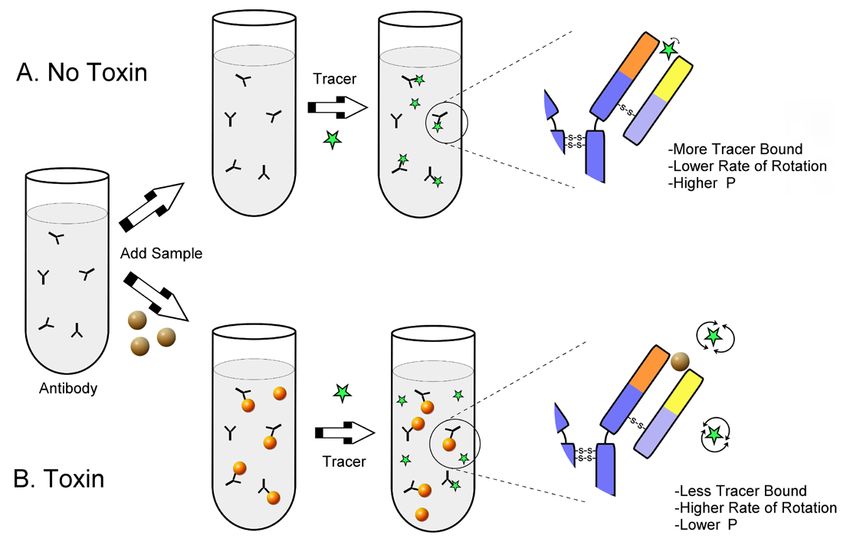
14Fluorescence polarization polarization reading instruments (e.g. the portable
immunoassay (FPI) Sentry200 from Ellie LLC/ Diachemix).
This is a newly developed immunoassay based on the The deficiency of such assays is the problem of cross-
indirect measurement of the changes of molecule reactivity which is not completely deleted and hence
rotation in a solution (Figure 6). There are only two further research is needed to evaluate this influence.
suppliers of FPI to determine a specific and limited Cross-reactivity is a general problem of immune
number of mycotoxins. methods that clean-up or determine mycotoxins.
Antigen-antibody reactions with metabolites or
Basically, a fluorochrome labeled mycotoxin with derivates of mycotoxins could not be eliminated in all
a low molecular weight acts as the antigen. The immune methods. IACs to clean-up deoxynivalenol
aggregation with the anti-mycotoxin antibody results (DON) and zearalenone (ZON) has been studies
in the formation of an immune complex, gaining in with regard to the cross-reactivity of antibodies for
weight and therefore slowing the rotation rate of the conjugated mycotoxin forms such as glycosides or
molecule. That causes an increase in polarization of acetylated forms (Tangni et al., 2010; Versilovskis et
emitted light which can be detected by fluorescence al., 2011)
Figure 6: Principle of fluorescence polarization immunoassay (FPI). A: Binding
the receptor molecule increases the weight of the fluorescence labeled ligand and
slowing its rotation which generates a polarization of light; B: With fewer receptors
bound, the rapid rotation of the fluorescence labeled ligand leads to a depolarization
of light (Maragos, 2009).
152.2.2.1 Thin layer chromatography (TLC) needs a well-controlled laboratory environment and
skilled laboratory personnel.
The first and well-established method for separation
and quantifying mycotoxins is the thin layer Various conditions may affect the result of the TLC
chromatography (TLC). TLC provides a less expensive analysis (Karunyavanij, 1991). For example, there
alternative to other LC-based methods. Especially are different coatings and binders for the plates
in developing countries it has an important role for depending on the analyte. The plate itself could be
surveillance purposes and control of regulatory glass, aluminum or plastic. Other factors are the purity
limit values (Gilbert and Anklam, 2002). Extensive of the standards, the manner of spotting the plate
investigations in the field of TLC led to highly sensitive and the development of samples as a chromatogram.
and good separating methods with relatively little The determination of the results can be visual or with
technical and methodological efforts (developing densitometry. Different spraying after developing the
tank, coated plate, UV-detector). In summary, after plate can enhance the visual effects. For example it
preparation of the sample it is spotted along with has been shown that a reaction of sterigmatocystin
standards on to a silica gel plate. It is then separated with aluminum chloride on the plate increases the
in a tank with mobile phase e.g. chloroform:acetyl fluorescence intensity up to 100-fold (Stack and
(9:1, v:v) or diethyl ether:methanol:water (96:3:1, Rodricks, 1971) . Other authors reviewed color
v:v:v). After developing the determination can be reaction with iodine starch or Fast Corinth V to get
achieved with long-wave UV-light (Holcomb et al., more sensitive results in the analysis of e.g. ZON
1992). Various applications of TLC are described (Turner et al., 2009). Lin et al. (1998) summarized
(Turner et al., 2009). Both one-dimensional and two- different detection techniques after TLC: UV-light of
dimensional TLC are used for quantitative and semi- long or short wavelength, fluorescence quencher,
quantitative determination of mycotoxins (Lin et al., autoradiography, vaporing of iodine or ammonium or
1998). Despite its ease and simplicity, the method exposition to X-ray (Lin et al., 1998).
Table 8: Detection limits for TLC-methods from www.eurofinsus.com.
Compound limit of detection [ppb]
aflatoxin B1, B2, G1, G2 2
ochratoxin A 200
T-2 10
Zearalenone 100
Table 9: Strength and weakness of thin layer chromatography (TLC).
TLC
strength • multiple analysis (Lin et al., 1998; Turner et al., 2009)
• cost efficient (Espinosa-Calderón et al., 2011; Turner et al., 2009)
• LOD: EU and US P
• rapid (Espinosa-Calderón et al., 2011; Lin et al., 1998)
• repeatable (Espinosa-Calderón et al., 2011)
• little or no clean-up (Pittet, 2005)
• no interfering of the mobile phase (Espinosa-Calderón et al., 2011)
weakness • solutions, reagents for coloring or enhancing the fluorescence
• increase of the costs when IAC is used for purification of the samples
16Fluorescence methods Pearson et al. (2004) investigated a high-speed
dual-wavelength sorting to reduce the aflatoxin and
Non-specific fluorescence methods
fumonisin contamination in yellow corn. The study
Basic rapid and easy to handle determination methods
was based on a sorting with near-infrared (NIR)
of mycotoxins commonly use the physicochemical
reflectance spectra (500-1.700 nm). A reduction
natures of mycotoxins e.g. the ability to stimulate the
of aflatoxin contamination with an average of 82%
autofluorescence. Tests like the “black light” test, also
(level of aflatoxins in corn > 10 ppb) and an average
called Bright Greenish Yellow Fluorescent (BGYF) test,
of 38% (level of aflatoxins in corn < 10 ppb) by high-
show mycotoxin producing fungi-infections in samples
speed sorting (filters at 750 nm and 1,200 nm) has
under UV-light (365 nm) rapidly, indirectly, with low
been reported (Pearson et al., 2004). A further study
equipment investment. However, these tests are not
using NIR in optical sorting showed the classification
specific. This analytical principle is used in automated
accuracies in post-harvest detection and removal of
sorting systems e.g. SORTEX from Bühler GmbH
aflatoxins and fumonisins contaminated maize kernel
(Germany) (www.buhlergroup.com ). On the basis of
(Wicklow and Pearson, 2006). Another rapid and
color or other optical properties, contaminated kernels
visual method described by Atas and coworkers used
and foreign materials are identified and separated from
hyperspectral imaging with UV and halogen excitation
the stream of seeds (Figure 7). Bühler provides different
to differentiate between aflatoxin contaminated and
SORTEX applications depending on the commodity to be
non-contaminated chili peppers (Atas et al., 2012).
analyzed e.g. SORTEX Z+ for rice, grain and beans.
The application of these optical techniques is
Other machines are the Detox Aflatoxin Laser Sorter
still limited to screening purposes due to high
from Best and the Nimbus sorting machine from TOMRA.
matrix dependence and the lack of appropriate
It makes it possible to detect aflatoxins in various grains
calibration materials.
and combines various lasers for detection.
Figure 7: Picture of seeds in the “black light” test. A Bright-Green-Yellow Fluorescence
(BGYF) will be reflected from Aspergillus flavus- infected seeds. The fluorescence is the
result of the reaction of kojic acid (a fungal metabolite) and a host peroxidase.
Courtesy Peter J. Cotty Agricultural Research Service, United States Department of Agriculture, School of Plant
Sciences, University of Arizona, Tucson http://cals.arizona.edu/research/cottylab/research/epidemiology.html
17Fluorescence and column separation is widely used in analysis of feed and food. Magnesium
silicate is also used as filler material and parting agent
In the early stages scientists introduced a by the industry (E 553a).
minicolumn technique to detect aflatoxin in peanuts
based on the principle of thin layer chromatography In principle, a small glass column is packed with various
(TLC) (Holaday, 1981). Contrary to TLC this method layers but generally including magnesium silicate. For
was faster and easier to use. A glass column (Table example, for sterigmatocystin analysis the minicolumn
11: Holaday “Dip” column) was packed with a is stuffed with glass wool and stacked with anhydrous
glass fiber plug to hold the packing material, silica sodium sulphate, neutral alumina, Florisil® and again
gel and another glass fiber plug and placed in a anhydrous sodium sulphate (Ramakrishna and Bhat,
beaker containing a developing solvent with the 1990). The column is purged with different organic
sample filtrate. After developing the minicolumn solvents (e.g. dichlormethane, hexane) under gravity.
was removed and determination was based on Thereafter, a methanol-sample solution is rinsed
visualization under UV-light (e.g. aflatoxins - blue or through the minicolumn. The mycotoxin adsorbs to
bluish - green color). The detection limit was 10 ppb. the layer in the column and can be detected under
UV-light. The determination is done by comparing the
Further developments led to the official technique column with a column treated the same way and with a
the Romer minicolumn (Table 11) which is packed standard only. They are also called “go-no-go” methods
with Florisil®. Florisil® is a magnesium silicate (MgO because of their semi-quantitative or quantitative but
– SiOH 15:85) with a particle size of 150 to 250 µm. It less sensitive determination (Egmond, 1986).
Table 10: Strength and weakness of Romer minicolumn method.
Romer minicolumn method
Strength rapid, little equipment and amounts of solutions, easy handling, no special
scientific knowledge
weakness sample preparation, less sensitive, less selective, only semi-quantitative, high
LOD (limit of detection)
18Table 11: Investigations in minicolumn methods to analyze mycotoxins. (Egmond,
1986; Holaday, 1981; International, 2000; Shotwell, 1983).
Holaday “Dip“ column (1968) Velasco column (1972) Romer minicolumn (official method
975.36 AACC-AOAC method)
detection limit 10 ppb detection limit 5 ppb detection limit 5-15 ppb
25 min > 15 min -
100 mm
glass fiber
150 mm
silica glass woll
alumina neutral
glass fiber
silica
4 mm glass woll
250 mm
Florisil®
Sand to pass No.30 sieve calcium sulfate
glass woll alumina neutral
5 mm
silica
Florisil®
calcium sulfate
glass woll
5 mm
• extraction with chloroform/ • clean-up with ferric chloride • extraction with acetone/water
acetone (97:3 v/v) solution (pH 4,6) (85:15 v/v) and filtering
• extraction with acetone/ • purification with sodium
water (85:15 v/v) hydroxide, ferric chloride and
chloroform
• sample in chloroform/acetone
(9:1 v/v) drain by gravity
through MC
• blue band 10 mm from • blue band in the interface of • blue band at the top of Florisil®
the lower end of the micro silica-Florisil® under UV light under UV light
column under UV light
19Fluorescence in solution used for the extraction followed by IAC (Chiavaro
Another fluorometric method to analyze mycotoxins et al., 2002). Determination was performed using a
is solution fluorometry. After extraction and clean- up xenon-lamp fluorometer from VICAM. The results
with IAC or SPE the elute will be filled in a cuvette, corresponded well to a reference RP-HPLC-method.
derivatized with e.g. bromine and then measured
with a fluorometer (Jansen et al., 1987). In one study, Malone et al. (2000) describes a similar method for
a sodium bicarbonate solution-methanol mixture was the quantification of aflatoxins in grains and raw
peanuts in comparison to LC-analysis and the results
were in good agreement. According to this method,
Table 12: Strength and weakness of
the fluorescence of the mycotoxins is enhanced by
solution fluorometry. bromine-derivatization.
Solution fluorometry
Labeling and derivatization
Strength rapid, easy handling, inexpensive, Combining enhancement of fluorescence and better
sensitive, multiple analysis separation of mycotoxins with add-on substances
is widely used in toxin analysis to receive more
Weakness derivatization, fresh derivatization
sensitive results. Methodological approaches using
solution every day, equipment,
different labeling or derivatization strategies are
calibration of the system
summarized in Table 13.
necessary
Table 13: Labeling or derivatization-methods to improve the results of
mycotoxin analysis.
Labeling/ derivatization Contact point Compound Reference
1,2-diamino-4,5- pre-column Monoliformin (Filek and Lindner, 1996)
dichlorbenze (DDB)
Iodine post-column Aflatoxins (Jansen et al., 1987; Lemke et al.,
1988; Shotwell, 1983)
Fluorescein T-2/ HT-2 (Lippolis et al., 2011; Maragos and
Thompson, 1999; Thompson and
Maragos, 1996)
Bromine post-column Aflatoxin (Espinosa-Calderón et al., 2011;
Stroka et al., 2000; Yuan, 2011)
Cyclodextrin Aflatoxins, DON, (Espinosa-Calderón et al., 2011;
ZON Francis et al., 1988; Galaverna et
al., 2008)
aluminium chloride TLC plate DON, (Egmond, 1986)
Sterigmatocystin
spraying on TLC-plate
and heating
trifluoroacetic acid pre-column Aflatoxins (Egmond, 1986; Espinosa-Calderón
et al., 2011)
o-phthalaldehyde pre-column Fumonisin (Shephard et al., 1990)
20Derivatization with fluoresceine, trifluoroacetic- Maragos et al. (2008) investigate a fluorometric
acid, iodine, bromine method to detect the non-fluorescence T-2 in maize.
The tracer fluorescein can be used in a fluorescence They derived T-2 with pyrene-1-carbonyl cyanide (T-2-
polarization immunoassay for the determination of Pyr) and studied the enhancement of the fluorescence
T-2 and HT-2-toxins in wheat. The labeling not only by adding different CD´s as buffer modifier in capillary
increases the sensitivity of the test system but also electrophoresis laser-induced-fluorescence. The
shortens the clean-up procedures and the incubation- most effective CD was heptakis (2,6-di-O-methyl)-b-
time (Lippolis et al., 2011). Other techniques are cyclodextrin (DIMEB) (Maragos et al., 2008).
the pre- and post-column derivatization with
trifluoroacetic-acid, iodine or bromine (Espinosa-
Calderón et al., 2011). Especially in the case of Specific fluorescence methods
bromine derivatization electrochemical cells can be
Another fluorescence method to analyze mycotoxins
used. Here the strong oxidizer bromine is induced by
is the fluorescence polarization immunoassay (FPI) as
what is known as KOBRA® cell. Jansen et al. (1987)
a rapid screening test. This method is based on the
show a 20-fold increase of the fluorescence intensity
indirect measurement of the changes in molecule
of aflatoxin B1 and G1 with post-column iodine
rotation of a solution as a function of the size of the
derivatization.
molecules. It is one of the methods which are actually
Derivatization with cyclodextrin pretty much in use in research and it shows great
A new and promising substance that combines the promises in the field of rapid, sensitive analysis of
selective separation with the enhancement of native mycotoxins. Commercial available test kits are from
fluorescence of mycotoxins is cyclodextrin (CD). The Diachemix and Aokin AG but only for a limited number
cyclic oligosaccharides are formed by 6(a-CD), 7 (b- of mycotoxins. The problem of cross-reactivity is not
CD), 8 (g-CD) glucose units linked by a-1,4-glycosidic yet completely solved.
bonds. They are cone-shaped. Besides their good
In principle, the fluorochrome labeled mycotoxin
solubility in water and dipolar solvents they are able
with a low molecular weight acts as the antigen.
to form inclusion-complexes as host for a wide range
Aggregation with the anti-mycotoxin antibody results
of hydrophobic compounds (guest). The complexation
in the formation of an immune complex, gaining in
affected the guest´s solubility, stability, physical and
weight and slowing the rotation rate of the molecule.
chemical properties. The inexpensive substance is
This causes an increase in polarization of emitted light
widely used in pharmaceutical products (solubility,
which can be detected by fluorescence polarization
stability) and in the textile industry (masking odours)
reading instruments.
(Galaverna et al., 2008).
The forming of an inclusion complex between Table 14: Strength and weakness of
different mycotoxins and cyclodextrins leads to fluorescence polarization immunoassay
an enhancement of the native fluorescence of
(FPI).
mycotoxins. This is described as result of the
interaction of the coumarin structured mycotoxins FPI
and cyclodextrin. The inclusion results not only in
changes of the polarity and intermolecular rotation Strength • easy handling
but also in interaction with quenchers (Galaverna et
al., 2008). Cucci et al. (2007) described a method to • portable
analyze aflatoxin M1 in milk with the use of b-CD.
After cyclodextrin was added the detection limit of
Weakness • in research
analysis were decreased from 25 ng/l to 5 ng/l. In
addition there was no need to clean-up the samples • expensive equipment
with IAC before analysis (Cucci et al., 2007).
212.2.3 Other methods in research use One biosensor method to determine mycotoxins
is surface plasmon resonance (SPR) (Gaag et al.,
2003; Schnerr et al., 2002; Tudos et al., 2003).
Laser-induced fluorescence (LIF)
Here the measured variable is the change in mass
Espinosa-Calderón et al. (2011) reviewed publications of mycotoxins which are immobilized at a surface
concerning the laser-induced fluorescence (LIF) as a of a sensor chip. The mass change results in the
fluorescence screening method for mycotoxins. The attachment of a specific antibody to the mycotoxins.
method is based on the detection of the analyte in Results have shown to be comparable to LC-MS
the mobile phase while passing through the detection and the sensor chip can be reused without loss
window of the LIF-detector. This method enables the of activity up to 500 times. Such SPR biosensor
analysis of samples with very low concentrations. protocols are described by Puiu et al. (2012) for
Because of the high costs for the LIF (laser, special the direct measurement of albumin-bound AFB1 in
dyes for labeling) however, this method is not widely blood samples.
used.
DNA-based and aptamer-based biosensors
Near infrared spectroscopy (NIR)
Dinckaya et al. (2011) published a DNA biosensor-
Another method used in practice and in research based method to analyze aflatoxin M1 in samples
is the near infrared spectroscopy (NIR). Petterson such as in milk. A thiol-modified single stranded DNA
and Aberg (2003) described the determination of (ss-HSDNA) probe was immobilized on a monolayer
deoxynivalenol in wheat kernel with a wavelength of cysteamine and gold nanoparticles prepared on
570 – 1.100 nm and the detection limit was 400 ppt. gold electrodes. The DNA biosensor particularly
NIR can be used for the determination of aflatoxins bound aflatoxin M1. The detection of the process
at levels between 200-500 ppb in sample with solid is carried out with electrochemical impedance
or liquid physical conditions. However, this method spectroscopy (EIS) and cyclic voltammetry (CV)
is not yet established for the detection of aflatoxin techniques. But there is no information if the
in human food at regulatory levels (Jagger et al., method is assignable within the aflatoxin M1
2013). limits of the national and international legislations
(Dinckaya et al., 2011).
Biosensor techniques Another form to use DNA in biosensors is aptamer-
based. Aptamers are peptide molecules or DNA
In the last decade, different immunochemical or RNA duplex structures that can bind a specific
assays and assays including biosensor techniques analyte. Chen et al. (2012) investigated a DNA
are investigated. Biosensors enable the detection of duplex structure with an anti-ochratoxin A-aptamer
an analyte in a sample because of the interaction including a fluorophore and a quencher. Binding
between the analyte and a biological sensitive ochratoxin A to this structure leads to an increase
element e.g. enzyme, tissues, nucleic acids or of the fluorescence. With this rapid and highly
antibodies. The interaction results in a signal which selective method (only 1 min per measurement)
can be detected by a transducer (e.g. optical or OTA can be determined with a limit of detection of
physicochemical detection) and is transformed in a 0.8 ppb (Chen et al., 2012). A similar method, the
utilizable measured variable. biosensor DNA-enzyme aptamer was described by
22Yang et al. (2012). The presence of OTA bound to the
DNAzyme hairpin leads to open the hairpin structure
2.3 POST-ANALYSIS
and activates a horseradish peroxidase-mimicking Companies that market commercially available rapid
DNAzyme. This process can be detected with screening assays usually also provide portable reader,
colorimetric measurement at 620 nm in microtiter fluorometer or fluorescence polarization reading
wells (Yang et al., 2012). Other aptamer-sensor-based instruments. Most of the readers allow for primary
assays are currently under investigation (Prabhakar et analysis with the possibility to exchange data or they
al., 2011; Wu et al., 2011). are linked to PC with special software.
A new way of post-analysis is the use of smart
Electronic nose
phones for quantification of aflatoxins in the field.
A new analytic approach based on biosensors are The app measures aflatoxins using a phone image of
electronic noses that open a new field for the rapid a color-changing test strip. The developer claims the
non-destructive analysis of mycotoxins (Cheli et measuring is more accurate than immunoassay tests.
al., 2007; Dell’Orto et al., 2007; Olsson et al., 2002; The results can be geo-tagged and uploaded to the
Tognon et al., 2005). The electronic nose, an array internet. The cloud application allows secure data
of biosensors detecting volatiles emanating, could storage, information management and compliance
distinguish between the presence and the absence of reporting [ www.mobileassay.com ].
the mycotoxins. The fungal growth and the production
of mycotoxins lead to biochemical changes resulting
in changes in the chemical composition of volatiles. Figure 8: Quantification of aflatoxin in
Different volatile molecules act within the electronic the field with a smart phone. Courtesy
nose and generate a special detectable electronic www.mobileassay.com
signal. Changes in the relative composition of the
molecules lead to changes in the electronic signal.
The investigation of Cheli et al. (2009) showed that
electronic noses can differentiate between aflatoxins
-positive and aflatoxins–negative samples but further
quantitative analysis are needed to evaluate the
real potential as reliable method in the practical
mycotoxins analysis (Cheli et al., 2009).
23You can also read