Neurotrauma: 2021 update - Daniel P. Perl
←
→
Page content transcription
If your browser does not render page correctly, please read the page content below
Free Neuropathology 2:4 (2021)                                                                                                                                                         Daniel P. Perl
doi: https://doi.org/10.17879/freeneuropathology‐2021‐3264                                                                                                                               page 1 of 8

                                                                                                                                                                                       Review

Neurotrauma: 2021 update
Daniel P. Perl

J. Edward Hebert School of Medicine, Uniformed Services University, Bethesda, MD, USA

Corresponding author:
Daniel P. Perl ∙ J. Edward Hebert School of Medicine ∙ Uniformed Services University ∙ Bethesda, MD 20814 ∙ USA
daniel.perl@usuhs.edu

Submitted: 12 February 2021                      ∙     Accepted: 09 March 2021                      ∙      Copyedited by: Shino Magaki                        ∙     Published: 15 March 2021

Abstract
Despite the interruptions and restrictions to the progress of science that the COVID‐19 pandemic has introduced,
2020 was marked by a number of important advances in the field of neurotrauma. Here, I will highlight what I
believe are among the most important contributions. This year there were notable advances towards providing
clinically useful information on neurotrauma outcome through the use of fluid biomarkers. I also introduce fas‐
cinating approaches to studying the role of microglia in nervous system repair and neuroinflammatory mecha‐
nisms leading to dysfunction through the use of colony‐stimulating factor 1 receptor inhibitors, especially
Plexxikon 5622 (PLX5622). Oral administration of this compound is able to deplete microglial elements and then,
following withdrawal from the drug, a new population of microglia then repopulates the brain. Use of this ap‐
proach in traumatic brain injury experimental models has produced important insights into the pathogenetic role
of microglia in responding to this process. Important new data on the nature and distribution of tau involvement
of neurons and astrocytes in cases of chronic traumatic encephalopathy (CTE) also appeared suggesting differ‐
ences and similarities to Alzheimer’s disease. Additionally, the use of tau‐specific PET scan ligands in at‐risk pop‐
ulations has suggested that this approach may be able to identify cases with CTE. Lastly, we note the death in
the past year of a major contributor to the field of neurotrauma neuropathology, Professor J. Hume Adams.

Keywords: Traumatic brain injury, Chronic traumatic encephalopathy, Biomarkers, Neuroinflammation, Microglia, Tau

Introduction                                                                                                that appeared during 2020, it is hard to see any sig‐
                                                                                                            nificant slowing of progress. I have the impression
      For virtually all of us, 2020 will mostly be re‐                                                      that with most scientists working within a telecom‐
membered as the year of COVID‐19. With numerous                                                             muting mode, rather than being active in the lab,
calls for mask wearing, social distancing, partial‐to‐                                                      many took the time to get to accumulated data wait‐
complete shutdowns of offices and laboratories (in‐                                                         ing to be analyzed, write papers waiting to be writ‐
cluding our own) and other similar restrictions, this                                                       ten, etc., and thus scientific output did not appear to
pandemic has definitely had a dampening effect on                                                           slow, and possibly even increased. It might be pre‐
everyone’s scientific productivity. Nevertheless, by                                                        dicted that following the achievement of wide‐
viewing the traumatic brain injury (TBI) literature                                                         spread vaccination allowing for a final full return to

Copyright: © 2021 The author(s). This is an open access article distributed under the terms of the Creative Commons Attribution 4.0 International License (https://creativecommons.org/licenses/by/4.0/),
which permits unrestricted use, distribution, and reproduction in any medium, provided the original author and source are credited, a link to the Creative Commons license is provided, and any changes are
indicated. The Creative Commons Public Domain Dedication waiver (https://creativecommons.org/publicdomain/zero/1.0/) applies to the data made available in this article, unless otherwise stated.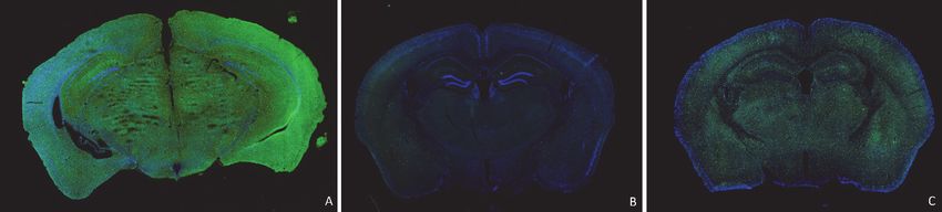
Free Neuropathology 2:4 (2021)                                                                          Daniel P. Perl
doi: https://doi.org/10.17879/freeneuropathology‐2021‐3264                                                page 2 of 8

our lab benches, there will likely be a lag in output        when the blood was drawn. The nature of that pro‐
as new experiments need to be put in place and ad‐           cess remains unclear, although in their discussion
ditional data collected. Time will tell. Nevertheless,       the authors noted literature pointing towards the el‐
in surveying the field of neurotrauma, many im‐              evation of NfL levels in association with axonal in‐
portant contributions were reported in 2020 and              jury. The use of isolated exosomes, brain‐derived
here I would like to pick some of what I believe were        membrane‐bound components that cross the
among the more important advances that were pub‐             blood‐brain‐barrier to enter the blood stream, is
lished. This is an individual viewpoint, reflecting my       particularly noteworthy. Since these exosomes can
own biases, and I will apologize, in advance, if I have      be linked back to a central nervous system origin, it
omitted any of your favorites on this year’s list. So,       suggests that this approach is capable of providing
among the more than 2,000 publications appearing             indicators of more specific brain‐related pathology
in the literature on neurotrauma in 2020, here are           and thus more closely reflect relevant biologic pro‐
those that I chose to highlight for you.                     cesses. Since the results were predictive for the de‐
                                                             velopment of depression and symptoms indicative
Contributions to the development of                          of PTSD, this result represents another piece of evi‐
                                                             dence for a biologic substrate of the long‐term per‐
fluid biomarkers for TBI: Finally some                       sistent behavioral consequences of TBI.
approaches that appear to be useful
                                                                    Further advances in the blood biomarker field
      Over the past few years, a large number of             were reported by Okonkwo et al. [2] in a study
groups have engaged in efforts to identify useful            where the predictive accuracy of determining glial
blood‐based biomarkers in the field of TBI that              fibrillary acidic protein (GFAP) levels was compared
hopefully would be able to reflect outcome, espe‐            with S100 calcium‐binding protein B (S100B) assays
cially with respect to predicting a transition to clini‐     in the blood of TBI patients. The U.S. Centers for Dis‐
cally persistent sequelae. By and large, the results of      ease Control and Prevention reports that each year
these efforts have been relatively inconsistent and          about 4,800,000 evaluations for TBI occur in emer‐
have not achieved robust clinical validity. However,         gency departments in the United States. About 80‐
in 2020 a group reported [1] that increases in exo‐          90% of these patients have mild forms of TBI (mTBI,
some and plasma levels of neurofilament light (NfL)          as defined by a Glasgow Coma Scale score of 13‐15)
chain protein in patients with repeated mild trau‐           and only 10% of the mTBI patients will subsequently
matic brain injuries (mTBIs) were predictive of sub‐         demonstrate abnormalities on computed tomogra‐
sequent development of chronic post‐concussion               phy (CT) studies of the head and thus need further
syndrome, post‐traumatic stress disorder (PTSD)              evaluation and monitoring. Accordingly, rapid relia‐
and depression. The study involved a cohort of 195           bly predictive studies are needed to lighten this
retired Service Members enrolled in the Chronic Ef‐          huge clinical diagnostic load and better identify
fects of Neurotrauma Consortium Longitudinal                 which patients need further CT scrutiny.
Study. The cohort received detailed assessments to
                                                                   In the United States, assays of serum GFAP
document and quantify episodes of prior mTBI and
                                                             have been approved by the U.S. Food and Drug Ad‐
were grouped into repetitive mTBI (3 or more epi‐
                                                             ministration (FDA) so that they can be used in a clin‐
sodes), 1‐2 mTBIs and controls with no TBI history.
                                                             ical setting to determine the need for a head CT
These assessments were obtained through inter‐
                                                             within 12 hours of an episode of mTBI. Alternatively,
views with the subjects. These data have particular
                                                             in Europe, approval has been granted for S100B as‐
importance, because they were obtained from re‐
                                                             says that are currently used to serve this clinical as‐
tired Service Members, and thus their TBI exposures
                                                             sessment function. Importantly, in the study now re‐
mostly occurred years prior to obtaining the bi‐
                                                             ported, plasma GFAP was determined using a newly
omarker assay.
                                                             developed point‐of‐care prototype assay that can be
     The results reported suggest that their assays          completed within 15 minutes. Serum S100B was de‐
are reflecting a chronic, ongoing pathologic process         termined by a more standard laboratory‐based ana‐
that extended from the mTBI exposures to the time            lytic method that requires transfer of the specimen
Free Neuropathology 2:4 (2021) Daniel P. Perl
doi: https://doi.org/10.17879/freeneuropathology‐2021‐3264 page 3 of 8
to the laboratory, adding considerably greater time tlefield locations. Assays on the playing field of con‐
to provide a report. The endpoint used for the 1,359 tact sport events might also find this approach quite
TBI patients in the study, who ranged from mild to attractive for clinicians involved in such evaluations.
more severe degrees of injury, was the ability to pre‐
dict the presence of structural abnormalities on a The role of microglia in repair of TBI
head CT scan. Using a predetermined cut‐off value
for serum GFAP of 22 pg/mL, this study found that Microglia are abundant cellular constituents of
GFAP levels were an excellent predictor of CT abnor‐ the brain that play important roles in brain develop‐
malities and could do so even on samples taken up ment, maintenance and the pathogenesis of most
to 24 hours from the time of trauma. GFAP assays disease states. We are taught to consider microglia
substantially outperformed that of S100B as a CT ab‐ as playing a central role in virtually all nervous sys‐
normality predictor. Further, the mean level of GFAP tem repair processes, including that of TBI. Indeed,
correlated with increasing numbers of distinct le‐ although the precise mechanism for their action re‐
sions identified on head CT, suggesting a dose re‐ mains unknown, microglia have been thought to
sponse. Indeed, patients with severe to moderate mediate ongoing neuroinflammatory damage to the
TBI (GCS 3‐12) had 10‐fold greater levels than the brain that is responsible for at least some of the
levels assayed on patients with mTBI (GCS 13‐15). long‐term functional sequelae of TBI and even as‐
Overall, the GFAP assay had a predictive sensi‐ pects of potential subsequent neurodegenerative
tivity of 0.987. It should be pointed out that the phenomena. Recently, a series of drugs have been
point‐of‐care assay, with its rapid reporting time (15 identified that inhibit colony‐stimulating factor 1 re‐
minutes) is much faster than the currently FDA ap‐ ceptor (CSF‐1R), which microglia depend upon for
proved lab‐based methodology and, by its nature, their survival. These drugs penetrate the brain after
can be performed in obscure locations without ac‐ oral administration and within two to three weeks
cess to laboratory facilities. It is my understanding are able to almost completely deplete the entire mi‐
that this new and faster technology is in the process croglial cellular compartment of the brain in an ex‐
of being evaluated by the FDA for approval as a clin‐ posed animal. Following withdrawal of the drug,
ical diagnostic tool. This promises to become an ex‐ there will be a steady repopulation of the brain by
tremely valuable approach for clinicians faced with microglia (Figure 1). Importantly, the repopulated
a staggering number of TBI patients to evaluate. A microglia appear to be a new rejuvenated popula‐
rapid point‐of‐care assay method will also be of tion of cells without a prior history of antigen expo‐
great value for military medicine where many TBIs sure. The primary agent used in such microglial re‐
are clinically evaluated downrange in isolated bat‐ population experiments is referred to as Plexxikon
(PLX) 5622.
Figure 1. Microglia are depicted in 8 weeks old CX3CR1‐GFP mice (microglia – green; DAPI – blue) A) control mouse, normal diet, B) 3
weeks following a diet containing 1200 mg/kg of PLX5622 (note virtual complete absence of microglia) and C) after 3 weeks on the PLX5622
diet then 2 weeks on the control diet (note the return of prominent green staining from the repopulated microglia). From ongoing studies
of K. Whiting and Z. Galdzicki of Uniformed Service University ‐ Neurosciences Program.Free Neuropathology 2:4 (2021) Daniel P. Perl
doi: https://doi.org/10.17879/freeneuropathology‐2021‐3264 page 4 of 8
The use of drugs such as PLX5622 have begun It may be predicted that the use of tools such
to be used to explore the specific roles that microglia as PLX5622 will greatly help in unraveling such is‐
play in the repair processes and sequelae of experi‐ sues. In the discussion, the authors wonder if
mental TBI, such as occurs with controlled cortical PLX5622 might even represent a clinically feasible
impact. Henry and colleagues [3] administered therapeutic approach to reducing some of the long‐
PLX5622 one month after controlled cortical impact term complications of TBI. PLX5622 has received
in mice and then withdrew the drug to allow for mi‐ limited FDA approval for clinical use in patients with
croglial repopulation. By 3 months post‐injury, they other non‐TBI indications. For those who are inter‐
noticed that the PLX5622 treated animals possessed ested in pursuing microglial depletion and repopula‐
a smaller cortical lesion, reduced hippocampal neu‐ tion, in a companion publication, this group has pro‐
ron cell death and decreased expression of NOX2 vided detailed protocols for such experiments in
and NLRP3 inflammasome‐associated neuroinflam‐ mice [5]. This would appear to be a valuable new
matory modulators, when compared to non‐treated tool for the investigation of the role of microglia,
animals receiving equivalent controlled cortical im‐ both positive and negative, in the pathogenesis of
pact injury. The PLX5622 treatment animals also not only neurotrauma but many other disease pro‐
showed improved long‐term motor and cognitive cesses [6, 7].
function. These intriguing experiments demon‐
strated that removal of microglia in the chronic Studies of the long‐term effects of sub‐
phase of TBI repair reduced subsequent neuroin‐ concussive blast exposure in breachers
flammation as well as lessening subsequent motor
and cognitive functional deficits. Further, it showed The effects of blast TBI on the brain have
that such inflammatory effects extended far longer mostly been studied either experimentally in small
than has been traditionally believed. animal models or in humans following a single sig‐
nificant blast event, typically related to exposure to
an improvised explosive device (IED). It should be
Willis and coworkers [4] also used PLX5622 for
kept in mind that, in the combat setting, blunt im‐
microglial depletion and repopulation in a mouse
pact TBI also commonly occurs in conjunction with
model of TBI (they too used closed cortical impact),
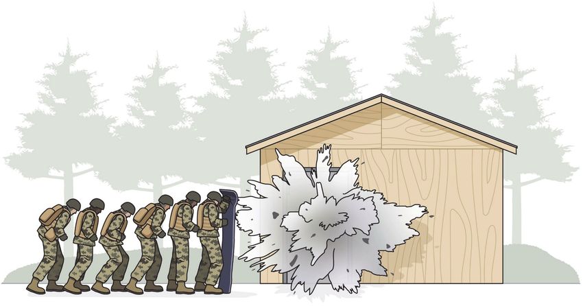
these blast injuries. Breaching represents a process
however these workers showed that removal of mi‐
where explosions are used to blast open doors and
croglia had little effect on the clinical outcome of
thus gain entry to buildings (Figure 2). As such,
their TBI model. Nevertheless, examination of the
breaching is associated with blast exposure in the
treated animals showed neuroprotective effects
absence of impact trauma. Stone and colleagues [8]
that appeared to aid in recovery. These beneficial ef‐
reported studies comparing a cohort of career
fects were mostly modulated through interleukin‐6
breachers (typically breaching instructors) to a
(IL‐6) signaling via the soluble IL‐6 receptor and its
matched but minimally exposed control group. Of
support of neurogenesis. These authors suggested
the 20 experienced breachers studied, they re‐
that the presence of activated microglia associated
ported having experienced an average of 4,628
with neurotrauma may not have a negative effect on
breaching blast exposures over their careers, as op‐
outcomes and that it would appear that, as they
posed to the control group (n=14) who experienced
state, “these cells lack an ability to support endoge‐
an average of 3 exposures. Keep in mind that these
nous repair processes.” Obviously, more needs to be
exposures are all considered to be sub‐concussive in
done to further dissect out the role that microglia
nature.
play in the brain’s response to TBI. The timing of
when the microglial removal and repopulation takes Using detailed neuroimaging approaches, they
place in these models appears to be critical to the found, somewhat surprisingly, evidence of a signifi‐
results obtained. cant degree of cerebral cortical thickening in the
breacher group. This change was widespread,
throughout the cerebral cortex. The nature of thisFree Neuropathology 2:4 (2021) Daniel P. Perl
doi: https://doi.org/10.17879/freeneuropathology‐2021‐3264 page 5 of 8
Figure 2. Breaching is a procedure whereby an explosive charge, typically placed on a door, is used by combatants to enter a building and
engage the enemy. Service Members participate in breaching in both training exercises and on the field of battle. Repeated training
exercises, such as portrayed here, expose participants to numerous sub‐concussive blast wave exposures, in the absence of impact TBI.
Courtesy of Sofia Echelmeyer.
cerebral cortical enlargement remains unclear, as no on moderate to severe TBI patients which have
neuropathologic studies of deceased career breach‐ demonstrated slightly elevated plasma cytokine lev‐
ers have yet to be reported. The authors also noted els in the acute phase post‐injury. Post‐mortem
differences between the heavily exposed and con‐ studies by Johnson and colleagues [9] have demon‐
trol groups related to regional blood flow, various strated that activated microglia can persist for dec‐
neuropsychological assessment results and serum ades after the initial traumatic incident. A recent
biomarkers, however none of these differences sur‐ study by Chaban and colleagues [10] published in
vived Bonferroni correction for multiple compari‐ 2020 looked at plasma levels of 12 different cyto‐
sons. This suggested that further studies using a kines in a cohort including 207 patients with mTBI
larger cohort would be instructive. Service Members and 82 matched uninjured community controls.
carrying out other duties, such as those who spend Plasma samples were drawn at admission for evalu‐
a career firing high caliber artillery ordinances or ex‐ ation of the TBI, as well as 2 weeks, 3 months and 12
plosive ordinance disposal personnel, may also have months following the injury. Brain magnetic reso‐
an equivalent degree of blast exposure, and it would nance imaging (MRI) was also performed on all the
appear that similar studies of these groups are war‐ participants. Comparing the mTBI group with the
ranted. controls, they found significant elevations in plasma
interferon gamma, IL‐8, macrophage inflammatory
TBl‐related neuroinflammatory mark‐ protein‐1 beta, monocyte chemoattractant protein‐
ers 1, IL‐17A, IL‐9, tumor necrosis factor, and basic fibro‐
blast growth factor at all time points, whereas a
Neuroinflammatory response to TBI is a subject number of the other cytokines remained un‐
that has received considerable attention in both ex‐ changed. The presence of persistent cytokine levels
perimental models and clinical settings. Clinically, did not correlate with the MRI findings, suggesting
studies of cytokine expression have mostly focused that the cytokine changes were not related to theFree Neuropathology 2:4 (2021) Daniel P. Perl
doi: https://doi.org/10.17879/freeneuropathology‐2021‐3264 page 6 of 8
extent of tissue damage. This is one of the few lon‐ Alzheimer’s disease and PART. In contrast, the tau‐
gitudinal studies of plasma neuroinflammatory positive astrocytes of CTE were indistinguishable
markers of patients with mTBI that showed evidence from those of ARTAG but distinctly different from
of persistent systemic inflammation lasting up to a the Alzheimer’s disease results. This led to the sug‐
year. gestion that CTE and ARTAG may share pathogenetic
mechanisms that separate these two diseases from
The nature of intracellular tau accumu‐ that of Alzheimer’s disease. Clearly, these are con‐
cepts that need to be explored further and may be
lations in cases of chronic traumatic of importance in sorting out the complex and con‐
encephalopathy (CTE) fusing nosology and diagnostic criteria for these var‐
ious neuropathologic entities. Obviously, additional
TBI, especially repeated impact TBI, constitutes work needs to be done and neuropathologists in‐
a risk factor for the development of chronic trau‐ volved in this area will follow it with interest.
matic encephalopathy (CTE), a tauopathy associated
with neurofibrillary tangles (NFTs) and tau accumu‐ Animal models of CTE and the use of
lation in astrocytes, primarily in the form of what are
PET ligand for tau for clinical diagnosis
referred to as thorn‐shaped astrocytes (TSAs). Arena
and colleagues [11] reported a detailed immuno‐ of CTE
histochemical study on the nature of tau accumula‐
Further on tau and its association with re‐
tions in NFTs and TSAs in cases of CTE and compared
peated TBI, Dickstein and colleagues [13] investi‐
them to staining results obtained in several other
gated various biomarkers in rats that were experi‐
forms of tau‐related neurodegenerative disorders
mentally exposed to repeated low‐level blast over‐
(Alzheimer’s disease, frontotemporal dementia,
pressures as well as military Service Members who
Pick’s disease) and other tau‐related conditions seen
had been exposed to IEDs on the battlefield and sub‐
in the elderly (age‐related tau astrogliopathy, or AR‐
sequently suffered persistent behavioral, cognitive
TAG, and primary age‐related tauopathy, or PART).
and/or memory complaints.1 In the rats, six weeks
This study showed that the NFTs of the CTE cases
following blast exposure, by Western blot, abnor‐
contained both 3R and 4R isoforms that are also
mally phosphorylated tau (Thr181, p‐tau) was
classically seen in association with the tangles of Alz‐
shown to be increased in the right anterior cortex
heimer’s disease and PART. The TSAs of CTE stained
and right hippocampus but not in contralateral loca‐
virtually entirely for 4R tau, similar to what is seen in
tions. By ten months post‐exposure, p‐tau levels had
cases of ARTAG but distinct from what has been ob‐
further increased and were now more likely to be
served in Alzheimer’s disease.
encountered bilaterally. Using immunohistochemis‐
try, involved animals primarily showed fine dendritic
The use of antibodies directed towards various
staining although some did show perikaryal accumu‐
post‐translational modifications of tau (primarily re‐
lations, a most unique observation among repeated
lated to the presence of specific phosphorylation
TBI experimental rodent models.
residues) also showed consistent similarities be‐
tween the TSAs of CTE and those of Alzheimer’s dis‐ The human military cases were studied by PET
ease and ARTAG. Further, the authors employed re‐ scanning using [18F]AV45 (flortaucipir), a PET ligand
cently developed anti‐tau antibodies that are config‐ that is reported to be selective for tau. Half of the
uration‐dependent [12] (GT‐7 and GT‐38) which ten blast‐exposed Service Members receiving PET
showed that the NFTs in the depths of sulci in the scans showed excessive accumulation of the ligand
cases of CTE were strongly immunoreactive, which at gray‐white matter junctions in frontal, parietal
is consistent with the staining reaction that is seen and temporal regions, a pattern the authors inter‐
in the NFTs of Alzheimer’s disease. However, in all preted to be “a typical localization of CTE tauopathy.”
but a few of the CTE cases, the astrocytes failed to 1 The first author on this paper, Dr. Dara Dickstein, works with me at the Uniformed Services
stain with these antibodies. In their discussion, the University and several of the other authors have been colleagues of mine on other TBI‐related
studies. While I am happy to recognize their contribution here, I don’t believe this seriously af‐
authors noted the similarity between the tau im‐ fects my decision that this paper deserves to be discussed among the more important publica‐
tions to appear in the neurotrauma literature in 2020.i
munophenotype seen in the NFTs of CTE and that ofFree Neuropathology 2:4 (2021) Daniel P. Perl
doi: https://doi.org/10.17879/freeneuropathology‐2021‐3264 page 7 of 8
In addition, levels of plasma NfL were elevated in the continues to remain virtually empty of drugs that
blast‐exposed subjects who showed excess carry evidence of improved clinical outcome.
[18F]AV45 retention on PET analysis.
This study suggests the potential value of the Finally
rat as an experimental model of tau accumulation
following repeated blast exposure. This stands in di‐ For those of us with an interest in the neuropa‐
rect contrast to the mostly negative findings in mice thology of neurotrauma, there is one additional
exposed to repetitive impact TBI. The reported re‐ event that occurred in 2020 that I felt was important
sults also point to the potential for further use of tau to note. On April 12, 2020, Professor James Hume
PET ligands in the study of human subjects at risk for Adams died [16]. Professor Adams was a pioneering,
CTE. Of note, since in the human studies they en‐ major contributor to our understanding of the neu‐
gaged living patients, no opportunity for neuropath‐ ropathology of neurotrauma, spending virtually all
ologic confirmation of the proposed diagnosis of CTE of his career studying the disorder. He trained in
could be made. neuropathology at the Institute of Psychiatry at the
Maudsley Hospital in London, where he initially
came in contact with several people with an interest
Follow‐up from last year’s Neuro‐ in neurotrauma, namely Sabina Stritch, Peter Daniel
trauma 2019 report and J.A.N. Corsellis. It is clear that the seeds for his
life‐long interest in neurotrauma were planted at
In my contribution last year, I highlighted a pa‐ this time. He subsequently took a position in Glas‐
per suggesting that the use of tranexamic acid treat‐ gow at the Department of Pathology, Western Infir‐
ment showed a significant lowering of mortality in a mary, eventually moving to the new facility at the
very large randomized placebo‐controlled clinical Southern General Hospital. While in Glasgow, he de‐
trial in patients with more severe forms of acute TBI veloped and ran the Glasgow Database of Human
[14]. I felt this was important in view of the dismal Head Injury, and its brain bank repository has repre‐
record, despite numerous attempts, for introducing sented a major resource for the study of the effects
therapeutic agents with evidence‐based positive re‐ of impact TBI on the human brain. Importantly, this
sults in the treatment of TBI patients. At the time, I unique facility represents one of the only available
was encouraged by the size of the study and their collections in which one would be able to character‐
positive results. Since tranexamic acid appears to be ize the long‐term effects of impact TBI. Over the
quite safe and is a very inexpensive drug, I was fur‐ years, this facility has been extensively used by the
ther encouraged by this approach. I also used this Glasgow group, now under the direction of Dr. Willie
publication to highlight the importance of vascular Stewart. Professor Adams was best known for his
pathology and bleeding in TBI pathophysiology, seminal contributions in describing and then further
morbidity and mortality. Despite my optimism over characterizing diffuse axonal injury (DAI), both clini‐
the positive results of prehospital administration of cally and experimentally. Much of this work was car‐
tranexamic acid in TBI patients, in 2020, Bossers et ried out in conjunction with colleagues in Glasgow
al. [15] reported the results of a multicenter cohort and at the University of Pennsylvania. The critical
study of using the drug to treat 1,827 severe TBI pa‐ concepts related to the effects of trauma on the
tients. This new study showed that prehospital ad‐ brain that he first proposed continue to be under‐
ministration of tranexamic acid produced increased stood and used to this day. Although in more recent
mortality in those patients receiving the drug. Of years he had been in retirement as an Emeritus Pro‐
course, one negative study does not settle matters fessor, the influence of this work continues and ac‐
here, but these results suggest that care must be cording to his obituary, the 60 papers he published
taken before proceeding in this direction. It would with the University of Pennsylvania group on neuro‐
appear that, once again, those who seek an effective trauma have been cited more than 9,000 times. In
therapy for TBI patients are left with few, if any, 2020, we lost a towering figure who contributed
therapeutic approaches showing efficacy. The greatly to the field of neurotrauma.
toolbox for clinicians dealing with the effects of TBIFree Neuropathology 2:4 (2021) Daniel P. Perl
doi: https://doi.org/10.17879/freeneuropathology‐2021‐3264 page 8 of 8
Acknowledgement those of the Uniformed Services University of the
Health Sciences, the United States Department of
The opinions expressed herein are those of the Defense, the United States Army, Navy or Air Force.
author and are not necessarily representative of
References
1. Guedes, V. A. et al. Exosomal neurofilament light: A prognostic 9. Johnson, V. E. et al. Inflammation and white matter degeneration
biomarker for remote symptoms after mild traumatic brain injury? persist for years after a single traumatic brain injury. Brain, 2013. 136(Pt
Neurology, 2020. 94(23): p. e2412‐e2423. 1): p. 28‐42.
2. Okonkwo, D. O. et al. Point‐of‐care platform blood biomarker testing 10. Chaban, V. et al. Systemic inflammation persists the first year after
of glial fibrillary acidic protein versus S100 calcium‐binding protein B for mild traumatic brain injury: Results from the Prospective Trondheim
prediction of traumatic brain injuries: A transforming research and Mild Traumatic Brain Injury Study. J Neurotrauma, 2020. 37(19): p.
clinical knowledge in traumatic brain injury study. J Neurotrauma, 2020. 2120‐2130.
37(23): p. 2460‐2467.
11. Arena, J. D. et al. Tau immunophenotypes in chronic traumatic
3. Henry, R. J. et al. Microglial depletion with CSF1R inhibitor during encephalopathy recapitulate those of ageing and Alzheimer's disease.
chronic phase of experimental traumatic brain injury reduces Brain, 2020. 143(5): p. 1572‐1587.
neurodegeneration and neurological deficits. J Neurosci, 2020. 40(14):
p. 2960‐2974. 12. Gibbons, G. S. et al. Detection of Alzheimer disease (AD)‐specific tau
pathology in AD and nonAD tauopathies by immunohistochemistry with
4. Willis, E. F. et al. Repopulating microglia promote brain repair in an IL‐ novel conformation‐selective tau antibodies. J Neuropathol Exp Neurol,
6 dependent manner. Cell, 2020. 180(5): p. 833‐846. 2018. 77(3): p. 216‐228.
5. Willis, E. F. and Vukovic, J. Protocol for brain‐wide or region‐specific 13. Dickstein, D. L. et al. Brain and blood biomarkers of tauopathy and
microglia depletion and repopulation in adult mice. STAR Protoc, 2020. neuronal injury in humans and rats with neurobehavioral syndromes
1(3): p. 100211. following blast exposure. Mol Psychiatry, 2020.
6. Qu, W. et al. Inhibition of colony‐stimulating factor 1 receptor early in 14. CRASH‐3 trial collaborators. Effects of tranexamic acid on death,
disease ameliorates motor deficits in SCA1 mice. J Neuroinflammation, disability, vascular occlusive events and other morbidities in patients
2017. 14(1): p. 107. with acute traumatic brain injury (CRASH‐3): A randomised, placebo‐
controlled trial. Lancet, 2019. 394(10210): p. 1713‐1723.
7. Tahmasebi, F. et al. The effect of microglial ablation and mesenchymal
stem cell transplantation on a cuprizone‐induced demyelination model. 15. Bossers, S. M. et al. Association between prehospital tranexamic acid
J Cell Physiol, 2021. 236(5): p.3552‐3564. administration and outcomes of severe traumatic brain injury. JAMA
Neurol, 2020.
8. Stone, J. R. et al. Functional and structural neuroimaging correlates of
repetitive low‐level blast exposure in career breachers. J Neurotrauma, 16. Graham, D. and Smith, C. Obituary of Emeritus Professor J Hume
2020. 37(23): p. 2468‐2481. Adams. Neuropathol Appl Neurobiol, 2020. 46(6): p. 618‐620.You can also read