Nevus lipomatosis superfi cialis of Hoff mann and Zurhelle
←
→
Page content transcription
If your browser does not render page correctly, please read the page content below
Our Dermatology Online
Letter to the Editor
Nevus lipomatosis superficialis of Hoffmann and
Zurhelle
Mariet Zacharias, Boothankad Chandregowda Sharathkumar
Department of Dermatology, Venereology and Leprosy, Kempegowda Institute of Medical Sciences Hospital and Research
Centre, K R Road, Makalakuta Circle, V V Puram, Bangalore, Karnataka, India
Corresponding author: Mariet Zacharias, MD, E-mail: mariet_z@hotmail.com
Sir, report showed, varying proportion of mature adipose
tissue around sub papillary vessels. Dermal collagen
Nevus lipomatosis superficialis was first described by appears thickened and vascularity appear greater than
Hoffmann and Zurhelle in 1921 [1]. This nevus has normal. Boundary between dermis and hypodermis
been described to be a rare benign hamartoma [2]. was ill defined (Fig. 4). Hence a diagnosis of Nevus
lipomatosis superficialis of Hoffman and Zurhelle was
Our patient a 20-year-old male who came to us with a made.
multiple raised lesion over the left flank. The lesions
were asymptomatic. The lesions were of varying The patient was assured the benign nature of the lesion
sizes ranging from 1 x 1 cm to 6 x 4 cm. Some of the and was explained the option of surgical removal. He
individual lesions have coalesced to form a single larger however was not willing for any surgical procedures at
lesion. The overlying skin has an irregular surface with the time.
some areas showing small openings. He reports that on
squeezing these lesions, white cheesy material extrudes The word nevus is derived from Latin meaning
out from these openings. The patient has had these “maternal impression”. It is used synonymously with
lesions since birth however they have been progressively hamartoma which is derived from Greek word hamartia
increasing in size in proportion to his growth. No similar meaning “error”. The nevus lipomatosis superficialis is
history in the family. On examination we observed a fat nevus. It has two known forms the classical type
multiple skin colored to hyperpigmented papules, and the solitary type. The classical type was described
plaques and nodules present unilaterally on the left by Hoffman and Zurhelle [1], and hence it has been
flank distributed linearly. The skin over few lesions named after them. The lesions are multiple, soft,
also had yellowish appearance. The lesions were well nontender, pedunculated, cerebriform, yellowish or
defined with varying sizes and shapes (1 x 1cm to 6 skin-colored papules or nodules [3]. The lesions may
x 4 cm) with an irregular surface and margin. (Fig. 1) coalesce. The most common site is the pelvic girdle
Few of the papules and plaques have coalesced to form area, i.e., the involvement of one hip or buttock [3-5].
a larger lesion. Some areas show a cerebriform surface The trunk and abdomen are rare sites of manifestation5
and comedo like openings. Most of the lesions were as was in our patient. The onset of the lesion is usually at
sessile with few being pedunculated. The lesions were birth or at infancy. The solitary lesions have a later onset
non tender. White cheesy material could be expressed with a predilection to the trunk [5]. The nevi maybe
out from the comedo like opening on applying pressure. associated with comedo-like lesions and hypertrichosis
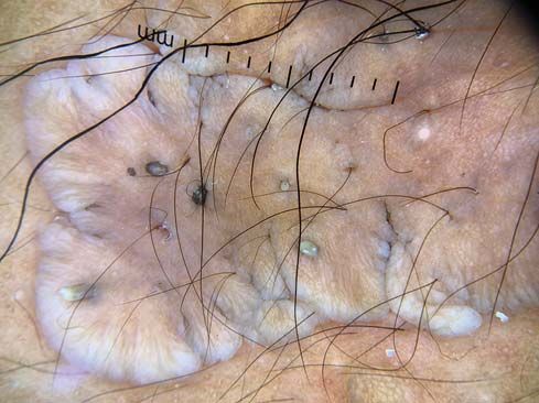
Dermoscopic evaluation was done using a DermLite over the nevus, angiokeratoma of fordyce, cafe-au-lait
DL4. Dermoscopy showed areas of cerebrifom pattern or vitiligo-like macules, hemangioma, and basal cell
with sulci and gyri, yellow structureless areas, comedo carcinoma [6]. Our patient had comedo-like lesions. It
like opening and pigment network (Figs. 2 and 3). A is considered to be a nevoid anomaly with the ectopic
3.5 mm punch biopsy was taken from the lesion and fat cells derived from the perivascular mesenchymal
sent for histopathological examination. The biopsy tissue. Histopathologically it has groups and strands
How to cite this article: Zacharias M, Sharathkumar BC. Nevus lipomatosis superficialis of Hoffmann and Zurhelle. Our Dermatol Online. 2021;12(e):e21.
Submission: 06.12.2020; Acceptance: 27.03.2021
DOI: 10.7241/ourd.2021e.21
© Our Dermatol Online e.2021 1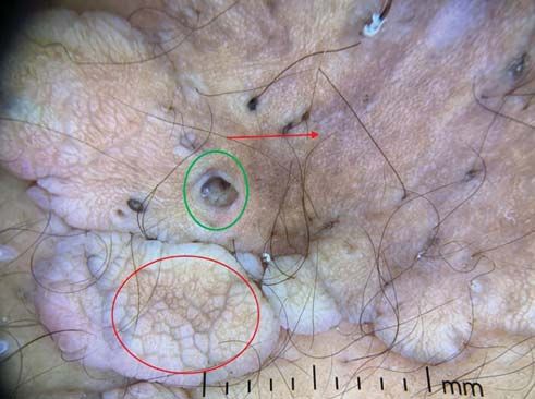
www.odermatol.com

Figure 1: Skin colored to yellowish papules, nodules and plaques over      Figure 3: Dermoscopy Green arrow- Yellow structureless area. Image
left flank with comedo like openings.                                      also shows comedo like openings and cerebriform surface.

                                                                           Figure 4: Histopathology image showing mature adipocytes in the
                                                                           dermis in the perivascular location.

                                                                           The condition is benign hence surgical removal is for
                                                                           cosmetic purposes.
Figure 2: Dermoscopy- Red circle- cerebriform pattern of sulci and gyri;
Green circle – commedo like opening; Red arrow- Pigment network.
                                                                           CONCLUSION
of mature fat cells which are found embedded among                         The above case has been reported due to the unusual
the collagen bundles of the dermis, often as high as                       characteristics observed in this benign hamartoma.
the papillary dermis. When there is large amount of                        Our patient had a presentation in the trunk. The
the mature fat cells distributed throughout the dermis                     lesion also showed comedo like openings on its
the hypodermis and dermis cannot be delineated [5].                        surface. Diagnosing this condition is important as it
                                                                           is completely benign and the patient can be assured
Dermoscopy of nevus lipomatosis superficialias is not                      of the same.
known to have any distinctive features. Most commonly
seen findings are the yellow structureless areas due to                    Consent
the fat deposition and the cerebriform surface due
to its irregular surface and comedo like openings.                         The examination of the patient was conducted according to the
These findings may be present in other conditions like                     principles of the Declaration of Helsinki.
seborrheic keratosis [7].
                                                                           The authors certify that they have obtained all appropriate patient
                                                                           consent forms, in which the patients gave their consent for images
Differential diagnosis to be considered include,
                                                                           and other clinical information to be included in the journal. The
nevus sebaceous, nevus commedonicus, focal dermal                          patients understand that their names and initials will not be
hypoplasia, connective tissue nevus, neurofibroma,                         published and due effort will be made to conceal their identity,
and acrochordons.                                                          but that anonymity cannot be guaranteed.

© Our Dermatol Online e.2021                                                                                                                2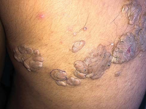
www.odermatol.com

REFERENCES                                                                        editors. Wolters Kluwer-Lippincott Williams & Wilkins; 2009.
                                                                             6.   Turan E, Yesilova Y, Ucmak D, Türkcü G, Celik OI, Gürel MS.
1.   Hoffmann E, Zurhelle E. Uber einen naevus lipomatodes cutaneous              Nevus lipomatosus cutaneus superficialis associated with nevus
     superficialis der linken glutaalgegend. Arch Dermatol Syphilol.              sebaceous of Jadassohn. Indian J Dermatol Venereol Leprol.
     1921;130:327-33.                                                             2014;80:194.
2.   Bhushan P, Thatte SS, Singh A. Nevus lipomatosus cutaneous              7.   Vinay K, Sawatkar GU, Saikia UN, Kumaran MS. Dermatoscopic
     superficialis: A report of two cases. Indian J Dermatol. 2016;61:123.        evaluation of three cases of nevus lipomatosus cutaneous
                                                                                  superficialis. Indian J Dermatol Venereol Leprol. 2017;83:383-6.
3.   Pujani M, Choudhury M, Garg T, Madan NK. Nevus lipomatosus
     superficialis: A rare cutaneous hamartoma. Indian Dermatol Online
     J. 2014;5:109-10.
                                                                              Copyright by Mariet Zacharias, et al. This is an open access article
4.   Abdullah M, Lateef A. Nevus lipomatosus superficialis. Our
                                                                              distributed under the terms of the Creative Commons Attribution License,
     Dermatol Online. 2020;11:412.
                                                                              which permits unrestricted use, distribution, and reproduction in any
5.   Ragsdale BD. Tumors with fatty, muscular, osseous, and/or                medium, provided the original author and source are credited.
     cartilaginous differentiation. Lever’s histopathology of the skin,       Source of Support: Nil, Conflict of Interest: None declared.
     10th ed. Elder DE, Elenitsas R, Jhonson BL, Murphy GF, Xu X

© Our Dermatol Online e.2021                                                                                                                             3
You can also read