Non-scanning CARS microscopy using wide-field geometry
←
→
Page content transcription
If your browser does not render page correctly, please read the page content below
1                  SPIE Proceedings, vol. 6442, 2007

          Non-scanning CARS microscopy using wide-field geometry
                I.Toytman1, K.Cohn1, T. Smith1, D. Simanovskii1and D. Palanker1,2
1
  Hansen Experimental Physics Laboratory, Stanford University, 445 Via Palou, Stanford, CA 94305
    2
      Department of Ophthalmology, Stanford University School of Medicine, 300 Pasteur Drive,
                                      Stanford, CA 94305

                                                     ABSTRACT

We report a wide-field Coherent Anti-Stokes Raman Scattering (CARS) microscopy technique based on non-phase-
matching illumination and imaging systems. This technique is based on a non-collinear sample illumination by broad
laser beams and recording image of sample at anti-Stokes wavelength using full-frame image detector. An amplified
Ti:Sapphire laser and an Optical Parametric Amplifier (OPA) provided picosecond pump and Stokes beams with
energies sufficient for CARS generation in an area of 100 µm in diameter. The whole field of view of the microscope
was illuminated simultaneously by the pump and Stokes beams, and CARS signal was recorded onto a cooled CCD,
with resolution determined by the microscope objective. Several illumination schemes and several types of thin sample
preparations have been explored. We demonstrated that CARS image of a 100x100 µm sample can be recorded with
submicrometer spatial resolution using just a few laser pulses of microJoule energies.
Keywords: Coherent anti-Stokes Raman scattering, Microscopy, Wide-field, Fast imaging, Chemically selective
imaging, Multiphoton microscopy, Scanning microscopy

                                              1. INTRODUCTION
Low natural optical contrast of biological cells and tissues often necessitates staining of the samples with various
chromophores and fluorescent markers. However, exogenous staining is limited in applications to living cells due to its
effect on cellular metabolism. Thus optical techniques for imaging of living cells with intrinsic chemical contrast and
high spatial resolution are of great interest. Mid-IR absorption microscopy can provide chemical selectivity, but it has
limited spatial resolution due to long (3-10 µm) wavelengths, and is difficult to apply in aqueous media due to high
water absorption. Raman microscopy overcomes these problems by generating chemically-selective signals in the visible
range, therefore providing resolution on the order of 1 micrometer. However, small cross section of spontaneous Raman
scattering limits the sensitivity of the method and requires high laser power and long exposures. Furthermore,
spontaneous Raman spectroscopy at visible wavelengths suffers from fluorescent background that reduces the contrast of
acquired images. These problems are circumvented in CARS microscopy, which is based on a nonlinear light generation
in a four-wave mixing process [1]. Utilization of visible and near IR (less than 1 µm) wavelengths makes spatial
resolution comparable to that of a conventional visible light microscope and allows for imaging of aqueous samples.
Since anti-Stokes component has a wavelength shorter than the pump wavelength, CARS signal is free of fluorescent
background. In addition, CARS signal can be much stronger than spontaneous Raman scattering due to coherent nature
of this emission process determined by constructive interference of signals from individual molecular vibrations over the
interaction length [2].
After theoretical description of the process by Maker and Terhune [3] and first implementation of CARS microscope by
Duncan et al [4] numerous studies were performed to increase the image brightness and resolution as well as the
acquisition speed [5-7]. Starting from Zumbush et al. [8] most of the CARS systems have been based on scanning
approach, in which two collinearly focused beams produce the signal in a small focal volume. In this geometry the
interaction length, over which CARS signal is generated is on the order of Rayleigh range Z0. For Gaussian beam with
waist W0 corresponding Rayleigh length is defined by

                                                          π ⋅ W02
                                                     Z0 =
                                                             λ
2 SPIE Proceedings, vol. 6442, 2007
In order to increase spatial resolution very tight focusing with large numerical aperture (NA) objectives is commonly
used. In this approach the focal spot size is comparable to the wavelength of the beam. With the pump and Stokes beams
focused to a spot with waist diameter W0 ~ 1 µm the Rayleigh length Z0 is on the order of several micrometers. The axial
and lateral extent of the focal zone defines the spatial resolution of scanning method. Relatively short interaction length
resulting from tight focusing provides high axial resolution close to the depth of field of a conventional microscope
objective, thus allowing for 3-dimensional imaging using layer-by-layer scanning of the sample.
We have explored a wide-field approach to CARS microscopy that would allow for simultaneous image acquisition
similarly to a conventional microscope with a focal plane array detector. Since the signals from all points of the sample
are recorded simultaneously, there are no strict requirements to pulse-to-pulse stability of the lasers. However, a
homogeneous illumination pattern is required to ensure similar conditions for CARS generation over the sample area.
First attempts to implement a wide-field system were made by Heinrich et al. [9]. In their illumination geometry phase-
matching conditions were satisfied within a sample. In our opinion, this resulted in a high level of background signal
generated in a bulk, which masked chemically specific CARS signal from the small-scale samples features.
Our approach is based on non-collinear non-phase-matching sample illumination geometry that would result in a very
low signal generation in the bulk. After refraction, or scattering of the laser beams on a small object, some portion of the
pump and Stokes beams will be deflected in a way that satisfies phase-matching condition. Therefore CARS signal can
be generated only inside the scattering objects and in their immediate vicinity. In this respect proposed illumination
scheme is similar to a dark-field illumination. Intensity of the generated CARS signal will depend on optical properties
of the scattering or refracting objects within the sample, as well as on their chemical composition. If the scattering center
exhibits Raman-active vibrational modes, CARS signal from that particular object will be strongly enhanced,
highlighting its chemical composition. Since this approach to CARS imaging relies on low scattering in the bulk of the
sample, it is best suitable for optically thin samples.
2. EXPERIMENTAL SETUP
2.1 Illumination scheme
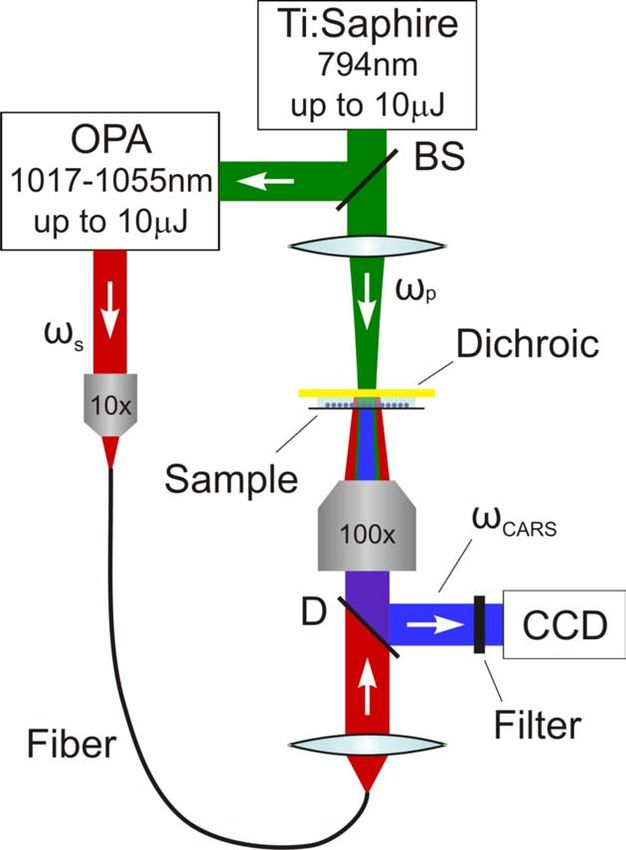
We tested the proposed technique using an experimental setup built around an inverted microscope (Axiovert 35, Carl
Zeiss), diagrammatically shown in Figure 1. The pump and Stokes beams were generated using Ti:Sapphire regenerative
amplifier (Regen) and Optical Parametric Amplifier (OPA) system (Spectra-Physics, CA). 794 nm radiation from the
Regen was used as a pump beam, and a frequency doubled signal from the OPA, tunable in the range from 1017 –
1055 nm was used as a Stokes beam.
Pump beam was focused to a 200 µm spot on the sample using 350 mm focal length lens, which formed practically
parallel beam on the sample. Stokes beam was focused to the same 200 µm spot on the sample plane through the
microscope objective. The 50x and 100x objectives with NA 0.45 and 0.73 respectively, produced wide range of Stokes
angles. While maximum Stokes angle was defined by the NA of the objective, the minimum angle was controlled by a
mask that was placed on the objective and blocked the central portion of Stokes beam, which was collinear to the pump.
Initially Stokes and pump beams were counter-propagating in the sample. After passing through the sample, Stokes
beam was reflected by a dichroic mirror and became co-propagating with the pump, though it had much broader angular
distribution. In this illumination geometry a small-angle scattering is sufficient to get fractions of the pump and Stokes
beams under phase-matching condition, necessary for efficient CARS generation.
Wide-field illumination technique requires much higher laser power in comparison to scanning methods to provide
sufficient energy density over the whole field of view. We found that for 200 µm focal spot at least ~10 µJ in pump and
Stokes beam is required to generate well detectable CARS signal. Regen and OPA produced pulses of the required
energy at repetition rate up to 1 kHz. Pulse duration of both pump and Stokes was about 1 ps, which for CARS
microscopy is proven to be optimal combination of pulse energy level and spectral width [10].
If sample is illuminated with coherent light portions of scattered, or reflected radiation may interfere with each other and
produce random intensity modulated pattern – speckles – on a detector. Unlike scanning CARS, where area illuminated
at any particular moment is relatively small (~1 µm) and speckles do not appear, coherent illumination of a 100 µm spot
in wide-field CARS produces significant speckling. To decrease speckling we coupled Stokes beam into a 1 m long3 SPIE Proceedings, vol. 6442, 2007
large-core (470 µm) multimode fiber that was randomly waggled from pulse to pulse. Averaging over 100 pulses was
sufficient to reduce intensity modulation caused by speckling to a few percent level.
Fig. 1. Experimental setup layout. BS: 20% beam splitter, D – dichroic mirror (separates CARS from Pump and Stokes),
Dichroic: dichroic mirror (reflects Stokes and transmits pump), 10x: Nikon 10x objective, NA = 0.25, 100x: Nikon
100x objective, NA = 0.73, Fiber: multimode fiber, CCD: CCD camera -Princeton Instruments LN-CCD-512
2.2 Image acquisition scheme
CARS signal was collected in forward direction (F-CARS) by the same microscope objective that was used to focus the
Stokes beam. Images formed by a microscope were recorded with a liquid nitrogen cooled CCD camera (512x512
pixels) with very low dark noise (less than 1 count/min). The imaged area size was 100x100 and 200x200 µm for 100x
and 50x objectives, respectively. The camera was replaced with a photomultiplier tube (PMT, Hamamatsu) for
measuring total amount of signal integrated over the whole field of view. It was especially convenient for measuring
power, spectral and angular dependencies of CARS, as well as for adjusting the delay between pump and Stokes beams.
A dichroic mirror transparent for pump and Stokes and highly reflective for CARS wavelengths and a filter that
transmitted light in a narrow range (600 – 700nm) and suppressed radiation beyond this range by more than 4 orders of
magnitude was installed in front of the detector to filter out residual pump and Stokes radiation.4 SPIE Proceedings, vol. 6442, 2007
3. RESULTS AND DISCUSSION
3.1 Proof of principle
To demonstrate capabilities of our method it would be indicative to compare images of objects of the same shape but
different chemical structure. For this purpose we used 6 µm polystyrene and quartz beads. Raman spectrum of
polystyrene has sharp peak at 3070 cm-1 corresponding to aromatic C-H stretch vibration, while quartz does not have any
pronounced features in this frequency range.
First, measurements were performed to confirm that the detected signal was produced as a result of CARS process.
Using PMT we measured CARS signal dependence on pump and Stokes beams intensities. As shown in Figure 2,
detected signal was found to be quadratically dependent on pump and linearly on Stokes beam intensity. Same behavior
was observed for both 50x and 100x objectives and for any size (0 to 5 mm) of the mask blocking central part of Stokes
beam.
10
9 Experimental data
CARS intensity, a.u.
8 Quadratic fit
7
6
5
4
3
2
1
0
0 2 4 6 8 10 12
2
Pump fluence, mJ/cm
a)
12
Experimental data
10
CARS intensity, a.u.
Linear fit
8
6
4
2
0
0.0 0.5 1.0 1.5 2.0 2.5 3.0
Stokes fluence, mJ/cm2
b)
Fig. 2. CARS signal from a polystyrene bead as a function of pump (a) and Stokes (b) fluences with quadratic and linear
fitting curves respectively.5 SPIE Proceedings, vol. 6442, 2007
To choose appropriate Stokes illumination angle range we compared CARS signal obtained from a single polystyrene
bead and from a 100 µm thick glass slide. As expected, without the mask strong CARS generation is observed in a glass
slide. This can be explained by the fact that there is a portion of Stokes beam, which is collinear with the pump within
the whole sample, and thus generates CARS signal in a homogeneous material. Increasing the mask diameter (and
thereby increasing the angular size of the cut-out central part of the Stokes beam) we were able to greatly suppress
CARS signal generated in the bulk material. For the blocked angle of 10o the background was reduced by more than an
order of magnitude. At the same time CARS signal produced by a polystyrene bead was reduced much less. In a first
approximation this reduction can be attributed to the reduced aperture of the collecting objective. For all further
measurements the angular size of the blocked area was set to 15o.
To evaluate chemical sensitivity of the method, CARS spectrum of polystyrene beads was measured by varying the
Stokes laser wavelength. For this measurement a number of polystyrene beads were deposited directly onto the dichroic
mirror. In Figure 3 Raman spectrum of polystyrene [11] and measured CARS signal are shown. Distinct Raman
resonance of polystyrene at 3070 cm-1, is evident on both spectra.
a)
6
CARS signal intensity, au
5
4
3
2
1
0
2500 2600 2700 2800 2900 3000 3100 3200
-1
Raman shift, cm
b)
Fig. 3. a) and b) Raman and CARS spectra of polystyrene respectively.6 SPIE Proceedings, vol. 6442, 2007
3.2 Chemically-selective imaging
To demonstrate chemically selective imaging a mixture of polystyrene and quartz beads floating in a thin (~30 µm) layer
of water between the dichroic mirror and a 3 µm Mylar film was imaged at two different wavelengths: on and off the
polystyrene Raman resonance. Images recorded at 3071cm-1 and 3116cm-1 are shown in Figure 4 a) and b), respectively.
Intensities of the pump and Stokes beams were kept at the same levels in both cases. As expected, brightness of the
quartz beads was practically the same in both cases. At the same time brightness of the polystyrene beads at resonance
was considerably higher than that out of resonance. It is interesting to note that even out of the resonance polystyrene
beads appeared significantly brighter than the quartz ones. One of the factors that could contribute to this effect is higher
refractive index of polystyrene beads (npolystyrene=1.59 vs. nquartz=1.46). This difference in indices of the particles
immersed in water (n=1.33), leads to stronger refraction of the laser beams in polystyrene beads, which is a key
condition for CARS generation under non-phase matching illumination geometry.
a) b)
Fig. 4. Acquired images of a mixture of polystyrene and quartz beads: a) image taken on resonance of polystyrene (Raman
shift 3071 cm-1), b) image taken off-resonance of polystyrene (Raman shift 3116 cm-1).
The ratio between resonant and non-resonant CARS signal of polystyrene in water, observed in this experiment, was
about 2, while dry beads have exhibited significantly higher spectral contrast of 5. This difference can be explained by
the fact that in our approach scattered pump and Stokes beams can generate CARS signal not only within the scattering
object, but potentially in the surrounding material as well. Apparently no such signal can be generated in the air;
however in water, surrounding the sample, its contribution could be substantial. Since water doesn't have pronounced
spectral features in the region of interest, signal from water will decrease the spectral contrast of the detected CARS
signal.
4. CONCLUSION
We have developed and successfully tested novel non-scanning wide-filed CARS imaging technique based on non
phase-matching illumination geometry that provides short image acquisition time, high spatial resolution, and chemical
selectivity. The validity of the method was proven by spectral and intensity measurements and CARS images of well-
defined test samples. Proposed method has potential advantages over the scanning CARS of being independent on laser
fluctuations, and capable of rapid image acquisition. Single pulse imaging could be especially useful for studying rapid
intracellular processes.7 SPIE Proceedings, vol. 6442, 2007
REFERENCES
1. R. J. H. Clark, R. E. Hester, Advances in Non-Linear Spectroscopy, John Wiley and Sons, New York, 1987.
2. B. S. Hudson, "New laser techniques for biophysical studies," Ann. Rev. of Biophys. Bioengin. 6, 135-150 (1977).
3. P. D. Maker, R. W. Terhune "Study of optical effects due to an induced polarization third order in the electric field
strength," Phys. Rev. 137, A801-A818 (1965).
4. M. D. Duncan, J. Reintjes, T. J. Manuccia, "Scanning coherent anti-Stokes Raman microscope," Opt. Lett. 7, 350-
352 (1982).
5. J. J. Song, G. L. Eesley, M. D. Levenson, "Background suppression in coherent Raman spectroscopy," Appl. Phys.
Lett. 29(9), 567-569 (1976).
6. J.-X. Cheng, A. Volkmer, L. D. Book, X. S. Xie, "An epi-detected coherent anti-Stokes Raman scattering (E-CARS)
microscope with high spectral resolution and high sensitivity," J. Phys. Chem. B, 105(7), 1277-1280 (2001).
7. M. Mueller, J. Squier, C. A. de Lange, G. J. Brakenhoff, "CARS microscopy with folded BoxCARS
phasematching," Journal of Microscopy 197(2), 150-158 (2000).
8. A. Zumbusch, G. R. Holton, X. S. Xie, "Three-dimensional vibrational imaging by coherent anti-Stokes Raman
scattering, " Phys. Rev. Lett. 82, 4142-4145 (1999).
9. C. Heinrich, C. Meusburger, S. Bernet, M. Ritsch-Marte, "CARS microscopy in a wide-field geometry with
nanosecond pulses," Journal of Raman Spectroscopy 37, 675-679 (2006).
10. J.-X. Cheng, X. S. Xie, "Coherent anti-Stokes Raman scattering microscopy: instrumentation, theory, and
applications," J. Phys. Chem. B 108, 827-840 (2004).
11. R. L. McCreery, http://www.chemistry.ohio-state.edu/~rmccreer/freqcorr/images/poly.htmlYou can also read