Norwegian Soils and Waters Contain Mesophilic, Plastic-Degrading Bacteria
←
→
Page content transcription
If your browser does not render page correctly, please read the page content below
microorganisms

Article
Norwegian Soils and Waters Contain Mesophilic,
Plastic-Degrading Bacteria
Colin Charnock

                                           Department of Life Sciences and Health, Faculty of Health Sciences, Oslo Metropolitan University, Postbox 4,
                                           St. Olavs Plass, 0130 Oslo, Norway; colin@oslomet.no

                                           Abstract: Plastic pollution has become one of the most critical environmental issues, as rapidly in-
                                           creasing production, compounded by persistence of plastic wastes in the environment, are outpacing
                                           efforts to keep ecosystems plastic-free. A switch to plastics more amenable to microbial attack is one
                                           of several possible responses. Against this background, the current study describes the isolation,
                                           enumeration and polyphasic characterization of plastic-degrading bacteria present in Norwegian
                                           terrestrial and aquatic habits. It shows that these bacteria are present in relatively high numbers, and
                                           that plastic-degrading capabilities are found in several taxa, most especially Streptomyces. Some iso-
                                           lates wereable to degrade several plastics. Notably, a Rhodococcus sp. and a Streptomyces sp. degraded,
                                           respectively, four and six of the eight plastics investigated and a number of other polymers relevant
                                           for plastic blends. The paper also has a methodological aspect, presenting various approaches for
                                           assaying plastic-degrading properties and a PCR/sequencing-based approach for the identification
                                           of potential polyethylene terephthalate-degrading genes. A candidate gene was detected in several
                                           Streptomyces isolates. The study shows that Norwegian environments are a rich source of bacteria
                                           with the ability to degrade bioplastics possibly representing a natural remediation capacity, as well
                                           as a potential source of useful enzymes.

                                           Keywords: plastic-degrading bacteria; Norway; environmental samples; biochemical characteriza-

                                    tion; Streptomyces sp.; Rhodococcus sp.
Citation: Charnock, C. Norwegian
Soils and Waters Contain Mesophilic,
Plastic-Degrading Bacteria.
Microorganisms 2021, 9, 94. https://       1. Introduction
doi.org/10.3390/microorganisms                  Plastics are a heterogenous collection of polymeric hydrocarbons. In common parlance
9010094                                    they are synthetic molecules. However, plastic-like molecules occur naturally; bacteria
                                           produce the polyhydroxyalkanoate-type polymers polyhydroxybutyrate (PHB) and Poly(3-
Received: 24 November 2020
                                           Hydroxybutyrate-co-3-Hydroxyvalerate) (PHBV) as storage molecules. Starch is commonly
Accepted: 17 December 2020
                                           used in the production of bio-based biodegradable plastics, and cellulose- and starch-based
Published: 3 January 2021
                                           materials can be used for some of the same functions as synthetic plastics, such as shopping
                                           and garbage collection bags. However, the main polymers that are currently considered
Publisher’s Note: MDPI stays neu-
                                           of greatest importance to our economy are usually synthetic and derived from petroleum
tral with regard to jurisdictional clai-
                                           and similar sources. These include polyurethane (PUR), polyethylene (PE), polyamide
ms in published maps and institutio-
nal affiliations.
                                           (PA), polyethylene terephthalate (PET), polystyrene (PS), polyvinylchloride (PVC) and
                                           polypropylene (PP). Such plastics are highly stable and do not readily undergo degradation.
                                           About 80% of the global plastic-usage is of synthetic origin [1,2]. Generally speaking, there
                                           are many factors which contribute to the degradation rate of plastics, including molecular
Copyright: © 2021 by the author. Li-       weight, melting temperature, glass transition temperature, the presence and types of
censee MDPI, Basel, Switzerland.           additives and monomer chemistry [3]. Large-scale production and distribution, careless
This article is an open access article     disposal methods and the inherent non-biodegradability of petroleum-based plastics are
distributed under the terms and con-       creating a major environmental concern, as plastics are accumulating in the environment
ditions of the Creative Commons At-        and have become a threat to the biosphere. Every major aquatic environment on the
tribution (CC BY) license (https://
                                           planet is now contaminated with plastic wastes, and marine plastic litter in particular is
creativecommons.org/licenses/by/
                                           considered a major challenge [4]. Microplastics and nanoplastics in aquatic environments
4.0/).

Microorganisms 2021, 9, 94. https://doi.org/10.3390/microorganisms9010094                             https://www.mdpi.com/journal/microorganisms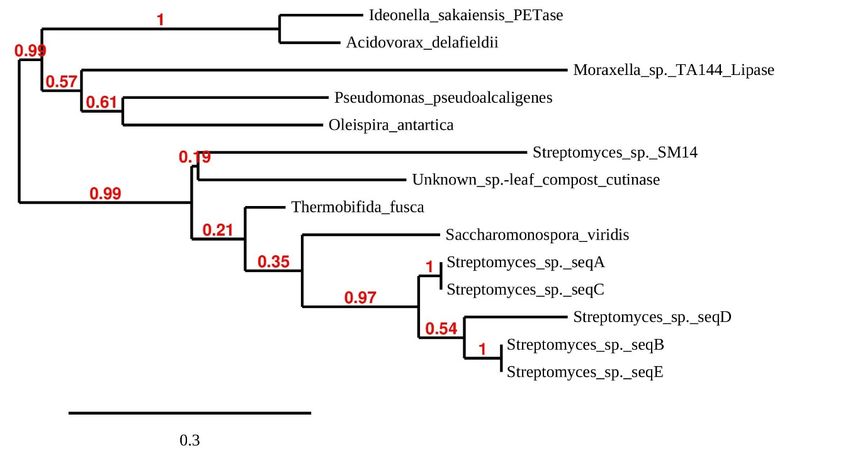
Microorganisms 2021, 9, 94                                                                                              2 of 18

                             are entering the food chain and ultimately end up in human intestines [4]. Several excellent
                             reviews exist summarizing the extent and nature of contaminating plastics in natural
                             ecosystems, and the possible if not probable negative effects they represent for the food
                             chain and health [5–7].
                                   High accumulation of low- or non-biodegradable plastics in the environment is driv-
                             ing an increased focus on readily biodegradable plastics, and the removal of plastics from
                             the environment using microbes and microbial enzymes is a related avenue of recent
                             research. Plastic production is a relatively new phenomenon, starting in essence in the
                             1950s–1960s; this is a very short time for the evolution of plastic-degrading enzymes, and
                             the selective pressure for doing so in the presence of more bioassimilable substrates may be
                             low. However, microbial enzymes capable of degrading or partly degrading a wide range
                             of plastics are known [7]. Microbial biodegradation will often involve consortia of multiple
                             species, some of which are capable of degrading high molecular weight polymers into
                             simpler forms (multipers, dimers) using exoenzymes; these are then broken down by other
                             microbes in the community [7,8]. Carboxylesterases (E.C. 3.1.1.1), lipases (E.C. 3.1.1.3) and
                             cutinases (E.C. 3.1.1.74) are those classes of enzymes chiefly associated with the hydrolysis
                             of plastics [9]. Most biodegradable plastics are polyesters (e.g., the polyhydroxyalkanoates,
                             polycaprolactone (PCL), poly(ethylene succinate) (PES), poly(1,4-butylene succinate) (PBS),
                             Poly(L-lactide) (PLLA) and polybutylene succinate-co-adipate). Polyesters can, in one
                             scheme, be further categorized into three groups: those bracketed above are examples of
                             aliphatic polyesters and are hydrolyzed by various ester-bond hydrolases. In addition,
                             there are aromatic polyesters, e.g., polyethylene terephthalate (PET), which are the least
                             biodegradable [10,11]. In an attempt to combine good material properties with biodegrad-
                             ability, aliphatic–aromatic copolyesters, such poly (butylene adipate-co-terephthalate) have
                             been developed [10,11]. Aliphatic polyesters alone or as copolymers (to improve processing
                             and end-point properties) have been used for many applications such as shopping bags,
                             film production in agriculture and as coatings. Some have been approved for in vivo
                             applications, including controlled drug delivery systems in bone tissue engineering [12].
                             Polyesterase acting on aromatic polyesters (primarily PET) was first reported for Ther-
                             mobifida fusca [13]. Although several PET-degrading enzymes are now known [14], there
                             is currently only a single report of the utilization of this plastic for bacterial growth. In
                             2016, the first PET-degrading microbe also capable of utilizing this synthetic plastic as a
                             source of carbon and energy was isolated from a PET-contaminated soil [15]. The bacterial
                             strain Ideonella sakaiensis 201-F6 was shown to grow on low-crystallinity PET films. Two
                             α/β-hydrolase fold enzymes, PETase (a unique cutinase-like enzyme) and MHETase, work
                             together to degrade PET in two steps. The residues constituting the substrate binding
                             site in enzymes with known PET-hydrolyzing ability are almost conserved [9,14,16,17].
                             However, PETase has a number of unique features near the catalytic center, and the active
                             site of PETase is wider than that of the other PET-hydrolyzing cutinases. Presumably
                             because of these features, PETase is 5.5–120-fold more efficient than previously reported
                             PET-hydrolyzing homologs [9].
                                   The present study, which is the first of its kind in Norway, has as its main aim to isolate,
                             identify and characterize the plastic-degrading potential of bacteria from environmental
                             samples in Norway. This will provide an insight into the types and distribution of these
                             bacteria, and to some extent their potential to process plastics entering the environment. It
                             may also identify novel enzyme activities with useful plastic-degrading capabilities.

                             2. Materials and Methods
                             2.1. Samples and Isolation and Enumeration of Bacteria
                                  The majority of strains originate from a systematic investigation of plastic-degrading
                             bacteria in nine soil, sediment and water samples (Table 1). The approach used here was as
                             follows: samples (about 10 g) were taken with a sterile spatula and transferred to a 50 mL
                             sample tube with a screw cork. Samples were transported cold and analyzed within 24 h.
Microorganisms 2021, 9, 94 3 of 18
All samples were taken in the period January to March, 2019. Salient information on the
nine samples is given in Table 1.
Table 1. Details on the nine sampling sites forming the core of the study.
Additional
Sample Sampling Site Coordinates Sample Appearance
Information
Soil from immediate vicinity of a
59◦ 400 12.8”N Dry soil, with a dark Sample was taken a few
1 small factory making plastic products
10◦ 470 45.0”E appearance. cm beneath the top soil.
to order.
Bank of a stream passing through an
Brown, wet material Sample was taken a
industrial area, close (20 m) to a small 59◦ 420 33.0”N
2 apparent mix of soil few cm beneath the top
factory making plastic products to 10◦ 510 10.7”E
and sand. layer
order
Natural marsh, several km from 59◦ 340 12.1”N Dark brown, watery Sample taken under
3
housing/industry. 10◦ 400 17.7”E sediment. shallow water.
Sample was taken a
Bank of a stream running through 59◦ 330 51.1”N
4 Clay-like material. few cm beneath the top
housing estate and construction site. 10◦ 440 02.6”E
layer
Sample was taken a
Bank of a river passing through 59◦ 360 34.1”N Brown, watery material
5 few cm beneath the top
agricultural area. 10◦ 450 51.2”E with plant debris.
layer
Edge of a small ditch below the outlet
Sample was taken a
of a drainage pipe probably leading 59◦ 360 30.6”N Mixture of sand and
6 few cm beneath the top
water from housing area and 10◦ 450 55.4”E clay-like material.
layer
surroundings.
Bank of a stream about 100 m in the Light, finely-grained Sample was taken a
59◦ 340 47.2”N
7 terrain below a sewage-treatment sand mixed with few cm beneath the top
10◦ 390 13.7”E
plant. clay-like material. layer.
Sample was taken a
River bank in an urban area close to 59◦ 540 45.7”N Coarse mix of sand and
8 few cm beneath the top
outlet to the Oslo fjord. 10◦ 450 26.8”E clay-like material.
layer.
River bank in an urban/industrialized 59◦ 540 46.8”N Mix of sand and Sample taken from top
9
area. 10◦ 490 40.7”E clay-like material. layer of sediment
The following approach was developed to isolate and detect plastic degrading bacteria:
samples 1.0 ± 0.1 g wet weight and a spatula of 2 mm glass beads were transferred to a
15 mL centrifuge tube (Corning™ from Sigma-Aldrich, St. Louis, MO, USA, Catalogue
number CLS430055). Thereafter, Maximum Recovery Dilutant (MRD; Oxoid, Hampshire,
UK, Catalogue number CM0733) was added to give a total volume of 10 mL. The mixture
was then vortexed vigorously (Test Tube Shaker, Labworld-online.com) using three 1 min
bursts and intermittent cooling. Subsequently, tubes were placed in a vertical position for
10 min to allow the largest particles to sink. One mL of the top layer was transferred to
a new tube and serially diluted 1/10 a further six times with MRD. From each dilution,
a sample (0.1 mL) was transferred to R2A (Oxoid, Hampshire, UK. Catalogue number
CM0906) and incubated at 22 ± 2 ◦ C for 14 days. After incubation, the dilution (plate)
for each sample showing 50–100 colonies or the number nearest to this, was chosen for
interpretation and screening for plastic-degrading bacteria. The number of different colony
types judged visually based on color, shape, pattern and consistency were noted. In the
initial screening, a portion of each colony was spotted onto agars containing the following
plastics: PHBV, PHB, PCL, PES, PBS and PLLA (see details in 2.2). Plates were incubated
at 22 ± 2 ◦ C and examined for appearance of zones of clearing around colonies over
a six-week period. The study also includes two strains isolated previously from a soil
in the vicinity of a small industrial unit (59◦ 240 49.5”N 10◦ 410 32.3”E) producing plastic
products. Here, soil particles were sprinkled onto PHB-containing agar, and two strains
with PHB-degrading capabilities were subsequently isolated from zones of clearing.Microorganisms 2021, 9, 94 4 of 18
2.2. Plastic-Containing Agar
The test media consisted of two layers (each made of about 20 mL agar solution in 9
cm petri dishes): a bottom layer consisting of vitamins and salts overlayed with an agar
containing the test plastic. The bottom layer contained 0.01 g casamino acids (Bacto™,
BD Diagnostics-TriPath, Burlington, USA, Catalogue number 223120), 0.01 g yeast extract
(Sigma-Aldrich, St. Louis, MI, USA, Y1625), 2.27 g M9 minimal salts (Sigma-Aldrich, m-
6030) and 15 g washed agar-agar (Merck, Kenilworth, NJ, USA, Catalogue number 101614)
in 1000 mL ion-exchange purified water. After sterilization by autoclaving and cooling to
circa 60 ◦ C, 1 mL Trace Metal Mix (Sigma-Aldrich, St. Louis, MI, United States, Catalogue
number 92949), 2.4 mg Mg2+ (from 10 mg/mL filter sterilized stock) and 0.4 mg Ca2+ (from
10 mg/mL filter sterilized stock) were added and plates were poured. The top agar layer
contained 0.1–0.2% (w/v) plastic in 15 g/L twice washed agar-agar (Merck, Darmstadt,
Germany, Catalogue number 101614) in 1 × PBS. Two different approaches, dependent on
the plastic form (powder or pellets), were used to include the plastic in the top agar: for fine
powders, a suspension was made in 10 × PBS at pH 7.2, the suspension was then added
dropwise to agar (in water) melted at 60 ◦ C with vigorous stirring to achieve 0.1–0.2% w/v
plastic and 1.5% agar in 1 × PBS. To achieve a homogenous suspension in 10 × PBS, the
plastic powder was added slowly to the liquid placed in an ultrasound-generating water
bath (S30 Elmasonic, Elmasoni). Repeated pipetting with a glass pasteur pipette was used
to break up small clumps of powder.
Plastic pellets were dissolved in dichloromethane (5 mL) and added dropwise under
sonication, at 40% amplitude with 45 s on/15 s off pulses (Vibra-Cell VCX130, Sonics,
Seattle, WA, USA), to 200 mL molten, autoclaved agar in 1 × PBS and containing 0.1 g/L
N-Lauroylsarcosine (Sigma-Aldrich, St. Louis, MO, USA, Catalogue number L5777).
The following eight plastics were included in the study: poly[(R)-3-hydroksybutyrate],
PHB (fine powder; Sigma-Aldrich, 363502); poly(3-hydroksybutyrate-co-3- hydroksyvaler-
ate), PHBV (fine powder; Materials Gateway, UK, sold as ENMAT Y1000P); poly(ethylene
succinate), PES (Pellets, average molecular weight = 10,000; Sigma-Aldrich, St. Louis,
MO, USA, Catalogue number 182036); poly(1,4-butylene succinate), extended with 1,6-
diisocyanatohexane, PBS (pellets; Sigma-Aldrich, Catalogue number 448028); polycapro-
lactone, PCL (Powder/flakes, MW 50,000; Polysciences 25090, Warrington, PA, USA);
poly(L-lactide), PLLA (Fine powder, MW ~1600–2400; Polysciences 18580-1, Tyskland);
Resomer® RG 502, Poly(D,L-Lactide-co-Glycolide), RES (Fine, amorphous powder: lac-
tide:glycolide 50:50 and ester terminated, MW 7000–17,000); polyethylene terephthalate,
PET (amorphous sheet; Goodfellow, ES303010, London, UK).
2.3. PCR Amplification and Sequencing Studies
Identification based on the 16S rDNA gene: The PCR reaction mixture (50 µL) con-
tained 3 µL of 25 mM MgCl2 (Promega), 1 µL dNTPS 10 mM (Promega), 10 µL HotStart
DNA polymerase buffer, 0.2 µL HotStart DNA polymerase 5 U/µL (Promega), 0.25 µL of
each of primers 27f (AGA GTT TGA TCA TGG CTC A) and 1492r (TAC GGT TAC CTT
GTT ACG ACT T) {100 µM stock of standard à la carte sequencing primers from MWG
Eurofins} and PCR-grade water to 50 µL. To provide the template, a flame-sterilized steel
pin was touched onto a bacterial colony and the pinpoint of material was transferred to the
reaction mix.
PCR conditions were: one cycle at 95 ◦ C (10 min) followed by 32 cycles of 95 ◦ C/min,
52 C/45 s and 72 ◦ C/1.5 min. This was followed by a final elongation of 72 ◦ C/12 min.
◦
Products were checked for purity by agarose electrophoresis and DNA concentrations
were measured using Qubit™ dsDNA BR Assay Kit (Thermo Fisher Scientific, Waltham,
MA, USA). PCR products were sequenced on both strands at least twice. Sequencing was
performed by a commercial laboratory (MWG Eurofins) using the PCR primers. Sequences
were aligned using Clustal Omega [18], and the consensus region of overlap was used
for purposes of identification. Sequences were analyzed and assigned to taxa using the
RDP Naive Bayesian rRNA Classifier Version 2.11 (RDP rRNA training set 18) with theMicroorganisms 2021, 9, 94 5 of 18
default 80% confidence threshold [19]. In addition, BLAST [20] was used to search the
NCBI database. The curated ‘Reference RNA sequence’ setting in BLAST was used to
assign the sequences to a named taxon.
Touchdown PCR amplification and sequencing of genes potentially encoding PET-
hydrolyzing enzymes: The reaction mixture (50 µL) contained 3 µL of 25 mM MgCl2
(Promega, Madison, WI, USA), 1 µL dNTPS 10 mM (Promega, Madison, WI, USA), 10 µL
HotStart DNA polymerase buffer, 0.05 µL HotStart DNA polymerase 5 U/µL (Promega,
Madison, WI, USA), 0.2 µL of the forward primer (GTCATCACCATCGACACCA) and
0.2 µL of the reverse primer (GTAGCG(G/C)GTGTCGTTGTC). A touchdown PCR was
performed with the following specifications: An initial denaturation 95 ◦ C/10 min was
followed by 36 cycles in which the annealing temperature was lowered from 65 ◦ C by 1 ◦ C in
each round until it reached 55 ◦ C (36 cycles: 95 ◦ C/30 s; 64–55 ◦ C/30 s; 72 ◦ C/60 s). A final
elongation step of 72 ◦ C/5 min completed the reaction. Quality control and measurements
of DNA concentration were carried out as described above. The primers were designed
based on alignments of the PET-hydrolyzing genes of Gram-positive (Thermobifida fusca)
and Gram-negative bacteria (I. sakaiensis NBRC 110686, Acidovorax delafieldii) presented with
references in Joo et al. [14], and amplify a region of approximately 480 bp in these genes.
The PCR product was checked for an amplicon of expected size by gel electrophoresis.
PCR products were sequenced on both strands at least twice. Sequencing was performed
by a commercial laboratory (MWG Eurofins, Ebersberg, Germany) using the PCR primers.
I. sakaiensis NBRC 110686 was used as the control for PCR, and its amplicon was also
sequenced to check the fidelity of the reaction. Sequences were aligned using Clustal
Omega [18] and the consensus region of overlap was used for further analysis. Open
reading frames were generated based on the sequences by comparison with the PET-
hydrolase enzyme from I. sakaiensis. Similarity searches on the sequences were performed
using BLASTP [20] to search the non-redundant protein sequence library at NCBI [21].
Additional searches were performed using FASTA [22] to search the manually annotated
section of UniProtKB [23].
The ORF with significant similarity to the control PETase sequence from I. sakaiensis
was aligned with putative PET-hydrolysis genes given in [14]. Sequences were aligned
using Clustal, and key residues were identified across the sequences by reference to the
literature (see Results and Discussion). A phylogenetic tree for PETase and PETase-like
enzymes was generated using the Phylogeny.fr [24,25] online tool (www.phylogeny.fr/)
in the a la carte mode: reference sequences were trimmed manually to approximately
equal size to those of the isolate sequences, and aligned using MUSCLE (v3.8.31) in the
program package [26]. After alignment ambiguous regions (i.e., containing gaps and/or
poorly aligned) were removed with Gblocks v0.91b [27]. The tree was constructed using
the maximum likelihood method [28], implemented with the approximate likelihood-
ratio test (aLRT) setting for branch support [29] included in the pipeline PhyML (v 3.0)
program. The default WAG substitution model for amino acids was chosen to account
for rate heterogeneity across sites [30]. Internal branch reliability was assessed using
the bootstrapping method (100 bootstrap replicates). Graphical representation of the
phylogenetic tree was achieved using the workflow TreeDyn tool [31].
2.4. Broth-Based Investigation of PET-Degrading Activity
In order to provide an additional indication of possible PET-hydrolysis, small pieces
(approximately 3 × 3 mm) of amorphous plastic hung from fishing line were suspended
in a dilute growth medium (g/L: 0.2 casamino acids; 1.26, yeast nitrogen base (Merck,
Kenilworth, NJ, USA, Y1251); 0.04 yeast extract) seeded with a pure culture of I. sakaiensis
NBRC 110686 or sample isolates. Bottles were observed over a period of 6 weeks (room
temperature, without shaking) with occasional changes of medium. Development of
growth and discoloration on the plastic were looked for. The technique is basically the
same as what was used in the original isolation of I. sakaiensis [15].Microorganisms 2021, 9, 94 6 of 18
2.5. API® ZYM (bioMérieux Inc.)
The Rhodococcus sp. was grown on BUG agar plates (Biolog Inc., Hayworth, CA,
USA) at 30 ± 1 ◦ C for 24 h. A thick suspension of isolated colonies was made in 0.85%
NaCl and this was used to create the inoculum (McFarland 5-6) exactly as described in
the product insert. Sixty-five µL of the inoculum was pipetted into each cupule and plates
were incubated at 31 ± 1 ◦ C. In each round of testing (two in total), plates were interpreted
after 20 h incubation.
The Streptomyces sp. was grown on oatmeal agar (Sigma-Aldrich, St. Louis, MO,
USA, Catalogue number O3506) for 7 days at 31 ± 1 ◦ C. A thick hyphal/spore suspension
was made in 0.85% NaCl and this was used to create the inoculum. Thereafter, the same
procedure as that used for Rhodococcus was followed.
2.6. API® 50CH (bioMérieux Inc.)
Rhodococcus/Streptomyces were grown as for the API® ZYM test. A suspension in API
50 CHB/E medium (the inoculum) was made exactly as described in the product insert.
Plates were inoculated without oil overlay in the cupule at 31 ± 1 ◦ C and read at 48 h and
6 days. The system was quality-controlled using Paenibacillus polymyxa, as suggested in the
package insert.
2.7. Assays for Additional Enzyme Activities
Sodium carboxymethyl cellulose (CMC) agar for detection of cellulase activity: 50%
R2A agar (Oxoid) was amended with CMC (Sigma-Aldrich, St. Louis, MO, USA, Catalogue
number 419338) at 0.5% w/v. Plates were inoculated to achieve well-isolated colonies. After
incubation at 31 ± 1 ◦ C for 72 h, plates were flooded with 0.1% Congo Red (Sigma-Aldrich,
St. Louis, MO, USA, Catalogue number C6767) for 15–20 min. Plates were then rinsed
carefully with 1 M NaCl. The NaCl was decanted and replaced, and plates were then
agitated gently for 20–30 min. The NaCl was again removed. Zones of clearing round
individual colonies against a red background of none-hydrolyzed CMC was taken as
an indication of cellulase activity. Paenibacillus polymyxa DSM 365 and 372 were used as
positive controls.
Tributyrin agar for the detection of esterase/lipase activity: 50% R2A agar (Oxoid,
Hampshire, UK) was amended with tributyrin (Sigma-Aldrich, St. Louis, MO, USA,
W222305) at 1% v/v with mixing. Plates were inoculated to achieve well-isolated colonies.
After incubation at 31 ± 1 ◦ C for 72–96 h, plates were examined for zones of clearing around
colonies indicating esterase/lipase activity. Burkholderia cepacia DSM9421 and Pseudomonas
aeruginosa DSM 1128 were used as positive control organisms.
Olive oil—Rhodamine B agar for the detection of lipase activity: R2A agar (Oxoid,
Hampshire, UK) was amended with olive oil (Sigma-Aldrich, O1514) as follows: 30 mL of
olive oil was emulsified (Vibra-Cell VCX130, Sonics) into 50 mL water containing 250 µL
of Tween 20 (Sigma-Aldrich, St. Louis, MO, USA, P1379). The emulsion was autoclaved,
cooled to 60 ◦ C and amended with 20 mL of a filter sterilized Rhodamine B solution
(1 mg/mL). Then, 50 mL of this mixture was added with stirring into 450 mL R2A agar
melted at 50 ◦ C. Plates were prepared and inoculated to achieve well-isolated colonies.
After incubation at 31 ± 1 ◦ C for 72–96 h, plates were examined for orange fluorescent
zones emanating from colonies and also zones of clearing around colonies, indicating lipase
activity. Burkholderia cepacia DSM9421 and Pseudomonas aeruginosa DSM 1128 were used as
positive control organisms.
Coconut oil—Rhodamine B agar for the detection of lipase activity: Coconut oil-agar
was made essentially as described for olive oil agar, except that 20 mL of melted coconut oil
(Green choice, Vantaa, Finland) was used. Analysis and interpretation were as described
for olive oil agar.
Starch agar for the detection of α-amylase activity: 50% R2A agar (Oxoid, Hampshire,
UK) was amended with starch (Sigma-Aldrich, 03967) at 0.5% w/v. Plates were inoculated
to achieve well-isolated colonies. After incubation at 31 ± 1 ◦ C for 72 h, plates were floodedMicroorganisms 2021, 9, 94 7 of 18
with Gram’s iodine. Colorless zones surrounding the colonies against a blue/purple
background of non-hydrolyzed starch was taken to indicate amylase activity. Paenibacillus
polymyxa DSM 365 and 372 were positive controls.
Skim milk powder agar for the detection of protease activity: 50% R2A agar (Oxoid,
Hampshire, UK) was amended with skim milk powder (Sigma-Aldrich, 70166) at 2.8%
w/v. Plates were inoculated to achieve well-isolated colonies. After incubation at 31 ± 1 ◦ C
for 72 h, plates were examined for zones of clearing around colonies indicating protease
activity. Burkholderia cepacia DSM 9241 and Pseudomonas aeruginosa DSM 1128 were used as
positive control organisms
DNase agar: Commercially available plates containing 0.2% w/v deoxyribonucleic
acid (Oxoid, Hampshire, UK, Catalogue number CM0321) were inoculated to achieve
well-isolated colonies. After incubation at 31 ± 1 ◦ C for 72 h, plates were flooded with 1N
HCl and allowed to stand for 5 min. Zones of clearing in the agar around colonies was
taken to indicate DNase activity. Staphylococcus aureus strains ATCC 25923 and DSM 799
were used as positive control organisms.
Hemolysis on sheep blood agar: Commercially available Columbia agar plates contain-
ing 7% sheep blood (Oxoid, Hampshire, UK, Catalogue number PB5039A) were inoculated
to achieve well-isolated colonies. After incubation at 31 ± 1 ◦ C for 72 h, plates were exam-
ined for hemolysis around single colonies. Streptococcus pyogenes ATCC 19615 (producing
beta hemolysis) was used as a positive control.
Test for beta-lactamase activity: Production and activity of beta-lactamase activity was
tested for using commercially available discs containing the chromogenic cephalosporin
Nitrocefin (Remel, San Diego, CA, USA). Using the kit protocol, a color change to pink/red
was taken as an indication of enzyme activity. Escherichia coli ATCC 35218 and S. aureus
DSM 2569 were used as positive controls.
Accession numbers: 16S rDNA sequences: MW269812-22. Putative PET-hydrolases:
MW281313-17.
3. Results and Discussion
3.1. Numbers, Identities and Range of Activities of Plastic Degrading Bacteria
For the nine environmental samples forming the core of the study, the CFU/g wet
weight ranged from 5 × 10⁴ (sample 3) to 6 × 10⁷ (sample 1). A single dilution/agar plate
(showing between 50–100 colonies) was chosen for further investigation. The number of
different colony-classes on this plate was visually adjudged to range from 6 (sample 3) to
19 (sample 7). In total, 88 different colony types across all nine samples were recognized,
and a representative was transferred to a range of six plastic-containing agars. Figure 1
shows some typical results from the agar screening tests. Six of the nine samples contained
one or more plastic-degrading bacteria. Colonies originating from samples 2, 6 and 8
did not produce clearing on any agars. In total, 9 of the 88 colony types showed clearing
on one or more of the plastic-containing agars. Summary data on their degradative
properties are shown in Table 2 (isolates designated iii-xi). In addition, two isolates (i-ii;
Table 2) from a previous, preliminary study were also characterized. Subsequently, activity
against additional plastics and other polymers were tested for. Attempts were not made
to determine the absolute numbers or proportion of different plastic degrading bacteria
arising from each sample. However, as mentioned, of the 88 different colony types tested,
9 (about 10%) showed the ability to degrade one or more of the 8 plastics used in the initial
screening.Number + 3 9 3 5 5 2 6 10 3 3 5
PCR product (PET-primers) − − − + − − − + − − +
Roman numerals (i–xi) denote bacterial isolates. * Isolates i–ii were obtained from a
previous investigation (see methods for their origin). Isolates iii–xi forming the core of the
Microorganisms 2021, 9, 94 present study, were isolated from the samples shown in Table 1 as described in the meth- 8 of 18
ods section. [+] = weakly positive reaction.
Figure Figure
1. Growth of isolates
1. Growth of and production
isolates of zones of
and production of clearing
zones ofonclearing
plastic-containing agars (a) agars (a)
on plastic-containing
Rhodococcus sp. (isolate vii; Table 2) on PES-agar (b) Streptomyces sp. (isolate iv; Table 2)
Rhodococcus sp. (isolate vii; Table 2) on PES-agar (b) Streptomyces sp. (isolate iv; Table on 2)
PHB-
on PHB-agar
agar (c) Streptomyces sp. (isolate viii; Table 2) on PHB-agar (d) Streptomyces sp. (isolate viii; Table 2)
(c) Streptomyces sp. (isolate viii; Table 2) on PHB-agar (d) Streptomyces sp. (isolate viii; Table 2) on
on PCL-agar.
PCL-agar.
Table 3 gives a detailed presentation of the sequencing-based identifications of the
Table 2. Polymer degrading profiles for environmental isolates.
11 isolates based on similarity searches of partial 16S rDNA gene sequences.
Isolate i–xi (sample of origin 1–9;—Table 1)
Agar Table 3. Identification of isolates based on partial sequencing of the 16S rDNA gene.
i* ii * iii (1) iv (9) v (7) vi (7) vii (7) viii (4) ix (5) x (5) xi (3)
Sequence
Isolate PHB +Phylogenetic
+ −
affiliation* + +
Closest + (Accession−number)
species + −Identity-% (query
− +
length(bp) coverage)
PHBV + + + + + + − + + − +
i 625
PES − Acidovorax−
− − −A. facilis LMG2193
− (EU024133)
+ + − 99.36 (100)
− −
ii 526
PBS − Streptomyces
− − − S.
−similanensis−KC-106**(AB773850)
+ [+] − 99.62 (100)
− −
iii 508
PCL − Streptomycetaceae
[+] − − S.−urticae NEAU-PCY-1
− (KY788226)
[+] + − 99.80 (100)
[+] −
iv 540
PLLA − −
Streptomyces− − −lunaelactis −
S. −
MM109**(KM207217) − − −
100 (100) −
v RES
592 − −
Streptomyces− − S.−alboniger DSM40043**(AY845349)
− + + − +
99.49 (100) −
vi Starch
510 Pseudomonas P. floridensis GEV388**(KY614191) 100 (100)
− + − + + − − + + − +
vii (amylase)
731 Rhodococcus R. fascians CF17 (X79186) 99.86 (100)
viii CMC1403 Streptomyces
− + + + +S. brevispora−BK160 (FR692104)
− + ‘ 99.86 (100)
− +
ix (cellulase)
528 Oxalobacteraceae Rugamonas rubra MOM 28/2/79 (HM038005) 99.43 (99)
x DNA422 Nakamurella N. lactea** DSM19367 (HE599561) 98.82 (100)
− − − − − − − − − − −
xi (DNase)
951 Streptomyces S. fulvissimus DSM40593** 100 (100)
Protein
− + − − − − − + − + −
(protease)
Tributyrin
+ + + [+] + − + + + − +
(esterase)
Olive oil
− + − − − − − − − − −
(lipase)
Coconut oil
− [+] − − − − + − − − −
(lipase)
Number + 3 9 3 5 5 2 6 10 3 3 5
PCR product
− − − + − − − + − − +
(PET-primers)
Roman numerals (i–xi) denote bacterial isolates. * Isolates i–ii were obtained from a previous investigation (see methods for their origin).
Isolates iii–xi forming the core of the present study, were isolated from the samples shown in Table 1 as described in the methods section.
[+] = weakly positive reaction.
Table 3 gives a detailed presentation of the sequencing-based identifications of the 11
isolates based on similarity searches of partial 16S rDNA gene sequences.Microorganisms 2021, 9, 94 9 of 18
Table 3. Identification of isolates based on partial sequencing of the 16S rDNA gene.
Phylogenetic Closest Species (Accession Identity-% (Query
Isolate Sequence Length (bp)
Affiliation * Number) Coverage)
i 625 Acidovorax A. facilis LMG2193 (EU024133) 99.36 (100)
S. similanensis KC-106 **
ii 526 Streptomyces 99.62 (100)
(AB773850)
S. urticae NEAU-PCY-1
iii 508 Streptomycetaceae 99.80 (100)
(KY788226)
S. lunaelactis MM109 **
iv 540 Streptomyces 100 (100)
(KM207217)
S. alboniger DSM40043 **
v 592 Streptomyces 99.49 (100)
(AY845349)
P. floridensis GEV388 **
vi 510 Pseudomonas 100 (100)
(KY614191)
vii 731 Rhodococcus R. fascians CF17 (X79186) 99.86 (100)
viii 1403 Streptomyces S. brevispora BK160 (FR692104) 99.86 (100)
Rugamonas rubra MOM 28/2/79
ix 528 Oxalobacteraceae 99.43 (99)
(HM038005)
N. lactea ** DSM19367
x 422 Nakamurella 98.82 (100)
(HE599561)
xi 951 Streptomyces S. fulvissimus DSM40593 ** 100 (100)
* From RDP classifier. Lowest affiliation with a score of 1.0 at default 80% confidence setting. ** Where there are other strains or species
with same total score.
In agreement with previous investigations [1,32–34], the present study shows that
the ability to degrade one or more plastic is a fairly common trait and is found across a
range of taxa of easily cultured species present in environmental samples. None of the 11
plastic-degrading isolates had effect on PLLA, whereas 9 caused clearing in PHBV, and this
plastic was the one that was the most commonly degraded by the bacteria (Table 2). Based
on similar clear-zone techniques, others have looked at populations of bacteria in different
environments. Urbanek et al. [1] reported the following order of degradation of plastics
by artic microorganisms: PCL > PBS > PLA (isomeric form not specified). The same order
was seen for microbes from agricultural soil in Thailand [33]. Looking at microorganisms
in different ecosystems, Nishida et al. [34] found high and approximately equal numbers
of PHB- and PCL-degraders. These results are all similar to those of the present work.
The complete order of biodegradation in the present study, when based on the number
of different isolates producing clearing in plastic-containing agar, was as follows PHBV >
PHB > PCL > PBS =PES >PLLA (Table 2). The general agreement with previous studies,
which together cover a wide range of clean environments, might suggest this order reflects
a general tendency in environmental bacterial isolates. If so, this could help inform future
choices on green plastics with respect to consequences on entering the environment.
There were no bacteria that degraded PHB and not PHBV, while the reverse was
not the case (Table 2). The naturally occurring aliphatic polyesters PHB and PHBV are
accumulated as carbon and energy storage materials in various microorganisms. PHB is
the most common and simplest form of PHA found in bacteria. PHBV is the copolymer
of 3-hydroxybutyrate (3-HB) and 3-hydroxyvalerate (3-HV) and is synthesized in bacteria
especially when the growth medium contains organic acids [35]. Higher biodegradability
of PHBV than PHB has been reported previously [36]. However, Boyandin et al. [37] found
for isolates from Vietnamese soils that the opposite was the case. It should be noted that
comparisons of this sort must be seen in the light of several other extenuating factors, such
as molecular weight, degree of crystallinity and, in this instance, the percentage of the
copolymer. Degradation in the cell would be required for mobilization as substrates for
growth, and this could explain why PHA-degradation was that most commonly found in
the present and other studies. Owing to their biodegradability, and physical properties
similar to most of the synthetic plastics, PHAs are considered as a green substitute for
petroleum-derived plastics. However, the limiting physical properties of PHB, such as brit-Microorganisms 2021, 9, 94 10 of 18
tleness, high crystallinity and instability during the melting stage, hinder its wide-spread
application [38]. PHBV has superior properties such as better thermal behavior, plasticity,
toughness and biodegradability, making it more attractive in the bioplastic market [39].
As discussed above and as seen in Table 2, both PHB and PHBV are readily degraded
by a broad spectrum of taxa. PCL is a biodegradable, synthetic semi-crystalline aliphatic
polyester made by the ring-opening polymerization of ε-caprolactone. It is a fossil-based
biodegradable polyester which is reportedly degraded by both aerobic and anaerobic
microorganisms [3]. It is used for blood bags, catheters and packaging materials [40].
The present and previous reports show in addition that PCL is readily hydrolyzed by
environmental strains, adding to its potential suitability as a green alternative. PBS and
PES are other types of aliphatic polyesters that are known to be degraded by microorgan-
isms. However, published reports on the degradation of these polymers are sparse. The
distribution of PES degrading microorganisms in the environment has been suggested
to be low compared to PHB- and PCL-degraders [3], and the same tendency was seen in
the present study. The ability to degrade the aliphatic polyesters PES and PBS was found
only in the single Rhodococcus isolate and in one of the Streptomyces isolates (isolate viii;
Table 2). These isolates were also two of only four that degraded PCL (Table 2), and only
these and one other isolate were able to degrade Resomer® RG 502, Poly (D,L-Lactide-
co-Glycolide (Table 2). Resomer® RG 502 is one of series of biodegradable polymers for
medical device applications research. These copolymers have been researched for a wider
range of applications than any other type of biodegradable polymers. They are polyesters
that biodegrade in the body by simple hydrolysis of the ester backbone to non-harmful
and non-toxic compounds and can be used in for example drug delivery systems (see
Introduction). Thus, although none of the isolates had an effect on PLLA, several could
degrade its useful copolymer. Ecological studies on the abundance of PLLA-degrading
microorganisms in different environments have confirmed that PLLA-degraders are not
widely distributed [3,41–43].
Aliphatic polyesters, e.g., cutin and suberin, also exist in nature as water-insoluble
polymeric materials occurring on higher plants. It has been reported that some named
fungal phytopathogens formed clear zones on emulsified PCL agar plates. This could
involve the serine hydrolase, cutinase. It is suggested that PCL is an analogue of natural
products of polyesters such as cutin, and as such the possibility of its use as a ‘model’
substrate for PET hydrolase and cutinase activities has been raised [44]. This is based on
the suggestion by Yoshida et al. [15] that PETase has close sequence identity to bacterial
cutinases, with Thermobifida fusca cutinase being the closest known structural representative.
The closest named relative of the PCL-hydrolyzing Rhodococcus isolate (Table 3) is
Rhodococcus fascians. Colony pigmentation (a dull yellow) is a further similarity. R. fascians is
a well-known phytopathogen which infects a wide range of plants, initiating the formation
of leafy galls [45]. If the isolate is a phytopathogen, its wide array of polymer-degrading
enzymes may be an aid to colonization of plants. Phytopathogens should be a focus
of future screening studies aimed at finding new plastic-degrading species and enzyme
specificities. Furthermore, the study conducted by Urbanek et al. [1] in comparable cold
climates to that of Norway, documents 113 bacterial and 8 fungal species which could
degrade variously poly (butylene succinate-co-adipate) (PBSA), PBS, PCL and PLLA. Of
these, Pseudomonas spp. and pertinently Rhodococcus spp. were found to have the highest
degradation capacity.
The closest named relative to the multi-polymer degrading Streptomyces sp. (Table 3) is
S. brevispora, which forms a distinct clade based on sequencing of the 16S rDNA gene with
S. laculatispora and S. druzdowski [46]. However, the sequencing-based delineation from
multiple Streptomyces species is not particularly significant in terms of percentage-identity.
Streptomyces spp. are well-known for the production of a great range of enzymes and
secondary metabolites, most particularly antibiotics. Perhaps unsurprisingly, there are
several publications relating to plastic-degrading properties in the genus [47,48]. With
respect to degradation of plastics and other non-plastic polymers, the Streptomyces isolateMicroorganisms 2021, 9, 94 11 of 18
had the most wide-ranging biodegradation profile (Table 2). This theme is returned to
below in terms of a possible PET-degradation potential.
3.2. Additional Characterizations of Multi-Polymer Degrading Rhodococcus and Streptomyces
Based on their plastic-degrading profiles, it was most interesting to single out for
further study the Rhodococcus isolate and Streptomyces isolate viii (Table 2). A series of addi-
tional biochemical tests to establish metabolic profiles (particularly hydrolytic enzymes)
were performed.
3.2.1. API® ZYM and Correlations to Observed Lipid/Polysaccharide Hydrolysis
As discussed in the Introduction section, esterases and lipases are among the major
classes of enzymes involved in plastic degradation. The API-ZYM kit assays for 20 en-
zyme activities, with several tests dedicated to distinguishing substrate C-chain lengths
for esterases and lipases. The use of tributyrin, a short-chain fatty acid triglyceride in
agar plates, will detect activities of esterases and true lipases. The use of triglycerides
with long-chain fatty acids (FA) such as olive oil instead is more selective for lipases, be-
cause activity towards substrates with fatty acid chains > C10 is a characteristic of these
enzymes [49]. Cutinases have been categorized as between esterases and true lipases,
because they are reported to have higher affinities for short-chain to middle-chain fatty
acid ester substrates with chain lengths up to C8 or C12 [50]. Because of this, established
lipase-specific screenings with olive oil agar may miss lipolytic enzymes with additional
polyesterase activity [49]. The application of coconut oil that contains, in contrast, a large
portion of C6-C14 FA esters [51] may bridge the gap between tributyrin and olive oil. Both
Streptomyces isolate viii and Rhodococcus were able to hydrolyze the 4-carbon 2-naphthyl
butyrate in the API ZYM test, which is the expected result for esterases and true lipases.
This finding was is in line with the observation that both isolates also gave zones of clear-
ing on tributyrin agar (Table 2). The Rhodococcus sp. gave strong hydrolysis of the kit
C8-substrate, but a much weaker result was seen with Streptomyces. Coconut oil contains
a large proportion of C6-C14 fatty acid esters and Rhodococcus also produced zones of
clearing in this agar. As mentioned, the major fatty acids in olive oil are longer than in
coconut oil (typically C16–C18) and neither Rhodococcus nor Streptomyces produced visible
zones of hydrolysis on olive oil agar. However, a weak lipase activity on the API® ZYM
C14-substrate was obtained for Rhodococcus, confirming the presence of enzymes with
longer-chain specificities. Thus, both isolates, particularly Rhodococcus, showed the ability
to produce classes of enzymes (esterases, lipases) also associated with plastic-degradation.
Furthermore, the results also show that the agar hydrolysis test and kit enzyme tests are
mutually informative.
Streptomyces, but not Rhodococcus, produced hydrolysis on CMC and starch agars
(Table 2), indicating cellulase (endo-beta-1,4-glucanase) and α-amylase (1,4-alpha-D-glucan
glucanohydrolase) activity, respectively. In accord with this, the API ZYM kit detected some
activity of both beta and alpha glycosyl hydrolases. This is a relevant finding with respect
to a potential for biodegradation and bioremediation of commonly used starch/cellulose-
based plastic-products and blends (see Introduction).
In addition to the API ZYM results for esterase/lipase and amylases discussed above,
other salient findings with respect to the present study and polymer hydrolysis, were that
neither isolate hydrolyzed the kit substrates designed to indicate the activity of trypsin and
α-chymotrypsin. This is in accord with the lack of hydrolysis on skimmed milk agar for
the Rhodococcus isolate. However, the Streptomyces isolate produced clearing in the agar.
This seeming discrepancy could be explained by the presence of other bacterial proteases.
3.2.2. API® 50CH
Supporting information on the general metabolic potentials of the isolates was ob-
tained using the API® 50CH assay. The results from the analysis suggest that even after
prolonged incubation, relatively few carbohydrates were metabolized by RhodococcusMicroorganisms 2021, 9, 94 12 of 18
under the conditions of the test, perhaps suggesting a metabolism more tailored to non-
carbohydrate substrates. Conversely, a wider range of substrates (about double that for
Rhodococcus) were definitively or probably metabolized by Streptomyces. With respect
to Rhodococcus, glycerol, L-arabinose, D-glucose, D-fructose, D-mannose, D-mannitol, D-
sorbitol, D-arabitol and possibly D-saccharose were metabolized. This pattern is highly
similar to that recorded for Rhodococcus luteus [52], which is a close phylogenetic neighbor
to the isolate in this study [53].
3.2.3. Additional Enzyme Tests
Hemolysins: Streptomyces produced zones of partial clearing around individual colonies.
Colonies of Rhodococcus did not produce any visible effect in the surrounding agar. The best
described Streptomyces hemolysin is the S-hemolysin produced by Streptomyces coelicolor [54].
It has been suggested that that this may assist the cell in nutrient and specifically iron
uptake. The hemolysin produced by the Streptomyces isolate in the present study could
potentially have a similar function and is a further example of the wide array of extracellular
factors produced by this bacterium.
Test for beta-lactamase activity: Streptomyces produced faint but definite pink col-
oration (score +: against +++ for control strains) on discs indicating enzyme activity. No
change in color was observed with Rhodococcus, indicating no enzyme activity.
3.3. Presence of Putative PET-Hydrolase Genes
All of the isolates from the present study were analyzed using a touchdown PCR/
sequencing- based approach for the presence of putative PET-degrading genes. It was
hypothesized that if a single, similar-sized product to the control strain was obtained with
these primers, it might be indicative of a PET-degrading potential. Only the I. sakaiensis
control and three of the isolates (iv, viii, xi; Table 2), all of which were Streptomyces, produced
a product. In each instance, a single product of the expected size was obtained. A large
number of laboratory strains representing many genera (results not shown) did not give
rise to PCR-products suggesting fidelity in the analysis. Given the relative specificity of the
system and the correct predicted size of the product, ORF of the sequences were generated.
The ORF with significant similarity to the control PETase sequence was identified. It should
be noted that isolates ii, iii and v, which are also Streptomyces, did not produce a product,
indicating that the targeted gene is probably not present in all members of the genus. In a
parallel study (unpublished results) looking at the presence of PCL-degrading bacteria in
the environment, five isolates resembling Streptomyces were found. Of these, two (both of
which were reliably assigned at the genus level by 16S rDNA sequencing) also produced a
correctly sized PCR-product. The PCR-product was sequenced for both isolates and the
PETase-like ORF was generated and is included. Figure 2 shows alignments of PETase
and other PET-hydrolyzing enzymes and is adapted from Joo et al. [14] to include the
Streptomyces ORF from this report. Key residues related to catalysis are highlighted in the
figure [9,14,16,17].Of these, two (both of which were reliably assigned at the genus level by 16S rDNA se-
quencing) also produced a correctly sized PCR-product. The PCR-product was sequenced
for both isolates and the PETase-like ORF was generated and is included. Figure 2 shows
alignments of PETase and other PET-hydrolyzing enzymes and is adapted from Joo et al.
Microorganisms 2021, 9, 94 [14] to include the Streptomyces ORF from this report. Key residues related to catalysis
13 ofare
18
highlighted in the figure [9,14,16,17].
Tf DTITTLDQPDSRAEQLNAALNHMINR---ASSTVRSRIDSSRLAVMGHSMGGGGTLRLAS 57
Sv DTNTRLDQPGQRGRQLLAALDYLVER---SDRKVRERLDPNRLAVMGHSMGGGGSLEATV 57
D ----------SRGRQLLAALDYLTQS---S--TVRTRIDSARLGVMGHSMGGGGSLEAAK 45
A DTNTTVDQPDSRGRQLLAALDYLTKT---S--SVRTRIDTSRLGVMGHSMGGGGTLEAAK 55
E ----------SRGRQLLAALDYLTKT---S--SVRTRIDTSRLGVMGHSMGGGGTLEAAK 45
B -TNTTVDQPDSRGRQLLSALDYLTQR---S--SVRTRVDATRLGVMGHSMGGGGSLEAAK 54
C -TNTTVDQPDSRGRQLLSALDYLTQR---S--SVRTRVDATRLGVMGHSMGGGGSLEAAK 54
Is DTNSTLDQPSSRSSQQMAALRQVASLNGTSSSPIYGKVDTARMGVMGWSMGGGGSLISAA 60
Ad DTNSTLDQPDSRSRQQMAALSQVATLSRTSSSPIYNKVDTSRLGVMGWSMGGGGSLISAR 60
Pp DTNTGFDQPPSRARQINNALDYLVDQNSRRTSPVNGMIDTDRLGVIGWSMGGGGTLRVA- 59
Oa SANSGFDQPASRATQLGRALDYVINQSNGSNSPISGMVDTTRLGVVGWSMGGGGALQLA- 59
Tf QRPDLKAAIPLTPWHLNK-NWSSVTVPTLIIGADLDTIAPVATHAKPFYNSLPSSISKAY 116
Sv MRPSLKASIPLTPWNLDK-TWGQVQVPTFIIGAELDTIASVRTHAKPFYESLPSSLPKAY 116
D SRTSLKAAVPLTAWDLNK-NWAQVRTPTLLVGADGDTVAPVASHSEPFYESLPGSLDKAY 104
A SRTSLKAAIPLTGWNLDT-TWPEVRTPTLIFGADGDTIAPVATHSEPFYQSLPSTLDKAY 114
E SRTSLKAAIPLTGWNLDT-TWPEVRTPTLIFGADGDTIAPVATHSEPFYQSLPSTLDKAY 104
B SRTSLKAAIPLTGWNTDK-TWPELRTPTLVVGADGDTVAPVATHSEPFYQSLPGSLDKAY 113
C SRTSLKAAIPLTGWNTDK-TWPELRTPTLVVGADGDTVAPVATHSEPFYQSLPGSLDKAY 113
Is NNPSLKAAAPQAPWDSST-NFSSVTVPTLIFACENDSIAPVNSSALPIYDSMSR-NAKQF 118
Ad NNPSIKAAAPQAPWSASK-NFSSLTVPTLIIACENDTIAPVNQHADTFYDSMSR-NPREF 118
Pp SQGRIKAAIPLAPWDTTNAR--SVQAPTLIFACQADIIAPVGVHASPFYNQLPNDIEKAY 117
Oa SGDRLSAAIPIAPWNQGGNRFDQIETPTLVIACENDVVASVNSHASPFYNRIPSTTDKAY 119
Tf LELDGATHFAPNIP---NKIIGKYSVAWLKRFVDNDTRYTQFLCPGPRDGLFGE-- 167
Sv MELDGATHFAPNIP---NTTIAKYVISWLKRFVDEDTRYSQFLCPNPTDRA----- 164
D LELKGASHSAPTTA---NTTIAK--------------------------------- 124
A VELRGATHFTPNSS---NTTIAK--------------------------------- 134
E VELRGATHFTPNSS---NTTIAK--------------------------------- 124
B LELRGASHFTPNSS---DTTIAKYS------------------------------- 135
C LELRGASHFTPNSS---DTTIAK--------------------------------- 133
Is LEINGGSHSCANSGNSNQALIGKKGVAWMKRFMDNDTRYSTFACENPNSTRVSDFR 174
Ad LEINNGSHSCANSGNSNQALLGKKGVAWMKRFMDNDRRYTSFACSNPNSYN----- 169
Pp VEISGGSHYCANGGGLNNDVLSRLGVSWMKRFLDNDTRYSQFLCGPNHTSDRR--- 170
Oa LEINGGSHFCANDGGSIGGLLGKYGVSWMKRFIDNDLRYDAFLCGPDHAANRS--- 172
Figure 2. Alignment of putative PET-degrading enzymes. Is, Ad, Pp, Oa, Tf and Sv are representations of the enzymes
from Ideonella sakaiensis (PETase), Acidovorax delafieldii, Pseudomonas pseudoalcaligenes, Oleispira antarctica, Thermobifida fusca
and Saccharomonospora viridis, respectively (see [14] and references therein). A, B and C are enzymes from Streptomyces
isolates iv, viii and xi (Table 2 in the present work). Sequences C and D are from Streptomyces spp. isolated in a separate
study of PCL-degrading bacteria. Color code: Residues marked in light red form a region close to the catalytic site, which
is shorter in the Streptomyces enzymes than in PETase. The residues marked yellow/grey form the motif G-X1-S-X2-G,
which is highly conserved in serine hydrolases. Residues S,D,H highlighted in turquoise are the canonical catalytic triad
of cutinases. Cysteine (C) residues, representing a potential for disulfide bond formation, are marked in pale green. The
extended loop is highlighted in grey. W marked in dark red is present in all sequences and is probably involved in binding
of substrate. However, the serine, S, also marked in dark red, is found exclusively in PETase. Serine at this position allows
rotation of W (‘wobble’), which has been shown to be important for effective substrate binding by the I. sakaiensis PETase
Figure 2 shows that I. sakaiensis PETase, other potentially PET-hydrolyzing enzymes
(previously reported) and Streptomyces enzymes A–E share high general sequence similarity,
and a number of key elements related to catalysis. A relatively high number of residues that
comprise the substrate-binding pocket are conserved or semi-conserved in all the sequences
shown in the figure. It is beyond the scope of this discussion to look at these in detail.
However, the reader is referred to Figure 2 in Chen et al. [9] for just such a comparison
of general primary structure similarity. The active site of α/β hydrolase superfamily
proteins, such as cutinases, esterases and lipases as well as I. sakaiensis PETase, contains the
highly conserved serine hydrolase sequence -(G)-X1-(S)-X2-(G). This is present in all of the
sequences (Figure 2), and in the alignments, two subclasses (marked in yellow and grey)
are recognized based on the identities of X1 and X2. Cutinases employ a canonical catalytic
triad to carry out ester bond hydrolysis consisting of Ser(S)-His(H)-Asp(D), and this is also
seen in all of the sequences (turquoise shading) [17]. However, the I. sakaiensis PETase is
known to form an additional disulfide bond between the Cys(C) residues shown in green
in the vicinity of the active site. This links two loops that harbor the catalytic acid (D) andMicroorganisms 2021, 9, 94 14 of 18
base (H) of the triad. This possibility is lacking in the Streptomyces enzymes. Deletion
of the disulfide bond in PETase significantly reduced enzyme activity [17]. Similarly, in
the area of primary structure marked in light red, the PETase enzyme has additional
residues compared to sequences A–E perhaps creating a larger active site which better
accommodates PET [9]. Joo et al. [14] compared the PETase enzyme with that of Thermobifida
fusca (both shown in Figure 2). Compared with the T. fusca-enzyme (and also with those
from Streptomyces), PETase has an ‘extended loop’ [14], shaded grey in the figure, which is
absent or shorter in the Streptomyces enzymes. The unique conformation of the extended
loop in PETase has been shown to be necessary for the formation of some subsites of the
substrate binding site. In addition, completely unique to PETase is the Ser(S) residue shown
in bold and shaded dark red. The other sequences have the larger His (H) at this position.
The relevance of this is as follows: the Trp(W) residue, also shaded red in the figure,
is strictly conserved across all homologous sequences and may play a role in substrate
binding [9]. However, it exhibits more than one conformation (wobble) exclusively in
PETase. Trp(W) wobbling is closely related to the binding of substrate and product and it
appears that His(H) and Trp(W) side chains would stack and restrict conformational change
in the latter. Because it is smaller, the Ser(S) residue in PETase yields necessary space for
Trp(W) rotation [9]. His(H) substitution for Ser(S) in PETase was shown experimentally
to partially compromise PETase activity [17]. The above presentation shows that several
critical features (e.g., additional disulfide bonds and the extended loop) contributing to
the excellent PET-hydrolyzing activity of PETase are lacking in the Streptomyces sequences,
and probably account for the lack of observed activity against amorphous PET in growth
Microorganisms 2021, 9, x FOR PEER REVIEW
medium (see below). Figure 3 shows a similarity tree of the enzymes presented in Figure142of 18
and additional sequences.
Figure 3. Maximum likelihood Figure 3. Maximum
phylogenetic tree oflikelihood phylogenetic treeenzymes
putative PET-hydrolyzing of putative PET-hydrolyzing
and some structuralenzymes
homologs. and some
The tree is based on sequences structural
aligned inhomologs. Theadditional
Figure 2 and tree is based on sequences
ones: Streptomycesaligned in Figure
sp. SM14 (from2 aand additional
marine ones: Strep-
sponge)
alpha/beta hydrolase (DAC80635). tomyces sp. SM14
Unknown (from aleaf
species: marine sponge)
compost alpha/beta
cutinase hydrolase
(G9BY57). (DAC80635).
Moraxella sp. lipaseUnknown species:
(P19833) for
rooting of the tree. Tree branch support values are shown in red at the nodes. The scale bar indicates the average number ofbranch
leaf compost cutinase (G9BY57). Moraxella sp. lipase (P19833) for rooting of the tree. Tree
support values are shown in red at the nodes. The scale bar indicates the average number of
amino acid substitutions per site.
amino acid substitutions per site.
The five Streptomyces enzymes (A–E) from the present study group together in their
own clade.TheSequences
five Streptomyces
A/E and enzymes
B/C were(A–E) from the
identical present
over study group
the sequenced together
range, althoughin their
all the isolates originate in different samples from different locations (Tables 1 and 2). The all
own clade. Sequences A/E and B/C were identical over the sequenced range, although
the isolatestree
phylogenetic originate in different
shares some samples
features from
with that differentby
produced locations (Tables
Joo et al. 1 and
[14] for 2). The
a larger
phylogenetic tree shares some features with that produced by Joo et al. [14] for a larger
number of possible PET-degrading enzymes, and suggests that the Streptomyces enzymes
A–E belong in these authors’ class I group of PETase-like enzymes. Similarity searches on
the A and E sequences using BLASTP (non-redundant database) produced essentially the
same percentage identity score with Streptomyces dienelactone hydrolase (E.C.3.1.1.45)
and ‘platelet-activating factor acetylhydrolase isoform II’ (a microbial lipase) families overYou can also read