Presentation brochure - Grupo Asís
←
→
Page content transcription
If your browser does not render page correctly, please read the page content below
Medicina pediátrica en pequeños animales

                                                            Presentation
                                                            brochure

                                                              Small animal surgery
           Small animal

                                                                                                  Rodolfo Brühl Day (Coordinator)
           surgery

                                                                                                  María Elena Martínez
                                                                                                  Pablo Meyer
                                                                                                  José Rodríguez Gómez

                                                             Lips
             The gastrointestinal tract

                                                             Tongue

                                                             Oesophagus

                                                             Stomach

                                                             Pancreas

                                                             Liver

                                                             Gallbladder
                      Surgery atlas, a step-by-step guide

                                                             Mesentery

                                                             Intestines

                                                             Surgery atlas, a step-by-step guide

                                                             The gastrointestinal tract
                                                             CliniCal Cases
LIBR0559

Small Animal Surgery
                                                        Small animal surgery            Rodolfo Brühl Day (Coordinator)
                                                                                        María Elena Martínez

The gastrointestinal
                                                                                        Pablo Meyer
                                                                                        José Rodríguez Gómez

                                                       Lips

tract. Clinical
                                                       Tongue

                                                       Oesophagus

                                                       Stomach

cases
                                                       Pancreas

                                                       Liver

                                                       Gallbladder

                                                       Mesentery

                                                       Intestines

                                                       Surgery atlas, a step-by-step guide

                                                       The gastrointestinal tract
                                                       CliniCal Cases

               Authors: Rodolfo Brühl Day (coord.),
                         María Elena Martínez, Pablo Meyer
                         and José Rodríguez Gómez.
               Format: 23 x 29.7 cm.
               Number of pages: 208.
               Number of images: 480.                                RETAIL
                                                                     PRICE
               Binding: hardcover.
                                                                     83 €

            This new book on veterinary surgery focuses on the gastrointestinal
            tract and accessory organs of digestion in small animals. Its
ebook       educational approach, through the description of 30 surgical cases,
available
            provides the reader with a better understanding when it comes to
            perform surgeries in dogs and cats with gastrointestinal disorders.
            Both simple cases and more complex ones are addressed, covering
            a wide range of situations that the veterinary surgeon may be faced
            with in the practice.
            For each case, the authors include the case history, the physical
            examination, the surgical preparation and technique, as well as
            additional considerations and tips when necessary.
The gastrointestinal tract. Clinical cases

Presentation of the book
It is the intention of the authors of this book to present a series of assorted surgical cases
related to the digestive system. Surgical situations of dogs and cats with more frequent
presentation, but because of that none less challenging, will be included. Some less com-
monly presentations, with their own puzzling demands, will be also addressed.

The surgical procedures described will comprise those related to the head and neck, tho-
se related to the thoracic cavity, and those involving the abdomen.

Specialised surgeons in this field with several years in academia and private practice will
explain, in most cases step by step, how these procedures were diagnosed, and later sol-
ved with the use of surgery.

Referral cases can sometimes be demanding and a team work, gathering different spe-
cialties, will be looked for. This means that continuous training, effort and updating are a
must in order to accomplish many more successful cases. The team work includes inter-
nists and staff support as well. Without them, little chance will the patients have in several
instances.

                                                                          Rodolfo Brühl Day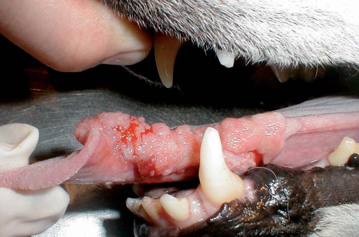
The authors

Rodolfo Brühl Day (coord.)
Dr Brühl Day (DVM) graduated from the Facultad de Ciencias Veterinarias (University of
Buenos Aires, Argentina) in 1977, with honours (Magna cum Laude) and Gold Medal for
best GPA. After a Residency in Small Animal Surgery in the Veterinary Medical Teaching
Hospital (University of California, Davis) in 1984, he has become a Charter Diplomate in
Small Animal Surgery from the Universidad de Buenos Aires (1998), specialist in Universi-
ty Teaching with orientation to Veterinary and Biological Sciences (2000), and a Diplomate
of the Latin-American College of Veterinary Ophthalmologists (2002).

He has taught in several universities throughout his extensive career (Universidad de Bue-
nos Aires, Facultad de Ciencias Veterinarias, Buenos Aires; University of California, Davis,
School of Veterinary Medicine, California, United States; and Ross University, School of
Veterinary Medicine, Saint Kitts, West Indies). Since 2008 he is Professor of Small Ani-
mal Surgery, Director of the Small Animal Medicine and Surgery Academic Program , and
Staff Surgeon at the Small Animal Clinic in St. George’s University (School of Veterinary
Medicine, Grenada, West Indies).

Dr Brühl Day has been awarded with many scholarships, awards and distinctions and has
contributed in a number of publications in books, journals and handouts. He has also par-
ticipated in courses, seminars and taken several CE courses throughout his career. Since
1995 he is a member of the Editorial Board of the scientific section Selecciones Veterina-
rias of Editorial Intermédica, Buenos Aires.

María Elena Martínez
Dr Martínez (DVM) graduated from the Facultad de Ciencias Veterinarias (University of
Buenos Aires, Argentina) in 1991. As a specialist in Small Animals Surgery and Anaesthe-
siology, she has been tutoring and teaching in the University of Buenos Aires from 1998 to
2006. In 2002, she became a Diplomate in Small Animal Surgery and is currently Head of
the Surgery Service in the course on Veterinary Neurology. She has gained experience in
several countries like United States (Missouri University), Brasil (Universidade do Estado
de Santa Catarina), and Colombia (Fundación Universitaria San Martín). She is a member
of Neurolatinvet and a founding member of Neurovet-Argentina (Argentinean Association
of Veterinary Neurologists).
The gastrointestinal tract. Clinical cases

Pablo Meyer
Dr Pablo Meyer (DVM) graduated from the Facultad de Ciencias Veterinarias (Universi-
ty of Buenos Aires, Argentina) in 1986. Since 2003, he is a Diplomate in Small Animal
Surgery, and lecturer on skin surgery and reconstruction in the specialisation course
on Surgery in small animals. He is also a surgeon of the Surgery Service of the Tea-
ching Hospital of the Facultad de Ciencias Veterinarias of the University of Buenos Aires
(HEMV-UBA), and lecturer at the Service of Oncology. Author of various works in this
field, he has participated in several conferences and contributed in specialised journals
focusing in surgery and oncology.

Collaborators
José Rodríguez, DVM, PhD
Graduate in Veterinary Medicine from the Complutense University of Madrid, Spain. Head
Tutor of the Department of Animal Pathology, University of Zaragoza, Spain. Veterinary
surgeon, Hospital Veterinario Valencia Sur, Valencia, Spain.

Sandra Mattoni, DVM
Resident Limited Status, Emergency and Critical Care, UC-Davis, California, US. Assistant
Professor, Small Animal Medicine, St. George’s University - School of Veterinary Medicine.
Grenada, West Indies. Medical Director, Centro de Cuidados Intensivos y Emergencias,
Buenos Aires, Argentina.

Eduardo Durante, BVSc, BVSc(Hons), MedVet, DVSc
Professor, Small Animal Surgery, Universidad Nacional de la Plata, Provincia de Bue-
nos Aires, Argentina. Professor, Small Animal Surgery and Senior Associate Dean, St.
George’s University - School of Veterinary Medicine, Grenada, West Indies.

Francesca Ivaldi, DVM, MSc
Associate Professor, Small Animal Surgery, St. George’s University - School of Veterinary
Medicine, Grenada, West Indies.
Communication services
Web site
 Online visualisation of the sample chapter.

 Presentation brochure in PDF format.

 Author´s CV.

 Sample chapter compatible with iPad.

 www.grupoasis.com/promo/gastrointestinal_surgery_cc
Small animal surgery            Rodolfo Brühl Day (Coordinator)
                                 María Elena Martínez
                                 Pablo Meyer
                                 José Rodríguez Gómez

Lips

Tongue

Oesophagus

Stomach

Pancreas

Liver

Gallbladder

Mesentery

Intestines

Surgery atlas, a step-by-step guide

The gastrointestinal tract
CliniCal Cases
Table of contents
1. Cases involving the oral cavity       3. Cases involving the digestive
   and pharynx                              organs in the abdomen
   Lip neoplasia                            Stomach foreign body
   Zygomatic gland mucocoele                Canine acute gastric dilatation-volvulus
   Linear foreign body entrapped under      Y-U pyloroplasty
   the tongue in a cat
                                            Chemical peritonitis due to traumatic
   Severe facial trauma                     rupture of the pancreas
   Cricopharyngeal achalasia                Mesenteric torsion
   Glossectomy                              Duodenal foreign body
   Transverse glossectomy                   Extrahepatic shunt
   Wedge glossectomy                        Multiple extrahepatic shunts
                                            and intrahepatic shunt
2. Cases involving the thoracic             Biliary peritonitis associated
   oesophagus                               with extrahepatic biliary rupture
                                            Biliary mucocoele
   Oesophageal foreign body in a dog
                                            Rupture of the gallbladder
   Linear foreign body in a cat
                                            Gallbladder lithiasis
   Combined technique for removal
   of a foreign body                        Caecal neoplasia
   Megaoesophagus                           Splenic torsion
   Hiatal hernia
                                         4. Techniques applied in
                                            gastrointestinal disorders
                                            Mouth examination
                                            Oesophagostomy tube placement for
                                            feeding (E-tube)
                                            Jejunostomy tube placement for feeding
                                            (J-tube)
Oral cavity and pharynx / Zygomatic gland mucocoele
Zygomatic gland Parotid gland
9
Sublingual gland Mandibular gland
Fig. 2. Salivary glands of the dog (with the zygomatic bone excised). Note the position of the zygomatic salivary gland in the orbital area.
Surgical preparation
After the placement of a peripheral intrave-
nous catheter, anaesthesia was induced
and, with the patient ready for intubation,
a non-painful bulge with an uneven surface
was observed in the aboral buccal vestibule
of the oral cavity. The oral mucosa in the
bulging area was slightly oedematous and
damaged due to self-chewing (Fig. 3).
Fig. 3. Patient intubated and mucocoele located at
the buccal vestibule of the oral cavity (arrow).
01_Head_neck.indd 9 02/06/15 09:18The gastrointestinal tract CliniCal Cases
Glossectomy Rodolfo Brühl Day, María Elena Martínez, Pablo Meyer
Prevalence
Technical difficulty
■■ Partial or total resection of the tongue. Case history
■■ Indicated for wounds, neoplasia, and/or
Name Helga
necrosis.
Species canine
Breed Samoyed
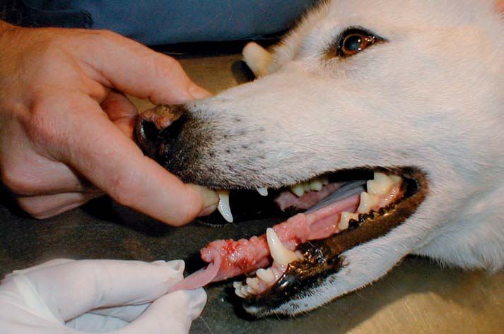
Clinical signs: difficulty to eat, intermittent bleeding from Sex female, spayed
the mouth.
Age 8 years old
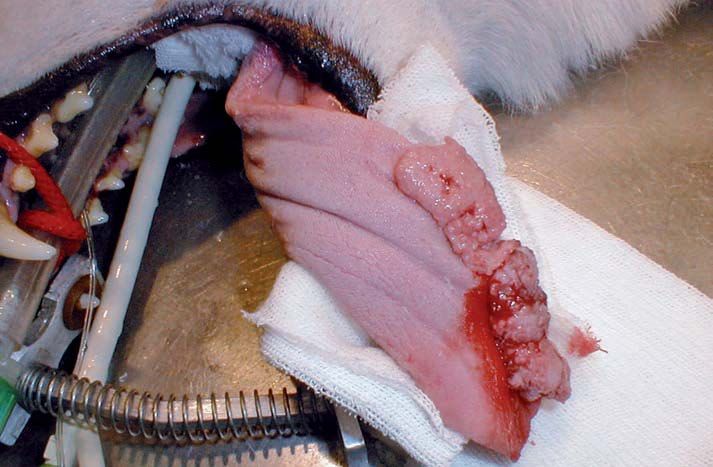
Physical examination
A short-acting anaesthesia allowed a thor-
ough evaluation of the patient, including the
aspect of the lesion, its extent (Fig. 1), the
presence of other disease manifestations
and involvement of regional lymph nodes.
34
Fig. 1. A thorough examination under general
anaesthesia is required in these cases.
The tumour occupied about 30 % of the
length of the tongue (left side), while 70 % of
it remained unaffected (Figs. 2 and 3).
Fig. 2. Detail of the tumour occupying the tongue.
01_Head_neck.indd 34 02/06/15 09:19Oral cavity and pharynx / Glossectomy
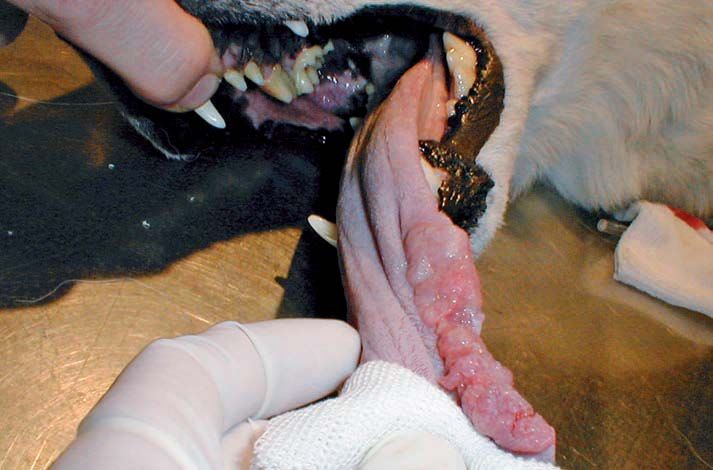
Surgical preparation
This is a clean-contaminated surgery because the surgical proce- The surgical field was prepared with an antiseptic solution of povi-
dure is performed in the oral cavity. done iodine 1:10 or chlorhexidine 1:30 diluted in saline. The whole
oral cavity was cleaned several times (Fig. 4), being careful enough
to block the pharynx with rolled gauze sponges to prevent fluid
See Table 1 in the case aspiration.
page 41
Transverse glossectomy
Physical examination prior to
surgery is of utmost importance.
35
Fig. 3. Size of the neoplasm, which affects nearly
30 % of the length of the tongue.
Fig. 4. Preparation for the surgery includes cleaning
the mouth with a diluted antiseptic solution.
9 01_Head_neck.indd 35 02/06/15 09:19The gastrointestinal tract CliniCal Cases
Surgical technique
Once the abdominal cavity was entered, the FB was located in the
ascending duodenum, which was exteriorised. Moistened laparot- This surgical procedure has three stages (aseptic/septic/aseptic)
omy sponges were placed surrounding the bowel loop to minimise
intestinal spillage into the cavity. In this procedure, the laparotomy
Aseptic stage. Moistened 4 × 4 gauze sponges are placed
sponge closer to the surgeon will receive the bowel loop for ease
around the exteriorised duodenum until it is incised.
of handling. Holding the intestinal loop close to the midline should
be avoided to prevent any intestinal content from leaking into the
abdominal cavity. Septic stage. Duodenotomy and FB removal.
Once the FB is found (Fig. 3), the rest of the small and large Aseptic stage. Once the sponges are removed and the gloves
bowel must be examined due to the possible presence of changed, the duodenum is closed. A new set of instruments,
another FB that may go unnoticed otherwise. small pack, will be used for the abdominal closure.
116
Fig. 3. The bowel loop is dilated cranial to the FB,
but has a normal size caudal to it.
Once the affected duodenum is isolated
(Fig. 4), the intestinal content (chyme) is
gently milked away from the lumen of the
duodenum. This manoeuvre minimises
spillage of chyme during the enterotomy
procedure.
Fig. 4. Isolated and packed segment of duodenum,
prepared to be incised.
03_Abdomen.indd 116 02/06/15 09:14Abdomen / Duodenal foreign body
To reduce the spillage of chyme, the intesti-
nal lumen must be clamped proximally and
distally before the enterotomy site is incised.
The assistant surgeon will place the index
and middle fingers of both hands in a scis-
sor-like grip at about 4 cm from each end to
achieve and carry out an atraumatic lumen
occlusion (Fig. 5). Doyen intestinal forceps
can also be used for the same purpose.
Fig. 5. Before the duodenum is incised, the assistant
uses the index and middle fingers of both hands to
clamp the intestinal lumen cranially and caudally to
the FB.
Do not use the thumb and index
fingers since they can apply too
much pressure to the intestinal
wall. Small (baby) Doyen intestinal
forceps are a better option for the
delicate duodenal wall and the
occasional lack of adequate space
within the abdominal cavity.
117
The incision is generally made in a healthy
segment of the intestine (Fig. 6). Then, the FB
has to be gently removed through this open-
ing. The length of the incision has to be made
according to the size of the FB to allow a
smooth removal without unnecessary traction
against the incised edges of the intestinal wall.
Fig. 6. Bowel wall incision with scalpel.
In this case, the extent of the incision had to
be enlarged. The surgeon extended it along
the long axis of the intestine using Metzen-
baum scissors to ensure the FB could be
removed without tearing the intestinal wall
(Figs. 7 and 8). A scalpel can also be used
in such cases.
Fig. 7. Enlargement of the incision in the intestinal
wall using scissors.
4 03_Abdomen.indd 117 02/06/15 09:14The gastrointestinal tract CliniCal Cases
Using a finger-trap pattern allows overlapping sutures to
The tube should be secured to the skin with a finger-trap suture pat- be tightened when pulling on the tube, thus decreasing any
tern using non-absorbable material. A syringe needle can be used possibility of removal.
to thread it through skin and around the tube (Fig. 15).
a b
c d
184
Fig. 15. (a) First, a suture loop is tied loosely to the skin, then around the tube in a finger-trap pattern. (b) Detail of the knot. (c) A cap is placed to close the tube, thus
preventing air from going into the oesophagus and stomach. (d) Completed finger-trap suture.
Neck bandage
Next step would be to protect the tube and skin incision with a neck
bandage, which has to be loose enough to allow free neck and
head movement. Having the distal end of the tube in a dorsal posi-
tion will facilitate to feed and medicate the patient through the tube.
Figures 17-26 show how to apply a neck bandage step by step.
Fig. 16. Immediate postoperative period. Oesophagostomy tube in place.
04_tecnique.indd 184 02/06/15 09:24Techniques / Oesophagostomy tube placement for feeding (E-tube)
Fig. 17. Two 4 × 4 gauze sponges are cut as shown. Fig. 18. Antibiotic ointment is applied to the skin incision.
185
Fig. 19. The gauze sponges are placed around the tube in opposite directions.
Fig. 20. The neck is bandaged to further protect the tube and assist local wound Fig. 21. The bandage has to be applied in a loose manner.
healing by preventing any contamination.
4 04_tecnique.indd 185 02/06/15 09:24The gastrointestinal tract CliniCal Cases
Omentum may be interposed between the jejunal loop and the
abdominal wall to increase adherence. Once inside the abdominal The feeding tube/catheter must always be inserted
cavity, the needle is passed through the wall of the selected jejunal following the direction of ingesta flow.
loop, entering through its antimesenteric side and exiting distally a
few centimetres further.
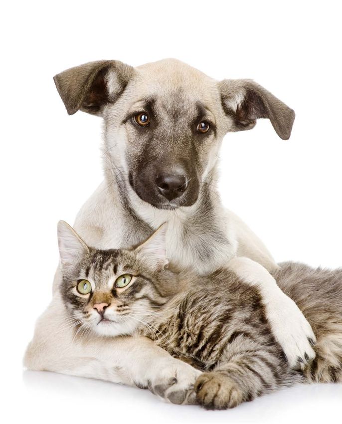
Since the catheter must always be inserted in an isoperistaltic di-
rection (same direction of ingesta flow), the needle must enter the The tube/catheter is fed into the needle again and passed through
bowel loop in an antiperistaltic direction (opposite direction of in- the intestinal lumen. The needle is then removed while the catheter
gesta flow). remains inside the jejunal lumen (Fig. 4).
a
Distal
190
Proximal
b
Distal
Proximal
Fig. 4. Insertion of a 5-Fr feeding tube/catheter into
the intestinal lumen using a 10-G needle.
04_tecnique.indd 190 02/06/15 09:25The publishing strength of Grupo Asís Editorial Servet, a division of Grupo Asís, has become one of the reference publishing com- panies in the veterinary sector worldwide. More than 15 years of experience in the publis- hing of contents about veterinary medicine guarantees the quality of its work. With a wide national and international distribution, the books in its catalogue are present in many diffe- rent countries and have been translated into nine languages to date: English, French, Por- tuguese, German, Italian, Turkish, Japanese, Russian and Chinese. Its identifying characteristic is a large multidisciplinary team formed by doctors and graduates in Veterinary Medicine and Fine Arts, and specialised designers with a great knowledge of the sector in which they work. Every book is subject to thorough technical and linguistic reviews and analyses, which allow the creation of works with a unique design and excellent contents. Servet works with the most renowned national and international authors to include the topics most demanded by veterinary surgeons in its catalogue. In addition to its own works, Servet also prepares books for companies and the main multinational companies in the sector are among its clients.
Servet (División de Grupo Asís Biomedia S.L.)
         Centro Empresarial El Trovador, planta 8, oficina I
    Plaza Antonio Beltrán Martínez, 1 • 50002 Zaragoza (Spain)
Tel.: +34 976 461 480 • Fax: +34 976 423 000 • www.grupoasis.com
You can also read