Primary aortic intimal sarcoma masquerading as intramural hematoma
←
→
Page content transcription
If your browser does not render page correctly, please read the page content below
Open Medicine 2021; 16: 1306–1310
Case Report
Xiaodong Fan#, Xiaofeng Chen#, Zhiqi Yang*#, Tianhui Zhang, Yuting Liao, Weixiong Fan,
Xiangguang Chen
Primary aortic intimal sarcoma masquerading as
intramural hematoma
https://doi.org/10.1515/med-2021-0337
received March 31, 2021; accepted July 24, 2021
1 Introduction
Abstract: Primary aortic intimal sarcoma is a very rare Primary aortic intimal sarcoma is a very rare disease with
disease and most patients present with features similar a poor prognosis [1,2]. The most frequently reported clin-
to those of atherosclerotic plaque and thrombus; how- ical symptoms are caused by embolic events, and most of
ever, primary aortic intimal sarcoma presents with fea- them present with features similar to those of athero-
tures similar to those of intramural hematoma (IMH) on sclerotic plaque and thrombus [2–7]. Here we report a
CT imaging and clinical presentation had never been pre- case of primary aortic intimal sarcoma masquerading as
viously reported. Here we report a case involving a 49- intramural hematoma (IMH) on CT imaging and clinical
year-old woman with primary aortic intimal sarcoma presentation and discuss the reasons for misdiagnosis.
masquerading as IMH on radiological images and clinical
presentation. We also discuss some of the diagnostic pit-
falls and hope that these diagnostic pitfalls will be very
useful for clinicians. 2 Case presentation
Keywords: thoracic aorta, intimal sarcoma, intramural A 49-year-old woman visited the emergency room for
hematoma acute chest pain radiating to the back. She had known
hypertension: the blood pressure in the left upper limb,
right upper limb, left lower limb, and right lower limb
was 210/65, 218/60, 176/90, and 175/90 mm Hg, respec-
tively. The D-dimer level was normal at initial admission.
An electrocardiogram showed a normal sinus rhythm.
Considering that the patient had typical clinical symp-
# Xiaodong Fan, Xiaofeng Chen and Zhiqi Yang contributed equally toms of acute chest pain radiating to the back, with
to this work. asymmetry of limb blood pressures, and a normal
D-dimer level and electrocardiogram, the diagnosis of
IMH was suspected. Chest nonenhanced computed tomo-
* Corresponding author: Zhiqi Yang, Department of Radiology,
Meizhou People’s Hospital, Meizhou, 514031, China; Guangdong
graphy (CT) was performed, which showed crescent-shaped
Provincial Key Laboratory of Precision Medicine and Clinical thickening of the thoracic aorta wall with the same
Translational Research of Hakka Population, Meizhou, 514031, attenuation as that of the lumen and linear calcification
China; Guangdong Provincial Engineering and Technology Research ingression (Figure 1a). Computed tomography angiography
Center for Molecular Diagnostics of Cardiovascular Diseases, (CTA) revealed an intimal flap and expanded false lumen
Meizhou, 514031, China, e-mail: y13643090854@163.com
with slight enhancement (Figure 1b–d). Digital subtrac-
Xiaodong Fan, Xiaofeng Chen, Tianhui Zhang, Weixiong Fan,
Xiangguang Chen: Department of Radiology, Meizhou People’s tion angiography showed mild stenosis in the thoracic
Hospital, Meizhou, 514031, China aortic lesion without an obvious intimal flap (Figure 2a).
Xiaofeng Chen, Xiangguang Chen: Guangdong Provincial Key Based on both the clinical symptoms and diagnostic test
Laboratory of Precision Medicine and Clinical Translational results, only thoracic IMH was considered by both radio-
Research of Hakka Population, Meizhou, 514031, China
logists and cardiothoracic surgeons; hence, thoracic
Xiaofeng Chen, Xiangguang Chen: Guangdong Provincial
Engineering and Technology Research Center for Molecular
endovascular aortic repair and not surgery was consi-
Diagnostics of Cardiovascular Diseases, Meizhou, 514031, China dered at that time. Then, an endovascular stent (30 mm ×
Yuting Liao: GE Healthcare, Guangzhou, 510623, China 200 mm, Medtronic) was implanted into the patient.
Open Access. © 2021 Xiaodong Fan et al., published by De Gruyter. This work is licensed under the Creative Commons Attribution 4.0
International License.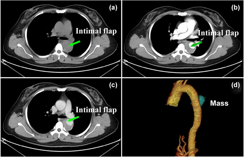
Primary aortic intimal sarcoma masquerading as IMH 1307
Figure 1: Axial nonenhanced CT images (a) showing a crescent-shaped thickening of aortic wall with the same attenuation as that of the
lumen and intimal flap. CTA (c and d) showing an intimal flap and expanded false lumen with slight enhancement, and correspond
reconstructed image (d) showing a mass-like false lumen, which was interpreted by radiologists as IMH.
Figure 2: Digital subtraction angiography image showing mild stenosis in the thoracic aortic lesion without an obvious intimal flap (a) and
the implanted stent (b).
Completion angiography demonstrated that the thoracic other medical centers during follow-up. However, seven
aorta and branches were patent, with no obvious endo- months later, the patient was readmitted to the hospital
leak or extravasation (Figure 2b). The prevalence of with complaints of dysphagia and dyspnea. Subsequent
acute chest pain radiating to the back decreased after magnetic resonance imaging (Figure 4) showed gradual
treatment, and the patient recovered well and was dis- enlargement of the false lumen with mediastinal exten-
charged home. sions and new lesions in the azygos vein. Angiogenic
Three months after endovascular stent graft implan- hemangioma was suspected, and endobronchial ultra-
tation, follow-up CTA (Figure 3) showed enlargement of sound biopsy was performed to confirm the lesion. Histo-
the false lumen with nonhomogeneous enhancement pathological evaluation indicated an intimal sarcoma
and mediastinal extensions. The patient was interpreted (Figure 5). Immunohistochemical staining for vimentin,
by radiologists as having an endoleak and periaortic/ CD10, CD68, and CD99 was positive, whereas that for
mediastinal hematoma, which were also considered by S-100, CD56, desmin, CD30, CD15, leukocyte common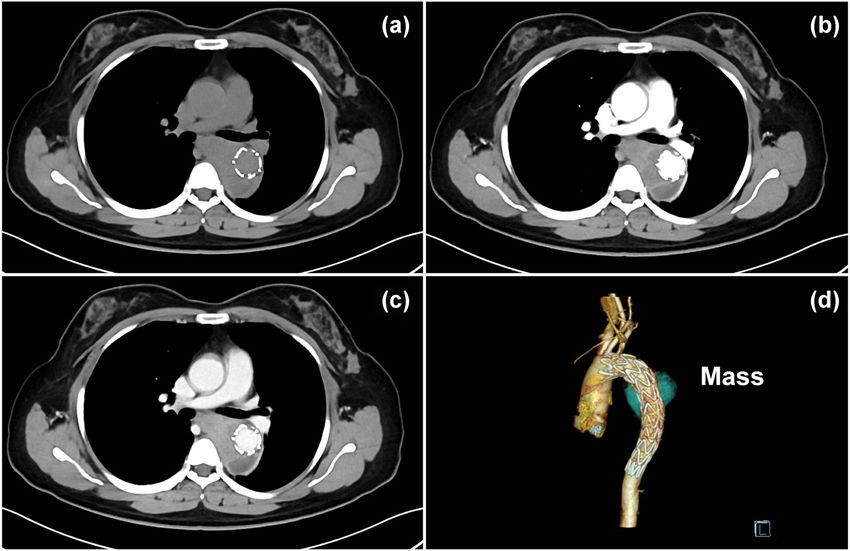
1308 Xiaodong Fan et al. Figure 3: Axial nonenhanced CT image (a) and CTA (b–d) showing enlargement of the false lumen three months after stent implantation, with nonhomogeneous enhancement and mediastinal extensions of the false lumen, which was interpreted by radiologists in our hospital and other medical centers during follow-up as an endoleak and periaortic hematoma. Figure 4: Magnetic resonance imaging showing gradual enlargement of false lumen with mediastinal extensions and a new lesion in the azygos vein. T1WI image (a) and T2WI image (d) showing the heterogeneous of “false lumen” with bleeding area (green arrow). Contrast- enhanced T1WI image (b) showing a nonhomogeneous enhancement of the “false lumen,” which correspond to the hyperintense regions in DWI (b = 800 s/mm2) image (c).
Primary aortic intimal sarcoma masquerading as IMH 1309
of the aorta on the non-contrast CT (Figure 1a and b) and
that was interpreted by radiologists as “intimal flap.”
According to the previously reported cases, the diag-
nosis of IMH depends on the identification of the intimal
tear, the false lumen, and the presence of most hyper-
dense intramural thrombus on unenhanced images [8–11].
However, their following features differed from those on
previous reports of IMH: First, the “false lumen” was lim-
ited in length and made it seem more like a mass, although
it had an intimal flap on CT images. Second, in contrast to
the typical feature of obviously enhanced blood in the
false lumen, the false lumen was filled with a slight and
nonhomogeneous enhancement mass. Third, the “false
lumen” tended to grow extravascularly, which manifests
as an aggressive tumor. In this case, stent implantation
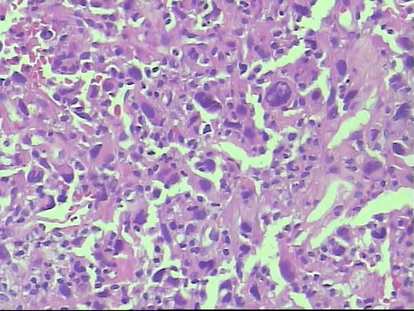
Figure 5: Histopathology of aortic tissues showed that the tumor
was composed of malignant spindle cells and demonstrated nuclear might have further facilitated sarcoma invasion into the
pleomorphism and atypia (hematoxylin and eosin staining ×200). mediastinum. Fourth, the aortogram demonstrated mild
stenosis in the thoracic aorta lesion without severe athero-
sclerotic changes in the aortic wall and obvious intimal
antigen, smooth muscle actin, CD34, CD61, and CD5 was
flap. Furthermore, the following features with regard to a
negative. Afterwards, she received adjuvant chemotherapy
leak and periaortic hematoma after stent graft implanta-
with a combined treatment of ifosfamide and epirubicin;
tion differed from those on previous reports [12,13]: first,
however, the general condition of the patient rapidly dete-
there was no open channel inside the stent graft and no
riorated after 1 cycle of adjuvant chemoradiotherapy, and
obvious backflow in the “false lumen.” Second, the “false
she died exactly 19 months after the initial hospitalization.
lumen” gradually became enlarged, increasing the ten-
dency of mediastinal invasion. Third, MRI showed a hetero-
Informed consent: Informed consent was obtained from
geneous mass with a false lumen and a new lesion in the
the patient’s family.
azygos vein. To our knowledge, no similar case has pre-
viously been reported in the English and Chinese literature.
According to previous studies, the prognosis of pri-
mary aortic intimal sarcoma is dismal [1,14–18]. Consis-
3 Discussion tent with these studies, the overall survival in this case
was 19 months, even after treatment with adjuvant che-
Primary aortic intimal sarcoma can be diagnosed only by motherapy. Therefore, clinicians must be vigilant for
postoperative histopathology and immunohistochemical primary aortic intimal sarcoma in patients with IMH
markers, while its preoperative diagnosis is challenging manifesting as acute chest and back pains, a localized
because of various clinical manifestations and no specific false lumen and intimal flap, slight and nonhomo-
imaging features. Based on previous case reports [2–7], geneous enhancement of the false lumen, and extra-
aortic intimal sarcoma often presents with features similar vascular extensions on CT images.
to those of atherosclerotic plaque and thrombus on CT
images, and the most frequently reported clinical symp- Acknowledgments: We are thankful to the patient and
toms are caused by embolic events. In contrast to prior patient’s family for cooperation and allowing us to use
studies, acute chest and back pain with asymmetry of patient’s medical records in our case report.
limb blood pressures and an expanded false lumen with
an intimal flap were the only features observed on CT Funding information: This study was partly supported by
images. Thus, as these features were similar to those of the Medical Research Foundation of Guangdong Province
IMH, this led to the confident diagnosis of “mimicking (B2021280).
IMH.” An episode of acute chest and back pains might
be explained by micro-calcification/micro-hemorrhage of Author contributions: Z.Q.Y. put forward the study
the tumor, which showed as a hyperintense foci at the center concepts, then X.D.F. and X.F.C. designed the study.1310 Xiaodong Fan et al.
Data acquisition was done by X.D.F. and X.F.C., T.H.Z., [7] Wu ZY, Weng LQ, Chen ZG, Chen YX, Li YJ. Primary aortic sar-
Y.T.L., W.X.F., and X.G.C. were responsible for proof- coma in arch and descending aorta: a case report and litera-
reading. All the authors were involved in data analysis ture review. J Thorac Dis. 2018;10(4):E289–95.
[8] Desinan L, Scott CA, Piai I, Mazzolo GM. Sudden death due to
and interpretation. X.D.F., X.F.C., and Z.Q.Y. were the
spontaneous rupture in splenic artery atypical dissection with
major contributors and contributed equally in writing features of vasculitis: case report and review of the literature.
the manuscript. All the authors read and approved the Forensic Sci Int. 2010;200(1–3):e1–5.
final manuscript. [9] Fisher ER, Stern EJ, Godwin JD 2nd, Otto CM, Johnson JA. Acute
aortic dissection: typical and atypical imaging features.
Radiographics. 1994;14(6):1263–71. discussion 71–4.
Conflict of interest: The authors declare that they have no
[10] Lawal OJ, Dhindsa HS, Loyd JW. A patient with altered mental
competing interests. status and possible seizure reveals an atypical aortic
dissection upon workup. Am J Emerg Med.
Data availability statement: The data cohorts used and/ 2014;32(5):488.e1–2.
or analyzed for the present study are available from the [11] Golledge J, Eagle KA. Acute aortic dissection. Lancet.
2008;372(9632):55–66.
corresponding author on reasonable request.
[12] Rohlffs F, Tsilimparis N, Mogensen J, Makaloski V, Debus S,
Kölbel T. False Lumen occlusion in chronic aortic dissection:
the new generation candy-plug II. Ann Vasc Surg.
2019;57:261–5.
References [13] Afzal AM, Alsahhar J, Podduturi V, Schussler JM.
Undifferentiated Intimal sarcoma of the inferior vena cava with
[1] Mo H, Kwon HM, Choi JS, Ahn SJ, Lee YS. Multiple embolic extension to the right atrium and renal vasculature. Case Rep
infarction due to a primary aortic intimal sarcoma. J Stroke. Cardiol. 2015;2015:812374.
2016;18(3):358–60. [14] Riles E, Gupta S, Wang DD, Tobin K. Primary cardiac angio-
[2] Staats P, Tavora F, Burke A. Intimal sarcomas of the aorta and sarcoma: a diagnostic challenge in a young man with recurrent
iliofemoral arteries: a clinicopathological study of 26 cases. pericardial effusions. Exp Clin Cardiol. 2012;17(1):39–42.
Pathology. 2014;46(7):596–603. [15] Rusthoven CG, Liu AK, Bui MM, Schefter TE, Elias AD, Lu X,
[3] Akiyama K, Nakata K, Negishi N, Henmi A. Intimal sarcoma of et al. Sarcomas of the aorta: a systematic review and pooled
the thoracic aorta; clinical-course and autopsy finding. Ann analysis of published reports. Ann Vasc Surg.
Thorac Cardiovasc Surg. 2005;11(2):135–8. 2014;28(2):515–25.
[4] Rhee MY, Myong NH, Park YB. Primary intimal sarcoma of the [16] Schmehl J, Scharpf M, Brechtel K, Kalender G, Heller S,
aorta: role of transesophageal echocardiography. Circ J. Claussen CD, et al. Epithelioid angiosarcoma with metastatic
2002;66(1):111–3. disease after endovascular therapy of abdominal aortic
[5] Sebenik M, Ricci A Jr, DiPasquale B, Mody K, Pytel P, Jee KJ, aneurysm. Cardiovasc Intervent Radiol. 2012;35(1):190–3.
et al. Undifferentiated intimal sarcoma of large systemic blood [17] Vinod P, Jabri A, Hegde V, Lahorra J, Cutler D. functional mitral
vessels: report of 14 cases with immunohistochemical profile stenosis: imposture of primary cardiac intimal sarcoma.
and review of the literature. Am J Surg Pathol. Cardiol Res. 2018;9(5):307–13.
2005;29(9):1184–93. [18] Shimogawara T, Ono S, Kobayashi K, Sasaki A, Shimizu H,
[6] Thalheimer A, Fein M, Geissinger E, Franke S. Intimal Matsui J. Aortic sarcoma mimicking a mycotic aneurysm in the
angiosarcoma of the aorta: report of a case and review thoracoabdominal aorta. J Vasc Surg Cases Innov Tech.
of the literature. J Vasc Surg. 2004;40(3):548–53. 2019;5(4):593–6.You can also read