Recombinant Oil-Body-Expressed Oleosin-hFGF5 in Arabidopsis thaliana Regulates Hair Growth - MDPI
←
→
Page content transcription
If your browser does not render page correctly, please read the page content below
G C A T
T A C G
G C A T
             genes
Article
Recombinant Oil-Body-Expressed Oleosin-hFGF5 in
Arabidopsis thaliana Regulates Hair Growth
Hongyu Wang 1,2 , Xinxin Lan 1 , Muhammad Noman 1, * , Ze Wang 1 and Jing Zhang 1

                                         1   College of Life Sciences, Jilin Agricultural University, Changchun 130118, China
                                         2   Key Laboratory of Straw Biology and Utilization, The Ministry of Education, Changchun 130118, China
                                         *   Correspondence: noman@jlau.edu.cn

                                         Abstract: FGF5 (Fibroblast Growth Factor) is a member of the fibroblast growth factor family, which
                                         not only regulates growth and development but also inhibits hair regeneration. The oil-body expres-
                                         sion vector pOTB-hFGF5 was constructed by the genetic engineering method and it was transformed
                                         into Arabidopsis by flora dip. T3 homozygous transgenic Arabidopsis was obtained after screening and
                                         propagation by the PCR and Western blot methods. The recombinant oil-body-expressed oleosin-
                                         hFGF5 can inhibit the proliferation of hair follicle epithelial cells and it exhibits the pharmacological
                                         activity of inhibiting hair regeneration in vivo by protein hybridization and imunohistochemistry. At
                                         the same time, the potential mechanism of recombinant oil-body-expressed oleosin-hFGF5 inhibiting
                                         hair growth was also revealed by RNA-Seq. This implies that the recombinant oil-body-expressed
                                         oleosin-hFGF5 has a good effect on inhibiting hair growth.

                                         Keywords: Arabidopsis; recombinant oil-body; oleosin-hFGF5; hair growth; RNA-Seq

                                         1. Introduction
                                              The oil body is a sub-organelle of 0.5–2.5 µm in oil crop seed [1], which is a spheroid
                                         composed of oil-body-associated proteins, triacylglycerol (TAG) and a monolayer of phos-
                                         pholipids [2]. Oil-body-associated proteins include caleosin, oleosin and steroleosin.
                                         Among them, oleosin is composed of the C-terminus of the variable region, the N-terminus
Citation: Wang, H.; Lan, X.; Noman,
                                         of the variable region and the middle hydrophobic region [3]. Therefore, the oil body
M.; Wang, Z.; Zhang, J. Recombinant
                                         expression system was established based on the structural characteristics of oleosin, which
Oil-Body-Expressed Oleosin-hFGF5
                                         is an efficient expression system for foreign proteins. The target gene is fused downstream
in Arabidopsis thaliana Regulates Hair
                                         of the oleosin gene and the promoter drives the expression of the oleosin-target gene in
Growth. Genes 2023, 14, 21. https://
                                         the transgenic seeds [4]. Since the fusion gene is expressed on the surface of the oil body, it
doi.org/10.3390/genes14010021
                                         is embedded with the target protein and can be separated by centrifugation. Meanwhile,
Academic Editor: Paola Vittorioso        based on its structural characteristics, the oil body has a function similar to that of liposome
Received: 24 October 2022
                                         and can carry functional substances as the delivery carrier. For example, a new transdermal
Revised: 2 December 2022
                                         drug delivery system (oil-body-linked oleosin-hEGF) was established, which accelerates
Accepted: 16 December 2022
                                         wound healing [5], and the specific expression of oleosin-hFGF10 in safflower oil body
Published: 22 December 2022              can not only promote the transdermal absorption but also significantly improve the effi-
                                         cacy of hFGF10 [6]. aFGF, bFGF and hEGF were expressed in the oil body using oleosin
                                         fusion technology, which has the effect of reducing the wound size and promoting wound
                                         healing [7–9]. The growth factors with poor stability are easily hydrolyzed by protease
Copyright: © 2022 by the authors.        in vivo and need to be stored at low temperature. However, seeds lose a lot of water after
Licensee MDPI, Basel, Switzerland.       maturation, which can weaken the hydrolysis of oil-body-related proteins by enzymes.
This article is an open access article
                                         Therefore, the growth factor can be expressed in seeds through oil body fusion technology
distributed under the terms and
                                         and can be stored in seeds for a long time.
conditions of the Creative Commons
                                              Fibroblast growth factor 5 (FGF5) is a member of the fibroblast growth factor family,
Attribution (CC BY) license (https://
                                         which has different biological effects when expressed in different tissues [10,11]. FGF5
creativecommons.org/licenses/by/
                                         promotes embryonic development in the early stage and the regeneration of endothelial
4.0/).

Genes 2023, 14, 21. https://doi.org/10.3390/genes14010021                                                    https://www.mdpi.com/journal/genes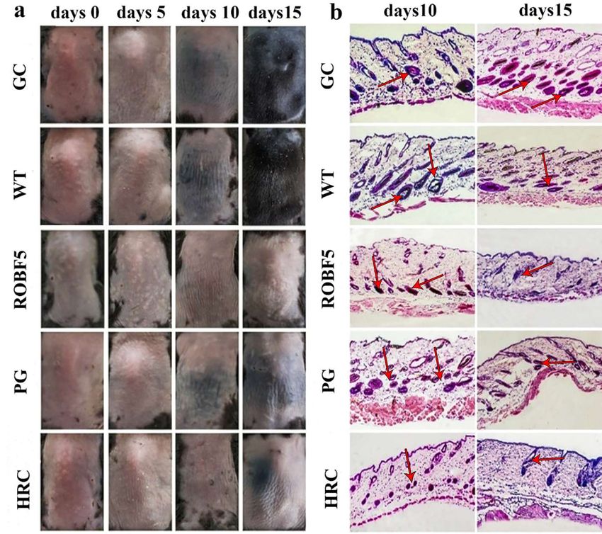
Genes 2023, 14, 21                                                                                            2 of 11

                     cells and accelerates the repair of vascular injury [12]. The most common biological function
                     of FGF5 is to regulate hair growth, so it is an important hair growth regulator [13,14], and
                     excessive hair growth occurs when FGF5 is knocked out [15]. The growth cycle of hair
                     is mainly divided into three stages, which are the growth stage, the degenerative stage
                     and the resting stage [16]. All stages are affected by various growth factors, and FGF5 is
                     involved in the regulation of the hair growth cycle [17]. The control of hair growth length
                     by FGF5 may be related to the control of the hair growth cycle [18]. It was confirmed
                     that FGF5 could promote the hair follicle from the growth phase to the rest phase as soon
                     as possible by the culture of hair follicle cells, so that the growth of the hair follicle was
                     inhibited. FGF5 can control the hair growth cycle, but it is unstable and easy to be degraded.
                     Therefore, the production method of FGF5 can be changed by oil body fusion technology
                     to improve its stability.
                           In this study, the oleosin-hFGF5 fusion gene was expressed on the surface of the oil
                     body in Arabidopsis thaliana. The activity of the recombinant oil body expressing hFGF5
                     was confirmed by cell proliferation in vitro and hair inhibition in vivo. At the same time,
                     the mechanism of recombinant oil body expressing hFGF5 and inhibiting hair growth was
                     analyzed to provide a reference for its application and development by RNA-Seq.

                     2. Materials and Methods
                     2.1. Construction of pOTB-rhFGF5 Vector
                          The human FGF5 (hFGF5) gene was optimized and synthesized by Sangon Biological
                     Company (Gene ID: 2250). The gene of hFGF5 was digested by Nco I and Hind III and
                     ligated to the pOTB vector using ligase T4 , and the recombinant vector pOTB-hFGF5 was
                     successfully constructed (Figure S7). It was transformed into Agrobacterium tumefaciens
                     EHA105 cells and the positive monoclonal strains were screened and identified by PCR
                     to prepare engineering strains for infection. PCR-specific primers are as follows: forward
                     primer 50 CATATGCACGGGGAGA30 and reserve primer 50 AAGCTTATCCAAAGCG 30 .
                     PCR reaction process: pre-denaturation at 95 ◦ C for 5 min, denaturation at 95 ◦ C for 30 s,
                     anneal at 56 ◦ C for 30 s, amplification for 30 cycles and extension at 72 ◦ C for 10 min.

                     2.2. Arabidopsis Transformation and Screening of Transgenic Lines
                           EHA105 cells were collected at 5000 rpm for 10 min and dissolved in floral dip solution
                     with OD600 value up to 0.8. Arabidopsis was transformed by floral dip, and the inflores-
                     cences were soaked in the infection solution for 5 min (1 L infection solution containing
                     3.1 g MS powder, 50 g sucrose, 0.5 g MES powder, 0.05% silwet, 10 µg 6-BA) and cultured in
                     dark conditions for 24 h, and then T1 seeds were harvested under normal light conditions.
                     T1 seeds were sown to get 4–6 leaves, which were sprayed with 0.1% glyphosate solution
                     and identified by PCR to screen the transgenic line. T3 transgenic seeds were harvested by
                     propagation. A total of 0.05 g of transgenic seeds was grounded by PBS buffer, and it was
                     centrifuged at 12,000 rpm for 5 min to collect the upper layer. The oil body was boiled and the
                     protein expression was identified by Coomassie brilliant blue staining and Western blot.

                     2.3. Recombinant Oil Body Extraction
                         The transgenic and wild-type seeds (each 200 mg) were placed in 1 mL of PBS buffer
                     and ground for 3 min. The extraction procedures of the oil body were referred to the
                     methods of Qiang et al. [9].

                     2.4. Cell Proliferation Experiment
                           The mouse vibrissa follicles were obtained from C57BL/6 suckling mice under aseptic
                     conditions. The hair follicle tissue was digested by 0.1% type I collagenase for 15 min.
                     Subsequently, the tissue was digested with 0.25% trypsin for 15 min, and the digestion was
                     terminated by adding an appropriate amount of high-sugar DMEM medium. These cells
                     were inoculated into the culture dishes pretreated with type I collagen. Cells were cultured
                     for 6 passages and then treated with drugs, including the control group (CG), wild-type oil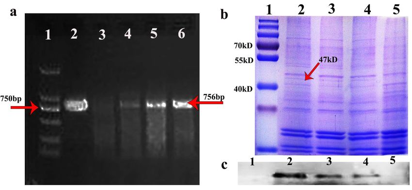
Genes 2023, 14, 21                                                                                        3 of 11

                     body group (WT), hFGF5 group (PG) and recombinant oil body expressing hFGF5 group
                     (ROBF5). The cells were starved in a K-SFM culture medium containing 0.1% fetal bovine
                     serum for 24 h. Additionally, then, the control group was treated with 0.01 M PBS, while
                     the concentrations of WT, PG and ROBF5 were 0, 6.25, 12.5, 25, 50 and 100 ng/mL. The
                     proliferation efficiency of cells was measured by MTT assay. MTT solution was added and
                     incubated for 4 h at 37 ◦ C, followed by adding 100 µL DMSO solution and incubation at
                     room temperature for 10 min.

                     2.5. Establishment of a Hair Loss Mice Model and Drug Treatment
                           The male mice of C57BL/6 aged 6 weeks (20 g–22 g) were randomly divided into
                     5 groups (n = 6 in each group): control group (CG) with 200 µL 0.01 M PBS, wild-type
                     oil body group (WT) with 200 µL wild-type oil body, recombinant oil-body-expressed
                     hFGF5 group (ROBF5) with 3.1 mg transgenic oil body containing 5 µg hFGF5, hFGF5
                     positive group (PG) with 200 µL solution containing 5 µg hFGF5 and hair retardant cream
                     group (HRC). The experimental animals were handled according to the “license for the
                     use of laboratory animals” of Jilin Agricultural University (SYJK 2018-0023). Mice were
                     anesthetized with isoflurane, and the back hair was shaved to form a 2 cm × 3 cm area
                     to make a hair loss model. The frequency of administration was once a day. All animal
                     experiments were approved by the Laboratory Animal Welfare and Ethics Committee of
                     Jilin Agricultural University (No.20201204001).

                     2.6. H&E Staining Analysis
                          The skin of mice was fixed with 4% paraformaldehyde overnight, and embedded
                     in paraffin after dehydration, transparency and embedding treatments for pathological
                     analysis. Paraffin blocks were cut into 5 µm sections, dewaxed, hydrated and stained with
                     an H&E staining kit. The shape and number of hair follicles and hair regeneration were
                     observed under an optical microscope.

                     2.7. Immunohistochemical Staining
                          The sections were soaked into the sodium citrate solution of 0.01 M and heated and
                     pressured for 5 min to repair the tissue antigens. The solution containing 80% methanol
                     and 3% H2 O2 was added to the sections for 15 min to inactivate endogenous horseradish
                     peroxidase (HRP). The sections were treated with 1% Triton of PBS, and then the sections
                     were blocked for 1 h by 5% BSA at room temperature and incubated overnight at 4 ◦ C with
                     polyclonal rabbit anti-cytokeratin 14 (Bioss, Shanghai, China, bsm-52054R, 1:200), and the
                     goat anti-rabbit HRP was incubated for 1 h. The positive protein was stained by DAB kit,
                     and coloration was observed and photographed under optical microscope.

                     2.8. Western Blot Experiment
                          The target protein was extracted and quantified by the BCA method. The protein was
                     separated by 12% SDS polyacrylamide gel (SDS-PAGE), transferred to PVDF membrane
                     and blocked for 120 min by 5% skimmed milk powder. The first antibodies were incubated
                     overnight at 4 ◦ C, washed 3 times, followed by incubation with the second antibody
                     goat anti-rabbit for 120 min and washed 3 times. The target bands were visualized using
                     electrochemiluminescence. The first antibodies were as follows: the rabbit anti FGF5
                     antibody (Bioss, Shanghai, China, bs-1257R, 1:1000), rabbit polyclonal anti-β-actin (Bioss,
                     Shanghai, China, bs-0061R, 1:5000) and rabbit polyclonal cytokeratin 14 (Bioss, Shanghai,
                     China, bsm-52054, 1:1000).

                     2.9. RNA-Seq Analysis
                          The skin from mice treated with the positive drug hFGF5 and recombinant oil body
                     samples was collected and sent to Orvison gene, Beijing, China, for transcriptome sequenc-
                     ing analysis, which was used to explore the genes of recombinant oil body regulating hair
                     growth and screen the related pathways. The expression of differential genes was analyzed
The skin from mice treated with the positive drug hFGF5 and recombinant oil body
samples was collected and sent to Orvison gene, Beijing, for transcriptome sequencing
analysis, which was used to explore the genes of recombinant oil body regulating hair
growth and screen the related pathways. The expression of differential genes was ana-
lyzed and further verified by qRT-PCR method. The pathway of recombinant oil body
Genes 2023, 14, 21
expressing hFGF5 and regulating hair regeneration was explored to elucidate its mecha-
4 of 11
nism.
2.10.further
and Statistical Analysis
verified by qRT-PCR method. The pathway of recombinant oil body expressing
hFGF5 and regulating hair regeneration was explored to elucidate its mechanism.
All experiments were replicated three times. The results were expressed as mean ±
standard
2.10. deviation.
Statistical The experimental data were analyzed through Graph Pad Prism 6.01
Analysis
software and statistics
All experiments were werereplicated
performed by ANOVA
three which
times. The was conducted
results by one-way
were expressed as
method,
mean * p < 0.05,deviation.
± standard ** p < 0.01The
andexperimental
*** p < 0.001. data were analyzed through Graph Pad
Prism 6.01 software and statistics were performed by ANOVA which was conducted by
one-way method, * p < 0.05, ** p < 0.01 and *** p < 0.001.
3. Results
3. Results
3.1. Oleosin-hFGF5 Was Successfully Expressed in Oil Body of Arabidopsis
3.1. Oleosin-hFGF5 Was Successfully Expressed in Oil Body of Arabidopsis
The positive seeds were screened with 0.1% glyphosate [7] (Yuanye Biotechnology,
The positive seeds were screened with 0.1% glyphosate [7] (Yuanye Biotechnology,
Shanghai) and using positive seed cDNA as template. hFGF5-specific primers were used
Shanghai) and using positive seed cDNA as template. hFGF5-specific primers were used
for PCR identification, and the target band of hFGF5 was amplified at 756 bp (Figure 1a),
for PCR identification, and the target band of hFGF5 was amplified at 756 bp (Figure 1a),
and then
and thenT3 T3transgenic
transgenicseeds
seedswere
were obtained
obtained byby propagation.
propagation. TheThe oil body
oil body waswas extracted
extracted
from the transgenic seeds and the target band was detected at 47 kDa by SDS-PAGE and and
from the transgenic seeds and the target band was detected at 47 kDa by SDS-PAGE
Westernblot
Western blot(Figure
(Figure1b,c).
1b,c).AAT3T3 transgenic
transgenic seed
seed waswas obtained
obtained via via
the the identification
identification of the
of the
target gene
target geneand
andprotein
proteinofoftransgenic
transgenic Arabidopsis.
Arabidopsis.
Figure
Figure 1.1. Identification
Identificationofoftransgenic
transgenic Arabidopsis.
Arabidopsis. (a) (a)
PCR-identified
PCR-identified using genomic
using genomicDNADNA as a as a
template and hFGF5-specific primers. line1: DNA marker, line2: hFGF5 positive
template and hFGF5-specific primers. line1: DNA marker, line2: hFGF5 positive control, line3: wild control, line3:
type type
wild Arabidopsis genomic
Arabidopsis as aasnegative
genomic a negativecontrol, line4–6:
control, line4–6:transgenic
transgenicArabidopsis
Arabidopsisgenomic
genomic DNA DNA as a
template and hFGF5-specific primers (Red arrow (right) indicates the target band of 756 bp).
as a template and hFGF5-specific primers (Red arrow (right) indicates the target band of 756 bp). (b)
SDS-PAGE
(b) SDS-PAGE of of
thethe
transgenic
transgenic Arabidopsis
Arabidopsis(Red
(Redarrow
arrowindicates
indicates the
the expressed proteinof
expressed protein of4747kD).
kD). (c)
Western
(c) Westernblotting
blottingidentified
identified the oleosin-hFGF5fusion
the oleosin-hFGF5 fusionprotein.
protein. line1:
line1: protein
protein mark,
mark, line2–4:
line2–4: fusion fusion
proteinfrom
protein fromtransgenic
transgenicArabidopsis,
Arabidopsis, line5:
line5: protein
protein fromfrom
wild wild
typetype Arabidopsis.
Arabidopsis.
3.2. Recombinant Oil-Body-Expressed hFGF5 Inhibits the Proliferation of Hair Follicle Epithelial
3.2. Recombinant
Cells In Vitro Oil-Body-Expressed hFGF5 Inhibits the Proliferation of Hair Follicle Epithelial
Cells In Vitro
When hair enters the growth stage, hair follicle epithelial cells proliferate and differen-
When hair
tiate rapidly enters
under the growth
the regulation stage, hair
of various follicle
growth epithelial
factors to formcells proliferate
hair fibers, whichand differ-
is the
entiate rapidly under the regulation of various growth factors to form hair
basis of hair regeneration. The recombinant oil-body-expressed hFGF5 had no significant fibers, which
effect on the epithelial cells of hair follicles at very low concentrations, and there was no sig-
is the basis of hair regeneration. The recombinant oil-body-expressed hFGF5 had no
nificant effect
significant on thebetween
difference epithelial
all cells of hair
treatment follicles
groups. at verywhen
However, low concentrations,
the concentrationand
wasthere
higher than 12.5 ng/mL, the inhibitory effect on hair follicle epithelial cells in various treat-
ment groups is significantly different, and the inhibitory effect of PG and ROBF5 increased
with the increase in concentration and was significantly higher than that of the CG and
WT groups (Figure 2). ROBF5 significantly inhibited cell proliferation in a dose-dependent
manner with the increase in protein concentration.
was no significant difference between all treatment groups. However, when the concen-
tration was higher than 12.5 ng/mL, the inhibitory effect on hair follicle epithelial cells in
various treatment groups is significantly different, and the inhibitory effect of PG and
ROBF5 increased with the increase in concentration and was significantly higher than that
Genes 2023, 14, 21 of the CG and WT groups (Figure 2). ROBF5 significantly inhibited cell proliferation
5 of 11 in a
dose-dependent manner with the increase in protein concentration.
Figure 2. Effects of recombinant oil body on the inhibition of hair follicle epithelial proliferation
Figure 2. Effects of recombinant oil body on the inhibition of hair follicle epithelial proliferation
(*** p < 0.001). CG: Control group (PBS), WT: wide-type oil body, PG: positive group (hFGF5), ROBF5:
(*** p < 0.001). CG: Control group (PBS), WT: wide-type oil body, PG: positive group (hFGF5),
recombinant oil body expressing hFGF5.
ROBF5: recombinant oil body expressing hFGF5.
3.3. Recombinant Oil-Body-Expressed Oleosin-hFGF5 Inhibits Hair Regeneration in Mice In Vivo
3.3. Recombinant Oil-Body-Expressed
The hair on the Oleosin-hFGF5
back of the mice began Inhibits
to turn black on theHair
10thRegeneration
day in the CGingroup,
Mice In
Vivo
WT group and ROBF5 group, and the black hair on the back of the mice continued to grow
The
in the CGhair
grouponand
the WT
back of the
group, mice
but began
black to turnwas
hair growth black
not on the 10th
obvious day
in the in the
ROBF5 CG group,
group,
WT group and ROBF5 group, and the black hair on the back of the mice continued
and it was also observed in the HRC group, while no black hair was observed in the PGto grow
group (Figure 3a), indicating that the hair regeneration was inhibited after the treatment
in the CG group and WT group, but black hair growth was not obvious in the ROBF5
with recombinant oil body on the 15th day. A histopathology analysis of the inhibition of
group, and it was also observed in the HRC group, while no black hair was observed in
hair growth showed that there were more hair follicles in the subcutaneous tissue of the
the
CGPG groupgroup
and (Figure
WT group,3a),andindicating
the black that
dots the hairinregeneration
existed wasindicating
the hair follicles, inhibitedthatafter the
treatment with recombinant oil body on the 15 th day. A histopathology analysis of the
there was a small amount of hair regeneration in the hair follicles by H&E staining on the
inhibition
10th day. Onof hair growththe
the contrary, showed
number that there
of hair were was
follicles more hair follicles
decreased, lacking inblack
the subcutaneous
dots in
tissue of follicles
the hair the CGingroup
the PGand WTROBF5
group, group,group
and and the HRC
blackgroup,
dots existed
indicating inthat
the the
hair follicles,
hair
indicating that there was a small amount of hair regeneration in the hair follicles by H&E
regeneration was weaker. On the 15th day, the new hair in the hair follicles of CG and WT
staining
groups wason the 10thelongated,
further day. On the andcontrary,
the volume the
of number of hair was
the hair follicles follicles was decreased,
increased compared lack-
to the 10th day; however, the hair growth was still inhibited and there was no obvious new
ing black dots in the hair follicles in the PG group, ROBF5 group and HRC group, indi-
hair in the hair follicles in the PG, ROBF5 and HRC groups (Figure 3b). This implies that
cating that the hair regeneration was weaker. On the 15th day, the new hair in the hair
recombinant oil body had the activity of inhibiting hair growth.
follicles of CG and WT groups was further elongated, and the volume of the hair follicles
was
3.4. increased comparedOil
Effect of Recombinant toBody
the 10th day;hFGF5
Expressed however, the hair growth
on Cytokeratin 14 was still inhibited and
there The
wascytokeratin
no obvious 14 new hair in the
is an important hairinfollicles
factor inhair
process of the regeneration.
PG, ROBF5 Theandpositive
HRC groups
(Figure 3b). This implies that recombinant oil body had the activity of inhibiting
staining rate of cytokeratin 14 in the CG and WT groups was significantly higher than in hair
growth.
the PG, ROBF5 and HRC groups on the 10th day by immunohistochemistry (Figure 4a).
The expression of cytokeratin 14 in the ROBF5 group was lower than that in the CG and WT
groups, and slightly higher than that in the PG and HRC groups (Figure 4a). On the 15th
day, there was no significant difference between the CG and WT groups, but the expression
of cytokeratin 14 was higher than in the PG, ROBF5 and HRC groups. The expression of
cytokeratin 14 in the PG, ROBF5 and HRC groups on the 15th day was higher than on the
10th day. The expression of cytokeratin 14 in hair follicle cells after different treatments was
detected by Western blot, which was consistent with the results of immunohistochemical
staining (Figure 4b,c). It has been proved that recombinant oil body can inhibit hair growth.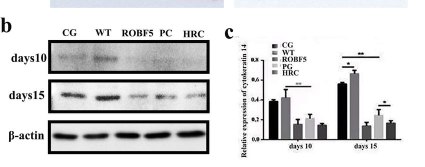
Genes
Genes2022,
2023,13,
14,x21
FOR PEER REVIEW 6 of
6 of1111
Figure
Figure3.3.Effect onon
Effect inhibiting hair
inhibiting growth
hair by recombinant
growth by recombinantoil body expressing
oil body expressinghFGF5 in vivo.
hFGF5 (a)
in vivo.
Hair regeneration on the back of mice in 15 days after administration (n = 6). (b) Pathological
(a) Hair regeneration on the back of mice in 15 days after administration (n = 6). (b) Pathological anal-
ysis by H&E
analysis staining
by H&E on day
staining on10day
and10day
and15day
(magnification 100 times).
15 (magnification CG: Control
100 times). groupgroup
CG: Control (PBS),(PBS),
WT:
wide-type oil body, PG: positive group (hFGF5), ROBF5: recombinant oil body expressing hFGF5,
WT: wide-type oil body, PG: positive group (hFGF5), ROBF5: recombinant oil body expressing hFGF5,
HRC: hair retardant cream (Red arrows show the staining).
HRC: hair retardant cream (Red arrows show the staining).
3.4.
3.5.Effect of Recombinant
Differentially OilGenes
Expressed Body Expressed hFGF5ofonRecombinant
by the Treatment Cytokeratin Oil-Body-Expressed
14 hFGF5
The cytokeratin 14 is an important factor in process of hair regeneration.
Compared with the CG group, 1412 genes were up-regulated and 1566 genes were The positive
staining rate of cytokeratin
down-regulated 14 in the
in the ROBF5 CG (Figure
group and WT5a), groups
andwas significantly
compared with thehigherWTthan in
group,
the PG, ROBF5 and HRC groups on the 10th day by immunohistochemistry
2086 genes were up-regulated and 2038 genes were down-regulated in the ROBF5 group (Figure 4a).
The expression
(Figure 5b), andofcompared
cytokeratin
with14the
in PG
the group,
ROBF5the group was lower
expression of 625than that
genes wasin up-regulated
the CG and
WT
andgroups,
601 genesandwere
slightly higher than that
down-regulated in the
in the PG and
ROBF5 groupHRC groups
(Figure (Figure
5c). 4a). Oninthe
Differences the
15th day, there level
transcriptional was ofnothe
significant
genes lead difference
to changes between the CG
in metabolic and WTafter
regulation groups, but the
the treatment
expression of cytokeratin
of the PG group and ROBF5 14 group,
was higher
whichthan in the PG,
ultimately ROBF5
produce and HRC
different groups.
effects. The
Compared
expression of cytokeratin 14 in the PG, ROBF5 and HRC groups on
with the WT group, the up-regulated genes and the down-regulated genes in the ROBF5 the 15th day was
higher thangroup
treatment on the 10th
were day. The
involved in expression of cytokeratin
the regulation 14 in hairpathways
of different signaling follicle cells after
by KEGG
different
analysis treatments
(Figure S1).was detected by Western blot, which was consistent with the results
of immunohistochemical staining (Figure 4b,c). It has been proved that recombinant oil
3.6. The
body canMechanism
inhibit hairofgrowth.
Recombinant Oil-Body-Expressed hFGF5 Inhibiting Hair Growth
Oil body treatment can affect the expression of key genes in the EGFR, TGF-β and
Wnt signaling pathways, and then regulate hair growth. Compared with the WT treatment
group, some differentially up-regulated genes in the ROBF5 group, such as E2F4, SMAD7,
Bax, E2F5 and CSNK1E were involved in the regulation of the EGFR, TGF-β and Wnt
pathways (Figure 6). Bax is mainly involved in the regulation of the EGFR pathway, which
causes the activation of the intracellular apoptosis pathway and inhibits hair growth (Figure
S2). The expression of Bax was up-regulated after ROBF5 treatment. E2F4, E2F5 and Smad7
are mainly involved in TGF-β, and Smad7 inhibits the TGF-β pathway by regulating the
activation of Smad2/3. Additionally, the up-regulation of E2F4 and E2F5 directly inhibits
the cell cycle, leading to cell apoptosis (Figure S3). CSNK1 is mainly involved in the
regulation of the Wnt pathway, which inhibits Wnt/β-catenin signal transduction. The
activation of the Wnt/β-catenin pathway was a classic intracellular signal transduction
pathway regulating hair growth (Figure S4). Some differentially up-regulated genes such as
ID2, SOS1 and PIK3R3 were involved in the regulation of the EGFR and TGF-β pathways
(Figure 6). PIK3R3 was involved in the regulation of the EGFR pathway, which can promote
the synthesis of intracellular proteins and cell proliferation (Figure S5). Compared with
the WT group, the recombinant oil body down-regulated PIK3R3 in the EGFR pathwayGenes 2023, 14, 21 7 of 11
(Figure 6). SOS1 is involved in the regulation of the EGFR pathway, which promotes the
genes expression of cell proliferation in the EGFR pathway, such as the ERK gene. The
overexpression of SOS1 can promote the activation of ERK and enhance the activity of cell
mitosis and differentiation (Figure S6). Compared with the WT group, the recombinant oil
body down-regulated SOS1, which could inhibit hair growth (Figure 6). ID2 participated in
the TGF-β pathway, which can promote DNA synthesis and accelerate cell proliferation and
differentiation. Compared with the WT group, the recombinant oil body down-regulated
Genes 2022, 13, x FOR PEER REVIEW
ID2 in the TGF-β pathway. The intracellular signal transduction was changed after the
treatment of the recombinant oil body, which affected the EGFR, TGF-β and Wnt pathways,
and then inhibited hair growth.
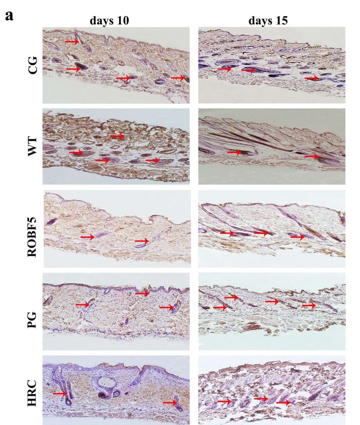
Figure 4. The expression of cytokeratin 14 analyzed in vivo by immunohistochemistry staining
Figure 4. The expression of cytokeratin 14 analyzed in vivo by immunohistochemistry
and Western blotting. (a) Immunohistochemistry staining of cytokeratin 14 (magnification of 100×),
and Western blotting. (a) Immunohistochemistry staining of cytokeratin 14 (magnification
the red arrow indicates a positive protein. (b) Western blot analyzed the expression of cytokeratin 14.
the red arrow indicates a positive protein. (b) Western blot analyzed the expression of cy
(c) The relative expression was calculated by the gray value of the Western blot (* p < 0.05, ** p < 0.01).
14. Control
CG: (c) Thegroup
relative expression
(PBS), wasoilcalculated
WT: wide-type by thegroup
body, PG: positive gray (hFGF5),
value ofROBF5:
the Western
recombinantblot (* p < 0
0.01). CG: Control group (PBS), WT: wide-type
oil body expressing hFGF5, HRC: hair retardant cream. oil body, PG: positive group (hFGF5), RO
combinant oil body expressing hFGF5, HRC: hair retardant cream.
3.5. Differentially Expressed Genes by the Treatment of Recombinant Oil-Body-Expresse
hFGF5
Compared with the CG group, 1412 genes were up-regulated and 1566 genGenes 2023, 14, 21 Genes 2022, 13, x FOR PEER REVIEW 8 of 11 8 of
Figure
Figure 5. Volcanic map5.of
Volcanic map of
differential differential
gene expressiongene expression
after after
treatment of treatment
recombinantof recombinant
oil body oil bod
Genes 2022, 13, x FOR PEER REVIEW expressing hFGF5, compared with the treatment of CG, WT and PG. (a) ROBF59 of
vs 11
CG, (b) ROBG
expressing hFGF5, compared with the treatment of CG, WT and PG. (a) ROBF5 vs CG, (b) ROBG5
vs WT, (c) ROBF5 vs PG. CG: positive control group; WT: wild-type oil body treatment group; P
vs WT, (c) ROBF5 vs PG. CG: positive control group; WT: wild-type oil body treatment group; PG:
hFGF5 treatment group; ROBF5: recombinant oil body expressing hFGF5 treatment group.
hFGF5 treatment group; ROBF5: recombinant oil body expressing hFGF5 treatment group.
3.6. The Mechanism of Recombinant Oil-Body-Expressed hFGF5 Inhibiting Hair Growth
Oil body treatment can affect the expression of key genes in the EGFR, TGF-β an
Wnt signaling pathways, and then regulate hair growth. Compared with the WT trea
ment group, some differentially up-regulated genes in the ROBF5 group, such as E2F
SMAD7, Bax, E2F5 and CSNK1E were involved in the regulation of the EGFR, TGF-β an
Wnt pathways (Figure 6). Bax is mainly involved in the regulation of the EGFR pathwa
which causes the activation of the intracellular apoptosis pathway and inhibits ha
growth (Figure S2). The expression of Bax was up-regulated after ROBF5 treatment. E2F
E2F5 and Smad7 are mainly involved in TGF-β, and Smad7 inhibits the TGF-β pathwa
by regulating the activation of Smad2/3. Additionally, the up-regulation of E2F4 and E2F
directly inhibits the cell cycle, leading to cell apoptosis (Figure S3). CSNK1 is mainly i
volved in the regulation of the Wnt pathway, which inhibits Wnt/β-catenin signal tran
duction. The activation of the Wnt/β-catenin pathway was a classic intracellular sign
transduction pathway regulating hair growth (Figure S4). Some differentially up-reg
lated genes such as ID2, SOS1 and PIK3R3 were involved in the regulation of the EGF
and TGF-β pathways (Figure 6). PIK3R3 was involved in the regulation of the EGFR pat
way, which can promote the synthesis of intracellular proteins and cell proliferation (Fi
ure S5). Compared with the WT group, the recombinant oil body down-regulated PIK3R
in the EGFR pathway (Figure 6). SOS1 is involved in the regulation of the EGFR pathwa
which promotes the genes expression of cell proliferation in the EGFR pathway, such
the ERK gene. The overexpression of SOS1 can promote the activation of ERK and enhan
the activity of cell mitosis and differentiation (Figure S6). Compared with the WT grou
the recombinant oil body down-regulated SOS1, which could inhibit hair growth (Figu
6). ID2 participated in the TGF-β pathway, which can promote DNA synthesis and acce
erate cell proliferation and differentiation. Compared with the WT group, the recomb
nant oil body down-regulated ID2 in the TGF-β pathway. The intracellular signal tran
duction was changed after the treatment of the recombinant oil body, which affected th
EGFR, TGF-β and Wnt pathways, and then inhibited hair growth.
Figure
Figure6.6.The
Theexpression
expressionanalysis
analysisofofup-regulated
up-regulatedand
anddown-regulated
down-regulateddifferential
differentialgenes
genesafter
after
treatment
treatmentofofrecombinant
recombinantoil oilbody.
body.(a)(a)
E2F4 gene
E2F4 relative
gene expression,
relative (b) Smad7
expression, genegene
(b) Smad7 relative ex-
relative
pression, (c) Bax
expression, genegene
(c) Bax relative expression,
relative (d) E2F5
expression, genegene
(d) E2F5 expression, (e) CSNK1E
expression, genegene
(e) CSNK1E relative ex-
relative
pression, (f) ID2 gene relative expression, (g) PI3K3R3 gene relative expression, (h) SOS1
expression, (f) ID2 gene relative expression, (g) PI3K3R3 gene relative expression, (h) SOS1 genegene rela-
tive expression (* p < 0.05, ** p < 0.01, *** p < 0.001). ROBF5: recombinant oil body expressing hFGF5
relative expression (* p < 0.05, ** p < 0.01, *** p < 0.001). ROBF5: recombinant oil body expressing
group, WT: wild-type oil body group.
hFGF5 group, WT: wild-type oil body group.
4.4.Discussion
Discussion
The
Thehalf-life
half-lifeofof
growth factor
growth is too
factor shortshort
is too to show effective
to show biological
effective activityactivity
biological in treat-in
ment,
treatment, so it is particularly important to find effective material carriers [7]. Oil bodyasasaa
so it is particularly important to find effective material carriers [7]. Oil body
new
newtype
typeofofsafe
safecarrier
carriermaterial
materialhashasbeen
beenwidely
widelyconsidered.
considered.The Therecombinant
recombinantproteins
proteins
expressed
expressedin in the
the surface of oil
surface of oilbody
bodyare areeasier
easiertoto purify
purify andand have
have a longer
a longer half-life
half-life com-
compared
pared with
with the the prokaryotic
prokaryotic expression
expression system.system.
The oil The
bodyoilexpression
body expression
system issystem
easy toisstore
easyand
to
store and transport,
transport, easy to process,
easy to process, reduces reduces
the cost the cost of purification
of purification and most and most importantly,
importantly, presents
presents low toxicity, among others. Therefore, an oil-body expression system was se-
lected to produce FGF5. Although hFGF5 is expressed in A. thaliana, its expression level is
only about 0.19%. In the future, we need to further modify the gene and vector through
genetic engineering technology to express in oil crops, so as to obtain transgenic seedsGenes 2023, 14, 21 9 of 11
low toxicity, among others. Therefore, an oil-body expression system was selected to
produce FGF5. Although hFGF5 is expressed in A. thaliana, its expression level is only
about 0.19%. In the future, we need to further modify the gene and vector through genetic
engineering technology to express in oil crops, so as to obtain transgenic seeds with high
expression. The oleosin-rhFGF5 was expressed on the surface of the oil body, and the
recombinant oil body expressing hFGF5 was proved to have the effect of inhibiting hair
growth by cell and animal experiments. The growth of hair was inhibited, and the damage
of hair follicles was also significantly attenuated, while the volume of hair follicles in
subcutaneous tissue was not significantly reduced after treatment with recombinant oil
body on the 15th day. This may be an advantage for the use of recombinant oil body
expressing hFGF5 to temporarily inhibit hair growth on diseased skin without causing
damage to hair follicles.
The differentially expressed genes screened by RNA-seq after recombinant oil body
treatment were involved in the EGFR, TGF-β and Wnt pathways to regulate hair growth,
which promoted cell proliferation and apoptosis. Among them, Bax was a classic gene
regulating apoptosis, which is up-regulated expression that can cause apoptosis [19]. The
overexpression of Smad7 can not only inhibit TGF-β signal transmission but can also affect
the development process of hair follicles. When Smad7 in large quantities was expressed
in hair follicle cells, the hair circulation was blocked from entering the growth period,
leading to hair follicle degeneration [20]. The CSNK1E gene was one of the important
candidate genes potentially regulating hair follicle development by sequencing the genome
of sheep developing hair follicles [21]. ID2 can be highly expressed in the specialized cells
in the basal epidermis and cytoplasm of the outer root sheath [22]. The hair growth-related
pathway and immune-related pathway were abnormally activated by the transcriptome
of NIH hairless mice, and the PIK3R3 gene was expressed in large quantities around the
skin hair follicles of hairless mice [23]. PIK3R3 may be used as an important target for
the potential regulation of hair growth. EGFR controls some genes that are involved in
epidermal differentiation, cell cycle and apoptosis [24]. The loss of EGFR leads to absence
of the LEF1 protein, specifically in the innermost epithelial hair layers. After activating the
Wnt pathway, it binds to the receptor on the cell membrane and initiates a cascade reaction
to up-regulate the expression of intracellular β-catenin, and the interaction between β-
catenin and the N-terminus of the intracellular LEF/TCF transcription factor blocks the
expression of downstream target genes, resulting in the hair going through a degeneration
stage. TGF-β2 dominates the hair degeneration cascade, which prompts epithelial cells to
up-regulate and activate stromal cell-secreted protein smad 9 and smad3, leading to the
loss of epithelial cells and inducing hair to enter the degenerative stage. The recombinant
oil body expressing hFGF5 could regulate the changes of these genes that play a role in
hair growth.
Supplementary Materials: The following supporting information can be downloaded at: https://
www.mdpi.com/article/10.3390/genes14010021/s1, Figure S1: The recombinant oil body expressing
hFGF5 regulates Bax and participates in the EGFR pathway. Figure S2: The recombinant oil body
expressing hFGF5 regulates E2F4, E2F5 and Smad7 and participates in the TGF-β pathway. Figure S3:
The recombinant oil body expressing hFGF5 regulates CSNK1 and participates in the Wnt pathway.
Figure S4: The recombinant oil body expressing hFGF5 regulates PIK3R3 and participates in the EGFR
pathway. Figure S5: The recombinant oil body expressing hFGF5 regulates PIK3R3 and participates
in EGFR pathway. Figure S6: pOTB-hFGF5 expression vector. Figure S7: The recombinant oil body
expressing hFGF5 regulates SOS1 and participates in the EGFR pathway. Table S1 Sequence of primers.
Author Contributions: Carried out the experimental work and wrote the draft, H.W.; performed data
statistics and figures, X.L. and J.Z.; writing—review and editing, M.N.; visualization, J.Z.; supervision,
J.Z.; project administration, Z.W. All authors have read and agreed to the published version of
the manuscript.
Funding: This research received no external funding.
Institutional Review Board Statement: Not applicable.Genes 2023, 14, 21 10 of 11
Informed Consent Statement: Not applicable.
Data Availability Statement: All data generated or analyzed during this study are available within.
Conflicts of Interest: The authors declare no conflict of interest.
Abbreviations
FGF: fibroblast growth factor; hFGF5: human fibroblast growth factor5; SDS-PAGE: sodium do-
decyl sulfate polyacrylamide gel electrophoresis; PBS: phosphate-buffered saline; H&E: hematoxylin
and eosin; WT: wild-type oil body group; CG: control group; ROBF5: recombinant oil-body-expressed
hFGF5 group; PG: hFGF5 positive group; HRC: hair retardant cream group.
References
1. Huang, A.H.C. Oil bodies and oleosins in seeds. Annu. Rev. Plant Biol. 1992, 43, 177–200. [CrossRef]
2. Siloto, R.M.; Findlay, K.; Lopez-Villalobos, A.; Yeung, E.C.; Nykiforuk, C.L.; Moloney, M.M. The accumulation of oleosins
determines the size of seed oil bodies in Arabidopsis. Plant Cell 2006, 18, 1961–1974. [CrossRef] [PubMed]
3. Beaudoin, F.; Wilkinson, B.M.; Stirling, C.J.; Napier, J.A. In vivo targeting of a sunflower oil body protein in yeast secretory (sec)
mutants. Plant J. 2000, 23, 159–170. [CrossRef] [PubMed]
4. Ling, H. Oleosin fusion expression systems for the production of recombinant proteins. Biologia 2007, 62, 119–123. [CrossRef]
5. Qiang, W.; Zhou, T.; Lan, X.; Zhang, X.; Guo, Y.; Noman, M.; Du, L.; Zheng, J.; Li, W.; Li, H.; et al. A new nanoscale transdermal
drug delivery system: Oil body-linked oleosin-hEGF improves skin regeneration to accelerate wound healing. J. Nanobiotechnology
2018, 16, 62–80. [CrossRef]
6. Li, W.; Yang, J.; Cai, J.; Wang, H.; Tian, H.; Huang, J.; Qiang, W.; Zhang, L.; Li, H.; Li, X.; et al. Oil Body-Bound Oleosin-rhFGF-10:
A Novel Drug Delivery System that Improves Skin Penetration to Accelerate Wound Healing and Hair Growth in Mice. Int. J.
Mol. Sci. 2017, 18, 2177. [CrossRef]
7. Yang, J.; Guan, L.; Guo, Y.; Du, L.; Wang, F.; Wang, Y.; Zhen, L.; Wang, Q.; Zou, D.; Chen, W.; et al. Expression of biologically
recombinant human acidic fibroblast growth factor in Arabidopsis thaliana seeds via oleosin fusion technology. Gene 2015, 566,
89–94. [CrossRef]
8. Yang, J.; Qiang, W.; Ren, S.; Yi, S.; Li, J.; Guan, L.; Du, L.; Guo, Y.; Hu, H.; Li, H.; et al. High-efficiency production of bioactive
oleosin-basic fibroblast growth factor in A. thaliana and evaluation of wound healing. Gene 2018, 639, 69–76. [CrossRef]
9. Qiang, W.; Gao, T.; Lan, X.; Guo, J.; Noman, M.; Li, Y.; Guo, Y.; Kong, J.; Li, H.; Du, L.; et al. Molecular Pharming of the
Recombinant Protein hEGF-hEGF Concatenated with Oleosin Using Transgenic Arabidopsis. Genes 2020, 11, 959. [CrossRef]
10. Allerstorfer, S.; Sonvilla, G.; Fischer, H.; Spiegl-Kreinecker, S.; Gauglhofer, C.; Setinek, U.; Czech, T.; Marosi, C.; Buchroithner, J.;
Pichler, J.; et al. FGF5 as an oncogenic factor in human glioblastoma multiforme: Autocrine and paracrine activities. Oncogene
2008, 27, 4180–4190. [CrossRef]
11. Clase, K.L.; Mitchell, P.J.; Ward, P.J.; Dorman, C.M.; Johnson, S.E.; Hannon, K. FGF5 stimulates expansion of connective tissue
fibroblasts and inhibits skeletal muscle development in the limb. Dev. Dyn. Off. Publ. Am. Assoc. Anat. 2000, 219, 368–380.
[CrossRef]
12. Ren, Y.; Jiao, X.; Zhang, L. Expression level of fibroblast growth factor 5 (FGF5) in the peripheral blood of primary hypertension
and its clinical significance. Saudi J. Biol. Sci. 2018, 25, 469–473. [CrossRef]
13. Higgins, C.A.; Petukhova, L.; Harel, S.; Ho, Y.Y.; Drill, E.; Shapiro, L.; Wajid, M.; Christiano, A.M. FGF5 is a crucial regulator of
hair length in humans. Proc. Natl. Acad. Sci. USA 2014, 111, 10648–10653. [CrossRef]
14. Hébert, J.M.; Rosenquist, T.; Götz, J.; Martin, G.R. FGF5 as a regulator of the hair growth cycle: Evidence from targeted and
spontaneous mutations. Cell 1994, 78, 1017–1025. [CrossRef] [PubMed]
15. He, X.; Chao, Y.; Zhou, G.; Chen, Y. Fibroblast growth factor 5-short (FGF5s) inhibits the activity of FGF5 in primary and secondary
hair follicle dermal papilla cells of cashmere goats. Gene Int. J. Focus. Gene Cloning Gene Struct. Funct. 2016, 575 Pt 2, 393–398.
[CrossRef] [PubMed]
16. Alonso, L.E. The hair cycle. Sitri. It. 2006, 119, 391–393. [CrossRef]
17. Kim, Y.S.; Kim, K.H.; Lee, W.J.; Jung, J.W.; Song, K.Y.; Suhr, K.B.; Lee, J.H.; Park, J.K. Growth Factors Influencing the Morphology
and Growth Rate of Hair Follicles in a Human Hair Organ Culture. Korean J. Dermatol. 1998, 36, 210–216.
18. Lin, H.J.; Qi, J.M.; Xu, N.; Ding, M.; Chen, Y.; Li, X.N.; Hu, Z.L. Progress in the Regulation of FGF5 on Hair Follicle Cycle. Chin. J.
Aesthetic Med. 2019, 11, 163–164.
19. Keum, D.I.; Pi, L.Q.; Hwang, S.T.; Lee, W.S. Protective effect of Korean Red Ginseng against chemotherapeutic drug-induced
premature catagen development assessed with human hair follicle organ culture model. J. Ginseng Res. 2016, 40, 169–175.
[CrossRef]
20. Millar, S.E. Smad7: Licensed to kill β-catenin. Dev. Cell 2006, 11, 274–276. [CrossRef]Genes 2023, 14, 21 11 of 11
21. Sulayman, A.; Tian, K.; Huang, X.; Tian, Y.; Xu, X.; Fu, X.; Zhao, B.; Wu, W.; Wang, D.; Yasin, A.; et al. Genome-wide identification
and characterization of long non-coding RNAs expressed during sheep fetal and postnatal hair follicle development. Sci. Rep.
(Nat. Publ. Group) 2019, 9, 8501. [CrossRef] [PubMed]
22. Hammond NJahoda, L.C. Id2, Id3, and Id4 proteins show dynamic changes in expression during vibrissae follicle development.
Dev. Dyn. 2010, 237, 1653–1661. [CrossRef] [PubMed]
23. Ji, Z.-H.; Chen, J.; Gao, W.; Zhang, J.-Y.; Quan, F.-S.; Hu, J.-P.; Yuan, B.; Ren, W.-Z. Cutaneous transcriptome analysis in NIH
hairless mice. PLoS ONE 2017, 12, e0182463–e0182471. [CrossRef] [PubMed]
24. Amberg, N.; Sotiropoulou, P.A.; Heller, G.; Lichtenberger, B.M.; Holcmann, M.; Camurdanoglu, B.; Baykuscheva-Gentscheva,
T.; Blanpain, C.; Sibilia, M. EGFR Controls Hair Shaft Differentiation in a p53-Independent Manner. iScience 2019, 15, 243–256.
[CrossRef] [PubMed]
Disclaimer/Publisher’s Note: The statements, opinions and data contained in all publications are solely those of the individual
author(s) and contributor(s) and not of MDPI and/or the editor(s). MDPI and/or the editor(s) disclaim responsibility for any injury to
people or property resulting from any ideas, methods, instructions or products referred to in the content.You can also read