Retrograde Femoral Intramedullary Nail Extraction Techniques. Report of Three Cases
←
→
Page content transcription
If your browser does not render page correctly, please read the page content below
CASE PRESENTATION

Retrograde Femoral Intramedullary
Nail Extraction Techniques.
Report of Three Cases
Jonatan A. Lobo, Sebastián Pereira, Fernando Bidolegui
Orthopedics and Traumatology Service, Hospital Sirio Libanés (Buenos Aires, Argentina)

Abstract
Background: The indication for osteosynthesis with a retrograde intramedullary nail in femur fractures has increased in recent
years and with it, the number of complications. Three surgical techniques are described for the management of the proximal frag-
ment of the broken intramedullary osteosynthesis. From March 2001 to January 2019, 321 osteosyntheses with retrograde femoral
intramedullary nails were performed at our institution. The implant rupture rate associated with nonunion was 0.9%. Minimally inva-
sive techniques were performed to remove the implant, preserving the soft tissues. Definitive reosteosynthesis was achieved with
the consequent consolidation in an average time of 140 days. Conclusions: The techniques used were simple, safe, minimally
invasive, and reproducible.
Keywords: Broken; nail; retrograde; femur; extraction.
Level of Evidence: IV

Técnicas de extracción de clavos endomedulares retrógrados de fémur rotos.
Presentación de tres casos

Resumen
Introducción: La indicación de colocar un enclavado endomedular retrógrado en las fracturas de fémur se ha incrementado
en los últimos años y, con ello, la cantidad de complicaciones. Presentamos la descripción de las técnicas quirúrgicas para el
manejo del fragmento proximal de la osteosíntesis endomedular rota. Desde marzo de 2001 hasta enero de 2019, se realizaron
321 enclavados endomedulares retrógrados de fémur en nuestra institución. La tasa de rotura del implante asociadas a una seu-
doartrosis fue del 0,9% (3 casos); se realizaron técnicas mínimamente invasivas y diferentes para la extracción del implante. En
tres pacientes, se extrajeron los clavos rotos satisfactoriamente, sin complicaciones asociadas a la técnica quirúrgica. Se logró la
reosteosíntesis definitiva con la consiguiente consolidación en un tiempo medio de 140 días. Conclusiones: Las técnicas utiliza-
das fueron simples, seguras, mínimamente invasivas y muy reproducibles.
Palabras clave: Rotura; clavo retrógrado; fémur; extracción.
Nivel de Evidencia: IV

Introduction
  The use of antegrade intramedullary osteosynthesis is considered the treatment of choice for diaphyseal femur
fractures; union rates close to 99% are obtained. 1 The indication for retrograde intramedullary nails has gained
ground, expanding the initial indications for this technique and achieving similar consolidation results. 2-4
  However, some risk factors predispose to implant failure and rupture, such as exposed fractures, intramedullary
nail diameter, unreamed nail placement, surgical technique, osteosynthesis design, etc. 5-7

 Received on June 6th, 2019. Accepted after evaluation on May 4th, 2020 • Jonatan A. Lobo, MD • lobojonatan@gmail.com          ID https://orcid.org/0000-0001-6665-0063
 How to cite this article: Lobo JA, Pereira S, Bidolegui F. Retrograde Femoral Intramedullary Nail Extraction Techniques. Report of Three Cases. Rev Asoc Argent Ortop Traumatol
 2021;86(3):XXXX. https://dx.doi.org/10.15417/issn.1852-7434.2021.86.3.986

           This Journal is licensed under Attribution-NonCommercial-ShareAlike 4.0 International
           Creative Commons (CC-BY-NC-SA 4.0).
                                                                                                              Rev Asoc Argent Ortop Traumatol 2021; 86 (3): 375-391 • ISSN 1852-7434 (online)   375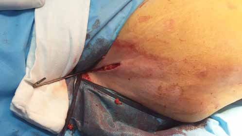
J. A. Lobo et al.

         In the case of a nonunion, the osteosynthesis material is subjected to an increased stress that predisposes it to
      fatigue and breakage.8
         Different surgical techniques have been described to extract the broken intramedullary nail, to preserve the soft
      tissues through minimally invasive approaches, without causing greater damage.
         The aim of this article is to describe the techniques used to extract the osteosynthesis, without opening the focus
      of the nonunion, using minimally invasive techniques.

      Materials and Methods
        321 retrograde intramedullary nailings of the femur were performed at our institution, between March 2001 and
      January 2019. 57.9% (186 cases) were diaphyseal fractures and 42% (135 cases) were supracondylar fractures. In
      this series, there were only three cases (0.9%) of implant rupture in the presence of a nonunion. We describe the
      different surgical techniques used for the management and extraction of the proximal fragment of the nail that has
      been broken.

      Case 1
         A 17-year-old male was admitted due to polytrauma after a motorcycle collision accident against a car, in which
      he suffered a transyndesmal fracture of the left ankle and a fracture of the right femur (type 32A2 of the AO clas-
      sification) associated with thorax trauma (Figure 1).

                          Figure 1. Anteroposterior and lateral radiographs of the femur. A 32A2 femur fracture is seen.

376   Rev Asoc Argent Ortop Traumatol 2021; 86 (3): 375-391 • ISSN 1852-7434 (online)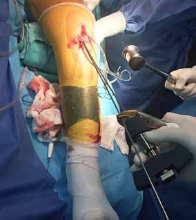
Broken Intramedullary Nails - Extraction Technique

  Initially, the femoral fracture was stabilized with an external fixator and the ankle fracture, with a plaster cast. On
the eighth day, the external tutor was replaced with an unreamed 9 mm x 350 mm retrograde femoral intramedul-
lary nail (Figure 2).

           Figure 2. Anteroposterior and lateral radiographs of the femur in the immediate postoperative period.
           Reduction and osteosynthesis were performed with an unreamed retrograde intramedullary nail.

   In the fourth month after surgery, the patient reported a sudden increase in pain in the operated limb, during
walking, without previous trauma. In the radiological control, nonunion and implant rupture were observed. A
series of combined conditions were identified that led to the failure of osteosynthesis. First, the choice of a smaller
diameter intramedullary nail due to a history of chest trauma. Second, the faulty design of the osteosynthesis mate-
rial, which presented a locking hole in the middle third and, added to this, the proximity of the fracture site to the
area of weakness of the osteosynthesis material.

                                                                      Rev Asoc Argent Ortop Traumatol 2021; 86 (3): 375-391 • ISSN 1852-7434 (online)   377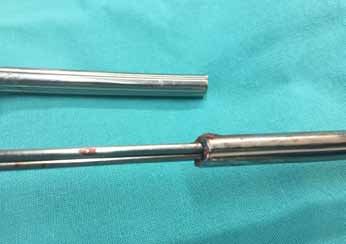
J. A. Lobo et al.

        These were the determining conditions for nonunion, material fatigue and implant rupture (Figure 3).

                Figure 3. Anteroposterior and lateral radiograph of the femur. Note the small diameter of the intramedullary
                nail, the nonunion over an area of implant weakness, and the breakage.

        The surgical plan consisted of the extraction of the intramedullary nail fragments and the new osteosynthesis
      with a larger diameter nail to provide the necessary stability and achieve consolidation over time.
        The blocks were extracted percutaneously. Through the transpatellar approach, the distal end of the nail was
      removed with the specific extractor.

378   Rev Asoc Argent Ortop Traumatol 2021; 86 (3): 375-391 • ISSN 1852-7434 (online)
Broken Intramedullary Nails - Extraction Technique
A 3 cm approach proximal to the greater trochanter was used to extract the proximal fragment. A threaded 3.8
mm guide pin was placed and, under fluoroscopic control, a correct entry point was made. The guide pin was re-
moved and then the olive-tipped guide wire was inserted through its non-olive end. The aim of this technique is to
thread and secure the olive tip at the proximal end of the implant (Figure 4). The olive-tipped guide wire should be
advanced from the greater trochanter to the distal femur approach to make the extraction in a retrograde and safe
way, through the transpatellar approach.
Figure 4. Intraoperative image showing the antegrade placement of the olive-tipped guide by threading the
proximal fragment of the broken nail.
Rev Asoc Argent Ortop Traumatol 2021; 86 (3): 375-391 • ISSN 1852-7434 (online) 379J. A. Lobo et al.
Next, a reamed 11mm x 380mm intramedullary retrograde nail was placed. Consolidation was achieved at four
months (Figure 5).
Figure 5. Radiographic control 4 months after surgery. Consolidation of the fracture is observed.
380 Rev Asoc Argent Ortop Traumatol 2021; 86 (3): 375-391 • ISSN 1852-7434 (online)Broken Intramedullary Nails - Extraction Technique
Case 2
A 79-year-old woman with a history of smoking and use of alendronate was admitted to the Emergency Service
with pain and functional impairment plus deformity of the lower right limb, after a fall from her own height. On
radiographs, a femoral shaft fracture, type 32A2 of the AO classification, was detected (Figure 6).
Figure 6. Anteroposterior and lateral radiographs of the right femur upon
admission. A 32A2 diaphyseal femur fracture is observed.
Rev Asoc Argent Ortop Traumatol 2021; 86 (3): 375-391 • ISSN 1852-7434 (online) 381J. A. Lobo et al.
Reduction and osteosynthesis with a reamed 11 mm x 340 mm intramedullary nail were carried out on the fourth
day of admission (Figure 7).
Figure 7. Immediate postsurgical radiographic control.
382 Rev Asoc Argent Ortop Traumatol 2021; 86 (3): 375-391 • ISSN 1852-7434 (online)Broken Intramedullary Nails - Extraction Technique
On radiological controls, a delay in union and its subsequent evolution to nonunion were observed, as well as
the inherent fatigue of the intramedullary nail, which presented a blocking hole close to the fracture focus. But,
as there were no symptoms or pain, the patient did not accept a second intervention. A year after the intervention,
she was admitted to the Emergency Service with pain and functional impairment, after a fall from her own height.
Implant rupture was observed (Figure 8).
Figure 8. Anteroposterior and lateral radiographs of the right femur upon admission to the
Emergency Service. Note the breakage of the intramedullary nail and the nonunion.
Rev Asoc Argent Ortop Traumatol 2021; 86 (3): 375-391 • ISSN 1852-7434 (online) 383J. A. Lobo et al.
The surgical plan consisted of removing the broken intramedullary nail to perform reosteosynthesis with a larger
diameter retrograde intramedullary nail. After removing the distal blocks percutaneously, the surgery continued
with the extraction of the distal nail fragment with a specific extractor through the transpatellar approach. The non-
olive guide wire was then introduced retrogradely and impacted up to the greater trochanter. A 3 cm approach was
made proximal to the trochanter in order to place a 3.8 mm guide pin and then drill with an 8 mm cannulated drill
bit. The guide was then removed and a “hook” bend was made at the tip (Figure 9).
Figure 9. Proximal approach to the greater trochanter through which the olive-tipped guide was
advanced and a “hook”-shaped bend was made.
The purpose of this technique is to “hook” the proximal fragment of the broken nail with the hook-shaped guide,
in order to be able to extract it retrogradely through the patellar approach (Figure 10).
Figure 10. Retrograde extraction of the hook-shaped guide once the proximal fragment of the broken nail has been
engaged.
384 Rev Asoc Argent Ortop Traumatol 2021; 86 (3): 375-391 • ISSN 1852-7434 (online)Broken Intramedullary Nails - Extraction Technique
Increased reaming and placement of the 14 mm x 340 mm retrograde intramedullary nail were carried out wi-
thout complications.
In the following radiological controls, consolidation was confirmed at 5 months (Figure 11).
Figure 11. Radiographic control 5 months after surgery. There are signs of bone consolidation.
Rev Asoc Argent Ortop Traumatol 2021; 86 (3): 375-391 • ISSN 1852-7434 (online) 385J. A. Lobo et al.
Case 3
A 52-year-old woman, smoker, obese with a body mass index of 42, and diabetic. She was admitted with poly-
trauma due to a motorcycle-car collision, in which she suffered a cranioencephalic traumatism, rib fractures, frac-
tures in both forearms and left humerus, a severe exposed dislocation-fracture of the right ankle and a diaphyseal
fracture of the left femur, type 32A2 of the AO classification.
Initial stabilization with an external tutor was carried out for the ankle and femur dislocation-fracture, and im-
mobilization with a plaster cast for the upper limbs. On the ninth day of hospitalization, the external tutor was
replaced with a 10 mm x 340 mm reamed retrograde intramedullary nail.
Two years after the accident, she was admitted to the Emergency Service with pain and difficulty walking, without
previous trauma. A nonunion associated with the breakage of the intramedullary nail was observed (Figure 12).
Figure 12. Radiographic control upon admission to the Emergency
Service. Implant rupture associated with nonunion is observed.
It was decided to remove the material and perform reosteosynthesis with a retrograde intramedullary nail using
a single approach.
The blocks were removed percutaneously and the distal nail fragment was removed through the patellar ap-
proach with the specific extractor. A non-olive intramedullary guide was then introduced retrogradely through the
proximal nail fragment until it passed the nail tip at approximately 6 cm (Figure 13A).
386 Rev Asoc Argent Ortop Traumatol 2021; 86 (3): 375-391 • ISSN 1852-7434 (online)Broken Intramedullary Nails - Extraction Technique
The aim of this technique is to introduce and impact another non-olive guide of a smaller diameter until the two
guides are obstructing each other (Figure 13B).
Once the “obstruction” had been achieved, the first guide that was initially placed was pulled, to later extract the
proximal nail fragment (Figure 13C), using a single approach and without complications (Figures 13D). In Figure
13E, the effective obstruction of both guides is seen.
A B C
E
Figure 13. A. Intraoperative image in which the correct placement
of the intramedullary guide is evaluated until it surpasses the tip
of the nail. B. Placement of the second guide of smaller diameter,
D achieving the obstruction of both guides to the proximal fragment
of the broken nail. C. Extraction of the proximal fragment by
pulling the first intramedullary guide. D. Correct retrograde
extraction of the fragment of the broken nail, through a single
approach. E. Image showing the effective locking of guides of
different sizes.
Rev Asoc Argent Ortop Traumatol 2021; 86 (3): 375-391 • ISSN 1852-7434 (online) 387J. A. Lobo et al.
The nail with the largest diameter was reamed and placed according to the technique (Figure 14).
The patient evolved favorably and consolidation was confirmed at six months in radiological controls (Fig-
ure 15).
Figure 14. Immediate postoperative follow-up with a larger diameter retrograde nail.
388 Rev Asoc Argent Ortop Traumatol 2021; 86 (3): 375-391 • ISSN 1852-7434 (online)Broken Intramedullary Nails - Extraction Technique
Figure 15. Follow-up 6 months after surgery. Signs of bone consolidation are observed.
Discussion
Many techniques to extract the distal fragment of a broken antegrade femur nail have been described, but there
is scant literature on this type of complication in retrograde femoral nails.
We described three techniques to extract the proximal fragment of a broken retrograde nail. The decision to per-
form one technique or another was based on its practicality and effectiveness, trying to protect the soft tissue and
gradually increasing the complexity of the techniques as needed.
Rev Asoc Argent Ortop Traumatol 2021; 86 (3): 375-391 • ISSN 1852-7434 (online) 389J. A. Lobo et al.
Initially, we attempted extraction using a single approach, placing two intramedullary guides through the
transpatellar approach, looking for their obstruction in the proximal fragment. If this technique was not effective,
as a second option, we placed the olive-tipped guide wire, by its non-olive end, through a new approach on the
greater trochanter, to perform the retrograde extraction. The next technique consists of placing the intramedul-
lary guide through the patellar approach, extracting it through the greater trochanter, and creating a hook over
the tip of the guide to “hook” the nail fragment.
In 1988, Franklin et al. 9 described a revolutionary technique for the extraction of broken nails. They used 50
cm long osteotomes to make a hook-shaped bend 2 cm from the tip. This technique achieves a change in osteo-
synthesis rescue surgeries, since it allows to preserve the soft tissues without raising the periosteum, which hap-
pens when the nonunion focus is opened to extract the distal fragment of the broken nail using basic instruments,
such as forceps, clamps or forceps. In conclusion, they report that this technique is extremely safe and effective,
and eliminates the need to open the focus to extract the nail fragment.
Authors such as Brewster et al., 10 Hak et al., 11 and Blake 12 described techniques for the extraction of the
broken osteosynthesis using two intramedullary guides. A hook-shaped guide and another smaller olive-tipped
guide, which had to be inserted beyond the tip of the broken nail, to then impact the other intramedullary guide
and thus increase the binding forces to be able to extract more efficiently. In their series, they do not describe
complications and, as an advantage, they mention the simplicity of the technique, which does not require new
approaches.
In 2004, Magu et al. 13 described an innovative technique to extract antegrade nails using an olive-tipped in-
tramedullary guide together with a washer through a transpatellar approach, seeking the obstruction of the latter
on the tip of the nail. The advantage of this technique over the others is that it generates a greater grip when
performing the extraction. This technique is easy to reproduce and is performed in an antegrade manner for
retrograde nail removal.
In 2006, Karladani14 published an extraction technique in which, in a first step, the olive-tipped intramedullary
guide was placed beyond the fragment of the broken nail, then a 12 mm long 3.5 mm screw was placed in the
locking hole crossing the first cortex, and was fixed only to the nail. Then, the olive-tipped intramedullary guide
was removed, looking for the olive to bind to the screw. In the literature review and case report published by
Abdelgawad and Kanlic, 15 in 2013, the authors mentioned this technique as their first option, since it was easily
reproducible and one of the safest. In our opinion, it is a risky and highly demanding technique, and a possible
complication is that the screw comes out of the locking hole of the nail and remains lodged in the intramedullary
cavity with the possibility of injuring the internal cortices of the femur during extraction.
In 2008, Acharya et al. 16 published a series of two cases of antegrade nail breakage, using an intramedullary
guide bent in the form of a hook to extract the distal fragment. They concluded that the technique could be used
to extract any intramedullary nail, but, in order to extract a retrograde nail, we have modified this technique by
making an approach on the greater trochanter to be able to extract the guide, make the fold in the shape of a hook
and thus be able to “hook” the broken proximal fragment and carry out the retrograde extraction in a satisfactory
and uncomplicated manner.
In the meta-analysis by Whalley et al., 17 35 articles with 11 techniques were cited. They did not reach a con-
clusion about which is the most effective technique and referred to the surgeon’s ability to choose the technique
that best suits each situation and the need to change techniques as the procedure is carried out.
Conclusion
The techniques we described are effective, simple, with minimal soft tissue aggression, and do not require so-
phisticated instruments. For this reason, we recommend considering these techniques when planning the extraction
of a broken retrograde nail, using materials and instruments that any operating room can have.
––––––––––––––––––
Conflict of interests: The authors declare they do not have any conflict of interests.
390 Rev Asoc Argent Ortop Traumatol 2021; 86 (3): 375-391 • ISSN 1852-7434 (online)Broken Intramedullary Nails - Extraction Technique
S. Pereira ORCID ID: https://orcid.org/0000-0001-9475-3158
F. Bidolegui ORCID ID: https://orcid.org/0000-0002-0502-2300
References
1. Winquist RA, Hansen ST Jr, Clawson DK. Closed intramedullary nailing of femoral fractures. A report of five
hundred and twenty cases. J Bone Joint Surg Am 1984;66(4):529-39. PMID: 6707031
2. Pereira S, Lugones A, Vindver GI, Bidolegui FM. Enclavado endomedular retrógrado en fracturas diafisarias
de fémur: indicaciones, técnica y resultados. Rev Asoc Argent Ortop Traumatol 2014;79(4):210-17. https://doi.
org/10.15417/340
3. Sanders R, Koval KJ, DiPasquale T, Helfet DL, Frankle M. Retrograde reamed femoral nailing. J Orthop Trauma
1993;7(4):293-302. https://doi.org/10.1097/00005131-199308000-00001
4. Janzing HM, Stockman B, Van Damme G, Rommens P, Broos PL. The retrograde intramedullary nail:
prospective experience in patients older than sixty-five years. J Orthop Trauma 1998;12(5):330-3. https://doi.
org/10.1097/00005131-199806000-00006
5. Benirschke SK, Melder I, Henley MB, Routt ML, Smith DG, Chapman JR, et al. Closed interlocking nailing
of femoral shaft fractures: assessment of technical complications and functional outcomes by comparison of a
prospective database with retrospective review. J Orthop Trauma 1993;7(2):118-22. PMID: 8459295
6. Tornetta P 3rd, Tiburzi D. The treatment of femoral shaft fractures using intramedullary interlocked nails with
and without intramedullary reaming: a preliminary report. J Orthop Trauma 1997;11(2):89-92. https://doi.
org/10.1097/00005131-199702000-00003
7. Pape HC, Remmers D, Regel G, Tscherne H. Pulmonale Komplikationen nach intramedullärer Stabilisierung langer
Röhrenknochen. Einfluss von Operationsverfahren, -Zeitpunkt und Verletzungsmuster [Pulmonary complications
following intramedullary stabilization of long bones. Effect of surgical procedure, time and injury pattern].
Orthopade 1995;24(2):164-72. PMID: 7753541
8. Moed BR, Watson JT. Retrograde intramedullary nailing, without reaming, of fractures of the femoral shaft in
multiply injured patients. J Bone Joint Surg Am 1995;77(10):1520-7. https://doi.org/10.2106/00004623-199510000-
00006
9. Franklin JL, Winquist RA, Benirschke SK, Hansen ST Jr. Broken intramedullary nails. J Bone Joint Surg Am
1988;70(10):1463-71. PMID: 3198670
10. Brewster NT, Ashcroft GP, Scotland TR. Extraction of broken intramedullary nails--an improvement in technique.
Injury 1995;26(4):286. https://doi.org/10.1016/0020-1383(95)90020-x
11. Hak DJ, McElvany M. Removal of broken hardware. J Am Acad Orthop Surg 2008;16(2):113-20. https://doi.
org/10.5435/00124635-200802000-00009
12. Blake SM. A technique for the removal of the distal part of a broken intramedullary nail. Ann R Coll Surg Engl
2009;91(2):169-70. https://doi.org/10.1308/rcsann.2009.91.2.169
13. Magu NK, Sharma AK, Singh R. Extraction of the broken intramedullary femoral nail--an innovative technique.
Injury 2004;35(12):1322-3. https://doi.org/10.1016/S0020-1383(03)00314-0
14. Karladani AH. Removal of a broken nail using a guide wire and a screw. Acta Orthop 2006;77(6):986-8. https://doi.
org/10.1080/17453670610013330
15. Abdelgawad AA, Kanlic E. Removal of a broken cannulated intramedullary nail: review of the literature and a case
report of a new technique. Case Rep Orthop 2013;2013:461703. https://doi.org/10.1155/2013/461703
16. Acharya M, Alani A, Almedghio S. The Fish Hook Technique of extracting broken intramedullary nails. Acta
Orthop Belg 2008;74(5):686-8. PMID: 19058706
17. Whalley H, Thomas G, Hull P, Porter K. Surgeon versus metalwork--tips to remove a retained intramedullary nail
fragment. Injury 2009;40(7):783-9. https://doi.org/10.1016/j.injury.2008.12.009
Rev Asoc Argent Ortop Traumatol 2021; 86 (3): 375-391 • ISSN 1852-7434 (online) 391You can also read