RNA-Puzzles Round IV: 3D structure predictions of - Index of
←
→
Page content transcription
If your browser does not render page correctly, please read the page content below
Downloaded from rnajournal.cshlp.org on June 13, 2020 - Published by Cold Spring Harbor Laboratory Press

        RNA-Puzzles Round IV: 3D structure predictions of
             four ribozymes and two aptamers
Authors
Zhichao Miao
      European Molecular Biology Laboratory, European Bioinformatics Institute (EMBL-
      EBI), Wellcome Genome Campus, CB10 1SD, UK
      Newcastle Fibrosis Research Group, Institute of Cellular Medicine, Faculty of Medical
      Sciences, Newcastle University, Newcastle upon Tyne, UK
      Translational Research Institute of Brain and Brain-Like Intelligence and Department
      of Anesthesiology, Shanghai Fourth People's Hospital Affiliated to Tongji University
      School of Medicine, Shanghai 200081, China

Ryszard W. Adamiak
        Institute of Bioorganic Chemistry, Polish Academy of Sciences, 61-704 Poznan,
        Poland
        Institute of Computing Science, Poznan University of Technology, 60-965 Poznan,
        Poland
Maciej Antczak
        Institute of Computing Science, Poznan University of Technology, 60-965 Poznan,
        Poland
        Institute of Bioorganic Chemistry, Polish Academy of Sciences, 61-704 Poznan,
        Poland
Michał J. Boniecki
        International Institute of Molecular and Cell Biology in Warsaw, Poland
Janusz Bujnicki
        International Institute of Molecular and Cell Biology in Warsaw, Poland
Shi-Jie Chen
        Department of Physics and Astronomy, Department of Biochemistry. MU Institute
        for Data Science and Informatics, University of Missouri-Columbia, USA.
Clarence Yu Cheng
        Department of Biochemistry, Stanford University School of Medicine, USA
Yi Cheng
        Department of Physics and Astronomy, Department of Biochemistry. MU Institute
        for Data Science and Informatics, University of Missouri-Columbia, USA.
Fang-Chieh Chou
        Department of Biochemistry, Stanford University School of Medicine, USA
Rhiju Das
        Department of Biochemistry, Stanford University School of Medicine, USA

Nikolay V. Dokholyan
       Department of Pharmacology, Penn State College of Medicine, Hershey, PA,
       17033, USA.
       Department of Biochemistry & Molecular Biology, Penn State College of Medicine,
       Hershey, PA, 17033, USA.
Feng Ding
       Department of Physics and Astronomy, Clemson University, Clemson, SC 29634,
       United States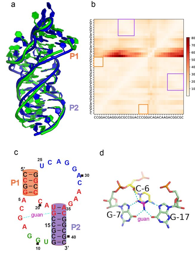
Downloaded from rnajournal.cshlp.org on June 13, 2020 - Published by Cold Spring Harbor Laboratory Press

Caleb Geniesse
        Department of Biochemistry, Stanford University School of Medicine, USA
Yangwei Jiang
        Department of Physics and Astronomy, Department of Biochemistry. MU Institute
        for Data Science and Informatics, University of Missouri-Columbia, USA.
Astha Joshi
        International Institute of Molecular and Cell Biology in Warsaw, Poland
        Present address: Department of Internal Medicine, Washington University in St.
        Louis School of Medicine, St Louis, MO 63110, USA.
Andrey Krokhotin
        Department of Biochemistry and Biophysics and Lineberger Comprehensive Cancer
        Center, University of North Carolina at Chapel Hill, Chapel Hill, NC 27599, USA.
        Departments of Pathology, Genetics and Developmental Biology, Howard Hughes
        Medical Institute, Stanford Medical School, Palo Alto, CA, 94305, USA
Marcin Magnus
        International Institute of Molecular and Cell Biology in Warsaw, Poland
        Present address: ReMedy-International Research Agenda Unit, Centre of New
        Technologies, University of Warsaw, Warsaw, Poland
Olivier Mailhot
        Institute for Research in Immunology and Cancer (IRIC), Department of Computer
        Science and Operations Research, Université de Montréal, Montréal, Québec, H3C
        3J7, Canada.
Francois Major
        Institute for Research in Immunology and Cancer (IRIC), Department of Computer
        Science and Operations Research, Université de Montréal, Montréal, Québec, H3C
        3J7, Canada.
Thomas H. Mann
        Department of Biochemistry, Stanford University School of Medicine, USA
        Present address: NOMIS Center for Immunobiology and Microbial Pathogenesis,
        Salk Institute for Biological Studies, La Jolla, CA, USA
Paweł Piątkowski
        International Institute of Molecular and Cell Biology in Warsaw, Poland
Radoslaw Pluta
        International Institute of Molecular and Cell Biology in Warsaw, Poland
        Present address: Institute for Research in Biomedicine (IRB Barcelona), The
        Barcelona Institute of Science and Technology, Barcelona 08028, Spain.
Mariusz Popenda
        Institute of Bioorganic Chemistry, Polish Academy of Sciences, 61-704 Poznan,
        Poland
Joanna Sarzynska
        Institute of Bioorganic Chemistry, Polish Academy of Sciences, 61-704 Poznan,
        Poland
Lizhen Sun
        Department of Physics and Astronomy, Department of Biochemistry. MU Institute
        for Data Science and Informatics, University of Missouri-Columbia, USA.
Marta Szachniuk
        Institute of Bioorganic Chemistry, Polish Academy of Sciences, 61-704 Poznan,
        Poland
Downloaded from rnajournal.cshlp.org on June 13, 2020 - Published by Cold Spring Harbor Laboratory Press

        Institute of Computing Science, Poznan University of Technology, 60-965 Poznan,
        Poland
Siqi Tian
        Department of Biochemistry, Stanford University School of Medicine, USA
Jian Wang
        Department of Pharmacology, Penn State College of Medicine, Hershey, PA,
        17033, USA.
Jun Wang
       School of Physics, Huazhong University of Science and Technology, Wuhan
       430074, Hubei, China
Andrew M. Watkins
        Department of Biochemistry, Stanford University School of Medicine, USA
Jakub Wiedemann
        Institute of Bioorganic Chemistry, Polish Academy of Sciences, 61-704 Poznan,
        Poland
        Institute of Computing Science, Poznan University of Technology, 60-965 Poznan,
        Poland
Yi Xiao
       School of Physics, Huazhong University of Science and Technology, Wuhan
       430074, Hubei, China
Xiaojun Xu
        Department of Physics and Astronomy, Department of Biochemistry. MU Institute
        for Data Science and Informatics, University of Missouri-Columbia, USA.
Joseph D. Yesselman
        Department of Biochemistry, Stanford University School of Medicine, USA
        Present address: Department of Chemistry, University of Nebraska-Lincoln, USA
Dong Zhang
        Department of Physics and Astronomy, Department of Biochemistry. MU Institute
        for Data Science and Informatics, University of Missouri-Columbia, USA.
Yi Zhang
       School of Physics, Huazhong University of Science and Technology, Wuhan
       430074, Hubei, China
Zhenzhen Zhang
        Department of Physics and Astronomy, Clemson University, Clemson, SC 29634,
        United States
Chenhan Zhao
        Department of Physics and Astronomy, Department of Biochemistry. MU Institute
        for Data Science and Informatics, University of Missouri-Columbia, USA.
Peinan Zhao
        Department of Physics and Astronomy, Department of Biochemistry. MU Institute
        for Data Science and Informatics, University of Missouri-Columbia, USA.
Yuanzhe Zhou
        Department of Physics and Astronomy, Department of Biochemistry. MU Institute
        for Data Science and Informatics, University of Missouri-Columbia, USA.
Tomasz Zok
        Institute of Computing Science, Poznan University of Technology, 60-965 Poznan,
        Poland
Adriana Żyła
        International Institute of Molecular and Cell Biology in Warsaw, Poland
Downloaded from rnajournal.cshlp.org on June 13, 2020 - Published by Cold Spring Harbor Laboratory Press

        Present address: Department of Macromolecular Physics, Faculty of Physics, A.
        Mickiewicz University, Poznan, Poland

Aiming Ren
        Life Sciences Institute,Zhejiang University, Hangzhou,Zhejiang 310058, China
Robert T. Batey
         Department of Biochemistry, University of Colorado at Boulder, Campus Box 596,
         Boulder, CO 80309-0596, USA
Barbara L. Golden
         Department of Biochemistry, Purdue University, West Lafayette, Indiana 47907,
         USA
Lin Huang
         Guangdong Provincial Key Laboratory of Malignant Tumor Epigenetics and Gene
         Regulation, Medical Research Center, Sun Yat-Sen Memorial Hospital, Sun Yat-
         Sen University, Guangzhou 510120, P. R. China
         RNA Biomedical Institute, Sun Yat-Sen Memorial Hospital, Sun Yat-Sen University,
         Guangzhou 510120, P. R. China.
         Cancer Research UK Nucleic Acid Structure Research Group, MSI/WTB Complex,
         The University of Dundee, Dow Street, Dundee DD1 5EH, UK.
David M. Lilley
         Cancer Research UK Nucleic Acid Structure Research Group, MSI/WTB Complex,
         The University of Dundee, Dow Street, Dundee DD1 5EH, UK.
Yijin Liu
         Cancer Research UK Nucleic Acid Structure Research Group, MSI/WTB Complex,
         The University of Dundee, Dow Street, Dundee DD1 5EH, UK.
Dinshaw J. Patel
         Structural Biology Program, Memorial Sloan Kettering Cancer Center, New York,
         NY, 10065, USA

Eric Westhof
       Architecture et Réactivité de l'ARN, Université de Strasbourg, Institut de Biologie
       Moléculaire et Cellulaire du CNRS, Strasbourg, France

*To   whom correspondence should be addressed:
Zhichao Miao: zmiao@ebi.ac.uk
Eric Westhof: e.westhof@ibmc-cnrs.unistra.fr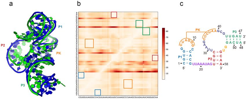
Downloaded from rnajournal.cshlp.org on June 13, 2020 - Published by Cold Spring Harbor Laboratory Press

Abstract
RNA-Puzzles is a collective endeavor dedicated to the advancement and improvement of
RNA 3D structure prediction. With agreement from crystallographers, the RNA structures are
predicted by various groups before the publication of the crystal structures. We now report
the prediction of 3D structures for six RNA sequences: four nucleolytic ribozymes and two
riboswitches. Systematic protocols for comparing models and crystal structures are
described and analyzed. In these six puzzles, we discuss a) the comparison between the
automated web servers and human experts; b) the prediction of coaxial stacking; c) the
prediction of structural details and ligand binding; d) the development of novel prediction
methods; and e) the potential improvements to be made. We show that correct prediction of
coaxial stacking and tertiary contacts is essential for the prediction of RNA architecture, while
ligand binding modes can be only predicted with low resolution and simultaneous prediction
of RNA structure with accurate ligand binding still remains out of reach. All the predicted
models are available for the future development of force field parameters and the
improvement of comparison and assessment tools.

Introduction

The RNA-Puzzles competition was initiated in 2011 following discussions and exchanges at
the annual meetings of the RNA Society. The blind assessment of RNA predictions was
started along broadly similar procedures for protein predictions organized within the Critical
Assessment of Protein Structure Prediction (CASP) experiment (Moult et al. 2014). While
CASP releases protein sequences publically, the RNA Puzzles experiment follows strict rules
of confidentiality. With the agreement of a structural biology group, an RNA sequence, with
the X-ray structure recently determined in that laboratory, is submitted to modeling groups
that all agree to keep the information confidential. Because the structures are unknown to
the modelers and program developers, and absent from databases, the procedure minimizes
bias in the prediction algorithms. The modeling groups have a deadline for submitting the
results of the prediction of the puzzle. The deadline depends on the delay between
submission and publication of the structure (with the public release of the coordinates) as
well as on the type of modeling exercise that is being tested (with or without manual
intervention). The prediction time for the automated web server is set to 48 hours, while
human experts have 3-4 weeks. The results of the comparisons between the experimental
and theoretical structures are then published together with all groups involved in the
enterprise. The puzzles are grouped into rounds based on the similarity of function and
approximate co-occurrence in prediction time. Three rounds of RNA-Puzzles have been
previously reported in the literature (Cruz et al. 2012; Miao et al. 2015, 2017). Over these
rounds, the RNA-Puzzles consortium, through numerous exchanges and discussions, has
fostered a better understanding between the experimental and theoretical groups, as well as
between the groups involved in the development of the modeling tools. Further, based on the
feedback from the evaluation, the modeling and assessment tools have improved in quality
and ease of use during the whole process.

Ribozymes (Doherty and Doudna 2000; Cech 2009) are RNA molecules that can catalyze
specific biochemical reactions, in a way similar to the action of protein enzymes, while

Downloaded from rnajournal.cshlp.org on June 13, 2020 - Published by Cold Spring Harbor Laboratory Press

riboswitches (Garst, Edwards, and Batey 2011; Breaker 2011) are regulatory segments of
protein-coding RNAs that bind a small molecule, resulting in a change in the protein
translation. Both ribozymes and riboswitches have biological functions achieved through
ligand/small molecule binding (Zhang, Lau, and Ferré-D’Amaré 2010). In particular, some
ribozymes and riboswitches may share similar structural folds, such as the c-di-GMP
riboswitch, the guanine riboswitch and the hammerhead ribozyme (Porter et al. 2017).
According to the Rfam database (Kalvari et al. 2018), 27 out of the 42 riboswitch families
have members with experimentally determined structures, while for the 31 ribozyme families
only only 6 have structures experimentally solved. Predicting these functionally important
RNA structures can provide significant structural insights to the RNA functions and help in
RNA engineering. In particular, the self-cleaving nucleolytic ribozymes became a hot topic in
the field of structural biology of the ribozyme (Ren et al. 2016). The fair assessment of the
computational prediction of these newly discovered ribozymes underscores a key aspect of
RNA structure prediction and function inference.

Here, we report our community effort to predict three-dimensional structures of ribozymes
and riboswitches. The six puzzles in this round of prediction include four ribozymes: the pistol
ribozyme, the hammerhead ribozyme, two twister-sister ribozymes, and two riboswitches:
the 5-hydroxytryptophan and the Guanidine-III riboswitch. A few additional puzzles were
carried out concomitantly with these six puzzles corresponding to non-coding RNA domains
with functions distinct from chemical catalysis or small-molecule recognition and will be
discussed in a separate manuscript. According to Round IV prediction results, we highlight:
a) the progress in automated web server approaches and the still existing gap between the
web servers and human experts; b) key rules that play important roles in prediction; c) the
existing bottleneck in ligand/ion binding prediction. The prediction methods developed within
our community will provide a practical resource to RNA biologists.

Results

Availability
All the prediction results are available at www.rnapuzzles.org. The structure manipulation
tools and all the assessment metrics together (Magnus et al. 2020) with the predicted data
are now available in an open-source repository at https://github.com/RNA-Puzzles.

The Six Prediction Targets

Puzzle 9: The 5-hydroxytryptophan aptamer
5-hydroxytryptophan (5-HTP) is a naturally occurring amino acid and chemical precursor of
serotonin, which is also marketed in many European countries for the treatment of major
depression. Porter et al. (Porter et al. 2017) introduced the aptamer as a scaffold for
genetically encodable small-molecule biosensor, which selectively binds to 5-HTP. The 71-
nucleotide (nt)-long sequence is:
Downloaded from rnajournal.cshlp.org on June 13, 2020 - Published by Cold Spring Harbor Laboratory Press

5’-
GGACACUGAUGAUCGCGUGGAUAUGGCACGCAUUGAAUUGUUGGACACCGUAAAU
GUCCUAACACGUGUCC-3’

This puzzle structure was only predicted by human experts. The 2 Å crystal structure was
deposited in the Protein Data Bank (PDB) (Berman 2000) (5kpy) after the prediction.
According to the scaffold analysis (Porter et al. 2017), the secondary structure of 5-
hydroxytryptophan riboswitch is similar to the guanine riboswitch (Batey, Gilbert, and
Montange 2004), di-GMP riboswitch (Kulshina, Baird, and Ferré-D’Amaré 2009) and
hammerhead ribozyme (Anderson et al. 2013), which have been reported. The 5-
hydroxytryptophan aptamer adopts a topology identical to that of the guanine riboswitch, with
a backbone Root Mean Square Deviation (RMSD) of 2.5 Å, Fig S1a.

Puzzle 15: The hammerhead ribozyme
Although the hammerhead ribozyme was the first of the small nucleolytic ribozymes to be
described (Uhlenbeck 1987), the initial structures were trapped in an inactive conformation
(Scott, Finch, and Klug 1995; Pley, Flaherty, and McKay 1994). The binding to the
pentavalent transition state analogue vanadate results in significant core rearrangements in
the structure (Mir and Golden 2016) compared to the initial structures. Considering the
availability of the solved hammerhead ribozyme structure, this puzzle was predicted by both
fully-automated web servers and human experts to benchmark the difference between these
two types of approaches. Two rounds of predictions were organized. In the first round, only
the RNA sequences were given for prediction; in the second round, the secondary structure,
based on the crystal structure, was given as additional information. Ligand or ion binding
information was not given in the prediction. The structure was predicted as two chains, 48 nt
+ 20 nt. The sequences are:

5’-GGGUACUUAAGCCCACUGAUGAGUCGCUGGGAUGCGACGAAACGCCCA-3’
5’-GGGCGUCUGGGCAGUACCCA-3’

The crystal structure was solved at resolution 2.95 Å and deposited in PDB (5di4). In the
crystal, the 5’ end of the first chain and the 3’ end of the second chain form intermolecular
base-pairs between symmetry-related molecules, forming, therefore, a dimer structure. Thus,
the known base-pairs between the 5’ end of the first chain and 3’ end of the second chain,
verified through functional studies are not formed between the expected strands in the crystal
structure. Unless explicitly stated, crystal contacts are not considered for comparison. Most
models rightly contained a regular stem between the 5’- and 3’-ends. Automatic comparisons,
therefore, lead to larger (and penalizing) deviations for those nucleotides (about 6). The
catalytically relevant part of the molecule is in the core of the structure.

Puzzle 17: The Pistol ribozyme
The recent discovery of pistol (this puzzle), twister-sister (Puzzles 19 and 20) and hatchet
(Puzzle 22) ribozymes (Weinberg et al. 2015) have invigorated the field of small self-cleaving
nucleolytic ribozymes. Pistol ribozyme studies have yielded significant insights into the G-U
cleavage mechanism, which is facilitated by its pseudoknot fold (Ren et al. 2016). The 62-
nucleotide (nt)-long sequence is:
Downloaded from rnajournal.cshlp.org on June 13, 2020 - Published by Cold Spring Harbor Laboratory Press

5’-
CGUGGUUAGGGCCACGUUAAAUAGUUGCUUAAGCCCUAAGCGUUGAUAAAUAUCAG
GUGCAA-3’

This puzzle was predicted by both web servers and human experts. After the prediction
deadline, the crystal structure (Ren et al. 2016) of resolution 2.73 Å was deposited in the
PDB (5k7c). The G53-U54 cleavage site is the key functional site and from a biological point
of view, the accuracy of predicted G53 interactions is most relevant.

Puzzle 19: The Twister sister ribozyme 1
Another self-cleaving ribozyme is twister-sister, which includes two coaxial helical stacks
organized by a three-way junction and two tertiary contacts (Liu, Wilson, and Lilley 2017). It
functions predominantly as a metalloenzyme. Both human expert prediction and automated
web server prediction were performed. The fold was predicted as a two-chain (40 nt + 22 nt)
structure with the following sequences:

5’-GCAGGGCAAGGCCCAGUCCCGUGCAAGCCGGGACCGCCCC-3’
5’-GGGGCGCGGCGCUCAUUCCUGC-3’

The crystal structure of resolution 2 Å in PDB (5t5a) was deposited after the prediction. This
puzzle was a good case to evaluate the prediction of coaxial stacking (Lescoute and Westhof
2006) as well as the long-distance tertiary contacts.

Puzzle 20: The Twister sister ribozyme 2
This is a variant of a twister-sister ribozyme that includes a four-way junction. As in Puzzle
19, the structure of Puzzle 20 contains several long-distance tertiary contacts and a number
of RNA-bound magnesium ions. These two structures adopt a similar global topology, but
they exhibit significant conformational differences (Fig S2), with a backbone RMSD > 10 Å.
Although Puzzle 19 prediction was organized before Puzzle 20, Puzzle 19 structure was not
made available as a template for homology modeling of Puzzle 20. The structure of Puzzle
20 was predicted both by automated web servers and by human experts. It was also
predicted as a two-chain structure (18 nt + 50 nt), with the longer sequences below:

5’-ACCCGCAAGGCCGACGGC-3’
5’-GCCGCCGCUGGUGCAAGUCCAGCCACGCUUCGGCGUGGGCGCUCAUGGGU-3’

Structures coming from the native and MnCl2-soaked crystals were solved and deposited in the
PDB as 5y85 (resolution 2.00 Å) and 5y87 (resolution 2.13 Å), respectively. The backbone
RMSD between these two structures is only 0.4 Å. Since both the solved structures (5y85
and 5y87) can be considered as ‘ground truth’ of the sequence, both of them were used as
reference structures in the later assessment.

Puzzle 21: The Guanidine-III Riboswitch
Barrick et al. identified the ykkC family as probable riboswitches more than a decade ago
(Barrick et al. 2004). It was eventually found that the ykkC family RNAs respond to guanidine
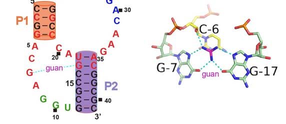
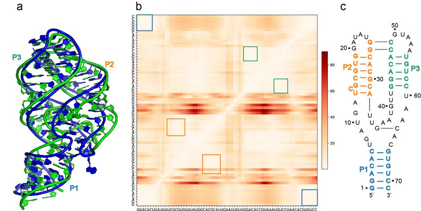
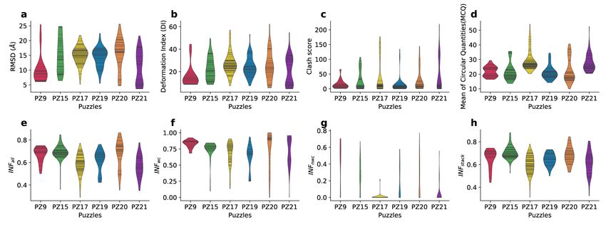
Downloaded from rnajournal.cshlp.org on June 13, 2020 - Published by Cold Spring Harbor Laboratory Press

(Nelson et al. 2017), and the ykkC family was later classified into three subclasses by Breaker
and colleagues, termed as the guanidine-I (Nelson et al. 2017), -II (Sherlock, Malkowski, and
Breaker 2017), and -III (Sherlock and Breaker 2017) riboswitches. This puzzle involves the
prediction of the structure of the guanidine-III riboswitch (Huang et al. 2017), which
corresponds to the Rfam family (RF01763) (Kalvari et al. 2018)). The 41-nucleotide (nt)-long
sequence is:

5’-CCGGACGAGGUGCGCCGUACCCGGUCACGACAAGACGGCGC-3’

After both web server and expert predictions were performed, two crystal structures were
deposited in the PDB: one structure solved in space group P3121 at 1.91 Å resolution (PDB:
5nwq) and another structure in space group P3212 at 2.94 Å resolution (PDB: 5nz6). The two
structures share a backbone RMSD of 0.58 Å and both were considered as reference
structures in the assessment, Fig S3. The structure is dominated by a pseudoknot fold and
the guanidine binding site is formed by a triplex left-handed helical core. This is the first
puzzle for which the identity of the binding ligand was provided before prediction.

Overall prediction results
The distributions of all the assessment metrics in the sets of all models submitted for every
puzzle are displayed in Fig 1. One important aspect to understand the predictions is based
on comparisons with the best model. From the RMSD distribution, the best-predicted models
start at RMSD values of 3.65 Å (PZ21) and 6.45 Å (PZ15), Fig 1a. But with consideration of
the sequence length as in the Deformation Index, the best prediction models for all the six
puzzles are comparable, Fig 1b, ranging from 4.75 (PZ21) to 8.64 (PZ9). The worst predicted
models peak at about 25.55 Å RMSD (PZ20). In Fig 1b, roughly two ensembles of models
can be distinguished for Puzzles 19 and 21. Although the RMSD range spreads, Puzzles 20
and 21 display lower RMSD predictions, while Puzzles 17 and 19 yielded predicted models
centered around 17 Å. Although most of the predicted structures show very low Clash scores,
still some outliers exist in the distribution, Fig 1c. Fig S4 displays the various metrics as a
function of RMSD. Deformation Index shows a distribution proportional to RMSD. For the
Clash score, the range is independent of RMSD, except for large outliers, which is not easy
to explain (Fig S4a).

The correct prediction of the interactions is the premise of accuracy in structure prediction.
From the distribution of INFwc (Interaction Network Fidelity for Watson-Crick base-pairs),
prediction of Watson-Crick interactions is near perfect in the best model for most of the
puzzles, ranging from 84% (PZ15) to 100% (PZ20), Fig 1e-h. However, the INFwc ranges are
rather large in several puzzles. The recurrent structural modules are normally formed by non-
Watson-Crick interactions. For example, guanidine, as a ligand, in Puzzle 21 is hydrogen-
bonded to the Hoogsteen edges of two guanine bases (Huang et al. 2017). The prediction of
non-Watson-Crick is more difficult than Watson-Crick base-pairs, thus, showing a much
wider range in the INFnwc distribution, except for Puzzles 17 and 21, for which the accuracy
of INFnwc is very low. The prediction of base stacking is highly related to the Watson-Crick
interactions and the distribution of INFstacking generally follows the same pattern as the
accuracy of Watson-Crick interactions INFwc. Interestingly, in Fig 1d, two groups of models
appear with very long tails in the MCQ distribution (PZ17 and PZ20).
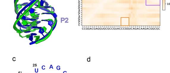
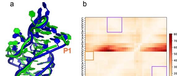
Downloaded from rnajournal.cshlp.org on June 13, 2020 - Published by Cold Spring Harbor Laboratory Press

Detailed prediction results
Below, the results specific for each RNA-Puzzle are described. Figures 2 to 7 contain a
superposition between the crystal structure and the model with the lowest RMSD, a
secondary structure diagram, and a heat map of the Deformation Index (DI) (Parisien et al
2009). In all figures, the secondary structure elements are colored and numbered (helix P1,
P2,..., pseudoknot PK). The corresponding colored numberings are indicated on the 3D
drawings. In the Deformation Index plots, the secondary structure elements are delimited by
colored rectangles within the heat maps. The RMSD is used as the primary ranking
parameter because of its simplicity. However, RMSDs spread errors over the whole structure
and one cannot tell which parts of the molecule were correctly modelled and which were not.
The Deformation Index attempts to alleviate this drawback in the following way. The
structures to be compared are superimposed locally three nucleotides at a time (following
the sequence) and the average deviations between the central nucleotide and all the other
nucleotides are calculated and plotted as a heat map. Thus, all domains globally
superimposable appear in white while those departing from that global core appear in red.

Puzzle 9: The 5-hydroxytryptophan aptamer (Fig 2)
Despite differences in local conformation, the 5-HTP riboswitch adopts the same topology as
the guanine riboswitch. Therefore, the guanine riboswitch structure was used correctly by
many groups as a global or local template in the prediction. Among the models, model 7 from
the Chen group demonstrates the lowest backbone RMSD as 3.74 Å (6.06 Å all-atom
RMSD), Fig 2. According to the structure comparison in Fig S1b (guanine riboswitch-based
similarity), the best model (Chen model 7) has a backbone RMSD of 1.54 Å from the
previously reported guanine riboswitch (Batey, Gilbert, and Montange 2004). Homology
modeling using templates may well capture the main topology but the local structural details
in the loops of P2 and P3 regions show a difference from the crystal structure. It is not easy
to evaluate how significant those changes are, since they occur in loop regions allowing
dynamical movements and often with high crystallographic temperature factors as discussed
previously (Miao et al. 2017; Magnus et al. 2020; Miao and Westhof 2017). The correct
modeling of such local structure variability continues to be a major challenge for the modeling
methods. In this puzzle, the crystal structure forms more turns around the ligand-binding
region compared to the best-predicted model. As the ligand information was not disclosed
during the prediction process, the structure was predicted as ligand-free. Knowing the ligand
information may still be beneficial to the prediction process. A similar case was detected in
Puzzle 10 (Round II): prediction groups pointed out that the kink-turn module in RNA can be
easily predicted if the binding protein (YbxF) information is given. Model 5 from the Das group
achieved the best INFall score; Das group modeling used FARFAR (Das, Karanicolas, and
Baker 2010) with two starting templates: residues A:30-40 and A:56-70 came from 2XNW,
while a tRNA T-loop from 3L0U furnished residues A:45-49. The RMSD values of the 34
submitted predictions range from 6.06 to 25.43 Å with an average value of 9.93 Å, as shown
in Table S1.

Puzzle 15: The hammerhead ribozyme (Fig 3)
The prediction of the hammerhead ribozyme structure in the state of binding to a transition
state analog is a difficult case for automatic prediction because the binding ligand was not
given in the prediction as prior knowledge. RMSD of the 74 submitted models ranges from
Downloaded from rnajournal.cshlp.org on June 13, 2020 - Published by Cold Spring Harbor Laboratory Press

6.45 Å to 24.78 Å. The predicted model of the lowest RMSD (6.45 Å) shows a topology similar
to the experimentally determined structure. Fig 3 and Table S2 contain the automatic
comparisons between crystal structure and models. As discussed above, the strands forming
stem I in hammerhead ribozymes involve intermolecular pairs between molecules in the
crystal while in models stem I forms intramolecularly. Thus, automatic comparisons in that
region are artifactual. This example shows the advantage of the Deformation Profile in which
local errors do not spread throughout the structure (see Fig. 3b). In Table S13, the last six
nucleotides on each of the strands are removed from the comparison. Most of the resulting
assessment parameters are much better. The corresponding Deformation Profile is shown
in Fig. 3c. As shown in Fig S5, the RNAComposer (Popenda et al. 2012; Antczak et al. 2016)
prediction shows better deformation profile and RMSD in the core structure (without stem I)
than the SimRNA (Boniecki et al. 2016) prediction. SimRNA has a better global RMSD
because of the orientation of the stem I region.

Puzzle 17: The Pistol ribozyme (Fig 4)
The Pistol ribozyme structure is dominated by a pseudoknot structure and Watson-Crick
interactions. According to Rfam, P1, P2 and PK regions of Pistol ribozyme have significant
covariations in the sequence alignment. The only uncertainty is the length of the pseudoknot:
Rfam (Kalvari et al. 2018) structure includes five base-pairs while R-scape (Rivas, Clements,
and Eddy 2017) predicts seven base-pairs, Fig S6. And, in the crystal structure, the
pseudoknot is made of six base-pairs. Despite this minor difference in the number of base-
pairs between Rfam prediction and the crystal structure, the best prediction demonstrated a
good accuracy with 5.15 Å RMSD (SimRNAweb server model 1 in the second round
prediction), Fig 4. But only seven out of the total 107 predicted structures have RMSDs better
than 10 Å, Table S3. Models from each prediction group are ranked according to their
estimated reliability. In this puzzle, the best RMSD model is SimRNA model 1 and the model
with the best INFall score (most interactions having been correctly predicted) is model 1 from
Das group (8.60 Å RMSD).

Puzzle 19: The Twister sister ribozyme 1 (Fig 5)
The RMSD values of the 55 submitted predictions range from 5.52 to 22.44 Å with an average
value of 14.33 Å, as shown in Table S4. The challenges in the prediction lie in: a) the correct
coaxial stacking of the P4 and P5 regions, b) the correct detection of long-distance contacts.
These two issues are linked to each other. Some models predict the stacking between P4
and P1 rather than P4 and P5 (for example, model 3 from Adamiak-Szachniuk group and
model 5 from the Bujnicki group), Fig S7. These models significantly deviate from the
reference structure but also do not capture the key tertiary interactions between P4 and P5.
As there is no gap between P4 and P5, the more likely stacking would have been predictable
from the rules in (Lescoute and Westhof 2006) and was predicted by other groups. As shown
in Fig 5, the best model (Chen group model 1) correctly predicts the global topology as well
as most of the contacts (seen from Deformation Profile). The only difference is that the
reference structure is more compact. The question of assessing structure compactness has
been brought up during the second RNA-Puzzles community meeting in 2018.
Downloaded from rnajournal.cshlp.org on June 13, 2020 - Published by Cold Spring Harbor Laboratory Press

Puzzle 20: The Twister sister ribozyme 2 (Fig 6)
Unlike Puzzle 19, Puzzle 20 includes four helices that form two coaxial stackings: between
P2 and P3, between P4 and P5 (even though P5 has no gap between P2). Although Puzzle
20 has a longer sequence than Puzzle 19, it shows better prediction accuracy: 10 out of 50
predicted models have
Downloaded from rnajournal.cshlp.org on June 13, 2020 - Published by Cold Spring Harbor Laboratory Press

RNAComposer models) have better RMSDs than the median score obtained for human
predictions. Interestingly, there is no such discrepancy between human and automatic
predictions in the INF scores, especially when considering Watson-Crick interactions only.
When it comes to the Clash score, 3D models delivered by the servers actually outperform
the predictions submitted in the human category. Nevertheless, even in the category of
stereochemical quality, both server and human expert predictions could be improved. An
analysis made with the use of the RCSB Maxit program (Berman 2000) shows that only 8
server-generated models (4 submitted by RNAComposer and 4 by RW3D) and 10 models of
Adamiak-Szachniuk group have correct stereochemical parameters.

Puzzle 17 ended up with 38 models (in the two rounds of prediction: in the first round of
prediction, no information on the 2D structure was given, while in the second round the 2D
structure was given) submitted by two servers (RNAComposer and SimRNAweb) and 69
predictions by 8 human groups. In this Puzzle, automatic predictions are qualitatively very
close to the human ones, as far as the RMSD, INF and DI measures are concerned. For
example, the lowest RMSD for server models is 5.15 Å (and for human models it is 7.13 Å)
and the highest is 21.52 Å for server models (and for human models it is 21.75 Å). 13 server
models obtained better scores than the human RMSD median value that reached 14.87 Å.
On the other hand, 21 human models were worse than the automatic RMSD median (16.09
Å). 3D models in the human category have on average better MCQ and Clash score values.
Only 23 models submitted in this Puzzle have correct stereochemistry. This set includes 12
automatic predictions submitted by RNAComposer and 11 human predictions (3 of Adamiak-
Szachniuk group and 8 by Das group).

The collection of RNA structure 3D models submitted for Puzzle 19 includes 28 automatic
models (from 5 servers: 3dRNA, LeeServer, RNAComposer, RW3D, and SimRNAweb) and
27 human models (from 7 expert groups). Here, the median values of all considered
measures for both categories are very similar. Although human predictions are more diverse
- the ranges of their RMSD, INF and DI values are wider than for automatic models. For
example, the latter have RMSDs in the range 9.59–19.58 Å, while the human models are in
the range 5.51–22.44 Å. The DI computed for automatic models takes values between 15.48
and 38.28 Å and that of human expert models between 7.48 and 53.18 Å. The INF scores
obtained for non-canonical and stacking interactions are very similar in both sets. Again, 11
server models are closer to the reference structure than the median human prediction (its
RMSD value is 14.75 Å). The subset of human models contains 12 submissions with RMSD
worse than the median score computed for server predictions which equal 15.25 Å. It should
be emphasized that RCSB Maxit reports no errors in 14 cases only which come from 9
automatic predictions (5 from RNAComposer and 4 from RW3D) and 5 human predictions
(all by Adamiak-Szachniuk group). It clearly shows that in all subsequent predictions the
check of stereochemical correctness should be performed. The RCSB Maxit program is a
reliable option.

In Puzzle 20, there were three servers and five human participants. Servers (RNAComposer,
RW3D, SimRNAweb) submitted 20 models, and human experts 35 models, respectively. In
the case of this challenge, automatic models’ qualities visibly differ from what human experts
predicted. The best RMSD for server submissions is 10.99 Å, while for human experts it is
4.70 Å. A significant difference can also be seen in the DI (14.01 Å vs 5.68 Å) and MCQ
(9.97° vs 15.45°) values between the best automatic and the best human predictions. The
Downloaded from rnajournal.cshlp.org on June 13, 2020 - Published by Cold Spring Harbor Laboratory Press

surprise is the best automatic prediction (RNAComposer, model 1) with INFwc=1, while the
best human model (Adamiak, model 5) has 0.98 for this measure. Only four server models
obtained lower RMSD scores than the median human prediction that has RMSD=16.05 Å.
On the other hand, five human submissions have higher RMSD than the median score
computed for automatic models which equals 16.64 Å. In this Puzzle, only 8 models pass the
RCSB Maxit test with no stereochemical errors reported. These are 3 automatic submissions
by RNAComposer and 5 human predictions by Adamiak-Szachniuk group.

Finally, Puzzle 21 was closed with 25 automatic predictions from 4 servers (3dRNA,
RNAComposer, RW3D, SimRNAweb) and 34 human predictions from 5 expert groups.
Server models submitted in this challenge differ significantly in quality from the majority of
human expert models. RMSD of automatic predictions is in the range 10.56-18.02 Å and the
median value reaches 15.99 Å; Deformation Index is between 19.58 Å and 39.46 Å with the
median equal to 31.46 Å. For human models, RMSDs are in 3.64-21.57 Å with the median
equal to 5.31 Å and DIs are in 4.74-54.79 Å with median 8.07 Å. INF computed for Watson-
Crick base-pairs is the same for both the best server and the best human model. But when
it comes to the evaluation of non-Watson-Crick interactions’ prediction, 3D models submitted
by human experts are the winners of this challenge. Only when the Clash score is considered,
automatic models perform better with their scores between 0 and 118.53, as compared to
7.52-218.94 range for human submissions. All models submitted in this Puzzle have
stereochemical errors. For the best models, as far as the stereochemistry is concerned (13
automatic predictions from RNAComposer and RW3D, 10 human predictions from Das and
Adamiak-Szachniuk groups), RCSB Maxit reported problems with phospho-sugar bonds
stereochemistry.

Methods

Results Assessment
At the first RNA-Puzzles community meeting, organized in 2016, it was decided to allow up
to 5 structural models, which should be ordered according to the expected reliability for each
prediction approach. Still, each research group may submit the results for multiple prediction
approaches, while automated web servers are separated from human expert prediction.

We assess the prediction accuracy by comparing each prediction model with the reference
structure of the lowest RMSD. Assessment metrics also include Deformation Index (Parisien
et al. 2009), which is a metric to normalize RMSD with the sequence length; Clash score
(Williams et al. 2018) shows the credibility of the structure in terms of atomic distance;
Interaction Network Fidelity (Parisien et al. 2009) (INF) is an evaluation of the prediction
accuracy of different types of interactions, which include Watson-Crick interaction, non-
Watson-Crick interaction and base stacking. Considering some new prediction approaches
may abstract RNA structure (such as the network model (Kim, Petingi, and Schlick 2013)),
we use the Mean of Circular Quantities (Zok, Popenda, and Szachniuk 2014) to assess the
accuracy in the torsion angle space.
Downloaded from rnajournal.cshlp.org on June 13, 2020 - Published by Cold Spring Harbor Laboratory Press

Summary of Structure Prediction methods
In the Supplementary Notes section, we briefly introduce 1) the updates and
improvement of the existing prediction approaches; 2) the development of novel
prediction approaches; 3) the development of automated web servers; 4) the
methods used and focus on the special treatments that were used in the prediction
of the present ribozymes and riboswitches.

Discussion
Overall, the results of RNA-Puzzles Round IV confirm observations made since earlier
rounds of the puzzles (reviewed in (Miao and Westhof 2017; Magnus et al. 2020)) . In every
test case, the current community of RNA structural modelers is able to produce blind
predictions of 3D structures that are in agreement with crystal structures (Figs. 2-7). An
important question then is whether these methods are ready to give biologically useful
models for the many RNAs that have not been experimentally characterized but are
suspected to have well-defined 3D structures. Only a very small fraction of the Rfam families
(Kalvari et al. 2018) have structure(s) solved, which is 99 families of the 3016 Rfam families.
Compared to the solved protein structures, the known RNA structure space is still limited.
Computational prediction of RNA 3D structure may therefore exert great potential in
understanding the unknown RNA structures, especially in generating potential structure
models to explain the molecular functions. Automatic web servers will likely be critical to
enable the broader RNA biology community to make such predictions, and so assessment
of these servers compared to expert models has been an important theme of recent rounds
of the RNA-Puzzles.

The prediction results in Round IV are compared with all the previously reported results in
Fig S13. One success achieved in Round IV is that all the best-predicted models have a
RMSD < 7 Å. The high atomic clash of otherwise accurate models has been pointed out since
the Round II prediction (Miao et al. 2015) and new structure relaxation approaches have
been developed. Still, it appears to be difficult to achieve both accurate structure prediction
and relaxation within the limited prediction time.

Since the first challenge of RNA-Puzzles, we have observed a growing interest in the
development and use of automated methods supporting selected stages of the modeling of
3D RNA structures, including web servers that predict RNA 3D models. In RNA-Puzzles
Round I (Cruz et al. 2012), the Major group tested MC-Fold/MC-Sym (Parisien and Major
2008), while the Das group applied their Rosetta SWA (Das 2013) algorithm. In Round II
(Miao et al. 2015), the spectrum of computing methods used in RNA 3D structure prediction
expanded to six programs (3dRNA, RNAComposer, DMD (Dokholyan et al. 1998; F. Ding et
al. 2008; Proctor, Ding, and Dokholyan 2011; Feng Ding and Dokholyan 2005; Krokhotin,
Houlihan, and Dokholyan 2015), ModeRNA (Rother et al. 2011), Rosetta FARFAR, MC-
Fold), some of them operating as fully-automated servers, while in Round III (Miao et al.
2017) there were five programs used (3dRNA, RNAComposer, RW3D, SimRNA, DMD).
These methods are still mostly used as the elements of the modeling procedure and are
complemented by expert knowledge, experimental data and manual or semi-automatic
Downloaded from rnajournal.cshlp.org on June 13, 2020 - Published by Cold Spring Harbor Laboratory Press

editing of the initial model. It was only in 2015 that rules for fully-automated prediction were
defined and, consequently, Puzzle 15 was opened in two categories, human and server.
Since then, both the structures predicted by human experts over weeks and models
simulated fully automatically within 2 days have been submitted for every puzzle. In the
current Round IV, a large gap remains between the accuracy of server models compared to
human expert models. Nevertheless, stereochemical quality (e.g., atom-atom clashes) of the
server models was similar to and in some puzzles, better than human expert models,
suggesting that further automation of expert procedures could help ameliorate these
pervasive stereochemical errors. In addition, this round of puzzles has inspired several
participating groups to improve automation of their methods, including the development of
new servers that are being tested in more recent RNA-Puzzles (see Methods).

Although ligand binding prediction and ligand pocket prediction have been discussed since
the previous round of RNA-Puzzles, the prediction of the RNA-ligand complex structure is
difficult when no structural template is available to generate a RNA model that can be
regarded as very close to the native RNA structure, and serve as a receptor to dock the
ligand. Thus, in Puzzles 9 and 21, corresponding to aptamers for 5-hydroxytryptophan and
guanidine, respectively (Fig S1, S8, and S14), the accurate prediction of ligand contacts was
still difficult. In Puzzle 21, the predicted structures within 5 Å RMSD from the crystal structure
demonstrate ligand binding positions that are close to the correct binding pocket, and the
model 1 of LORES prediction from Das group correctly predicts the contacts with G7
nucleotide (Fig S14). In terms of a more incisive evaluation of accuracy, we still do not have
a systematic program to analyze the quality of the ligand-binding pocket and the ligand-
binding interactions for RNA. A major difficulty lies in the analysis of various interaction types
between ligand and RNA. RNA-ligand binding remains an important aspect of both prediction
and evaluation, and will be further discussed in studies summarizing results on later puzzles
(e.g., PZ23, a Mango fluorogenic aptamer).

In addition to testing the predictive ability of new automated servers, Round IV provided
information on the ability of expert and automated server predictors to identify homologous
templates and ligand binding sites. To better understand the prediction performance, the
human vs. server results are compared in Fig S12, while radar plots discussing each puzzle
are summarized in Fig S15-S20. The diagrams generally show that polygon shapes are
larger on diagrams with human predictions (the larger is the polygon, the better is the
corresponding model). This is especially evident in Fig S20 (Puzzle 21) which shows
automatically predicted 3D models scored considerably worse than those predicted manually
by Das, Chen, and Adamiak-Szachniuk groups. In some cases, radar diagrams show clear
winners e.g., the Bujnicki model 1 in Puzzle 20. In other cases, radar plots highlight the
relative weakness of each prediction approach.

In the Round IV prediction, we have included two twister sister ribozyme puzzles (PZ19,
PZ20). Although belonging to the same sequence family, the two puzzles demonstrate
significant structural differences. The Guanidine-III riboswitch in Puzzle 21 is also different
from the previous solved Guanidine-II riboswitch structures (Huang, Wang, and Lilley 2017;
Reiss and Strobel 2017) and Guanidine-I riboswitch (Reiss, Xiong, and Strobel 2017).
Contrarily, some newly solved RNA structures could adopt the same structural topology as
structures present in the database, e.g., 5HTP riboswitch (5kpy) is similar to guanine
riboswitch (4fe5), which was in fact its ‘parent’ in an in vitro selection. It is crucial to
Downloaded from rnajournal.cshlp.org on June 13, 2020 - Published by Cold Spring Harbor Laboratory Press

understand how different sequences in the same family result in structural differences. And
RNA-Puzzles aims to understand if such structural differences can be captured by existing
prediction approaches. According to the results of PZ19 and PZ20, current prediction can
already predict the difference between the two twister sister ribozymes. In our next RNA-
Puzzles, manuscript, we will further discuss such structural variances in other RNA families,
including the T-box (Puzzles 10, 26, 27 and 28).

Ions are important for stabilizing RNA structures by screening the electrostatic repulsions
among the backbone phosphate groups, and divalent ions are known experimentally to be
more efficient than monovalent ions (Woodson 2005). Post-blind-prediction studies by the
Ding group showed that explicit modeling of divalent ions in all-atom DMD simulations could
significantly enhance the sampling of near-native structures and improve the accuracy of
predicted 3D structures (Fig. S9C). Additional studies to incorporate explicit modeling of
Mg2+ into the CG RNA simulations could further improve the prediction accuracy and
efficiency of multi-scale RNA modeling approaches.

Conclusions
The aims of the RNA-Puzzles collective work is to provide a snapshot and evaluation of the
state-of-the-art in RNA 3D structure modeling. In Round IV, blind tests on ribozymes and
riboswitch aptamers indicate that accurate prediction still requires an appropriate
combination of different prediction strategies, including the fragment/structure library-based
methods and the de novo prediction/force field-based simulation methods. Automated
predictions from web servers still remain worse than human expert models for many cases.
In future rounds, we speculate that further improvements may be achieved through machine-
learning-based contacts prediction (Senior et al. 2020), integration of experimental data or
better ranking of the predicted models. As discussed before (Miao and Westhof 2017),
secondary structure (the knowledge of the helical Watson-Crick pairs) alone is far from
sufficient to model RNA structures without prediction of non-WC base pairs. Non-WC base
pairs, atomic clashes, and ligand binding site prediction still have room for improvement.
Alternative structure comparison metrics, which are not sequence-length-dependent, may
benefit the RNA-Puzzles assessment in later puzzles.

Acknowledgements
Z.M. was supported by The Single Cell Gene Expression Atlas grant 108437/Z/15/Z from the
Wellcome Trust. E.W. was supported by the French National Program Investissement
d’Avenir (Labex NetRNA) administered by the Agence National de la Recherche (ANR-10-
LABX-0036_NETRNA). Y.X. was supported by NSFC under Grant No. 31570722 and
11874162. M.S. and her group were supported by the Polish National Science Center (NCN;
2016/23/B/ST6/03931) and by the statutory funds of Poznan University of Technology,
Poland. S-J.C was supported by NIH R01-GM117059 (S-J.C) and NIH R01-GM063732 (S-
J.C). R.D. and his group were supported by N.I.H. R35 GM122579 and R21 CA219847.
D.M.J.L. group was funded by Cancer Research UK program grant A18604. R.T.B. and his
group were funded by NIH R01-GM073850. J.M.B. and his group were supported by the
Polish National Science Center (NCN; 2012/04/A/NZ2/00455 to J.M.B. and
2015/17/N/NZ2/03360 to M.M), and by the Foundation for Polish Science (FNP;
Downloaded from rnajournal.cshlp.org on June 13, 2020 - Published by Cold Spring Harbor Laboratory Press

POIR.04.04.00-00-3CF0/16). Support from the National Institutes for Health
(5R01GM123247, 2R01 GM114015, and 1R35 GM134864 to N.V.D.) and the Passan
Foundation are acknowledged. The project described was also supported by the National
Center for Advancing Translational Sciences, National Institutes of Health, through Grant
UL1 TR002014 (J.W. and N.V.D.). The content is solely the responsibility of the authors and
does not necessarily represent the official views of the NIH.

The authors thank the Lee group at the Korea Institute for Advanced Study and an
anonymous group for their participation in RNA-Puzzles. The authors also thank the
members of the Bujnicki group for technical contributions, including Stanisław Dunin-
Horkawicz (IIMCB), Grzegorz Chojnowski (IIMCB), Wayne Dawson (IIMCB), Dorota
Matelska (IIMCB), Catarina Almeida (IIMCB), Bharat Madan (IIMCB), Grzegorz Lach
(IIMCB).

Author contributions
EW conceived and supervised the project, coordinated the collaboration and wrote the
manuscript. ZM organized the prediction, analyzed the results, built the website and wrote
the manuscript. All other authors predicted the structures, analyzed the results and wrote the
manuscript.

References
Anderson, Michael, Eric P. Schultz, Monika Martick, and William G. Scott. 2013. “Active-
    Site Monovalent Cations Revealed in a 1.55-Å-Resolution Hammerhead Ribozyme
    Structure.” Journal of Molecular Biology. https://doi.org/10.1016/j.jmb.2013.05.017.
Antczak, Maciej, Mariusz Popenda, Tomasz Zok, Joanna Sarzynska, Tomasz Ratajczak,
    Katarzyna Tomczyk, Ryszard W. Adamiak, and Marta Szachniuk. 2016. “New
    Functionality of RNAComposer: An Application to Shape the Axis of miR160 Precursor
    Structure.” Acta Biochimica Polonica 63 (4): 737–44.
Barrick, J. E., K. A. Corbino, W. C. Winkler, A. Nahvi, M. Mandal, J. Collins, M. Lee, et al.
    2004. “New RNA Motifs Suggest an Expanded Scope for Riboswitches in Bacterial
    Genetic Control.” Proceedings of the National Academy of Sciences.
    https://doi.org/10.1073/pnas.0308014101.
Batey, Robert T., Sunny D. Gilbert, and Rebecca K. Montange. 2004. “Structure of a
    Natural Guanine-Responsive Riboswitch Complexed with the Metabolite
    Hypoxanthine.” Nature 432 (7015): 411–15.
Berman, H. M. 2000. “The Protein Data Bank.” Nucleic Acids Research.
    https://doi.org/10.1093/nar/28.1.235.
Boniecki, Michal J., Grzegorz Lach, Wayne K. Dawson, Konrad Tomala, Pawel Lukasz,
    Tomasz Soltysinski, Kristian M. Rother, and Janusz M. Bujnicki. 2016. “SimRNA: A
    Coarse-Grained Method for RNA Folding Simulations and 3D Structure Prediction.”
    Nucleic Acids Research 44 (7): e63.
Breaker, Ronald R. 2011. “Prospects for Riboswitch Discovery and Analysis.” Molecular
    Cell 43 (6): 867–79.
Cech, T. R. 2009. “Evolution of Biological Catalysis: Ribozyme to RNP Enzyme.” Cold
    Spring Harbor Symposia on Quantitative Biology 74 (October): 11–16.
Cruz, José Almeida, Marc-Frédérick Blanchet, Michal Boniecki, Janusz M. Bujnicki, Shi-Jie
    Chen, Song Cao, Rhiju Das, et al. 2012. “RNA-Puzzles: A CASP-like Evaluation of
Downloaded from rnajournal.cshlp.org on June 13, 2020 - Published by Cold Spring Harbor Laboratory Press
RNA Three-Dimensional Structure Prediction.” RNA 18 (4): 610–25.
Das, Rhiju. 2013. “Atomic-Accuracy Prediction of Protein Loop Structures through an RNA-
Inspired Ansatz.” PloS One 8 (10): e74830.
Das, Rhiju, John Karanicolas, and David Baker. 2010. “Atomic Accuracy in Predicting and
Designing Noncanonical RNA Structure.” Nature Methods 7 (4): 291–94.
Ding, Feng, and Nikolay V. Dokholyan. 2005. “Simple but Predictive Protein Models.”
Trends in Biotechnology. https://doi.org/10.1016/j.tibtech.2005.07.001.
Ding, F., S. Sharma, P. Chalasani, V. V. Demidov, N. E. Broude, and N. V. Dokholyan.
2008. “Ab Initio RNA Folding by Discrete Molecular Dynamics: From Structure
Prediction to Folding Mechanisms.” RNA. https://doi.org/10.1261/rna.894608.
Doherty, Elizabeth A., and Jennifer A. Doudna. 2000. “Ribozyme Structures and
Mechanisms.” Annual Review of Biochemistry.
https://doi.org/10.1146/annurev.biochem.69.1.597.
Dokholyan, Nikolay V., Sergey V. Buldyrev, H. Eugene Stanley, and Eugene I.
Shakhnovich. 1998. “Discrete Molecular Dynamics Studies of the Folding of a Protein-
like Model.” Folding and Design. https://doi.org/10.1016/s1359-0278(98)00072-8.
Garst, Andrew D., Andrea L. Edwards, and Robert T. Batey. 2011. “Riboswitches:
Structures and Mechanisms.” Cold Spring Harbor Perspectives in Biology 3 (6).
https://doi.org/10.1101/cshperspect.a003533.
Huang, Lin, Jia Wang, and David M. J. Lilley. 2017. “The Structure of the Guanidine-II
Riboswitch.” Cell Chemical Biology 24 (6): 695–702.e2.
Huang, Lin, Jia Wang, Timothy J. Wilson, and David M. J. Lilley. 2017. “Structure of the
Guanidine III Riboswitch.” Cell Chemical Biology 24 (11): 1407–15.e2.
Kalvari, Ioanna, Joanna Argasinska, Natalia Quinones-Olvera, Eric P. Nawrocki, Elena
Rivas, Sean R. Eddy, Alex Bateman, Robert D. Finn, and Anton I. Petrov. 2018. “Rfam
13.0: Shifting to a Genome-Centric Resource for Non-Coding RNA Families.” Nucleic
Acids Research 46 (D1): D335–42.
Kim, Namhee, Louis Petingi, and Tamar Schlick. 2013. “Network Theory Tools for RNA
Modeling.” WSEAS Transactions Mathematics 9 (12): 941–55.
Krokhotin, Andrey, Kevin Houlihan, and Nikolay V. Dokholyan. 2015. “iFoldRNA v2: Folding
RNA with Constraints: Fig. 1.” Bioinformatics.
https://doi.org/10.1093/bioinformatics/btv221.
Kulshina, Nadia, Nathan J. Baird, and Adrian R. Ferré-D’Amaré. 2009. “Recognition of the
Bacterial Second Messenger Cyclic Diguanylate by Its Cognate Riboswitch.” Nature
Structural & Molecular Biology 16 (12): 1212–17.
Lescoute, Aurélie, and Eric Westhof. 2006. “Topology of Three-Way Junctions in Folded
RNAs.” RNA 12 (1): 83–93.
Liu, Yijin, Timothy J. Wilson, and David M. J. Lilley. 2017. “The Structure of a Nucleolytic
Ribozyme That Employs a Catalytic Metal Ion.” Nature Chemical Biology 13 (5): 508–
13.
Magnus, Marcin, Maciej Antczak, Tomasz Zok, Jakub Wiedemann, Piotr Lukasiak, Yang
Cao, Janusz M. Bujnicki, Eric Westhof, Marta Szachniuk, and Zhichao Miao. 2020.
“RNA-Puzzles Toolkit: A Computational Resource of RNA 3D Structure Benchmark
Datasets, Structure Manipulation, and Evaluation Tools.” Nucleic Acids Research 48
(2): 576–88.
Magnus, Marcin, Michał J. Boniecki, Wayne Dawson, and Janusz M. Bujnicki. 2016.
“SimRNAweb: A Web Server for RNA 3D Structure Modeling with Optional Restraints.”
Nucleic Acids Research 44 (W1): W315–19.
Miao, Zhichao, Ryszard W. Adamiak, Maciej Antczak, Robert T. Batey, Alexander J. Becka,
Marcin Biesiada, Michał J. Boniecki, et al. 2017. “RNA-Puzzles Round III: 3D RNA
Structure Prediction of Five Riboswitches and One Ribozyme.” RNA 23 (5): 655–72.
Miao, Zhichao, Ryszard W. Adamiak, Marc-Frédérick Blanchet, Michal Boniecki, Janusz M.
Bujnicki, Shi-Jie Chen, Clarence Cheng, et al. 2015. “RNA-Puzzles Round II:
Assessment of RNA Structure Prediction Programs Applied to Three Large RNA
Structures.” RNA 21 (6): 1066–84.You can also read