Roadmap of ultrafast x-ray atomic and molecular physics
←
→
Page content transcription
If your browser does not render page correctly, please read the page content below
Mathematisch-Naturwissenschaftliche Fakultät

Linda Young | Kiyoshi Ueda | Markus Gühr | Philip H Bucksbaum
Marc Simon | Shaul Mukamel | Nina Rohringer | Kevin C Prince
Claudio Masciovecchio | Michael Meyer | Artem Rudenko
Daniel Rolles | Christoph Bostedt | Matthias Fuchs | David A Reis
Robin Santra | Henry Kapteyn | Margaret Murnane | Heide Ibrahim
François Légaré | Marc Vrakking | Marcus Isinger | David Kroon
Mathieu Gisselbrecht | Anne L’Huillier | Hans Jakob Wörner
Stephen R Leone

Roadmap of ultrafast x-ray atomic and
molecular physics

Suggested citation referring to the original publication:
Journal of Physics B: Atomic, Molecular and Optical Physics 51 (2018), Art. 032003
DOI https://doi.org/10.1088/1361-6455/aa9735
ISSN (print) 0953-4075
ISSN (online) 1361-6455

Postprint archived at the Institutional Repository of the Potsdam University in:
Postprints der Universität Potsdam
Mathematisch-Naturwissenschaftliche Reihe ; 668
ISSN 1866-8372
https://nbn-resolving.org/urn:nbn:de:kobv:517-opus4-424238
DOI https://doi.org/10.25932/publishup-42423
Journal of Physics B: Atomic, Molecular and Optical Physics

J. Phys. B: At. Mol. Opt. Phys. 51 (2018) 032003 (45pp)                                                 https://doi.org/10.1088/1361-6455/aa9735

Roadmap

Roadmap of ultrafast x-ray atomic and
molecular physics
Linda Young1,2,22, Kiyoshi Ueda3, Markus Gühr4,5, Philip H Bucksbaum5,6,
Marc Simon7, Shaul Mukamel8, Nina Rohringer9,10, Kevin C Prince11 ,
Claudio Masciovecchio11, Michael Meyer12, Artem Rudenko13,
Daniel Rolles13, Christoph Bostedt1, Matthias Fuchs5,14, David A Reis5,
Robin Santra9,10, Henry Kapteyn15,16, Margaret Murnane15,16,
Heide Ibrahim17 , François Légaré17, Marc Vrakking18 ,
Marcus Isinger19 , David Kroon19, Mathieu Gisselbrecht19,
Anne L’Huillier19, Hans Jakob Wörner20 and Stephen R Leone21
1
  Argonne National Laboratory, 9700 S. Cass Avenue, Argonne, IL 60439, United States of America
2
  Department of Physics and James Franck Institute, University of Chicago, 5720 South Ellis Avenue,
Chicago, IL 60637, United States of America
3
  Institute of Multidisciplinary Research for Advanced Materials, Tohoku University, Sendai 980-8577, Japan
4
  Physics and Astronomy, Potsdam University, Karl-Liebknecht-Strasse24/25, 14476 Potsdam-Golm, Germany
5
  Stanford Pulse Institute, SLAC National Accelerator Laboratory, 2575 Sand Hill Road, Menlo Park, CA
94025, United States of America
6
  Department of Physics, Stanford University, 382 Via Pueblo Mall, Stanford, CA 94305-4060, United
States of America
7
  Sorbonne Universités, UPMC Univ Paris 06, CNRS, UMR 7614, Laboratoire de Chimie Physique-
Matière et Rayonnement, F-75005 Paris, France
8
  Department of Chemistry, University of California, Irvine, CA 92697-2025, United States of America
9
  Center for Free-Electron Laser Science, DESY, Notkestrasse 85, 22607 Hamburg, Germany
10
   Department of Physics, University of Hamburg, Jungiusstrasse 9, 20355 Hamburg, Germany
11
   Elettra Sincrotrone Trieste, Strada statale 14, Basovizza (Trieste), Italy
12
   European XFEL GmbH, Holzkoppel 4, 22869 Schenefeld, Germany
13
   J.R. Macdonald Laboratory, Department of Physics, Kansas State University, Manhattan, Kansas 66506,
United States of America
14
   Department of Physics and Astronomy, University of Nebraska, Lincoln, NE 68588-0299, United States of
America
15
   JILA, Center for Atomic, Molecular & Optical Physics, Boulder, CO 80309, United States of America
16
   Department of Physics and Electrical and Computer Engineering, University of Colorado Boulder,
Boulder, CO 80309-0440, United States of America
17
   Institut National de la Recherche Scientifique, Centre Énergie, Matériaux, et Télécommunications, 1650
Boulevard Lionel-Boulet, Varennes, Qc, J3X1S2, Canada
18
   Max-Born Institute, Max Born Strasse 2 A, 12489 Berlin, Germany
19
   Department of Physics, Atomic Physics, Lund University, Box 118, SE-221 00 Lund, Sweden
20
   ETH Zürich, Laboratorium für Physikalische Chemie, Vladimir-Prelog-Weg 2, 8093 Zürich Switzerland
21
   Department of Chemistry, University of California Berkeley, Berkeley CA 94720-1460, United States of
America

E-mail: young@anl.gov

Received 12 June 2017, revised 31 August 2017
Accepted for publication 31 October 2017
Published 9 January 2018

22
     Guest editor of the roadmap.

                 Original content from this work may be used under the terms
                 of the Creative Commons Attribution 3.0 licence. Any
further distribution of this work must maintain attribution to the author(s) and
the title of the work, journal citation and DOI.

0953-4075/18/032003+45$33.00                                                       1                 © 2018 IOP Publishing Ltd Printed in the UK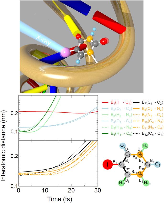
J. Phys. B: At. Mol. Opt. Phys. 51 (2018) 032003                                                    Roadmap

Abstract
X-ray free-electron lasers (XFELs) and table-top sources of x-rays based upon high harmonic
generation (HHG) have revolutionized the field of ultrafast x-ray atomic and molecular physics,
largely due to an explosive growth in capabilities in the past decade. XFELs now provide
unprecedented intensity (1020 W cm−2) of x-rays at wavelengths down to ∼1 Ångstrom, and
HHG provides unprecedented time resolution (∼50 attoseconds) and a correspondingly large
coherent bandwidth at longer wavelengths. For context, timescales can be referenced to the Bohr
orbital period in hydrogen atom of 150 attoseconds and the hydrogen-molecule vibrational
period of 8 femtoseconds; wavelength scales can be referenced to the chemically significant
carbon K-edge at a photon energy of ∼280 eV (44 Ångstroms) and the bond length in methane
of ∼1 Ångstrom. With these modern x-ray sources one now has the ability to focus on individual
atoms, even when embedded in a complex molecule, and view electronic and nuclear motion on
their intrinsic scales (attoseconds and Ångstroms). These sources have enabled coherent
diffractive imaging, where one can image non-crystalline objects in three dimensions on ultrafast
timescales, potentially with atomic resolution. The unprecedented intensity available with
XFELs has opened new fields of multiphoton and nonlinear x-ray physics where behavior of
matter under extreme conditions can be explored. The unprecedented time resolution and pulse
synchronization provided by HHG sources has kindled fundamental investigations of time
delays in photoionization, charge migration in molecules, and dynamics near conical
intersections that are foundational to AMO physics and chemistry. This roadmap coincides with
the year when three new XFEL facilities, operating at Ångstrom wavelengths, opened for users
(European XFEL, Swiss-FEL and PAL-FEL in Korea) almost doubling the present worldwide
number of XFELs, and documents the remarkable progress in HHG capabilities since its
discovery roughly 30 years ago, showcasing experiments in AMO physics and other
applications. Here we capture the perspectives of 17 leading groups and organize the
contributions into four categories: ultrafast molecular dynamics, multidimensional x-ray
spectroscopies; high-intensity x-ray phenomena; attosecond x-ray science.
Keywords: ultrafast molecular dynamics, x-ray spectroscopies and phenomena, table-top
sources, x-ray free-electron lasers, attosecond phenomena

(Some figures may appear in colour only in the online journal)

Contents

1. Introduction                                                                                          3
2. Ultrafast molecular dynamics                                                                          5
3. Multidimensional x-ray spectroscopies                                                                11
4. High-intensity x-ray phenomena                                                                       17
5. Attosecond science with table-top sources                                                            27
5. Attosecond science with table-top sources                                                            28

                                                               2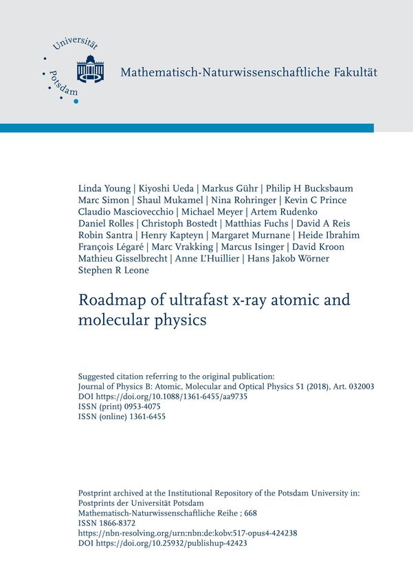
J. Phys. B: At. Mol. Opt. Phys. 51 (2018) 032003                                                                                   Roadmap

1. Introduction

Linda Young

Argonne National Laboratory and University of Chica-
go      The roadmap starts with topics generally familiar to
the AMO community: femtochemistry viewed with an x-ray
probe, multidimensional spectroscopy extended to the x-ray
regime, and then winds toward the less commonly encoun-
tered areas of high-intensity x-ray phenomena and attosecond
science. The first category describes how accelerator-based
x-ray sources are used to probe ultrafast molecular dynamics:
understanding ultra-intense x-ray pulse interactions with
matter as a prelude to x-ray free-electron laser (XFEL) probes
of femtosecond molecular dynamics (Ueda, section 2), opti-
cally induced molecular dynamics probed with XFELs (Gühr
and Bucksbaum, section 2), and the novel use of long-pulse,
monochromatic x-rays from a synchrotron source to induce
                                                                       Figure 1. Phase space covered by the XFEL and HHG sources
and probe ultrafast inner-shell molecular processes (Simon,            discussed in this roadmap. For XFELs, the pulse duration represents
section 2). The second category describes multidimensional             that of a single pulse, whereas for HHG, the range spans both single
x-ray spectroscopies enabled by XFELs: a theoretical per-              pulses and pulse trains spaced by the period of the driver laser. The
spective where analogy to NMR, infrared and optical reali-             numbers in each island indicate the number of photons/pulse/1%
zations is used to highlight the potential of x-rays to monitor        bandwidth. Research to extend the limits of all represented
                                                                       quantities, photon energy, time scale and photon number per pulse,
the phase and dynamics of non-equilibrium valence wave-                is pursued for both XFEL and HHG sources. The emphasis for
packets (Mukamel, section 3), a discussion of routes from an           XFELs is to extend the time scale to the attosecond regime and
atomic x-ray laser to control of stimulated Raman processes            photon energy above 20 keV; the emphasis for HHG is to extend the
with XFELs (Rohringer, section 3), and an account of the               photon energy range to hard x-ray and photon number per pulse. For
                                                                       properties not represented by these basic quantities, XFELs seek
realization of coherent control and four-wave mixing in the
                                                                       enhanced temporal coherence and synchronization with external
XUV regime using only the fully coherent, seeded XFEL,                 sources, and both sources seek increased average power and
FERMI (Prince and Masciovecchio, section 3). The third                 controlled polarization.
category deals with high-intensity x-ray phenomena created
in XFELs in systems of increasing complexity: nonlinear                spectroscopies in the attosecond domain (Leone,
multiphoton processes and polarization control in atoms                section 5).     To guide the reader we have sketched the
(Meyer, section 4), charge and nuclear dynamics after inner-           ultrafast photon source capabilities, dynamical phenomena
shell absorption in molecules (Rudenko and Rolles,                     and experimental techniques discussed in this roadmap.
section 4), imaging and scattering in nanoscale clusters               Source capabilities and phenomena that can be studied are
(Bostedt, section 4), hard x-ray nonlinear optics (Fuchs and           inextricably linked. In figure 1, the performance of accel-
Reis, section 4), and, to describe quantitatively these phe-           erator-based XFEL and HHG sources are shown. Both are
nomena, the theory of electronic structure under extreme
                                                                       labeled with photons/pulse/1% bandwidth for individual
conditions of x-ray irradiation (Santra, section 4).        The
                                                                       pulses. The complementarity of the two classes of sources is
final category addresses table-top and attosecond x-ray sci-
                                                                       clear. Most XFELs are based upon self-amplified spontaneous
ence as enabled by high harmonic generation (HHG) sources.
                                                                       emission (SASE) radiation, lack longitudinal coherence,
Technical frontiers in HHG include extension to x-ray
                                                                       struggle to obtain pulse lengths shorter than a few femtose-
wavelengths, enhancement of the single pulse energy and
increase of the average power for short-wavelength radiation.          conds and struggle to synchronize with external sources.
We start with a general perspective on table-top-scale ultrafast       HHG sources, on the other hand, have exquisite temporal
coherent x-ray science that leads toward a future that can be          coherence and pulse duration, but are challenged to obtain
‘smaller, cheaper and (ultra)faster’ (Kapteyn and Murnane,             large photon numbers per pulse, to extend their reach to short
section 5), followed by a description of a route to high-              wavelengths and to obtain high average power. At the carbon
average-power soft x-ray ultrashort pulses via mid-infrared            K-edge XFELs produce many orders of magnitude more
drive lasers (Ibrahim and Légaré, section 5), a discussion of          photons/pulse/1% bandwidth than HHG sources. Figure 2
attosecond and femtosecond XUV science (Vrakking,                      maps the fundamental dynamical phenomena observed in
section 5), quantitative studies of photoionization time delays        isolated atoms and molecules, as well as the collective phe-
in atoms (Isinger, Kroon, Gisselbrecht and L’Huillier,                 nomena occurring in condensed phases, onto their respective
section 5), evolution of attosecond spectroscopies from the            time and length scales. Figure 3 maps the experimental
XUV to the x-ray regime and from isolated molecules to the             techniques enabled versus photon number per pulse and
liquid phase (Wörner, section 5), and finally a description of          photon energy. Overlaid are some research areas that are
soft x-ray transient absorption and the first multidimensional          beneficiaries of these studies in atomic and molecular physics

                                                                   3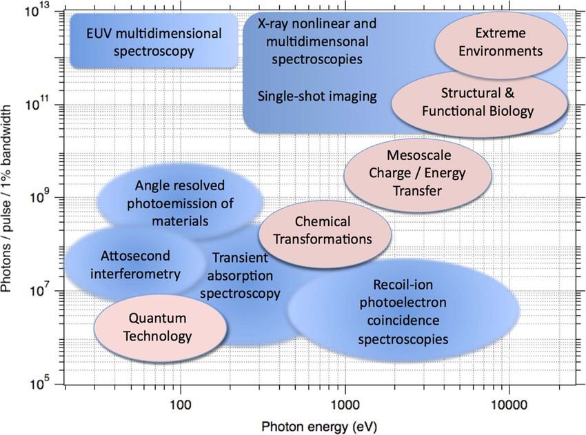
J. Phys. B: At. Mol. Opt. Phys. 51 (2018) 032003                                                                                        Roadmap

                                                                            Figure 3. Experimental techniques used in ultrafast x-ray science
Figure 2. Fundamental atomic, molecular and electronic phenomena            mapped onto photon number per pulse and photon energy typically
probed on ultrafast timescales (blue). Fundamental collective phenom-       used (blue). The high-fluence regime enables nonlinear x-ray
ena in the condensed phase probed on ultrafast timescales (pink).           spectroscopies and single-shot imaging, potentially with atomistic
                                                                            resolution. Low fluences are employed to remain in the linear x-ray
and the accompanying source and methodology development.                    absorption regime to probe ultrafast transient processes. (Saturation
                                                                            fluence for a carbon atom at 290 eV, just above the K-edge, is
The incredible progress of the past few years and the logical               ∼1010 photons/microns2.) Overlaid are research areas addressed
paths for source improvement augur a very exciting future for               with ultrafast x-ray methodologies that stem from understanding
ultrafast x-ray science.                                                    fundamental atomic and molecular physics processes (pink).

Acknowledgment                                                              irrevocable worldwide license in said article to reproduce,
The submitted manuscript has been created by UChicago                       prepare derivative works, distribute copies to the public, and
Argonne, LLC, Operator of Argonne National Laboratory                       perform publicly and display publicly, by or on behalf of the
(“Argonne”). Argonne, a U.S. Department of Energy Office                     Government. The Department of Energy will provide public
of Science laboratory, is operated under Contract No. DE-                   access to these results of federally sponsored research in
AC02-06CH11357. The U.S. Government retains for itself,                     accordance with the DOE Public Access Plan. http://energy.
and others acting on its behalf, a paid-up nonexclusive,                    gov/downloads/doe-public-access-plan

                                                                        4
J. Phys. B: At. Mol. Opt. Phys. 51 (2018) 032003 Roadmap
2. Ultrafast molecular dynamics
2.1. Probing ultrafast structural and electronic dynamics with
XFELs
Kiyoshi Ueda
Tohoku University
Status. Currently, two hard XFELs, LCLS in USA and
SACLA in Japan, are in operation for users, and a few more
will open for operation this year. The ultrashort and intense
x-ray pulses of these XFELs are revolutionizing the field of
ultrafast imaging, allowing determination of so far unknown
structures of transient species, proteins and any matter
undergoing reactions. Also, since XFEL pulses are giving
access to a new regime of x-ray intensities, they are opening up
new avenues in studying the interaction between intense x-rays
and various forms of matter. Understanding ultrafast reactions
induced by XFEL pulses is of fundamental interest, as well as
of crucial importance for structural determination with XFELs.
AMO science with XFELs may also be classified into the
two groups described above. The first group is the
investigation of the interaction of the intense XFEL pulse
with atoms, molecules and clusters. In the early experiments
at XFEL facilities, the target samples were just irradiated by a
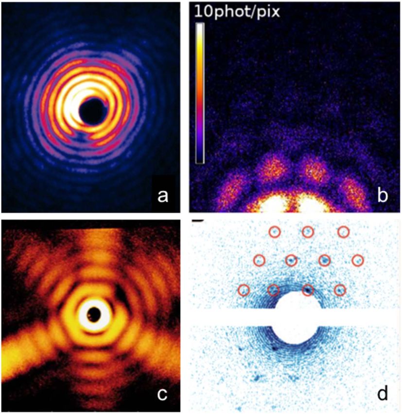
Figure 4. Upper panel, a schematic view of the radio-sensitizing effect
single XFEL pulse and the products (ions, electrons and of 5-iodouracil (5IU), a nucleobase analog of biological relevance.
fluorescent photons) were detected. Such experiments The work described in [6] illustrates how the molecule breaks apart
revealed new phenomena whenever the photon energy of and what ionic fragments are formed via the breakage of the molecular
the XFEL pulse and/or its pulse energy entered into a new edifice shortly after the inner-shell ionization, shedding light on the
regime. The findings may be summarized as follows. X-ray role of energetic ions in the initiation of damaging reactions. Lower
panel, time evolution of interatomic distances in 5IU after XFEL
absorption initially creates an inner-shell hole in a specific irradiation, obtained by model simulations; upper left, interatomic
atomic site. Then electronic relaxations, or Auger cascades, distances of I–C (red line), O–C (sky-blue lines), H–C (dark-green
follow. An XFEL pulse is so intense that it can cause multiple line) and H–N (light-green lines) pairs; lower left, interatomic
overlapping cycles of deep inner-shell photoemission and distances of C–C (black lines) and C–N (orange lines) pairs,
Auger cascades. Competitions between sequential photoemis- illustrating that most atoms other than hydrogen remain intact during
the XFEL pulse duration of 10 fs. Reprinted figure with permission
sion and Auger cascades have been studied extensively using from [6], Copyright 2016 by the American Physical Society.
rare-gas atoms as a target [1–3]. To study the coupled motion
of electrons and ions induced by an intense XFEL pulse, on
and simulations [7, 8]. The nanoplasma formation is expected
the other hand, a single molecule composed of a small
to be a general phenomenon, as it is expected to occur
number of atoms is an ideal target since various levels of
whenever nanometer-size particles are irradiated by an intense
theoretical modeling and experimental methods are available
or can be developed. Indeed, to study competition between XFEL pulse. In connection to structural studies using XFELs,
Auger cascades, charge redistribution and Coulomb explo- the above described investigations for atoms, molecules and
sions, a series of studies have been carried out for molecules clusters may be regarded as a fundamental study of radiation
that contain up to twelve atoms with one heavy atom as an damage at the atomic level (see figure 4).
x-ray absorber [4–6]. To study XFEL-induced reactions Nowadays, a combination of optical laser and XFEL
beyond these molecular model systems, atomic clusters are pulses is available at XFEL facilities. Consequently, one can
ideal objects because their size can be varied in a controlled probe the XFEL-induced reactions described in the previous
way from a single atom to a bulk-like macroscopic object. paragraph, employing an optical laser pulse as a probe.
When an atomic cluster is exposed to an intense x-ray pulse, Another way to probe XFEL-induced reactions is to use
many free electrons are created by sequential inner-shell XFEL pulses for both pump and probe. A split-and-delay
photoionization of many individual atoms followed by Auger assembly was introduced to XFEL facilities for such pump–
cascades. Because a number of electrons escape from the probe experiments. Recently, XFEL facilities started to
cluster, the cluster becomes highly charged and starts trapping provide double pulses at two different x-ray photon energies
electrons that are emitted from the individual atoms. A with variable time delay. This operation mode has some
nanoplasma is thus formed. These nanoplasma formation advantages over the use of the split-delay assembly since,
processes have been studied by a combination of experiments e.g., one can set the two photon energies above and below a
5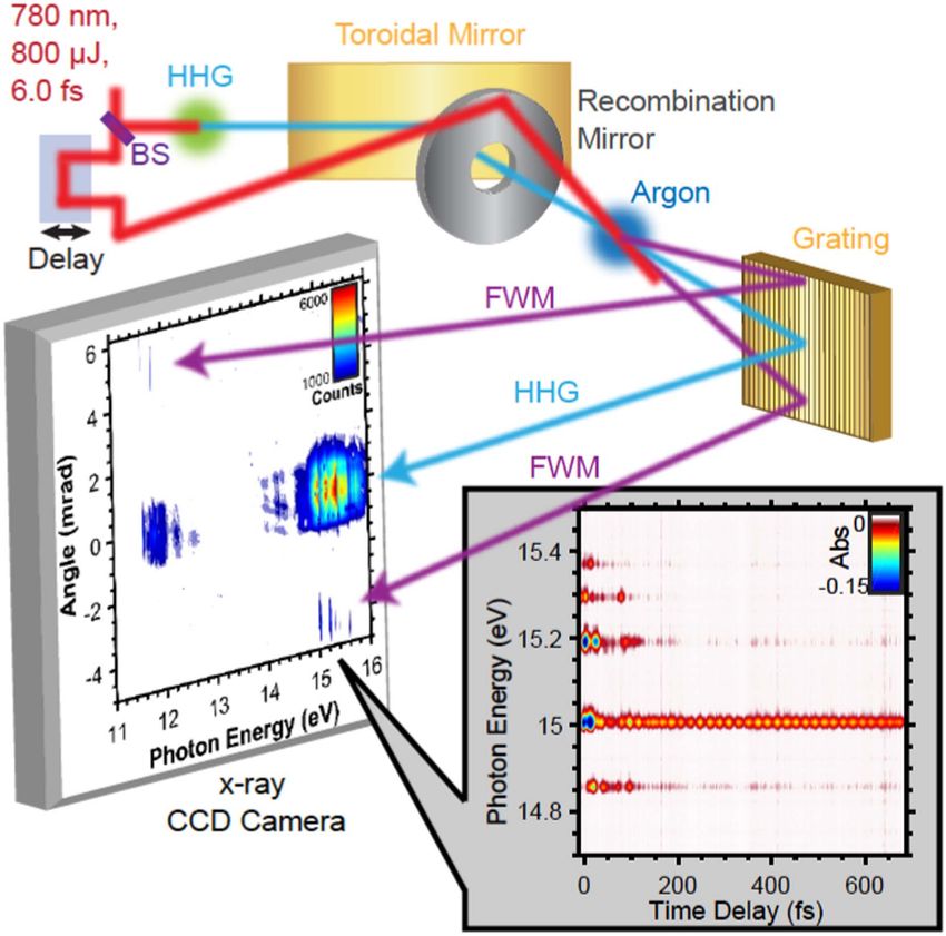
J. Phys. B: At. Mol. Opt. Phys. 51 (2018) 032003 Roadmap
certain edge of a certain atom. Such time-resolved studies on Advances in science and technology to meet challenges. The
XFEL-induced reactions have just started at XFEL facilities XFEL facilities can provide x-ray pulses with durations down
[9] and are in progress. to a few femtoseconds. This is also the case for double-pulse
The availability of optical lasers at XFEL facilities operations. Employing these ultrashort double pulses one can
opened a route to the second group of AMO science at achieve a time resolution of a few femtoseconds. Producing
XFELs, i.e. investigations of ultrafast reactions induced by pulses of duration below a femtosecond is also technically
light, or an optical laser, employing an XFEL pulse as a feasible. So far, the time resolution achieved for the
probe. For such studies on gas-phase samples, ions or combination of optical laser and XFEL pulses is often
electrons are usually detected because of small cross sections limited by the duration of the optical laser, say a few tens of
of x-ray scattering. X-ray diffraction experiments of isolated femtoseconds. In principle, the technology of producing an
molecules in photoreaction have, however, become feasible in ultrashort pulse down to a few femtoseconds (a few optical
the last few years [10], demonstrating that watching atomic cycles) is available and thus the time resolution in a few
motion in an isolated molecule is no longer a dream. X-ray femtoseconds should be within reach. To record ions together
imaging experiments for rare-gas clusters have also been with electrons in coincidence in a momentum-resolved manner
combined with optical laser-pump techniques and have just is also technically feasible, as has been demonstrated in
started to probe the structural evolution of clusters heated by experiments with synchrotron sources and high-repetition
an optical laser [11]. optical lasers. The reason why such measurements were
limited for the XFEL experiments (see, e.g. [4–6] for
multiple ion coincidence) is their low repetition rates. This
Current and future challenges. As noted above, most situation will dramatically improve when European XFEL, the
experiments with XFELs are nowadays based on a first high repetition rate XFEL, and LCLS-II will be in
combination of optical laser and XFEL pulses or two XFEL operation for users. X-ray detectors and sample injectors that
pulses. Thanks to the development of an arrival timing can accommodate in these high repetition rate XFELs will also
monitor for XFEL and optical laser pulses, which allows us to become available in time.
correct temporal jitters between the optical laser and XFEL
pulses, one can investigate light-induced ultrafast reactions in Concluding remarks. To probe electrons and atoms in action
real time, at a time resolution of tens of femtoseconds, which is no more a dream thanks to recent advances in technology
is comparable to that for the XFEL-pump and XFEL-probe related to time-resolved measurements with XFELs and
experiments. To fully explore electronic dynamics or detection techniques. Solving interplay between electronic
interplay between the electronic and structural dynamics, and atomic motions, which governs photochemistry, is now
however, it is desirable to have better time resolution. Various within reach.
kinds of electronic decays (Auger and interatomic Coulombic
decays as well as laser-enabled Auger decay) may occur on a Acknowledgments. The author is grateful to all the authors
time scale of femtoseconds to tens of femtoseconds. Nuclear of [2–6, 8, 9] for fruitful collaborations, and the XFEL
dynamics, especially involving the motion of hydrogen strategy program of MEXT, five-stars Alliance, and TAGEN
atoms, may also take place on a similar time scale and the project for support.
system may undergo reactions passing through conical
intersections. All these processes contribute to the charge
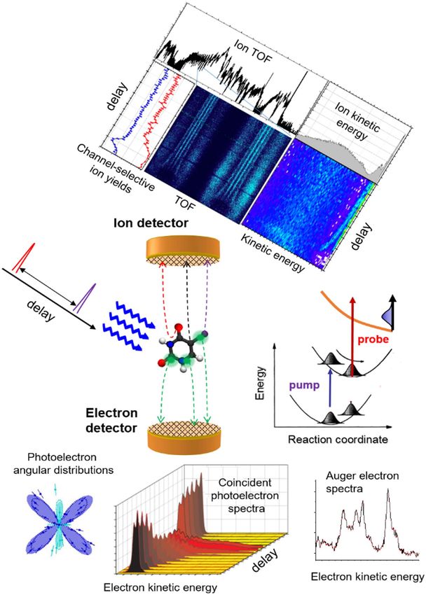
2.2. Methods for probing molecular dynamics with XFELs
redistribution in the molecule or cluster. The electronic
wavepacket may also be created when more than one
electronic state is populated via ultrafast electronic decay or Markus Gühr1,2 and Philip H Bucksbaum2
irradiation by an ultrashort optical laser pulse. Then the
1 2
wavepacket motion may be even faster. To see all of these Potsdam University Stanford PULSE Institute
dynamics, a pump–probe scheme with time resolution of a
few femtoseconds or less than a femtosecond would be Status. Photoexcited molecular dynamics is at the heart of
desirable. many processes in nature, from light harvesting and atmospheric
To fully extract information about ultrafast structural and chemistry to photoprotection of living organisms. Light interacts
electronic dynamics with the ultimate time resolution with molecular electrons, which couple to nuclei to initiate
discussed above, the signal detection scheme also needs to ultrafast and concerted motion of both the electrons and the
be improved. For molecules, if the number of events are atomic geometry. In only femtoseconds to picoseconds the
ideally less than one per single XFEL pulse, then one can subsequent steps for light-energy conversion are determined by
record electrons and ions in coincidence, in a momentum- this motion. Ultrashort hard and soft x-ray pulses provide
resolved manner. Such kinematically complete measurements valuable insights to understand the connections between the
should be a challenge for the aforementioned time-resolved initial motion and the ultimate chemical changes in excited
study. For clusters, single-shot imaging combined with ion molecules [12]. Time-resolved hard x-ray scattering experiments
and electron spectroscopy at ultimate time resolution should have revealed light-induced changes in the geometry, while soft
be a challenge. ultrafast x-ray spectroscopy is most sensitive to the electronic
6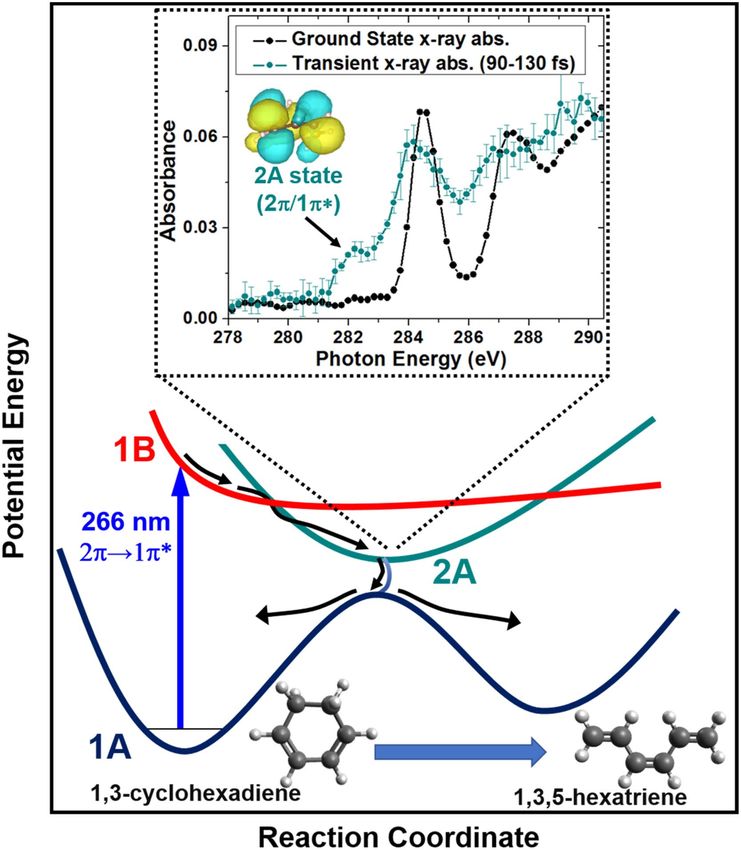
J. Phys. B: At. Mol. Opt. Phys. 51 (2018) 032003                                                                                     Roadmap

degrees of freedom. Large inner-shell binding energy differences
make x-ray spectra sensitive to both the type and location of
atoms in a molecule. Complementary information from ultrafast
scattering and spectroscopy can elucidate fundamental processes
such as charge transfer, light harvesting and photo-induced
molecular damage. Many well-developed x-ray spectroscopy
and scattering techniques have been extended to measurements
in the femtosecond time domain, and the first decade of ultrafast
molecular experiments in XFELs has had several science
successes, some of which are highlighted here.

Early science success at XFELs. Fundamental charge transfer
has been studied using pulsed x-rays to selectively excite the
iodine in ionized methyl iodide while monitoring the fragment
charge state [13]. Excited state isomerization of acetylene has
been investigated with coincidence methods [14, 15]. More
complex nonadiabatic dynamics were also investigated with
larger molecules. A longstanding controversy over the detailed
photoprotection mechanisms of nucleobases was resolved
through a combination of femtosecond Auger spectroscopy
and time-resolved x-ray absorption [16]. Figure 5 highlights the
extreme sensitivity of these methods to the non-radiative ππ*–
nπ* relaxation mechanism [17]. Due to the high lone-pair
localization, a strong oxygen 1s–n absorption feature results in          Figure 5. Transient near-edge absorption spectrum of thymine.
ππ*–nπ* relaxation. In liquids, nonadiabatic processes in                 Excitation of the molecule with ultraviolet light leads to strong
metallo-organic complexes have been investigated. In the case of          oxygen 1s–n absorption due to the half-full oxygen lone-pair orbital
Fe(CO)5, for example, resonant inelastic x-ray scattering (RIXS)          n. Reprinted by permission from Macmillan Publishers Ltd: Nature
                                                                          Communications [17], Copyright 2017.
[18], revealed the singlet spin nature of the photoproduct
Fe(CO)4.
     Hard x-ray scattering from optically excited or aligned              attosecond pulses are required. Accelerator-based schemes have
molecules gives insight into the transient geometry. Rovi-                already demonstrated x-ray pulse pairs with tunable wavelengths
bronic wavepackets in iodine, metal compounds in liquids                  and delays. The full scientific potential of these schemes will be
and organic ring openings have all been observed with                     explored with sub-femtosecond x-ray pulses.
unprecedented time resolution [10, 19, 20]. Simultaneously,                    Charged particle coincidence detection in combination
ultrafast electron diffraction has also made considerable                 with light excitation is another emerging method highlighted in
progress in gas-phase scattering with atomic resolution [21].             sections 2.1 and 4.2. Angle-resolved photoelectron spectrosc-
                                                                          opy suffers poor spectral stability inherent to self-amplified
                                                                          stimulated emission sources. Spectrally stable seeded sources
Source parameters. The success of the first XFEL                           at high photon energies are desirable. Seeding has been
experiments relied on microjoules to millijoules of pulse                 implemented the FERMI VUV FEL (see section 2.3).
energy over wavelengths from 0.1 to 100 nm. Pulse lengths                      An increased repetition rate, reduced pulse duration, and
can be as short as a few femtoseconds. Pulse timing jitter for            spectral stability would improve most existing ultrafast x-ray
laser—x-ray pump–probe experiments can be well under 50 fs.               probe schemes. In FELs, a higher repetition rate accompanies
Focused intensities up to 1020 W cm−2 or more permit nonlinear            increased average x-ray flux since the lasing dynamics generally
studies, and the high peak fluence enables single-shot diffraction         requires that the energy per pulse be kept approximately at the
based on the principle of ‘diffract before destroy.’ The repetition       μJ to mJ level. Higher average power will benefit x-ray
rate in copper-waveguide FELs (LCLS, SACLA, FERMI) can                    absorption experiments as well as x-ray emission and facilitate
exceed 100 Hz, while superconducting accelerators (FLASH)                 emission and RIXS in dilute liquid samples as well as in the gas
can provide 103 to 104 higher repetition rates. New sources will          phase.
soon be available (European XFEL, Swiss-FEL, PAL-FEL,                          Shorter pulses in conjunction with high pulse energy at
LCLS-II), expanding the capacity for femtosecond x-ray science.           wavelengths from the carbon edge near 300 eV to the hard
                                                                          x-ray range above 4 keV will broaden the applicability of
Current and future challenges. Ultrafast photoabsorption in               ultrafast x-ray methods not only for electronic wavepackets
molecules creates an electronic wavepacket. Probing its                   but also to study fast nuclear dynamics. This development
dynamics with x-rays could reveal both the location of the                must be accompanied by advanced optical x-ray cross-
excitation within the molecule and its local chemical                     correlation methods to utilize the time resolution available in
environment. Nonlinear x-ray optics protocols and their                   ultrafast pulses. Improved spectral stability from self-seeding
early success are described in section 3. Ultimately, multiple            or even high gain harmonic generation will improve the

                                                                      7
J. Phys. B: At. Mol. Opt. Phys. 51 (2018) 032003                                                                              Roadmap

quality of data in absorption and RIXS experiments. Those              rate accelerator will be limited to 5 keV in the fundamental;
have been hampered by the attenuation of x-ray monochro-               the highest photon energy will be derived from the warm
mators in SASE FELs. With seeding, those experiments could             LINAC at 100 Hz repetition rate.
utilize the full-spectrum ‘pink’ beam, or have much higher                  The stability should improve with repetition rate. If
throughput when using a monochromator. This in turn would              timing, spectrum and pulse energy can be controlled better,
allow the use of more dilute samples or enable more                    single-shot data collection may be unnecessary for many
systematic studies, which are beyond the scope of current              experiments. Many of the challenges identified above could
beamtimes.                                                             be resolved in next-generation machines.
     Raising the hard x-ray cut-off energy of x-ray FELs will               For example, several methods are under development to
increase the spatial resolution of scattering measurements,            produce sub-femtosecond x-ray pulses. The XLEAP project
opening a path to measure both motion and transient                    at SLAC is based on laser-manipulation of the electron bunch
geometries in excited states such as molecules approaching             phase space to produce isolated sub-femtosecond x-rays
conical intersections.                                                 precisely timed to an external laser. XLEAP will begin
     An important new challenge is in the area of calculation,         commissioning this year and, if successful, might become a
modeling, and simulation, and how they can help us                     standard operation at LCLS-II [22].
understand delay-dependent experimental observables such                    In addition, coincidence techniques will be useful over an
as charged-particle correlations or x-ray scattering patterns.         enlarged spectral range at the European XFEL and LCLS-II.
Core-ionized states must be incorporated in electronic-                Emission and RIXS experiments will benefit from the flux
structure codes that work together with valence excited                increase due to the higher repetition rates, easing constraints
molecules. The field of nonlinear x-ray optics is a fertile             on the sample concentration and systematic studies.
ground for theory (sections 3.1 and 4.5). Our conceptual
understanding of electron wavepackets is just beginning, and           Concluding remarks. The new field of femtosecond x-ray
we have much to learn about the manifestation of electron              probes of molecules is beginning to have an impact in
correlation in the time domain as well as how to interpret             molecular physics, chemistry, and biology. New sources that
experimental observables and display the resulting molecular           will be available to the research community in the next decade
movies in attosecond experiments.                                      will expand access to these unique probes and allow new
     An equally important challenge for the new field of                transformative methods.
ultrafast x-ray science in FELs is how to broaden participation
in the molecular physics and chemistry communities.                    Acknowledgments. MG is funded by the Volkswagen
Currently, experiments at FEL facilities require a high level          foundation via a Lichtenberg professorship. PHB is
of sophistication with experienced teams. The knowledge and            supported through the Stanford PULSE Institute, SLAC
manpower needs are high compared to synchrotrons, which                National Accelerator Laboratory by the US Department of
can discourage investigations by small groups interested in            Energy, Office of Basic Energy Sciences, Atomic, Molecular,
specialized or non-traditional scientific questions. This barrier       and Optical Science Program.
to use must shrink in the future. Furthermore, the new
generation of superconducting high average power x-ray
                                                                       2.3. Ultrafast dynamics with tender x-rays
SASE FELs will add the challenges of massive data rates
because each laser shot must be stored for later sorting,
binning, and further analysis.                                         Marc Simon
     If these social and technical barriers can be lowered,
higher stability FELs could attract a large community to use           CNRS and Pierre and Marie Curie University
more fully the potential of these revolutionary machines in all
aspects of science.                                                    Status. There is a revival of studies on the processes
                                                                       occurring after absorption of a tender x-ray photon
                                                                       (2–10 keV) by isolated atoms or molecules. Historically,
Advances in science and technology to meet challenges.                 this energy domain has been intensively studied before the
FEL sources are improving as the community gains                       soft x-ray domain, mainly because it was technically easier to
experience with their characteristics. In addition, new                deal with x-ray tubes than discharge lamps. Later on, the field
sources are coming on line with higher photon energies,                was declining in terms of number of scientists in favor of the
higher repetition rates, and higher stability of many crucial          soft x-ray domain, which was taking advantage of technical
variables. The PAL-FEL and Swiss-FEL will begin operating              improvements offered by synchrotron radiation facilities.
within the next year. Both employ a Cu LINAC and thus are              Using synchrotron radiation, there are, for the moment, only
limited to low repetition rates. The European XFEL and                 five groups in this research field around the world with their
LCLS-II use superconducting accelerators, and will be in full          own experimental setup: one group in Ljubljana (Slovenia),
operation within the next few years. At the European XFEL,             one group in Argonne (US), one group in RIKEN-SPring 8
the full wavelength range up to 25 keV will be delivered at the        (Japan), one group in PETRA III (Germany) and our group in
full repetition rate of 2700 pulses spaced by 220 ns within 10         Paris. This research field on single x-ray photon absorption
macrobunches per second. At LCLS-II, the high repetition               spectroscopy is also boosted by the huge interest in the

                                                                   8
J. Phys. B: At. Mol. Opt. Phys. 51 (2018) 032003 Roadmap
community on the results of multiphoton absorption in this
x-ray domain obtained at different XFELs: LCLS (US),
SACLA (Japan) and soon at European XFEL in Hamburg
(Germany). Recent technical developments in high-brilliance
third-generation synchrotron radiation facilities, electron or
x-ray spectrometers and COLTRIMS (cold target recoil ion
momentum spectroscopy) now allow investigations with
tremendously higher performance than before. This field is
attracting more and more scientists and leading to interesting
discoveries. A review of our recent results can be found
in [23].
One of the main interests of the tender x-ray domain
comes from the short core-hole lifetimes (one femtosecond or
less) used as an internal clock for the studies of ultrafast
processes. After resonant inner-shell excitation, when the
intermediate state is dissociative, ultrafast nuclear motion
occurring in a time shorter than the core-hole lifetime has
been observed in different chlorinated compounds. Because
of the cascade Auger effect, the ultrafast nuclear motion
occurring in the first intermediate state is amplified through
the different dissociative intermediate states and leads to the
ultrafast dissociation as illustrated in figure 6: the Auger
electron is emitted by the fragment and not by the molecule
[24, 25]. Because of the short core-hole lifetimes and large
kinetic energies of the emitted Auger electrons, just above
threshold large post collision interactions (Coulomb interac-
tions between the photoelectron, Auger and the ion) as well as
recapture of the photoelectron have been observed [23].
Below the ionization threshold, large lifetime broadening
induces overlap in energy of the different discrete resonances
located below the inner-shell ionization threshold, corresp- Figure 6. Potential energy curves for different steps of the KLL
onding to the promotion of the inner-shell electron to an Auger cascade following Cl 1s→σ* excitation. Wave-function
empty molecular orbital. Photon absorption leads to the distributions are shown in different colors for up to 8 fs after x-ray
photon absorption with 1 fs increments.
coherent resonant excitation of different electronic states
decaying to the same final states, giving rise to pronounced
electronic lifetime interferences, as recently studied [26]. lifetimes. These studies on molecules have started in
Because Auger cascades occur after deep inner-shell collaboration with the University of Gothenburg (Sweden).
ionization, multiply charged ions are produced that then On the CS2 molecule, a strong evolution of the RIXS
explode by Coulomb repulsion. The vector momenta of the spectral profile with the excitation energy much detuned
ions and the photoelectrons have been recorded in a multi- below the lowest discrete resonance has been observed. With
coincidence COLTRIMS setup revealing interesting features theoretical support, we understood this evolution as the onset
such as fragmentation dependence of the localization/ of electron dynamics triggered by a coherent excitation of
delocalization of a deep inner-shell hole in a symmetric multiple electronic states [31].
molecule [27].
At high photon energy, the large kinetic energy of the Current and future challenges. One of the challenges is to
escaping photoelectron induces large translational recoil control ultrafast fragmentation in the sub-femtosecond time
observed via the atomic Doppler Auger effect [28], illustrated scale; the effect of the nuclear wavepacket propagation on the
in figure 7 as well as vibrational recoil, which we have started first excited states in a time shorter than one femtosecond
to study in collaboration with Turku University (Finland). plays a crucial role in the ultrafast fragmentation observed.
Double core hole (DCH) states have recently received Measurements on van der Waals clusters should exhibit nice
growing interest, mainly because larger chemical shifts than surprises. Interatomic Coulomb decays, charge transfer
with single core hole states are expected. We have shown that dynamics, the solvation effect, etc, will probably be an
conventional electron spectroscopy in the tender x-ray important field in the future. X-ray emission or hard x-ray
domain can be used to study DCH states: one core electron photoelectron spectroscopy (HAXPES) from organometallic
is promoted into an empty orbital and another core electron is molecules, isolated or in solution, should soon be a field of
simultaneously ejected into the continuum and detected interest.
[29, 30]. We can then have access to the different states Access to higher photon energies will allow reaching
converging toward DCH states and determine their core-hole deeper inner shells with shorter core-hole lifetimes. The
9J. Phys. B: At. Mol. Opt. Phys. 51 (2018) 032003 Roadmap
the electronic states and the possibility to record fixed-in-space
photon x-ray emission. Photon–ion coincidences could be
decisive for the understanding of the radiative Auger process;
an x-ray photon and a slow Auger electron are emitted
simultaneously and share the excess of energy.
Probing the non-radiative electronic relaxation of doubly
excited states as has been performed for radiative relaxation
[35] would be very interesting.
Advances in science and technology to meet challenges.
The future of research in this field very much depends on
optical issues, synchrotron facilities upgrades and technical
improvements of end-stations. Sophisticated high-resolution
monochromators on a large photon energy range should allow
significant improvement of the measurement’s resolution. The
low emittance of modern synchrotron radiation facilities using
multi-bend-achromat magnets is very promising; a large
Figure 7. Schematic diagram of the atomic Doppler Auger physical increase of the photon flux even in the hard x-ray regime up to
phenomenon. Reprinted by permission from Macmillan Publishers 60 keV or more could be achieved. An increase in this limit is
Ltd: Nature Communications [28], Copyright 2014.
becoming more and more desirable for new beamlines
extending toward harder x-rays. HAXPES spectrometers are
core-hole clock method applied to tens of attoseconds for the moment limited to the detection of maximum 15 keV
lifetimes should allow us to reach dynamics occurring in a of electron kinetic energies. An increase in this limit will be
time shorter than 10 attoseconds. Another alternative would needed for HAXPES measurements at high photon energy.
be to use large photon detunings; electron dynamics in the Large increases in the collection efficiencies of electron and
tens of attosecond time scale will become measurable, as x-ray spectrometers are needed to perform photon–ion and
already observed in the hundred of attoseconds time scale Auger electron–ion coincidences. Such crucial developments
[31]. Compton scattering should be possible to study with have already started for x-ray spectrometers.
hard x-rays. Using a high flux photon beamline, it has been
already possible to elucidate the role played by Compton
scattering in the He++ formation in the 8–28 keV photon Concluding remarks. This research domain should continue
energy range [32, 33]. Non-dipole effects are becoming to rapidly grow in the future. The recent availability of intense
strong in the hard x-ray regime. Angular distribution XFELs in the hard x-ray domain is additionally stimulating
measurements of the photoelectrons are mandatory to extract any type of spectroscopies in this energy domain in order to
non-dipole parameters [34]. understand and to predict interesting processes.
Photon–ion and Auger electron–ion coincidences are, for
the moment, impossible to measure in this energy domain Acknowledgments. Maria Novella Piancastelli, Renaud
because the luminosity of the x-ray or electron spectrometers are Guillemin, Loïc Journel, Oksana Travnikova, Tatiana
not high enough. These kinds of coincidences would give Marchenko, Iyas Ismail, Gildas Goldsztejn, Ralph Püttner,
important information such as the fragmentation dependence on Denis Céolin and Jean-Pascal Rueff are warmly acknowledged.
10J. Phys. B: At. Mol. Opt. Phys. 51 (2018) 032003 Roadmap
3. Multidimensional x-ray spectroscopies The nonlinear response of valence electrons to multiple
cores excited at variable delays provides a unique window
3.1. Multidimensional nonlinear x-ray spectroscopy of into electron structure and correlations. The complex nature
molecules of excited-state dynamics leads to characteristic patterns in
nonlinear 2D correlation plots, which provide signatures of
Shaul Mukamel strongly coupled electron and nuclear dynamics. γ-ray pulses
could open up time domain nuclear spectroscopy.
University of California, Irvine
Current and future challenges. Time-resolved, off-resonant
Status. Multidimensional spectroscopic techniques, first scattering (diffraction) provides movie-like snapshots of the
developed in nuclear magnetic resonance [36], and charge density. By tuning the x-ray beam to be resonant with
gradually extended to higher frequency (infrared, and core excitations, these experiments reveal a qualitatively
optical) regimes [37, 38], probe the electronic structure and
higher level of many-body information related to the coupling
nuclear dynamics of molecules through their response to
of various core excitations connected by delocalized valence
sequences of short pulses with variable, carefully timed
states. Diffraction can be extended to multiple dimensions by
delays. By using femtosecond to attosecond x-ray pulses
photon coincidence measurements obtained by subjecting the
resonant with core transitions in selected atoms, these
techniques can be extended to probing core electronic states molecule to sequences of pulses [42]. These require single-
and couplings, the real-time tracking of impulsively created molecule photon counting and provide information on charge
valence electronic wavepackets and electronic coherences. fluctuations through multi-point correlation functions of the
Nonlinear experiments that combine x-ray and optical beams charge density. Note an important qualitative difference:
have been reported in atoms and crystals, and all x-ray wave coherent multidimensional spectroscopy involves several
mixing measurements in molecules are on the horizon [39]. perturbations followed by a single measurement, whereas
Thanks to their broad bandwidth (10 eV for a 100 multidimensional diffraction consists of a series of
attosecond pulse), x-ray pulses can create coherent super- measurements. Off-resonant x-ray pulses interact with
positions of a large number of electronic and vibrational states matter through the charge density while resonant x-ray
that are localized at a target atom, and monitor their evolution pulses interact with the current density; hybrid combinations
on a very short time scale. 2D x-ray spectroscopy may be of off and on resonance multipulse experiments are possible.
used to investigate the interactions between core excitations The breakdown of the adiabatic (Born Oppenheimer)
[40]. Since core energy levels are highly element specific, this approximation in strongly coupled electron-nuclear dynamics
technique can provide structural and dynamical information may be monitored through electronic coherences generated at
with high spatial, temporal, and spectral resolutions not conical intersections (CoIns). Nonlinear x-ray spectroscopies
feasible with optical pulses. can track the ultrafast passage through conical intersections
Apart from the direct study of core excitations, core and reveal the time evolution and couplings of electronic
resonant excitations offer a fast and versatile way to trigger coherences. At conical intersections, the energy splitting of
valence electronic excitations impulsively at selected posi- the electronic states involved in the coherence can be read
tions and times and monitor their subsequent dynamics via from the Raman shift and the coherent oscillation period
stimulated Raman processes. Notable advantages of Raman reveals this time-dependent level splitting averaged over the
signals are that they do not require phase control of the pulses nuclear wavepacket [43]. These offer a novel window into the
and their ability to probe valence excitations that are more electronic and nuclear dynamics as well as the coupling
chemically relevant than core excitations. Sequences of
between various core and valence level excitations. This
coherent broadband x-ray pulses thus offer new windows
technique could further detect direct signatures of geometric
into the dynamics of nuclei and electrons in molecules.
(Berry) phase effects in molecular dynamics near CoIns.
Resonant core transitions provide a short time window,
Diffraction from a molecule prepared in a coherent
limited by the core lifetime. Valence excitations prepared by a
superposition state can arise from diagonal as well as off-
Raman process provide a much longer observation window.
Ultrafast processes such as intersystem crossing and diagonal elements of the charge-density operator. The former,
radiationless decay monitoring are ubiquitous in complex known as charge densities, are obtained in conventional
molecules but have evaded complete understanding due to the diffraction and used to probe the structure of molecules in a
extreme temporal and spectral parameter regimes necessary given electronic state. The latter, known as transition charge
for their observation. Tunable, intense attosecond x-ray pulses densities, are associated with electronic and vibrational
can probe such ultrafast processes and reveal the nature of coherence and carry additional dynamical information about
elementary photophysical and photochemical events [41]. electronic excitations and their delocalization [44]. They are
Multidimensional x-ray techniques may be used to study naturally created during the passage through conical inter-
charge and energy transfer in photosynthetic complexes, sections. Nonlinear techniques such as sum frequency
donor–acceptor aggregates and the coherent control of long- generation and time-resolved diffraction can be used for
range electron transfer. imaging the transition charge densities.
11J. Phys. B: At. Mol. Opt. Phys. 51 (2018) 032003 Roadmap Advances in science and technology to meet challenges. detect it in coincidence with its twin that does not interact X-ray pulses produced via high-harmonic generation are easier with matter, could improve the detection sensitivity as well as to create and of much higher quality than XFEL pulses, but they the temporal and spectral resolutions. Photon statistics [53] are of significantly lower intensity, making it harder to use them and coincidence detection such as Hanbury-Brown–Twiss in higher-order nonlinear spectroscopies. They are further measurements detect higher-order light intensity correlations. limited to XUV and soft x-ray
J. Phys. B: At. Mol. Opt. Phys. 51 (2018) 032003                                                                                    Roadmap

electronic coherence. The basic building block of these
nonlinear x-ray techniques is stimulated resonant Raman
scattering (see figure 8). Despite the ultrahigh intensities
necessary for realizing stimulated x-ray emission
(1017–1018 W cm−2 in the soft and 1019–1020 W cm−2 in
the hard x-ray range), amplified spontaneous x-ray emission
[58] and stimulated resonant inelastic x-ray Raman scattering
(sRIXS) [59] have been demonstrated in atomic Ne in the soft
x-ray spectral range. Moreover, stimulated emission was
demonstrated in the hard x-ray range in Cu [60] as well as Mn
salts in solution [61], where chemical shifts of the K-α
emission were demonstrated in highly amplified x-ray
emission. More importantly, similarly to the ‘diffract before
destroy’ approach of femtosecond serial crystallography, the
chemical information of the oxidation state of the different
Mn compounds was demonstrated to be preserved. These
                                                                        Figure 8. Schematic diagram of UV x-ray nonlinear pump–probe
studies, demonstrating coherent amplification and Raman
                                                                        spectroscopy to observe long-range charge transport in biologically
gains of several orders of magnitude, are encouraging to                relevant samples. A UV pulse initiates charge migration on a specific
realize nonlinear optical x-ray pump–probe spectroscopy of              site in the molecule. Two attosecond x-ray pulses that are tuned to
optically dense samples in the liquid or solid phase in future          specific inner-shell absorption resonances of metallic centers in the
XFEL sources.                                                           molecule serve as a probe by inducing two stimulated electronic
                                                                        Raman scattering events at two different sites in the molecule. The
                                                                        nonlinear x-ray spectroscopic signal is obtained by varying the delay
                                                                        between the two x-ray probe pulses and monitoring the Raman gain.
Current and future challenges. The current feasibility of               Representation of Re-modified Azurin Molecule with courtesy to S
nonlinear x-ray Raman spectroscopy is limited by the                    Mukamel and Yu Zhang. Adapted with permission from [56].
attainable peak brilliance, limited temporal coherence,                 Copyright 2014 American Chemical Society.
control of pulse duration, and realizable experimental
geometries in present-day XFEL sources. Current proof-of-               pulses are generally heavily attenuated. The x-ray optical
principle experiments of sRIXS, the building block of                   cross-correlation measurement relies on a change of index of
nonlinear optical spectroscopy, focus on optically thick                refraction of an insulator by the x-ray pulse and typically
samples that are specifically chosen to maximize the                     requires an unattenuated x-ray beam. Hence, nondestructive
amplification gain. As opposed to the studies in neon,                   cross-correlation and timing techniques have to be developed
where a Raman gain of up to eight orders of magnitude has               that are suitable for the sRIXS setup. Moreover, experimental
been demonstrated [59], unaligned molecular targets at                  beamlines that allow measuring the incoming FEL spectrum
currently obtainable peak brilliances will give rise to soft            for every single pulse are required for detection of small
x-ray Raman amplification of only a few per cent [62, 63]. In            Raman gain.
contrast to the more sophisticated nonlinear spectroscopies                  Recent proof-of-principle studies to demonstrate sRIXS
involving higher-order x-ray susceptibilities, the direct signal        in CO gas were performed with two temporally overlapping
of stimulated Raman scattering is not background free and               XFEL SASE pulses, one tuned to the O π* pump transition,
measured in homodyne detection with the transmitted x-ray               the other to the Stokes-shifted transition. The ∼50 fs long
probe beam that stimulates the Stokes transition. Therefore, it         SASE pulses, in addition to driving resonant transitions, also
is particularly challenging to measure these small Raman                resulted in the creation of higher charged molecular and
gains with spectrally highly fluctuating SASE pulses.                    atomic ions, with absorption bands overlapping with the
     In the soft x-ray range in gas-phase targets the Raman             spectral area of the outgoing sRIXS signal, thereby producing
gain could be strongly enhanced by several orders of                    a strong background (absorption dips in the spectral region of
magnitude by pre-aligning the molecular target [62].                    sRIXS), that precluded the unambiguous demonstration of
Although impulsive rotational laser alignment of a molecular            Raman gain. In a second experiment the SASE-pump pulse
gas target was recently attempted in the sRIXS setup, the               was substituted with a self-seeded beam with overall less
technical challenge is to find the temporal overlap of the               pulse energy but comparable photon flux on the pump
rotational revivals and the x-ray pulse. Current timing                 transition. Despite the self-seeding, the pulses showed large
techniques that allow for an a posteriori characterization of           shot-to-shot spectral variations. We, however, demonstrated
the relative arrival times of an external optical laser and the         that post-sorting the pair of pulses according to their total
XFEL pulses rely on an x-ray optical cross-correlation                  pulse energy and the electron-beam energy resulted in very
measurement on a solid target, which typically alters the               stable, reproducible spectral averages, so that relative
x-ray spectrum. This destructive technique is therefore not             differences of >5% are measurable using this pulse
suited for sRIXS, where pump and signal x-ray beams are co-             combination [63]. A conclusive demonstration of sRIXS
propagating along the same direction. Moreover, in the course           was, however, not demonstrated in this experiment. A
of the sRIXS process in optically thick samples, the x-ray              comparison to our comprehensive theoretical model showed

                                                                   13
You can also read