Sarcoglycan A mutation in miniature dachshund dogs causes limb-girdle muscular dystrophy 2D
←
→
Page content transcription
If your browser does not render page correctly, please read the page content below
Mickelson et al. Skeletal Muscle (2021) 11:2
https://doi.org/10.1186/s13395-020-00257-y
RESEARCH Open Access
Sarcoglycan A mutation in miniature
dachshund dogs causes limb-girdle
muscular dystrophy 2D
James R. Mickelson1* , Katie M. Minor1, Ling T. Guo2, Steven G. Friedenberg3, Jonah N. Cullen3,
Amanda Ciavarella4, Lydia E. Hambrook4, Karen M. Brenner5, Sarah E. Helmond6, Stanley L. Marks7 and
G. Diane Shelton2
Abstract
Background: A cohort of related miniature dachshund dogs with exercise intolerance, stiff gait, dysphagia,
myoglobinuria, and markedly elevated serum creatine kinase activities were identified.
Methods: Muscle biopsy histopathology, immunofluorescence microscopy, and western blotting were combined to
identify the specific pathologic phenotype of the myopathy, and whole genome SNP array genotype data and
whole genome sequencing were combined to determine its genetic basis.
Results: Muscle biopsies were dystrophic. Sarcoglycanopathy, a form of limb-girdle muscular dystrophy, was
suspected based on immunostaining and western blotting, where α, β, and γ-sarcoglycan were all absent or
reduced. Genetic mapping and whole genome sequencing identified a premature stop codon mutation in the
sarcoglycan A subunit gene (SGCA). Affected dachshunds were confirmed on several continents.
Conclusions: This first SGCA mutation found in dogs adds to the literature of genetic bases of canine muscular
dystrophies and their usefulness as comparative models of human disease.
Keywords: Canine, Genetics, Myopathy, Sarcoglycanopathy, Gene mutation
Background discovery of their genetic loci. The sarcoglycan complex
Muscular dystrophies are a heterogenous group of her- is part of the dystrophin-glycoprotein complex, com-
editary degenerative myopathies with variable inherit- posed of four heavily glycosylated glycoproteins (α, β, γ,
ance patterns. In people, the most common forms and δ-sarcoglycan) which help maintain sarcolemmal in-
include the X-linked Duchenne and Becker muscular tegrity [3]. Disorders (also termed sarcoglycanopathies)
dystrophies associated with mutations in the dystrophin resulting from mutated genes encoding the sarcoglycan-
gene (DMD), and the limb-girdle muscular dystrophies sarcospan complex disrupt sarcolemmal integrity and
(LGMD) associated with autosomal dominant (LGMD1) cause LGMD types 2C-2F, respectively.
or autosomal recessive (LGMD2) mutations in several Mutations in the dystrophin gene (DMD) result in a
genes [1, 2]. Further sub-classification of LGMDs is wide range of clinical severity in human patients, includ-
based on an alphabetical system following the order of ing the milder Becker type MD (BMD) to the severe Du-
chenne type MD (DMD). Similarly, patients with LGMD
* Correspondence: micke001@umn.edu may have a wide range of clinical phenotypes including
1
Department of Veterinary and Biomedical Sciences, College of Veterinary both the severe DMD and milder BMD types with onset
Medicine, University of Minnesota, Saint Paul, MN 55113, USA from childhood to young adult. A phenotype with
Full list of author information is available at the end of the article
© The Author(s). 2021 Open Access This article is licensed under a Creative Commons Attribution 4.0 International License,
which permits use, sharing, adaptation, distribution and reproduction in any medium or format, as long as you give
appropriate credit to the original author(s) and the source, provide a link to the Creative Commons licence, and indicate if
changes were made. The images or other third party material in this article are included in the article's Creative Commons
licence, unless indicated otherwise in a credit line to the material. If material is not included in the article's Creative Commons
licence and your intended use is not permitted by statutory regulation or exceeds the permitted use, you will need to obtain
permission directly from the copyright holder. To view a copy of this licence, visit http://creativecommons.org/licenses/by/4.0/.
The Creative Commons Public Domain Dedication waiver (http://creativecommons.org/publicdomain/zero/1.0/) applies to the
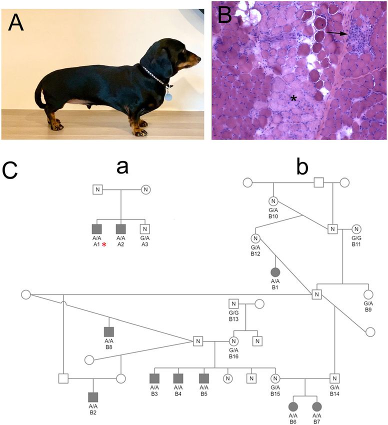
data made available in this article, unless otherwise stated in a credit line to the data.Mickelson et al. Skeletal Muscle (2021) 11:2 Page 2 of 11 exercise intolerance, recurrent rhabdomyolysis, and MD for many years [20, 21]. We report here the discov- myoglobinuria has also been described [4, 5]. The serum ery of the first mutation in the canine sarcoglycan alpha creatine kinase (CK) activities are usually mildly to subunit (SGCA) gene in young adult miniature dachs- markedly elevated. Muscle biopsies from most sarcogly- hunds that leads to a form of LGMD with clinical signs canopathy patients show dystrophic features. Immunore- that include subclinical myopathy with hyperCKemia, activity for the mutated sarcoglycan subunit is often exercise intolerance, progressive weakness, variable dys- reduced or absent, but mutations in one sarcoglycan phagia and pneumonia, and myoglobinuria. often result in decreased or absence of the other sarco- glycans as well as dystrophin [6]. Thus, it is difficult to Methods reach a diagnosis of a specific sarcoglycanopathy based Animals on immunoreactivity alone. The initial diagnostic muscle biopsies or stained cryosec- Several well-described forms of muscular dystrophy tions from four young miniature dachshund dogs were are found in domestic dogs, with at least fifteen DMD submitted by veterinary specialists in Australia to the mutations found in over 10 breeds [7–18], as well as two Comparative Neuromuscular Laboratory, University of sarcoglycan delta subunit (SGCD) mutations [19], re- California San Diego. The veterinarians reported that all ported to cause dystrophic clinical phenotypes similar to four dogs had persistently elevated CK activities and a DMD, BMD, and LGMD, respectively, as described in myopathic clinical phenotype varying from generalized people. The DMD mutation responsible for X-linked weakness and exercise intolerance with myoglobinuria to muscular dystrophy in golden retriever dogs has been a pharyngeal dysphagia (Fig. 1). Additional familial infor- model for testing new therapeutics for Duchenne type mation revealed relationships in which one of the initial Fig. 1 Miniature dachshund dystrophic phenotype and pedigrees. A A miniature dachshund from Australia that was evaluated for persistently elevated creatine kinase activity and exercise intolerance. B Representative H&E stained cryosection from an affected dachshund showed degenerative (asterisk) and regenerative (arrow) changes consistent with a dystrophic phenotype. C One small and one large family that formed the primary basis for this report are presented. Males are designated as squares and females as circles. Cases are solid symbols, controls are open symbols labeled with an “N,” and dogs with unknown phenotypes are open symbols. The case (dog A1) with whole genome sequence data is indicated with an asterisk. Genotypes for the functional SGCA variant are provided for all dogs with available samples
Mickelson et al. Skeletal Muscle   (2021) 11:2                                                                Page 3 of 11

four affected dachshunds came from one small pedigree            Western blotting was performed by standards methods
(dog A1, Fig. 1), and three came from a large multi-           using extracts from the triceps muscle of an affected dog
generational pedigree with extensive interbreeding (dogs       with a confirmed mutation in α-sarcoglycan (starred dog
B1, B2, and B3, Fig. 1).                                       in Fig. 1) or from archived cryopreserved canine control
  Further investigation found six additional affected dogs     triceps muscle. Protein bands were separated using
which could be placed in the pedigrees. One (dog A2)           NuPage Bis-Tris 4–12% gradient gels (Invitrogen). Pri-
was a sib to the original affected dog of pedigree A,          mary antibodies included a rabbit polyclonal antibody
while the five others (dogs B4, B5 B6, B7, and B8) could       against α-sarcoglycan (1:2000; [25]) and monoclonal
be placed in pedigree B, often as sibs to other affected       antibodies against β-sarcoglycan (1:1000), γ-sarcoglycan
dogs. Many parents and unaffected siblings were also           (1:1000), and against β-actin (1:2000, Sigma A2066) as a
identified. All parents were unaffected, and when tested,      loading control. Secondary antibodies included
CK activities from the parents and unaffected siblings         peroxidase-conjugated goat anti-mouse IgG (1:20,000,
were within the reference ranges.                              Jackson ImmunoResearch Lab, 115-035-062) and
  Muscle biopsies from two other dogs, originating in          peroxidase-conjugated goat anti-rabbit IgG (1:20,000,
California and South Africa, respectively, were submitted      Thermo Scientific, 31460). Protein bands were detected
to the Comparative Neuromuscular Laboratory. Both              using Super Signal West Dura Extended Duration Sub-
dogs also had markedly elevated CK activities, and clin-       strate (Thermo Scientific).
ical signs similar to the initial Australian dogs.
                                                               Genomic data collection and analysis
                                                               Eight cases and four unaffected dogs originating from
Histopathology, immunofluorescence microscopy, and             Australia, and the case originating from California, were
western blotting                                               genotyped on the Illumina CanineHD BeadChip 230 K
Biopsies from the biceps femoris, vastus lateralis, and tri-   array. Fourteen dachshund controls collected from unre-
ceps muscles were collected from 6 affected dogs under         lated projects conducted at the University of Minnesota
general inhalational anesthesia by an open biopsy pro-         were genotyped on the Axiom Canine HD Array 710 K.
cedure. The biopsy specimens were either unfixed and           The SNP genotype data was imputed using a cosmopol-
shipped under refrigeration to the Comparative Neuro-          itan reference panel of 49 wolves and 2,871 dogs from
muscular Laboratory by an express mailing service or           183 breeds, including 292 dachshunds genotyped at 710,
were processed by a local laboratory that shipped the          000 SNPs using Beagle 4.1 [26]. Five hundred ten thou-
stained cryosections to the Neuromuscular Laboratory           sand and nine SNPs remained after pruning the target
for evaluation. Upon receipt, unfixed biopsies were            and reference panels to remove SNPs with more than 2
snap-frozen in isopentane pre-cooled in liquid nitrogen        alleles, minor allele frequency less than 0.001, and dis-
and stored at − 80 °C until further processed. In              cordant genotypes > 0.05 within a multi-breed panel of
addition, biopsies were immersion fixed in 10% buffered        360 dogs genotyped at both 230 K and 710 K. PIHAT
formalin and processed into paraffin. Cryosections             values for SNPs across the genome obtained from
(8 μm) were stained or reacted with a standard panel of        PLINK 1.9 were used to estimate the proportion of iden-
histological and histochemical stains and reactions [22].      tity by descent between dogs with unavailable pedigree
  Additional cryosections (8 μm) were cut and stained          information [27]. Haplotypes derived from the imputed
for indirect immunofluorescence as previously described        data were used to narrow the genomic region flanking a
[23]. Sections were incubated with mouse monoclonal            positional and biological candidate gene.
antibodies against the rod (1:100, NCL-DYS1, Novocas-
tra Laboratory) and carboxy terminus (1:100, NCL-              Whole genome sequencing
DYS2, Novocastra Laboratories) of dystrophin, β-               A PCR-free library was prepared from a case from pedi-
sarcoglycan (1:100, NCL-βSG, Novocastra Laboratories),         gree A and sequenced in one lane of an Illumina HiSeq
γ-sarcoglycan (1:100, γSG, Novocastra Laboratories),           4000 sequencer by GeneWiz (South Plainfield, NJ
utrophin (1:20,NCL-DRP2, Novocastra Laboratories),             07080). Briefly, a fragment library with an average insert
and developmental myosin heavy chain (1:20, NCL-               size of ∼ 680 bp was prepared from which ∼ 125 million
MHCd, Novocastra Laboratories), or with rabbit poly-           2 × 150 bp paired-end reads were generated, corre-
clonal antibodies against laminin α2 (1:200; [24]) and α-      sponding to roughly 17× genome-wide coverage. The
sarcoglycan (1:200; [25]). Stainings were visualized using     reads were mapped against the dog reference genome
goat anti-rabbit HRP (1:20,000, Invitrogen #31460) or          assembly (CanFam3.1) as described [28, 29] and are
goat anti-mouse HRP (1:20,000, Jackson Immune La-              available in NCBI’s Short Read Archive (Accession num-
boratory #115-035-003) by previously described im-             ber SRR12537602). Variants identified in the critical
munofluorescent methods [23].                                  interval from the SNP genotype data in the case were
Mickelson et al. Skeletal Muscle (2021) 11:2 Page 4 of 11
compared to those of control genomes from the Univer- Both dogs had markedly elevated CK activities and a
sity of Minnesota’s private WGS database containing dystrophic phenotype on muscle biopsies.
289 dogs of 45 diverse breeds (including 10 dachshunds
from unrelated projects). Databases containing variants Histopathology, immunofluorescence microscopy, and
identified in WGS of more than 1300 dogs of 126 di- western blotting
verse breeds [30, 31] were subsequently searched for the Pathological changes in muscle biopsies from the four
presence of an identified mutation. affected miniature dachshunds were dystrophic in nature
regardless of the clinical presentation (Fig. 1B). Immuno-
Genotyping assay fluorescent antibody staining (Fig. 2A) showed a normal
The primers used in PCR amplification of the SGCA pattern for the dystrophin rod domain and patchy stain-
segment containing the reported mutation were 5′- ing with the antibody against the c-terminus of dys-
CGTGTCTTTGTGCACACCTT-3′ and 5′-GGGGAC trophin. Staining for utrophin and laminin α-2 was
TGAGATACCCACAA-3′. The 410 bp amplicon was similar to control muscle. Clusters of regenerating fibers
submitted for Sanger sequencing and the resultant se- were highlighted with the antibody against developmen-
quence was analyzed for the presence or absence of the tal myosin heavy chain (dMHC). Staining for α-
mutation with Sequencher 5.1 software (Gene Codes sarcoglycan and γ-sarcoglycan was absent and staining
Corporation, Ann Arbor, MI). was decreased for β-sarcoglycan. A sarcoglycanopathy
was suspected based on the staining results. Western
Results blotting (Fig. 2B) confirmed absent staining of protein
Clinical findings bands for α, β, and γ-sarcoglycans.
Muscle biopsies were received from four Australian
miniature dachshunds (3 male and 1 female) ranging in Genomic analysis
age from 7 to 17 months. Placement of these dogs (A1, Possible relationships between the pedigrees of Fig. 1C
B1, B2, B3) within the two pedigrees of Fig. 1 is de- and other dachshunds with SNP array genotyping data
scribed in the “Methods” section. No clinical signs of were estimated via PIHAT calculations (based on allele
myopathy were apparent in two of the dogs, but mark- sharing across the genome). The average PIHAT in the
edly elevated CK activities (10,000 to 174,041 IU/L; ref- 27 dog cohort was 0.08. This analysis indicated that the
erence 200–400 IU/L) were noted incidentally on pre- two affected dogs from family A do not have a signifi-
anesthetic blood work. On retesting, the CK activities cant close relationship to dachshunds in family B (PIHA
were persistently elevated. Another affected dog pre- T < 0.05). In addition, the additional case from Califor-
sented for myoglobinuria and weakness after a long nia had no significant relationships to the established
walk. The CK activity was markedly elevated (785,000 pedigrees (PIHAT < 0.05).
IU/L). Myoglobinuria resolved within a few days but the As an autosomal recessive trait was suspected, we first
CK activity was persistently elevated. One month after used a genotypic analysis model in which all nine cases
presentation, the owner reported chronic poor exercise were required to be homozygous for the minor allele,
tolerance. The fourth affected dog was presented for while controls could have all three genotypes. 444 SNPs
evaluation of chronic pneumonia and surgical correction met the criteria, with 112 of them lying in an ~ 19 Mb
of a porto-azygous shunt. Pre-anesthetic blood work segment on chromosome 9 between bp positions 18,928,
showed a CK activity of 40,828 IU/L that decreased to 814–37,617,735. When these 444 SNPs were ranked by
18,309 IU/L following surgical correction of the shunt. p value, 18 of the top 25 SNPs and 35 of the top 50 most
Muscle biopsies from all four dogs showed degenerative significant SNPs were in a ~ 12-Mb segment of chromo-
and regenerative changes consistent with a muscular some 9 (bp positions 18,958,229 to 30,966,212), with p
dystrophy (Fig. 1B). Five additional dogs within the pedi- values ranging from 9.4 × 10⁻⁵ to 2.1 × 10⁻⁷) (Table 1).
grees of Fig. 1 had persistently and markedly elevated A similar result was seen in a straight recessive model,
CK activities but tissue was not available for biopsy con- in which all nine cases were required to be homozygous
firmation of a dystrophic phenotype. The CK activities, for the minor allele and the controls could be either het-
where available from unaffected littermates and parents, erozygous or homozygous for the major allele (Table 2).
were within the reference range and no clinical signs of In this scenario, 32 of the top 50 SNPs were in the same ~
weakness described. 12 Mb region of chromosome 9 identified above. Further,
Tissue samples from two additional affected miniature in either analysis, only nine SNPs (seven on chromosome
dachshunds, both female and approximately 1 year of 9 and two on chromosome 34) met a criterion in which
age, were subsequently obtained from California and no controls could be homozygous for the minor allele.
South Africa. One female presented for chronic dyspha- Phasing of the SNP genotype data from this chromo-
gia and the other for exercise intolerance and dysphagia. some 9 segment revealed a haplotype in which all 9 casesMickelson et al. Skeletal Muscle (2021) 11:2 Page 5 of 11 Fig. 2 Immunofluorescent staining and western blotting from a dystrophic miniature dachshund. A Immunofluorescent staining of muscle cryosections from a representative dystrophic miniature dachshund. Staining for the rod-domain of dystrophin and laminin α-2 was similar to control muscle while staining for the dystrophin C-terminus was patchy. Utrophin was not increased. Numerous regenerating myofibers were highlighted with the antibody against developmental myosin heavy chain (dMHC). Staining for α- and γ-sarcoglycans was absent and decreased for β-sarcoglycan. B Western blotting confirmed all three sarcoglycans were absent. β-actin was used as loading control were homozygous (Fig. 3), where a minimally-conserved ~ this segment, including sarcoglycan A (SGCA, ENSC 1.8-Mb haplotype ranged from CFA9:24,792,165 to 26, AFG00000017013), at position CFA9:26,164,863–9:26,174, 644,060 Mb. The figure also shows that the central region 864 Mb. No significantly associated SNPs in the vicinity of of the minimally-conserved haplotype is common across the genes for other subunits of the sarcoglycan-sarcospan miniature dachshunds. A high frequency of many of the complex (SGCB, CFA13:44,972,473–44,987,184; SGCG, alleles comprising the affected haplotype exists throughout CFA25:15,249,622–15,430,044; SGCD, CFA4:53,263,866– the control population and it is flanking SNPs that tag the 53,820,231; and SSPN, CFA27:21.359,164–21,392,382) affected haplotype. Fifty-one genes with unique IDs are in were identified.
Mickelson et al. Skeletal Muscle   (2021) 11:2                              Page 6 of 11

Table 1 Top 50 SNPs in a genotypic model
CHR                  SNP                         A1   A2   AFF     UNAFF      P
9                    9:25951885                  C    T    9/0/0   0/8/10     2.134e−07
9                    9:25983462                  C    G    9/0/0   0/7/11     2.134e−07
9                    9:26288724                  C    T    9/0/0   0/6/12     2.134e−07
9                    9:26292362                  G    A    9/0/0   0/6/12     2.134e−07
9                    9:26334051                  A    G    9/0/0   0/6/12     2.134e−07
9                    9:26345821                  T    C    9/0/0   0/7/11     2.134e−07
9                    9:26347986                  C    T    9/0/0   0/8/10     2.134e−07
34                   34:10784553                 G    A    9/0/0   0/12/6     2.134e−07
34                   34:10786591                 C    A    9/0/0   0/12/6     2.134e−07
9                    9:25213000                  T    C    9/0/0   1/6/11     2.134e−06
9                    9:26313263                  C    T    9/0/0   1/6/11     2.134e−06
18                   18:39450835                 G    A    9/0/0   1/11/6     2.134e−06
9                    9:24972813                  C    G    9/0/0   1/7/10     4.267e−06
9                    9:24974987                  G    A    9/0/0   1/7/10     4.267e−06
9                    9:24994921                  T    G    9/0/0   1/7/10     4.267e−06
9                    9:25040066                  G    C    9/0/0   1/7/10     4.267e−06
9                    9:25046429                  T    G    9/0/0   1/10/7     4.267e−06
9                    9:25049565                  G    A    9/0/0   1/10/7     4.267e−06
9                    9:26020165                  A    G    9/0/0   1/10/7     4.267e−06
9                    9:26398124                  C    T    9/0/0   1/8/9      6.401e−06
18                   18:39447278                 C    T    9/0/0   1/9/8      6.401e−06
9                    9:25373707                  C    T    9/0/0   2/4/12     1.174e−05
4                    4:27820959                  T    C    9/0/0   2/10/6     1.387e−05
4                    4:27831539                  T    C    9/0/0   2/10/6     1.387e−05
4                    4:27868268                  A    G    9/0/0   2/10/6     1.387e−05
9                    9:30966212                  C    T    9/0/0   2/6/10     1.387e−05
34                   34:10891254                 G    A    9/0/0   2/10/6     1.387e−05
34                   34:10893260                 A    C    9/0/0   2/10/6     1.387e−05
9                    9:19304983                  C    T    9/0/0   2/8/8      1.984e−05
9                    9:26289840                  A    G    9/0/0   2/8/8      1.984e−05
32                   32:10752374                 T    C    9/0/0   2/8/8      1.984e−05
1                    1:106899476                 A    G    9/0/0   2/11/5     2.347e−05
9                    9:25394028                  C    G    9/0/0   2/11/5     2.347e−05
9                    9:24966631                  C    G    9/0/0   2/7/9      3.136e−05
9                    9:25053412                  A    G    9/0/0   2/9/7      3.136e−05
9                    9:25065255                  T    C    9/0/0   2/9/7      3.136e−05
9                    9:25444400                  T    C    9/0/0   2/7/9      3.136e−05
9                    9:25493635                  A    G    9/0/0   2/7/9      3.136e−05
34                   34:10909969                 C    A    9/0/0   2/9/7      3.136e−05
4                    4:26209208                  C    T    9/0/0   3/4/11     5.868e−05
6                    6:28794412                  G    T    9/0/0   3/11/4     5.868e−05
4                    4:26426217                  A    G    9/0/0   3/5/10     9.388e-05
6                    6:28865921                  G    T    9/0/0   3/12/3     9.388e−05
9                    9:18958229                  G    A    9/0/0   3/10/5     9.388e−05
Mickelson et al. Skeletal Muscle         (2021) 11:2                                                                                               Page 7 of 11

Table 1 Top 50 SNPs in a genotypic model (Continued)
CHR                    SNP                             A1                    A2                    AFF                     UNAFF                      P
9                      9:19012795                      A                     T                     9/0/0                   3/10/5                     9.388e−05
9                      9:19023729                      T                     G                     9/0/0                   3/10/5                     9.388e−05
9                      9:19122430                      A                     G                     9/0/0                   3/10/5                     9.388e−05
9                      9:19123171                      T                     G                     9/0/0                   3/10/5                     9.388e−05
9                      9:24989017                      A                     G                     9/0/0                   3/10/5                     9.388e−05
9                      9:25380184                      A                     C                     9/0/0                   3/5/10                     9.388e−05
The table lists the top 50 SNPs in which all nine cases were required to be homozygous, while controls could have all three genotypes. The chromosome (CHR),
position (SNP), minor allele (A1), major allele (A2), number of affected dogs homozygous for the minor allele/heterozygous/homozygous for the major allele (AFF),
number of unaffected dogs homozygous for the minor allele/heterozygous/homozygous for the major allele (UNAFF), and the p value for association (P)
are provided

Whole genome sequencing and mutation identification                                but has also been reported in humans with DMD [32]
Both the human and canine SGCA genes have eight                                    and in dogs with XLMD [33]. Similar to the dachshunds
protein-encoding exons. Analysis of WGS from the                                   of this report, a markedly elevated CK activity was also
vicinity of SGCA in a case (Fig. 1) identified a premature                         an incidental finding on pre-anesthetic blood screening
stop codon in exon 3; specifically, CFA9:26,166,312 G >                            prior to neuter from a family of Labrador retrievers with
A; c.G224A; p. W75*. This variant was not found in any                             dystrophin-deficient muscular dystrophy [34]. No clin-
of the more than 1300 dogs across published WGS data-                              ical evidence of myopathy was described by the owners
bases [29, 30]. The predicted stop codon at residue 75 is                          or detected on physical examination. However, muscle
expected to truncate 80% of the 387 amino acid sarco-                              biopsies were dystrophic, and dystrophin protein was ab-
glycan A protein. Interestingly, a flanking haplotype                              sent on western blotting and markedly decreased to ab-
identical to that found in homozygosity in all nine cases                          sent on immunostaining. Detection of a markedly and
was also present in the control population; however, the                           persistently elevated CK activity should be an indicator
SGCA c.G224A mutation genotype in such control dogs                                of an underlying congenital myopathy in young dogs.
demonstrated the wild type allele in these haplotypes.                               Although a sarcoglycanopathy was suspected based on
There were no other homozygous coding variants in                                  immunostaining and western blotting, the specific sarco-
SGCA and no other unique coding variants in the 1.8-                               glycan protein responsible could not be determined, as
Mb haplotype.                                                                      α, β, and γ-sarcoglycan were all absent or reduced. The
                                                                                   dystrophic pattern on the muscle biopsies and the clin-
Pedigree and population screening                                                  ical presentations also did not aid in the differentiation
SGCA c.G224A variant genotypes from the available                                  of the type of dystrophy. Pedigree analysis suggested an
members of the pedigrees were entirely consistent with                             autosomal recessive disorder, so an X-linked form of
autosomal recessive inheritance, with available parents                            MD (canine XLMD) was less likely, even though staining
being obligate heterozygotes, and other relatives being                            of the carboxy terminus of dystrophin was patchy. This
heterozygotes or homozygous wild type (Fig. 1C). The                               patchy staining can be secondary to the sarcoglycan defi-
two additional dachshund cases from South Africa and                               ciency. While the severity of clinical signs was milder
California were homozygous for the SGCA c.G224A vari-                              than that described in a previous publication of δ-
ant as well. One hundred and ten dachshunds available                              sarcoglycan deficiency in Boston terrier dogs [19], patho-
from submissions to the University of Minnesota for un-                            logical changes in muscle biopsies and the persistently
related projects were all clear of the mutation.                                   high CK activity were similar.
                                                                                     Whole genome SNP array genotyping data identified a
Discussion                                                                         region on CFA9 containing the SGCA gene where all
Here we describe the first canine cases of LGMD2D with                             cases were homozygous. A highly probable functional
a mutation in the α-sarcoglycan gene. In people, age of                            variant in the SGCA gene was identified from WGS,
onset of LGMD2D is reported to be from childhood to                                where a premature stop codon is predicted to truncate
young adult and in the miniature dachshunds of this re-                            approximately 80% of the protein. A recessive inherit-
port, onset was from 7 to 17 months of age. Similar to                             ance pattern was confirmed by targeted genotype assays
people with LGMD2D [4, 5], the clinical features varied                            that demonstrated that all affected dogs were homozy-
in severity among the affected dogs ranging from sub-                              gous for the variant, unaffected relatives were heterozy-
clinical myopathy with hyperCKemia to more general-                                gous or homozygous for the reference allele, and the
ized weakness and myoglobinuria. Such variability in the                           unaffected parents were all heterozygous. Although the
severity of clinical signs is not restricted to the LGMDs                          two identified pedigrees did not have close genetic
Mickelson et al. Skeletal Muscle (2021) 11:2 Page 8 of 11 Table 2 Top 50 SNPs in a recessive model CHR SNP A1 A2 AFF UNAFF P 9 9:25951885 C T 9/0 0/18 2.134e−07 9 9:25983462 C G 9/0 0/18 2.134e−07 9 9:26288724 C T 9/0 0/18 2.134e−07 9 9:26292362 G A 9/0 0/18 2.134e−07 9 9:26334051 A G 9/0 0/18 2.134e−07 9 9:26345821 T C 9/0 0/18 2.134e−07 9 9:26347986 C T 9/0 0/18 2.134e−07 34 34:10784553 G A 9/0 0/18 2.134e−07 34 34:10786591 C A 9/0 0/18 2.134e−07 9 9:24972813 C G 9/0 1/17 2.134e−06 9 9:24974987 G A 9/0 1/17 2.134e−06 9 9:24994921 T G 9/0 1/17 2.134e−06 9 9:25040066 G C 9/0 1/17 2.134e−06 9 9:25046429 T G 9/0 1/17 2.134e−06 9 9:25049565 G A 9/0 1/17 2.134e−06 9 9:25213000 T C 9/0 1/17 2.134e−06 9 9:26020165 A G 9/0 1/17 2.134e−06 9 9:26313263 C T 9/0 1/17 2.134e−06 9 9:26398124 C T 9/0 1/17 2.134e−06 18 18:39447278 C T 9/0 1/17 2.134e−06 18 18:39450835 G A 9/0 1/17 2.134e−06 1 1:106899476 A G 9/0 2/16 1.174e−05 4 4:27820959 T C 9/0 2/16 1.174e−05 4 4:27831539 T C 9/0 2/16 1.174e−05 4 4:27868268 A G 9/0 2/16 1.174e−05 9 9:19304983 C T 9/0 2/16 1.174e−05 9 9:24966631 C G 9/0 2/16 1.174e−05 9 9:25053412 A G 9/0 2/16 1.174e−05 9 9:25065255 T C 9/0 2/16 1.174e−05 9 9:25373707 C T 9/0 2/16 1.174e−05 9 9:25394028 C G 9/0 2/16 1.174e−05 9 9:25444400 T C 9/0 2/16 1.174e−05 9 9:25493635 A G 9/0 2/16 1.174e−05 9 9:26289840 A G 9/0 2/16 1.174e−05 9 9:30966212 C T 9/0 2/16 1.174e−05 32 32:10752374 T C 9/0 2/16 1.174e−05 34 34:10891254 G A 9/0 2/16 1.174e−05 34 34:10893260 A C 9/0 2/16 1.174e−05 34 34:10909969 C A 9/0 2/16 1.174e−05 1 1:120378888 C T 9/0 3/15 4.694e−05 4 4:26209208 C T 9/0 3/15 4.694e−05 4 4:26216500 A G 9/0 3/15 4.694e−05 4 4:26426217 A G 9/0 3/15 4.694e−05 6 6:28794412 G T 9/0 3/15 4.694e−05
Mickelson et al. Skeletal Muscle         (2021) 11:2                                                                                               Page 9 of 11

Table 2 Top 50 SNPs in a recessive model (Continued)
CHR                     SNP                             A1                    A2                    AFF                    UNAFF                      P
6                       6:28865921                      G                     T                     9/0                    3/15                       4.694e−05
6                       6:70411742                      T                     A                     9/0                    3/15                       4.694e−05
9                       9:18928814                      C                     T                     9/0                    3/15                       4.694e−05
9                       9:18958229                      G                     A                     9/0                    3/15                       4.694e−05
9                       9:19012795                      A                     T                     9/0                    3/15                       4.694e−05
9                       9:19023729                      T                     G                     9/0                    3/15                       4.694e−05
The table lists SNPs in which all nine cases were required to be homozygous for the minor allele and the controls could be either heterozygous or homozygous
for the major allele. The table lists the top 50 SNPs in which all nine cases were required to be homozygous, while controls could have all three genotypes. The
chromosome (CHR), position (SNP), minor allele (A1), major allele (A2), number of affected dogs homozygous for the minor allele/heterozygous plus homozygous
for the major allele (AFF), number of unaffected dogs homozygous for the minor allele/heterozygous plus homozygous for the major allele (UNAFF), and the p
value for association (P) are provided

relationships, affected dachshunds with the identical mu-                          terminal half of the protein that serve as glycosyla-
tation to that found in the Australian families have now                           tion sites, and a phosphorylatable serine residue near
been found in South Africa and California, suggesting                              the C-terminus. Several thousand human SGCA vari-
dispersal by a common founder. The relatively small 1.8-                           ants have been identified and at least 30 are associ-
Mb-affected haplotype further supports a hypothesis that                           ated with human LGMD (UniProtKB - Q16586;
the SGCA arose several generations ago.                                            March 1, 2020). These functional variants are spread
  The canine and human SGCA amino acid se-                                         across residues 30–284, with over half of them in
quences are both 387 amino acids long and the resi-                                the first third of the sequence. The canine variant
dues share 90% identity. Fifteen of the 39 amino                                   database lists 111 SGCA variants, but only one of
acid differences between these species are in the first                            them affects the coding sequence, a missense coding
60 N-terminal residues. Functional protein domains                                 variant with a SIFT score that does not predict dam-
of note are a single transmembrane alpha helix near                                age to the protein structure. In any event, a stop
the C-terminal, 2 asparagine residues in the C-                                    codon mutation that truncates 80% of the SGCA

    Fig. 3 Haplotypes in the region of interest on CFA9. SNP coordinates are provided along the top row. A pink cell indicates the minor allele and a
    white cell indicates the major allele. Identification numbers on the far left refer to the dogs within the pedigrees of Fig. 1. The CFA9 haplotype in
    which all 9 cases were homozygous spanned from 24,792,165–26,644,060 Mb. The affected haplotype is denoted with a red highlight (left side),
    unaffected haplotypes have a blue highlight, and gray haplotypes are identical to the affected haplotype but do not have the SGCA c.G224A
    mutation. The SGCA gene, located from positions (26,164,863–26,174,864 Mb), is indicated by an orange bar along the top
Mickelson et al. Skeletal Muscle (2021) 11:2 Page 10 of 11
protein is extremely likely to produce a nonfunc- CA 92093-0709, USA. 3Department of Veterinary Clinical Sciences, College of
tional protein even if the mRNA is not degraded and Veterinary Medicine, University of Minnesota, Saint Paul, MN 55113, USA.
4
Advanced Vetcare, Kensington, Victoria, Australia. 5Centre for Animal Referral
a partial polypeptide is translated. and Emergency, Collingwood, Victoria, Australia. 6Animal Referral Hospital,
Homebush, New South Wales, Australia. 7Department of Medicine and
Conclusions Epidemiology, School of Veterinary Medicine, University of California, Davis,
CA, USA.
This combined clinical, pathological, immunofluores-
cence, western blotting, and molecular genetic approach Received: 22 September 2020 Accepted: 14 December 2020
has identified a sarcoglycan A subunit mutation in mini-
ature dachshund dogs that results in a sarcoglycanopa-
thy, a form of limb-girdle muscular dystrophy. This first References
1. Liewluck T, Milone M. Untangling the complexity of limb-girdle muscular
SGCA mutation found in dogs adds to the literature of dystrophies. Muscle Nerve. 2018;58:167–77.
genetic bases of canine muscular dystrophies and their 2. Bushby KM, Beckmann JS. The limb-girdle muscular dystrophies--proposal
usefulness as comparative models of human disease. for a new nomenclature. Neuromuscul Disord. 1995;5:337–43.
3. Leyva-Leyva M, Sandoval A, Felix R, Gonzalez-Ramirez R. Biochemical and
Further, this demonstrates that persistent CK elevations functional interplay between ion channels and the components of the
should not be dismissed in young animals without clin- dystrophin-associated glycoprotein complex. J Membr Biol. 2018;251:535–
ical evidence of myopathy. 50.
4. Mongini T, Doriguzzi C, Bosone I, Chiadò-Piat L, Hoffman EP, Palmucci L.
Alpha-sarcoglycan deficiency featuring exercise intolerance and
Abbreviations
myoglobinuria. Neuropediatrics. 2002;33:109–11.
CFA9: Canis familiaris chromosome 9; DMD: Duchenne muscular dystrophy;
5. Ceravolo F, Messina S, Rodolico C, Strisciuglio P, Concolino D.
DMD: Dystrophin gene; GWAS: Genome-wide association analysis;
Myoglobinuria as first clinical sign of a primary alpha-sarcoglycanopathy. Eur
LGMD: Limb-girdle muscular dystrophy; SGCA: Sarcoglycan A subunit;
J Pediatr. 2014;173:239–42.
SNP: Single nucleotide polymorphism; WGS: Whole genome sequence
6. Kirschner J, Lochmüller H. Sarcoglycanopathies. Handb Clin Neurol. 2011;
101:41–6.
Acknowledgements
7. Sharp NJH, Kornegay JN, Van Camp SD, Herbstreith MH, Secore SL, Kettle S,
We are grateful to all owners, breeders, and clinicians who made this work
Hung WY, Constantinou CD, Dykstra MJ, Roses AD, Bartlett RJ. An error in
possible. The authors also thank Dr. Rodger Davies, Wild Coast Veterinary
dystrophin mRNA processing in golden retriever muscular dystrophy, an
Hospital, Eastern Cape, South Africa, for submitting tissue samples and
animal homologue of Duchenne muscular dystrophy. Genomics. 1992;13:
clinical information.
115–21.
8. Schatzberg SJ, Olby NJ, Breen M, Anderson LV, Langford CF, Dickens HF,
Authors’ contributions Wilton SD, Zeiss CJ, Binns MM, Kornegay JN, Morris GE, Sharp NJ. Molecular
JRM: Devised genetic mapping plan, analyzed data, and wrote the analysis of a spontaneous dystrophin 'knockout' dog. Neuromuscul Disord.
manuscript. KMM: Devised and performed genetic mapping plan, analyzed 1999;9:289–95.
results, performed mutation genotyping, and reviewed the manuscript. LTG: 9. Walmsley GL, Arechavala-Gomeza V, Fernandez-Fuente M, Burke MM, Nagel
Performed immunostaining and western blotting, analyzed results, and N, Holder A, Stanley R, Chandler K, Marks SL, Muntoni F, Shelton GD, Piercy
reviewed the manuscript. SGF: Developed variant database and analyzed the RJ. A Duchenne muscular dystrophy gene hot spot mutation in dystrophin-
variant data. JNC: Developed variant database and analyzed the variant data. deficient Cavalier King Charles Spaniels is amenable to exon 51 skipping.
AC: Provided medical and pedigree information and reviewed the PLoS One. 2010 Jan 13;5(1):e8647.
manuscript. LEH: Provided medical and pedigree information and reviewed 10. Smith BF, Yue Y, Woods PR, Kornegay JN, Shin JH, Williams RR, Duan D. An
the manuscript. KMB: Provided medical and pedigree information and intronic LINE-1 element insertion in the dystrophin gene aborts dystrophin
reviewed the manuscript. SEH: Provided medical and pedigree information expression and results in Duchenne-like muscular dystrophy in the corgi
and reviewed the manuscript. SLM: Provided medical information and breed. Lab Investig. 2011;91:216–31.
reviewed the manuscript. GDS: Devised pathology, immunofluorescence, and 11. Kornegay JN, Bogan JR, Bogan DJ, et al. Canine models of Duchenne
immunoblotting plan; analyzed results; and wrote the manuscript. The muscular dystrophy and their use in therapeutic strategies. Mamm Genome.
authors read and approved the final manuscript. 2012;23:85–108.
12. Jenkins CA, Forman OP. Identification of a novel frameshift mutation in the
Funding DMD gene as the cause of muscular dystrophy in a Norfolk terrier dog.
The authors have no sponsored funding to report. Canine Genet Epidemiol. 2015;2:7. https://doi.org/10.1186/s40575-015-0019-
4. eCollection 2015.
Availability of data and materials 13. Atencia-Fernandez S, Shiel RE, Mooney CT, Nolan CM. Muscular dystrophy in
The WGS of the affected dog is available under BioProject title: “Sarcoglycan the Japanese Spitz: an inversion disrupts the DMD and RPGR genes. Anim
A mutation in miniature dachshund dogs”. Accession number SRR12537602. Genet. 2015;46(2):175–84. https://doi.org/10.1111/age.12266. Epub 2015 Jan
23.
Ethics approval and consent to participate 14. Nghiem PP, Bello L, Balog-Alvarez C, Mata López S, Bettis A, Barnett H,
The study was approved under IACUC protocol 1903-36865A at the Univer- Hernandez B, Schatzberg SJ, Piercy RJ, Kornegay JN. Whole genome
sity of Minnesota. sequencing reveals a 7 base-pair deletion in DMD exon 42 in a dog with
muscular dystrophy. Mamm Genome. 2017;28:106–13.
Consent for publication 15. Sánchez L, Beltrán E, de Stefani A, Guo LT, Shea A, Shelton GD, De Risio L,
Not applicable. Burmeister LM. Clinical and genetic characterisation of dystrophin-deficient
muscular dystrophy in a family of Miniature Poodle dogs. PLoS One. 2018;
Competing interests 13(2):e0193372.
The authors have no competing interests to declare. 16. Mata López S, Hammond JJ, Rigsby MB, Balog-Alvarez CJ, Kornegay JN,
Nghiem PP. A novel canine model for Duchenne muscular dystrophy
Author details (DMD): single nucleotide deletion in DMD gene exon 20. Skelet Muscle.
1
Department of Veterinary and Biomedical Sciences, College of Veterinary 2018;8(1):16.
Medicine, University of Minnesota, Saint Paul, MN 55113, USA. 2Department 17. Shrader SM, Jung SW, Denney TS, Smith BF. Characterization of Australian
of Pathology, School of Medicine, University of California San Diego, La Jolla, Labradoodle dystrophinopathy. Neuromuscul Disord. 2018;28(11):927–37.Mickelson et al. Skeletal Muscle (2021) 11:2 Page 11 of 11
18. Barthélémy I, Calmels N, Weiss RB, Tiret L, Vulin A, Wein N, Peccate C,
Drougard C, Beroud C, Deburgrave N, Thibaud JL, Escriou C, Punzón I,
Garcia L, Kaplan JC, Flanigan KM, Leturcq F, Blot S. X-linked muscular
dystrophy in a Labrador Retriever strain: phenotypic and molecular
characterisation. Skelet Muscle. 2020;10(1):23.
19. Cox ML, Evans JM, Davis AG, Guo LT, Levy JR, Starr-Moss AN, Salmela E,
Hytönen MK, Lohi H, Campbell KP, Clark LA, Shelton GD. Exome sequencing
reveals independent SGCD deletions causing limb girdle muscular
dystrophy in Boston terriers. Skelet Muscle. 2017;7(1):15.
20. Nghiem PP, Kornegay JN. Gene therapies in canine models for Duchenne
muscular dystrophy. Hum Genet. 2019;138:483–9.
21. Kornegay JN. The golden retriever model of Duchenne muscular dystrophy.
Skelet Muscle. 2017;7:9.
22. Dubowitz V, Sewry CA, Oldfors A. Histological and histochemical stains and
reactions. In: Dubowitz V, Sewry CA, Oldfors A, editors. Muscle Biopsy: a
practical approach. 4th ed. Oxford: Saunders Elsevier; 2013. p. 16–27.
23. Guo LT, Moore SA, Forcales S, Engvall E, Shelton GD. Evaluation of
commercial dysferlin antibodies on canine, mouse and human skeletal
muscle. Neuromuscul Disord. 2010;20:820–5.
24. Leivo I, Engvall E. Merosin, a protein specific for basement membranes of
Schwann cells, striated muscle, and trophoblast, is expressed late in nerve
and muscle development. Proc Natl Acad Sci USA. 1988;85:544–8.
25. Lui LA, Engvall E. Sarcoglycan isoforms in skeletal muscle. J Biol Chem. 1999;
274:38171–6.
26. Scheet P, Stephens M. A fast and flexible statistical model for large-scale
population genotype data: applications to inferring missing genotypes and
haplotypic phase. Am J Hum Genet. 2006;78:629–44.
27. Purcell S, Neale B, Todd-Brown K, et al. PLINK, a tool set for whole-genome
association and population-based linkage analysis. Am J Hum Genet. 2007;
81:559–75.
28. Meurs KM, Friedenberg SG, Kolb J, Saripalli C, Tonino P, Woodruff K, Olby
NJ, Keene BW, Adin DB, Yost OL, DeFrancesco TC, Lahmers S, Tou S, Shelton
GD, Granzier H. A missense variant in the titin gene in Doberman pinscher
dogs with familial dilated cardiomyopathy and sudden cardiac death. Hum
Genet. 2019;138:515–24.
29. Shelton GD, Minor KM, Li K, Naviaux JC, Monk J, Guzik E, Guo LT, Porcelli V,
Gorgoglione R, Lasorsa FM, Persico AM, Leegwater PJ, Mickelson JR, Palmieri
L, Naviaux RK. A mutation in the mitochondrial aspartate/glutamate carrier
leads to intramitochondrial oxidation and am inflammatory myopathy in
Dutch Shepherd Dogs. J Neuromuscular Disease. 2019;6:485–501.
30. Jagannathan V, Drögemüller C, Leeb T, et al. Dog Biomedical Variant
Database Consortium (DBVDC). A comprehensive biomedical variant
catalogue based on whole genome sequences of 582 dogs and eight
wolves. Anim Genet. 2019;50:695–704.
31. Plassais J, Kim J, Davis BW, Karyadi DM, Hogan AN, Harris AC, et al. Whole
genome sequencing of canids reveals genomic regions 494 under selection
and variants influencing morphology. Nat Commun. 2019;10:1489.
32. Vainzof M, Passos-Bueno R, Takata RI, et al. Intrafamilial variability in
dystrophin abundance correlated with difference in the severity of the
phenotype. J Neurol Sci. 1993;119:38–42.
33. Kornegay JN, Spurney CF, Nghiem PP, et al. Pharmacologic management of
Duchenne muscular dystrophy: target identification and preclinical trials.
ILAR J. 2014;55:119–49.
34. Vieira NM, Guo LT, Estrela E, Kunkel LM, Zatz M, Shelton GD. Muscular
dystrophy in a family of Labrador retrievers with no muscle dystrophin and
a mild phenotype. Neuromuscul Disord. 2015;25:363–70.
Publisher’s Note
Springer Nature remains neutral with regard to jurisdictional claims in
published maps and institutional affiliations.You can also read