SHAPE-CONSISTENT GENERATIVE ADVERSARIAL NETWORKS FOR MULTI-MODAL MEDICAL SEGMENTATION MAPS
←
→
Page content transcription
If your browser does not render page correctly, please read the page content below
SHAPE-CONSISTENT GENERATIVE ADVERSARIAL NETWORKS FOR MULTI-MODAL
MEDICAL SEGMENTATION MAPS
Leo Segre * Or Hirschorn* Dvir Ginzburg Dan Raviv
Tel Aviv University
ABSTRACT
arXiv:2201.09693v2 [eess.IV] 4 Feb 2022
Image translation across domains for unpaired datasets has
gained interest and great improvement lately. In medical
imaging, there are multiple imaging modalities, with very dif-
ferent characteristics. Our goal is to use cross-modality adap-
tation between CT and MRI whole cardiac scans for semantic
segmentation. We present a segmentation network using syn-
thesised cardiac volumes for extremely limited datasets. Our
solution is based on a 3D cross-modality generative adversar-
ial network to share information between modalities and gen-
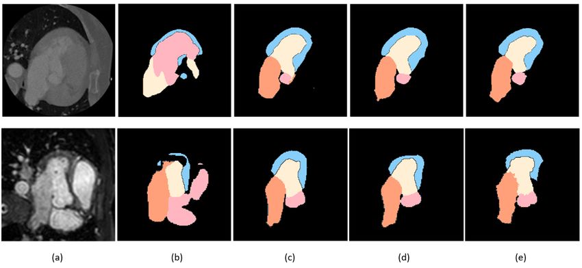
erate synthesized data using unpaired datasets. Our network Fig. 1. Segmentation results generated by our method on car-
utilizes semantic segmentation to improve generator shape diac CT images (top) and MRI (bottom). a) Examples of test
consistency, thus creating more realistic synthesised volumes images, b) Segmentation results without preprocess augmen-
to be used when re-training the segmentation network. We tations, c) Segmentation results without synthesized data, d)
show that improved segmentation can be achieved on small Segmentation results of our full method, e) The ground truth.
datasets when using spatial augmentations to improve a gen-
erative adversarial network. These augmentations improve
the generator capabilities, thus enhancing the performance of
the Segmentor. Using only 16 CT and 16 MRI cardiovascular However, to achieve satisfying results, a sufficiently large
volumes, improved results are shown over other segmentation number of training samples is required. As demonstrated in
methods while using the suggested architecture. Our code is figure 1, the results of semantic segmentation when the train-
publicly available1 . ing set is too small are significantly degraded. Segmented
medical imaging datasets are hard to acquire and use due to
strict regulations, lack of support from hospitals in acquiring
1. INTRODUCTION the data, and high costs of medical imaging services. As
medical images acquired using different modalities have very
Semantic segmentation is a key perceptual function in com- different characteristics, it is especially challenging to obtain
puter vision, aiming to densely categorize an image into data for new imaging modalities. This also applies to images
meaningful distinguished areas. In the medical imaging do- from the same modality, captured using different scanning
main, semantic segmentation is vital, providing tools for machines.
diagnostics, treatment planning, and prognosis. For disease To overcome the lack of data, a common way to generate syn-
diagnostics and surgical needs, multiple imaging modalities thesized data is to use augmentations on the original data [4].
are available such as MRI, CT, and X-ray. Another way to generate synthesized data is using a Genera-
Traditional machine learning methods, such as atlas and tive Adversarial Network (GAN). This can be done directly
model-based methods, showed good performance in cardiac by separate GAN for each domain, but it is even more ef-
image segmentation. However, they usually require features fective to use cross-modality GAN and share information
engineering which differs between image modalities [1]. In between modalities and generate more accurate synthesized
contrast, deep learning algorithms show promising results data [5, 6]. Although some models used GANs as a proxy
while implicitly discovering features from the data. They task to the segmentation objective on very small datasets [5],
have been widely adopted for various tasks, from 2D binary the mentioned method used 2D slices instead of 3D volumes
segmentation [2] to multi-class 3D segmentation [3]. thus discarding crucial information regarding the spatial con-
* Equal contribution sistency in the Z-axis. This paper presents a method to use
1 github.com/orhir/3D-Shape-Consistent-GAN deep learning based segmentation on a limited dataset byusing augmentations and generating 3D synthesized shape- 3.1. Synthetic Data Generation
consistent data. The presented method provides significant
improvements in the limited training data domain, with an First, all scans aligned to RAS+ orientation and following
average accuracy increase of 15.9% in the final segmentation Cheng Chen et al. [5] we manually cropped the MRI scans
score. Additionally, we provide our code for future works. around the heart. To align between the CT scans and MRI
scans, we also resized the CT scans into 256x256xZ, where
Z preserves the original scan width-depth ratio. Then, using
TorchIO [4] we randomly applied numerous 3D transforma-
2. RELATED WORK
tions on both the cardiac volumes and labels, specifically de-
signed for medical data: normalization, random anisotropy -
Using deep learning for domain adaptation in a super- Simulates an image that has been acquired using anisotropic
vised or unsupervised manner has recently gained popu- spacing and resampled back to its original spacing, random
larity [5, 7, 8, 9]. As the availability of training data from the elastic deformation - A random displacement according to a
same set of subjects in both source and target modalities is grid of control points, random affine - applies affine transfor-
undesirable (requires multiple scans from each subject), an mation and resamples back to the original spacing. We ob-
unsupervised cross-modal image synthesis, without pairing served the best results for 200 3D synthetic augmented scans,
training data approach is beneficial [7]. as more synthetic scans resulted in significantly longer run
Since the rise of deep learning, image to image transla- times and negligible improvements.
tion is usually formulated as a pixel to pixel mapping us-
ing CNN’s (Convolutional Neural Network) encoders and
decoders [6, 10, 8]. CycleGAN had wide success, where 3.2. Volume to Volume Translation
bi-directional image translations are learned by two GANs
For two unpaired domains A and B, we employ generative
separately, and the consistency constraint between trans-
adversarial networks using a generator G and a discrimina-
forms is enforced to preserve semantic information between
tor D for each domain. The volume to volume translation is
transformed outputs [11]. The good results and robustness
based on CycleGAN U-Net architecture enhanced to 3D vol-
Cycle-GAN showed for many applications made it a popu-
umes using 3D convolutions. The generator transforms the
lar backbone in future works. Thus, many unpaired image
input domain A to the other domain B, the notation for this
to image transformations are based on this framework with
transformation is GB (XA ). The discriminator competes with
additional constraints to further regularize the transformation
the generator, trying to distinguish between a fake volume
process. Although CycleGAN was initially proposed for 2D
GB (XA ) and a real volume xB . L2 loss is used to minimize
images, GANs have also been applied in 3D [12].
the generator’s objective of creating realistic volumes, noted
For medical image processing, adversarial learning has pre-
as Ladv,A .
sented great efficiency on a variety of tasks [13]. Using
We adopt the CycleGAN’s approach of cycle consistency
synthetic data as augmented training data helps the segmen-
loss. Thus, we force the generators to reconstructed synthe-
tation network as seen for brain MRI and CT images [9].
sized volumes GA (GB (xA )) and xA to be identical. We en-
Zizhao Zhang et al. [6] used a segmentation network to force
courage the transformed volumes to preserve content from the
shape consistency of the output transformed domain, using
original volume using L1 loss:
a private massive data set. As massive datasets are usually
hard to acquire in this domain, our work avoids this demand, 1 X
presenting great results using a public dataset with only 20 Lcycle,A = − |xA − GA (GB (xA ))|
N i
CT and 20 MRI samples. SIFA [5] used a novel approach of
fusing feature and image appearance adaptation and applied Using the above constraints can lead to geometrically
it to cross-modality segmentation of cardiac volumes, but was distorted transformations. The cycle consistency loss isn’t
limited to 2D slices, losing z-axis shape data. enough to prevent spatial distortions. It is possible for gener-
ator B to create a distortion F and for generator A to apply
the reverse transformation F −1 leading back to the original
3. METHODS shape. Thus, a shape consistent constraint is needed to reduce
the spatial distortion.
The method is applied in three phases. First, a segmentor is We suggest using a 3D segmentor to preserve shape consis-
trained with the original data. Then, a 3D cycle and shape tency. The segmentor maps xi → Y , where i is the domain A
consistent GAN network is trained to create synthesized car- or B. To constrain the geometric invariance of the generated
diac volumes, which is used later to train the segmentor in volume we optimize the Cross Entropy+Dice loss [14] of the
phase 3 and achieve the goal of improving the 3D segmentor generated domain and its labels:
network. Figure 2 illustrates the proposed architecture for
domain adaptation in cardiac volumes. Lspatial,A = LCE,A + LDICE,AFig. 2. Overview of our domain adaptation and segmentation architecture. The architecture above is duplicated to handle both
domains, a duplication where ”A” is CT and ”B” is MRI and vise versa. The flow is based on three chronological phases as
described in training strategies.
k k
P
2 X i∈I ui vi 3.4. Training Strategies
LDICE,A =− P k
P k
|K| i∈I ui + i∈I vi
k∈K
Training the network consists of three phases, as we observed
where u is the softmax output of the network and v is a one hot
pre-training and fine-tuning the segmentor and generator re-
encoding of the ground-truth segmentation yA of the volume
sults in better performances.
xA . The generator’s total loss function is composed of all the
First, the segmentor is pre-trained for 100 epochs using only
above constraints:
the original data. Then the generator and discriminator are
trained leaning on the pre-trained segmentor from phase 1.
LA = λadv Ladv,A + λcycle Lcycle,A + λspatial Lspatial,A
The goal of this phase is to train a generator that can gener-
where λi is a trade-off parameter. ate synthesized data in addition to the preprocessed synthe-
sized volumes. The generator and discriminator are trained
for 50 epochs without the spatial loss, and then for another
3.3. Segmentation 150 epochs with the spatial loss enforcing shape consistency.
The segmentation network in our solution is based on a 3D U- Last, after having a trained shape consistent generator, we
net architecture [3]. In our architecture, there are two identi- train the segmentation network using the augmented and syn-
cal segmentors, one for each domain. Although each segmen- thesised 3D volumes for 100 epochs.
tor has its own source domain, both CT and MRI segmentors
have the same target domain - cardiac segmentation labels.
Thus, the segmentation task can be implemented on the orig-
inal volume or on a generated synthetic volume, in both cases 3.5. Network Configurations and Implementations
the ground-truth labels are identical. Formally, given input
voxel XA from domain A and its labels map YA , we define Our network consists of segmentor, generator and discrimi-
the following. nator modules for each domain.
The segmentation network is a 3D U-net consisted of 4
SA (XA ) = SB (GB (XA )) = YA downsampling convolutions (maximum downsample rate is
16) and 4 upsampling using nearest interpolation, with con-
Using this approach, we can train the segmentor with ei-
volutions of 3x3x3 kernel and stride 1.
ther real and synthesised data, it is important since segmen-
The discriminators follow PatchGAN configurations [15],
tation networks usually require a lot of data to be trained on.
consists of 3 convolutional layers with kernels 4x4x4 and
Hence, the losses of the segmentors are defined by Lseg and
stride 2, and 2 convolutional layers with stride 1. For the first
Lseg,syn . Both are Cross Entropy+Dice losses, as used in
4 layers, each convolutional layer is followed by a normaliza-
Fabian Isensee et al..
tion layer and leaky ReLU with 0.2 slope parameter.
The generator is based on CycleGAN’s U-net, adjusted to
Lseg (XA , YA , SA ) = Lspatial (YA , SA ) 3D volumes, using a skip-connection U-net, as it achieves
Lseg,syn (XA , YA , SB ) = Lspatial (YA , SB (GB (XA ))) faster convergence and locally smooth results [6]. We apply 5
downsampling with 3x3x3 kernel and stride 2, and upsample
For each voxel of the input, the segmentor evaluates a vec- using nearest interpolation with 3x3x3 kernel and stride 1.
tor of probabilities - one for each label. SA (XA )i,Yi denotes We implemented our framework in PyTorch, and the training
the output of SA (XA ) on voxel i regarding to the probability was done on 8 NVIDIA Quadro RTX 8000 GPUs. All the
of the ground-truth label of this voxel. argmax(SA (XA )i ) networks were optimized using the Adam optimizer with a
will provide the label prediction of voxel i. learning rate of 2 × 10⁻⁴ .Table 1. Segmentation performance comparison. The fifth Table 2. Ablation study on our suggested net evaluating F1
and sixth rows show the boosted results by using shape con- score. The ”Mode” column states the switched off compo-
sistent synthetic data, comparing SynSeg-Net, AdaOutput, nent.
PnP-AdaNet, SIFA and our method (using 4 labels and 7 la-
bels), respectively. Mode CT MRI
Preprocessed Synthesized Volumes 58.7 61.7
Model CT MRI Cross-Domain Synthesized Volumes 81.0 81.9
SynSeg-Net 49.7 58.2 Shape Consistency 86.2 80.5
AdaOutput 51.9 59.9 Full method 88.2 81.2
PnP-AdaNet 54.3 63.9
SIFA 63.4 74.1
Ours - 4 labels 88.2 81.2 (RVC), pulmonary artery (PA). Our method achieves 85.0%
Ours - 7 labels 85.0 81.8 CT and 81.8% MRI dice score. CT dice score is slightly de-
creased compared to 4 labels segmentation(3.6%). The MRI
dice score slightly increased. The task of segmenting all 7
4. RESULTS labels is more valuable since segmenting only 4 labels is not
practical for real-life applications. Thus, our method shows
promising results in segmenting multiple labeled volumes.
We use the Multi-Modality Whole Heart Segmentation
To observe the effectiveness of our suggested architecture we
(MMWHS) Challenge 2017 dataset for cardiac segmenta-
conducted ablation experiments as shown in table 2. First,
tion [16, 17]. This dataset consists of unpaired 20 CT and
we trained the network on the original dataset without pre-
20 MRI volumes with 7 segmented labels. We divided the
processed augmentations, the score of both CT and MRI are
dataset as commonly used to 80% training data and 20%
very low compared to the full method. This is expected since
test data. As Cheng Chen et al. is a major leading paper in
the original dataset is extremely small, but it also proves the
this field and particularly on this dataset, we aligned our test
effectiveness and importance of our augmentations. Another
and train samples to it and followed its test protocol for a
ablation study is to completely leave out the synthesized data
better comparison. To evaluate the performance of the net-
and train the segmentor using only the preprocessed data. It
work segmentation accuracy we employ the commonly-used
stands out that there is a major difference between CT and
Dice similarity coefficient. As in previous works, the score
MRI results in this case, While the CT domain performed
is an unweighted average of the labels dice score, where
a 7.2% improvement when used the synthesised data, the
each label’s dice score is calculated separately. We compare
MRI domain shows the best result without any generated data
our method with the SOTA unsupervised domain adaptation
at all. A possible explanation is that the MRI scans in this
methods which utilize either feature alignment, image align-
dataset are more diverse than the CT scans. Where in the
ment, or their mixtures as shown in table 1. As part of our
MRI domain some scans are originally oriented to (P,S,R)
method we generated total of 400 3D augmented scans, other
and other scans to (L,S,P) and the size differs between scans,
methods provided with a total of 21,600 2D augmented slices
the CT scans are homogeneous in terms of orientation and
prepossessed as in [5]. Although the original dataset contains
size. We aim to better understand how to generate MRI syn-
7 labels for each volume, earlier works aimed to segment
thesized scans in future studies. In the last ablation study,
only 4 labels: ascending aorta (AA), left atrium blood cavity
we did not use any shape consistency loss, which means we
(LAC), left ventricle blood cavity (LVC), and myocardium of
generated data without any segmentation information. It can
the left ventricle (MYO). Thus, we first trained our network
be seen that the shape consistency constraint improves the
to segment only those 4 labels and compared the results to
score in both domains.
previous unsupervised networks. All four previous compared
models are using 2D slices for the segmentation and as can
be seen in table 1, SIFA achieved on this dataset 63.4% CT 5. CONCLUSION
and 74.1% MRI dice score. Our method which is the only
method in table 1 that uses a 3D segmentor achieves 88.2% We have presented a method for Whole Heart Segmentation
CT and 81.2% MRI dice score. This shows empirically that from a limited dataset using augmentations and generated
using 3D convolutions has an implicit effect of smoothness shape consistent synthetic data. Our results on the MICCAI
and z-coherency between the different slices. 2017 Multi-Modality Whole Heart Segmentation challenge
As a second step, we aimed to segment all 7 labels and test show excellent performance for CT scans. As can be ob-
the results compared to the 4 labels segmentation of our net- served, using 3D segmentation rather than 2D segmentation
work. In addition to the first 4 labels, we also segmented: on each Z-axis slice, achieve boosted results on both modali-
right atrium blood cavity (RAC), right ventricle blood cavity ties. This is empirical proof of the added z-axis shape data.[7] Raviteja Vemulapalli, Hien Van Nguyen, and Shao-
hua Kevin Zhou, “Unsupervised cross-modal synthesis
6. COMPLIANCE WITH ETHICAL STANDARDS of subject-specific scans,” in 2015 IEEE International
Conference on Computer Vision (ICCV), 2015, pp. 630–
This research study was conducted retrospectively using hu- 638.
man subject data made available in open access by MMWH
[8] Ming-Yu Liu, Thomas Breuel, and Jan Kautz, “Unsu-
Challenge 2017. Ethical approval was not required as con-
pervised image-to-image translation networks,” 2018.
firmed by the license attached with the open access data.
[9] Konstantinos Kamnitsas, Christian Baumgartner, Chris-
7. ACKNOWLEDGMENTS tian Ledig, Virginia F. J. Newcombe, Joanna P. Simp-
son, Andrew D. Kane, David K. Menon, Aditya Nori,
This work is partially funded by the Zimin Institute for Engi- Antonio Criminisi, Daniel Rueckert, and Ben Glocker,
neering Solutions Advancing Better Lives, the Israeli consor- “Unsupervised domain adaptation in brain lesion seg-
tiums for soft robotics and autonomous driving, the Nicholas mentation with adversarial networks,” 2016.
and Elizabeth Slezak Super Center for Cardiac Research and
Biomedical Engineering at Tel Aviv University and TAU Sci- [10] Phillip Isola, Jun-Yan Zhu, Tinghui Zhou, and Alexei A.
ence Data and AI Center. Efros, “Image-to-image translation with conditional ad-
versarial networks,” 2018.
8. REFERENCES [11] Jun-Yan Zhu, Taesung Park, Phillip Isola, and Alexei A.
Efros, “Unpaired image-to-image translation using
[1] Chen Chen, Chen Qin, Huaqi Qiu, Giacomo Tarroni, cycle-consistent adversarial networks,” 2020.
Jinming Duan, Wenjia Bai, and Daniel Rueckert, “Deep
learning for cardiac image segmentation: A review,” [12] David Abramian and Anders Eklund, “Generating fmri
Frontiers in Cardiovascular Medicine, vol. 7, pp. 25, volumes from t1-weighted volumes using 3d cyclegan,”
2020. 2019.
[2] Yundong Zhang, Huiye Liu, and Qiang Hu, “Trans- [13] Yongsheng Pan, Mingxia Liu, Chunfeng Lian, Tao
fuse: Fusing transformers and cnns for medical image Zhou, and Yong Xia, Synthesizing Missing PET from
segmentation,” 2021. MRI with Cycle-consistent Generative Adversarial Net-
works for Alzheimer’s Disease Diagnosis: 21st Interna-
[3] Özgün Çiçek, Ahmed Abdulkadir, Soeren S. Lienkamp, tional Conference, Granada, Spain, September 16-20,
Thomas Brox, and Olaf Ronneberger, “3d u-net: 2018, Proceedings, Part III, pp. 455–463, 09 2018.
Learning dense volumetric segmentation from sparse
annotation,” in Medical Image Computing and [14] Fabian Isensee, Jens Petersen, Andre Klein, David Zim-
Computer-Assisted Intervention – MICCAI 2016, Se- merer, Paul F. Jaeger, Simon Kohl, Jakob Wasserthal,
bastien Ourselin, Leo Joskowicz, Mert R. Sabuncu, Gregor Koehler, Tobias Norajitra, Sebastian Wirkert,
Gozde Unal, and William Wells, Eds., Cham, 2016, pp. and Klaus H. Maier-Hein, “nnu-net: Self-adapting
424–432, Springer International Publishing. framework for u-net-based medical image segmenta-
tion,” 2018.
[4] Fernando Pérez-Garcı́a, Rachel Sparks, and Sébastien
Ourselin, “Torchio: A python library for efficient load- [15] Zhengxia Zou, Sen Lei, Tianyang Shi, Zhenwei Shi, and
ing, preprocessing, augmentation and patch-based sam- Jieping Ye, “Deep adversarial decomposition: A uni-
pling of medical images in deep learning,” Computer fied framework for separating superimposed images,” in
Methods and Programs in Biomedicine, vol. 208, pp. 2020 IEEE/CVF Conference on Computer Vision and
106236, Sep 2021. Pattern Recognition (CVPR), 2020, pp. 12803–12813.
[5] Cheng Chen, Qi Dou, Hao Chen, Jing Qin, and [16] Xiahai Zhuang and Juan Shen, “Multi-scale patch and
Pheng Ann Heng, “Unsupervised bidirectional cross- multi-modality atlases for whole heart segmentation of
modality adaptation via deeply synergistic image and mri,” Medical Image Analysis, vol. 31, pp. 77–87, 2016.
feature alignment for medical image segmentation,”
2020. [17] Xiahai Zhuang, “Challenges and methodologies of fully
automatic whole heart segmentation: A review,” Journal
[6] Zizhao Zhang, Lin Yang, and Yefeng Zheng, “Translat- of healthcare engineering, vol. 4, pp. 371–408, 09 2013.
ing and segmenting multimodal medical volumes with
cycle- and shape-consistency generative adversarial net-
work,” 2019.You can also read