SHARK NEW ANTIGEN RECEPTOR (IGNAR): STRUCTURE, CHARACTERISTICS AND POTENTIAL BIOMEDICAL APPLICATIONS - MDPI
←
→
Page content transcription
If your browser does not render page correctly, please read the page content below
cells
Review
Shark New Antigen Receptor (IgNAR): Structure,
Characteristics and Potential Biomedical Applications
Salma Nassor Juma 1,2 , Xiaoxia Gong 1,2 , Sujie Hu 1,2 , Zhengbing Lv 1,2 , Jianzhong Shao 3 , Lili Liu 1,2, *
and Guiqian Chen 1,2, *
1 College of Life Science and Medicine, Zhejiang Sci-Tech University, Hangzhou 310018, China;
salmanassor60@yahoo.com (S.N.J.); 17826819184@163.com (X.G.); husj5322@163.com (S.H.);
zhengbingl@zstu.edu.cn (Z.L.)
2 Zhejiang Provincial Key Laboratory of Silkworm Bioreactor and Biomedicine, Hangzhou 310018, China
3 College of Life Science, Zhejiang University, Hangzhou 310058, China; shaojz@zju.edu.cn
* Correspondence: llliu@zstu.edu.cn (L.L.); gqchen@zstu.edu.cn (G.C.)
Abstract: Shark is a cartilaginous fish that produces new antigen receptor (IgNAR) antibodies. This
antibody is identified with a similar human heavy chain but dissimilar sequences. The variable
domain (VNAR) of IgNAR is stable and small in size, these features are desirable for drug discovery.
Previous study results revealed the effectiveness of VNAR as a single molecule or a combination
molecule to treat diseases both in vivo and in vitro with promising clinical applications. We showed
the first evidence of IgNAR alternative splicing from spotted bamboo shark (Chiloscyllium plagiosum),
broadening our understanding of the IgNARs characteristics. In this review, we summarize the
discoveries on IgNAR with a focus on its advantages for therapeutic development based on its
peculiar biochemistry and molecular structure. Proper applications of IgNAR will provide a novel
Citation: Juma, S.N.; Gong, X.; Hu,
avenue to understand its special presence in cartilaginous fishes as well as designing a number of
S.; Lv, Z.; Shao, J.; Liu, L.; Chen, G. drugs for undefeated diseases.
Shark New Antigen Receptor
(IgNAR): Structure, Characteristics Keywords: IgNAR; shark; antibody; therapeutic application
and Potential Biomedical
Applications. Cells 2021, 10, 1140.
https://doi.org/10.3390/
cells10051140 1. Introduction
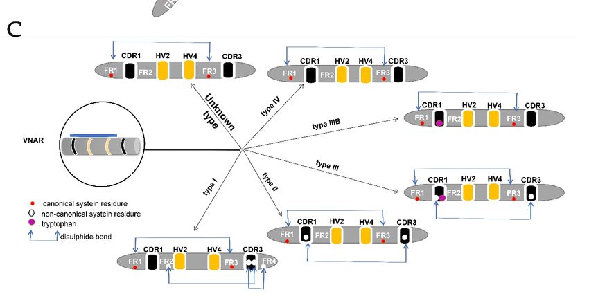
Most conventional antibodies (IgG) are heterodimers (Figure 1A) with two heavy chains
Academic Editor: Igal Ifergan
(VHs) and two light chains (VLs) [1]. Structurally, IgGs are distributed into antigen-binding
fragment (Fab) and Fragment-crystallizable (Fc) portion. Fab portion contains one constant
Received: 4 April 2021
domain of the heavy chain (C1) and one constant domain of the light chain (CL), as well
Accepted: 5 May 2021
as one variable domain of heavy chain (VH) and one variable domain of light chain (VL).
Published: 8 May 2021
The variable domain of each chain is responsible for antigen interactions due to the existence
of paratope (the antigen-binding site) [1,2]. The variable domains of IgG are linked by a
Publisher’s Note: MDPI stays neutral
flexible peptide into an antiparallel sheet (Figure 1A). Each variable domain has three loops
with regard to jurisdictional claims in
published maps and institutional affil-
of complementarity-determining regions (CDRs). The disulfide bond in the variable domain
iations.
between the framework region one (FR1) and the framework region three (FR3) is formed by
a canonical cysteine residue [3]. Fc portion [4] contains two constant domains (C2 and C3)
of the heavy chain for their biological activities [5]. The characteristic flexibility of the IgG is
characterized by the hinge region in the middle part of the heavy chain [6].
Copyright: © 2021 by the authors.
Licensee MDPI, Basel, Switzerland.
This article is an open access article
distributed under the terms and
conditions of the Creative Commons
Attribution (CC BY) license (https://
creativecommons.org/licenses/by/
4.0/).
Cells 2021, 10, 1140. https://doi.org/10.3390/cells10051140 https://www.mdpi.com/journal/cells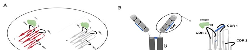
Cells 2021,10,
Cells2021,  10,1140
                1140                                                                                                                 23of
                                                                                                                                       of13
                                                                                                                                          14

       Figure1.
      Figure  1. Structure
                 Structure of
                           of the
                               the antibodies.
                                   antibodies. (A)
                                               (A) IgG
                                                   IgG has
                                                       has two
                                                            two identical
                                                                 identical heavy
                                                                           heavy chains
                                                                                  chains and
                                                                                         and two
                                                                                             two identical
                                                                                                 identical light
                                                                                                           light chains.
                                                                                                                 chains. (B)
                                                                                                                         (B) camelids’
                                                                                                                             camelids’
       antibodies  contain only   a heavy  chain without  the C1   domain.   (C) shark antibody-containing    only heavy
      antibodies contain only a heavy chain without the C1 domain. (C) shark antibody-containing only heavy chains with   chains with
       hypervariable regions 2 and 4, and a flexible hinge region between C3 and C4. Alternative spliced form of IgNAR in
      hypervariable regions 2 and 4, and a flexible hinge region between C3 and C4. Alternative spliced form of IgNAR in bamboo
       bamboo sharks with only C1, C4, and C5 domains; the variable domain matched the complete form of IgNAR with an
      sharks with only C1, C4, and C5 domains; the variable domain matched the complete form of IgNAR with an extra disulfide
       extra disulfide bond between CDR1 and CDR3 and HV2 and HV4 chains with the antigen in the long CDR3 as shown in
      bond
       VHH.between CDR1 and CDR3 and HV2 and HV4 chains with the antigen in the long CDR3 as shown in VHH.

                                         The existence of an antibody with only a heavy chain was first reported from camelids
                                   2. Characteristic of Variable of IgNAR
                                   in 1993 [7]. Two years later, a publication announced that sharks possess this type of
                                         Different
                                   antibody        shark belong
                                             [8]. Sharks  species to
                                                                   [18], including the
                                                                     cartilaginous   fishbanded
                                                                                          and thiswobbegong    (Orectolobus
                                                                                                   fish identified           macula-
                                                                                                                   with isotype IgM,
                                   tus), spiny  (Squalus  acanthias),   bamboo    (Chiloscyllium   plagiosum),   and  nurse
                                   IgW, and IgNAR [9], however, a constant domain (C1–C5) of IgNAR are identified to be      (Gingly-
                                   mostoma
                                   closely    cir-ratum)
                                           related        sharks produce
                                                    to primordial            different
                                                                   IgW isotype    [10]. VNARs [15,18]. Variable domains of sharks
                                   are formed by four hypervariable loops: CDR1 and CDR3, somatic mutations result in the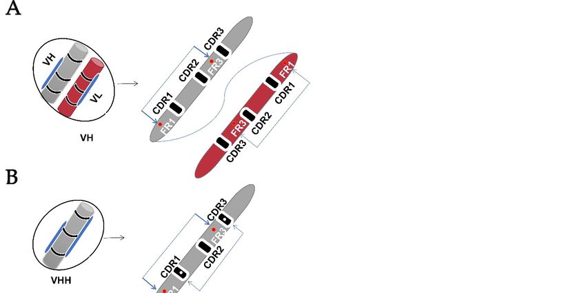
Cells 2021, 10, 1140                                                                                             3 of 13

                            The variable domain of camelids and sharks identified with molecular weights of approx-
                       imately 15 kDa and 12 kDa that are referred to as VHH and VNAR, respectively (Figure 1C);
                       the antigen-binding site is formed in only one single domain of the heavy chain [1]. Inter-chain
                       disulfide bonds joint the structure of VHH and VNAR domain [11–13]. The camelid antibody
                       consists of the constant domain two (C2) and constant domain three (C3) with a hinge region
                       between the variable region and the constant domain two (C2), and constant domain one (C1)
                       is absent due to a donor splice site mutation [1] (Figure 1B). The shark antibody (Figure 1C)
                       consists of C1–C5 domains and lacks canonical hinge regions between the variable region and
                       the constant domains; the structural flexibility of IgNAR is caused by the linkage of a disulfide
                       bridge to the constant domain (C3–C4) [14]. In this review, we will include our interesting
                       recent discovery of spliced form from white-spotted bamboo sharks (Chiloscyllium plagiosum)
                       that attained its flexible hinge region between constant domain (C1) and constants domain
                       (C4), where the flexible hinge region structure in nurse sharks identified between constant
                       domain (C3) and constant domain (C4) [15]. The variable regions in CDRs of different sharks
                       vary due to the presence of extra cysteines, which is used to classify VNARs as the com-
                       mon method to date. With the reference to amino acid sequences, the sizes of the CDR3 of
                       VH are shorter compared to those of VHH and VNAR [11]. VNAR was discovered to be
                       sharing structural homology with immunoglobulin light chain and T-cell receptor variable
                       regions [16,17].

                       2. Characteristic of Variable of IgNAR
                             Different shark species [18], including the banded wobbegong (Orectolobus macula-
                       tus), spiny (Squalus acanthias), bamboo (Chiloscyllium plagiosum), and nurse (Gingly-
                       mostoma cir-ratum) sharks produce different VNARs [15,18]. Variable domains of sharks
                       are formed by four hypervariable loops: CDR1 and CDR3, somatic mutations result in the
                       deletion of CDR2 [14], this position of CDR2 replaced by very short strand referred to as
                       HV2 [19]. The HV4 is sited between HV2 and CDR3, this HV4 is believed to contribute
                       to antigen binding [14,20]. 8 β-strands form the antigen-binding site of VNAR instead
                       of 10 as in mammalian variable domains, this making VNAR being smallest (12 kDa)
                       antigen-binding domain known in the vertebra to date [16,21].
                             Two canonical cysteine residues hold two beta-sheets in the framework regions (FRs)
                       1 and framework regions (FRs) 3. In addition to these canonical cysteines, CDRs may have
                       non-canonical/extra cysteine residues that form additional disulfide bonds within the variable
                       domain [22]. These features differentiate VH from the VHH and VNAR domains (Figure 2),
                       and to date are used as a means of classifying VNAR based on the presence or absence of
                       these extra cysteine residues within the hypervariable region [5,11,12,14,15,18,23]. VNAR
                       is classified into four types (I, II, III, and IV), the subtypes are further divided based on the
                       number of additional cysteines contained [18]. However, there are discovered IgNARs that
                       did not fit any of these described types due to the VNAR domain mutation rate [22].
                             The type I variable domains of IgNAR had been reported only in nurse sharks (Gin-
                       glymostoma cirratum) [12,14], but have now been also identified in the wobbegong shark
                       (Orectolobus ornatus) [18]. VNAR type 1 has non-canonical cysteine residues in FR2 and
                       FR4, and two paired residues in CDR3 [14].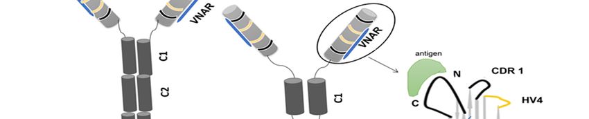
iable domain [22]. These features differentiate VH from the VHH and VNAR domains
                                    (Figure 2), and to date are used as a means of classifying VNAR based on the presence or
                                    absence of these extra cysteine residues within the hypervariable region
                                    [5,11,12,14,15,18,23]. VNAR is classified into four types (I, II, III, and IV), the subtypes are
Cells 2021, 10, 1140                further divided based on the number of additional cysteines contained [18]. However,     4 of 13

                                    there are discovered IgNARs that did not fit any of these described types due to the VNAR
                                    domain mutation rate [22].

        Figure
      Figure 2. 2. Variable
                Variable    domain
                         domain     structure.
                                  structure.   (A)
                                             (A)   IgG
                                                 IgG   antibody
                                                     antibody    variable
                                                               variable   domain
                                                                        domain     (VH)
                                                                                 (VH)   with
                                                                                       with   a disulfide
                                                                                            a disulfide    bond
                                                                                                         bond   connection
                                                                                                              connection    be-tween
                                                                                                                          be-tween
        the
      the   variable
          variable   domain
                    domain    and
                            and   two
                                two    conserved
                                     conserved    canonical
                                                canonical   cysteine
                                                          cysteine   residues
                                                                   residues inin the
                                                                               the   framework
                                                                                   framework     region
                                                                                               region    (FR1)
                                                                                                       (FR1)   and
                                                                                                             and   (FR3).
                                                                                                                 (FR3).   (B)
                                                                                                                        (B)   HCAbs
                                                                                                                            HCAbs
      variable domain (VHH) with an extra cysteine residue forming a disulfide bond between CDR1 and CDR3 despite the
      conserved canonical cysteine residues. (C) Variable domain (VNAR) of IgNAR lacking a CDR2, with vari-ous numbers of
      noncanonical cysteine residues that give rise to different types of VNAR and hypervariable regions (HV2 and HV4).

                                        The non-canonical cysteine residues in CDR3 form disulfide bonds with non-canonical
                                  cysteine residues within the FR2 and FR4 [16]. Type II VNAR forms intramolecular disulfide
                                  bonds between CDRI and CDR3 due to the presence of an additional cysteine residue. Types
                                  II VNARs have finger-like CDR3s that can bind into pockets or grooves, for example, active
                                  site clefts of enzymes [14]. Type III and type II VNAR is similar, possessing non-canonical
                                  cysteine residue in CDR1 and CDR3 [5]. These types of VNARs were hypothesized to fight
                                  against pathogens during the developmental stages of sharks but further developed to be
                                  matured classes of VNAR after maturation, which provides a more expansive immune
                                  repertoire [12,23]. Type IV VNAR lacks non-canonical disulfide bonds but contains only
                                  two cysteine residues that hold VNAR together [11]. This provides flexibility to the antigen-
                                  binding site of type IV VNAR [12,14]. Another structurally different VNAR type named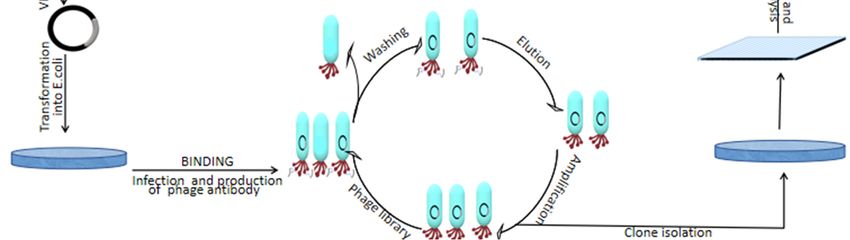
Cells 2021, 10, 1140                                                                                              5 of 13

                       type IIIb has been reported as a type IV VNAR with a tryptophan residue in CDR1 like the
                       one in type III VNAR [5,14], lacking non-canonical cysteine residues (and consequently
                       non-canonical disulfide bonds). Some VNARs cannot be characterized in any of the four
                       known types [22], which were isolated from different shark species and displayed good
                       binding abilities to antigens [18]. Therefore, more researches are required to reveal these
                       VNARs with peculiar cysteine arrangement that fall in neither of the four known types.
                            VNARs types can be significantly different among various species of sharks [22],
                       while some reported sharks such as small-spotted catsharks, banded hound sharks, and the
                       wobbegong sharks contained a novel variant without non-canonical cysteine residue [24].
                       Current studies revealed the existence of various VNARs with unknown types caused
                       by the inconsistency of cysteine numbers as well as locations [22], while some studies
                       revealed the therapeutic advantages of unknown VNAR types of different shark species [18].
                       The criteria used for VNAR classification should be reviewed because previous studies
                       were mainly in nurse sharks that possess type 1 other shark species are not. Therefore,
                       advancements in classifying VNAR using various methods are important to miss out on
                       VNAR that give out immune responses.
                            An unknown type of VNAR has been identified. A large phage library was constructed
                       from six adult nurse sharks (Ginglymosto macirratum) for diversification of the VNAR library
                       because previous studies showed types I VNAR are possessed by nurse sharks. Canonical
                       cysteines located at 21 and 82 amino acid were used as the main criterion to characterize
                       type I–IV VNAR. Eventually, around 5% of the total VNARs did not fit into any known
                       types, while 30% of the VNAR possess various numbers of cysteines with neither type I
                       nor type II behavior [22]. In our recent discovery, the presence of two canonical cysteines
                       located at 22 and 83 amino acid were used as a means of classification of type I-IV VNARs.
                       Approximately, 0.3% of the total VNARs did not match any of the four known VNARs
                       types [15].
                            Another unknown VNAR type was identified by randomization of CDR3 from banded
                       hound sharks (Triakis scyllium). This VNAR displayed the therapeutic potential. Hen egg-
                       white lysozyme gives successful isolation of antigen-specific IgNAR variable region without
                       immunization of target antigen. Amino acid sequences of CDR3 had either one or two
                       cysteine residues (behavior of type I-IV VNAR), but this VNAR was contradictory to previ-
                       ously reported results from (Ginglymostoma cirratum) nurse shark [25]. Again, Zielonka et al.
                       isolated VNAR of unknown types. This VNAR domain was successfully developed using
                       yeast surface display from a CDR3-randomized of bamboo shark (Chiloscyllium plagiosum),
                       showing high-affinity characteristics [19]. Therefore, various means of classification are
                       recommended as some studies produce VNARs with therapeutic advantages but nowhere
                       to be found among the VNARs types.
                            Categorization of IgNAR on reviewing variable domain only seems to be not sufficient
                       due to the higher variability in the CDRs regions [24]. The new method has proposed the
                       possibility of using the C1 domain sequence to identify IgNAR clusters, and this is because
                       the constant domain (C1) showed distinctiveness to other constant domains (C2-C5) of IgW.
                       The peculiarity of C1 appeared to be an important factor to be considered in characterizing
                       IgNAR clusters [24].
                            Compared to the other four constant domains, the C1 domain revealed higher stability
                       with the good antigen-binding ability to its variable region [15], thus the C1 domain plays a
                       significant role in structural alterations that increase affinity against a specific antigen [10].
                       A flexible (non-canonical) linker between VNAR and C1 domain gives out the dimerization
                       that produces a wide angle of VNAR and binding to multiple epitopes [14]. A unique
                       form of C1 domain was identified from the study of characterization of complete IgNAR
                       heavy chain constant domain of brown-banded bamboo sharks (Chiloscyllium punctatum)
                       as well as evolutionary relationship determination with different species. This IgNAR was
                       discovered with the presence of two distinct IgNAR types designated as (IgNAR-1 CH
                       type and the IgNAR-2 CH type) and 13 unique C1 sequences [24]. The results of the study
                       were related to our recent discovery in white-spotted bamboo (Chiloscyllium plagiosum)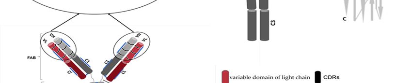
Cells 2021, 10, 1140                                                                                            6 of 13

                       sharks [15]. We identified two types of C1 domains, one with a short α-helix and the other
                       without a short α-helix. C1 with a short α-helix hypothesized to have higher stability than
                       the other. Based on these findings, it suggests using constants domain (C1) sequences for
                       comparing novel clusters of IgNAR types in future studies on cartilaginous fish [24].

                       3. Alternatives Spliced Constant Domain of IgNAR
                             The constant region of shark species is less studied, but recent evidence revealed a
                       primary role in stability maintenance of the antibody [24]. Shark constant domains are
                       identical to those in IgG’s constant domains except for their unstructured loop; however,
                       these unconventional IgNAR domains are very stable compared to the domains of con-
                       ventional antibodies. IgNARs can maintain biological activities in shark blood containing
                       350 mmol/L urea and 1000 mOsm/kg of osmotic salt ions [26].
                             Our recent discovery [15] revealed a splicing alternative form in the white-spotted
                       bamboo (Chiloscyllium plagiosum) shark. Surprisingly, only C1, C4, and C5 domains were
                       present during the cloning of the complete IgNAR sequence form with 5 constant domains,
                       but C2 and C3 were absent. We designated this spliced sequence as IgNARshort ∆C2-C3
                       (Figure 1C). According to phylogenetic analysis, the identified spliced sequences of white-
                       spotted bamboo (Chiloscyllium plagiosum) belonged to the IgNAR family and closely related
                       to the brown-banded bamboo shark (Chiloscyllium punctatum). VNAR of spliced form might
                       have the antigen-specific binding ability as a structural model prediction was consistent with
                       the structural characteristics of IgNAR. Conserved tryptophan and cysteine residues were
                       also present which could be involved in the formation of disulfide bonds and structural folds.
                       The interchain disulfide bond between C1 and C4 is predicted to be the result of an unpaired
                       cysteine in the flexible hinge region of IgNARshort (∆C2-C3) [15]. Therefore, we speculated
                       the flexibility attained in spliced form between C1-C4 is due to an unpaired cysteine contained.
                       This result is consistent with “Matthias J. Feige” and colleague study, the stalk of IgNAR
                       flexibility is maintained by a disulfide bond between C3 and C4 caused by unpaired cysteine
                       residue [10]. With references to previous studies as well as our bamboo shark results [15],
                       IgNAR full sequences of shark species needed to be examined to diverse our knowledge of
                       this antibody.

                       4. VNAR Complex Structure Responding to Different Proteins
                            The structural elements of the IgNAR constant domain could also influence the
                       folding pathway of the antibody which increases the stability. Thermostability of variable
                       region of IgNAR isolated from different sharks species are superior to that of conventional
                       antibodies [27]. Immunization of a horn shark (Heterodontus francisci) with recombinant
                       human tumor necrosis factor-alpha rhTNFα produced VNAR with higher thermostability
                       behavior due to its ability to identify rhTNFα and neutralize it in vitro [28]. This attribute
                       makes it a suitable candidate as a therapeutic antibody, mainly due to the ability to
                       transplant the stable structural motif into other domains.
                            Inhibition of full T cell activation can be attained by blocking the interaction with
                       inducible costimulator (ICOS) through Induced costimulatory ligand (ICOSL). The study
                       of Marina Kovaleva1 and colleagues revealed the reduction of inflammation to a murine
                       model of non-infectious uveitis by inhibiting this ICOSL using specific VNARs that rec-
                       ognized human ICOSL isolated from an immunized nurse shark. The anti-mouse ICOSL
                       VNAR Fc that was constructed showed a high affinity for inducible T-cell co-stimulator
                       ligand (ICOSL) and high penetration of the cornea in a mouse model of uveitis. Results
                       of their experiments demonstrated the efficiency and potentiality of the VNAR binding
                       domain for the treatment of auto-inflammatory conditions [29]. Obinna C. Ubah and
                       colleagues designed VNAR fusion anti-hTNF-α Quad-X™ that showed improvement in
                       potency over Humira® [30]. These findings from previous studies identified the usefulness
                       of the VNAR complex to the protein that is a good target for those diseases.
                            However, we should keep in mind the other side of the previous findings, despite the
                       immune responses of the isolated VNAR to bind with complex structure and responding to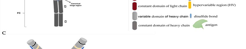
Cells 2021, 10, 1140                                                                                                  7 of 13

                        different proteins, but not all VNARs of shark species were tested to produce an adaptive
                        immune response [26]. Crouch et al. used human serum albumin (HSA) and (HEL)
                        as antigens to immunize spotted catsharks (Scyliorhinus canicula), after 37 weeks of the
                        immunization process, they did not find evidence of antigen-specific VNAR [31].

                        5. Therapeutic Biomedical Applications of IgNAR
                        Production of Shark IgNAR
                             IgNARs can be produced either by immunized, non-immunized [32–34] or semi-
                        synthetic library form [35,36]. The desired gene of the shark antibody can be inserted
                        into bacteria after insertion into a vector as a transporting agent. Displaying technology
                        can either be yeast, ribosome, or phage [32] followed by selection and analysis. In our
                        laboratory, we are immunizing sharks and isolate VNARs from the phage library technique
                        introduced [37]. In this approach, antibody-antigen binding is displayed by inserting
                        antibody DNA into a vector and introducing phenotype and genotype into phage genomes,
                        together with the construction of a large library for the particular antibody of interest and
                        higher affinity to antigen [38], to produce VNAR antibody from immunized shark [37,39].
                             Once the antigen proteins are ready, they will be injected into sharks to induce an
                        immune response (Figure 3). After several months, blood samples and shark spleen are
                        taken for transcriptome and mass spectrum analyses. Phagemid vector can be used to carry
                        specific antibody fragments to transform E. coli cells and generate a library. The genome
                        encoding an antibody protein of interest derived from the phage coding protein genes
                        leads to the display of proteins on the outside of bacterial cells (showing the phenotype
                        of interest) carrying the genes in their genome (showing the genotype of interest). Only
                        antibodies from the phage library that fit and bind to the antigen are recovered while the
                        others are washed away. Then, the selection of clones followed by screening using an
                        ELISA binding assay to identify antibody-antigen specificity [38]. After the screening, the
 Cells 2021, 10, 1140   identified IgNARs are further evaluated for efficacy using animal disease models to8test   of 14
                        their in vivo functions (Figure 3).

                         Figure3.3.IgNAR
                        Figure      IgNARproduction
                                          productionusing
                                                       usingaaphage
                                                                phagedisplay
                                                                       displaylibrary.
                                                                               library.Specific
                                                                                        Specificantigens
                                                                                                 antigensare
                                                                                                          areused
                                                                                                             usedto
                                                                                                                  toimmunize
                                                                                                                     immunize
                         sharks and induce an immune response about 3–6 months and then subcloned VNAR into the
                        sharks and induce an immune response about 3–6 months and then subcloned VNAR into the
                         phagemid vector for phage antibody display assays. The identified antibodies after the screening
                        phagemid vector for phage antibody display assays. The identified antibodies after the screening can
                         can be used to evaluate their efficacy in vitro and in vivo.
                        be used to evaluate their efficacy in vitro and in vivo.

                        6.6.Immunoglobulin
                             ImmunoglobulinVNAR VNARin  inDrug
                                                          DrugDiscovery
                                                                  Discovery
                               Differentcompanies
                               Different  companiesandandacademic
                                                          academicinstitutions
                                                                     institutionshave
                                                                                   havebeen
                                                                                        beenconsidering
                                                                                             consideringusing
                                                                                                          usingVNAR
                                                                                                                VNAR
                         shark  libraries to identify potential therapeutics  for human  diseases [18]. The unique
                        shark libraries to identify potential therapeutics for human diseases [18]. The unique fea-fea-
                         tures of VNAR may be useful for drug discovery; the isolated VNAR called V13 produced
                         using phage display to select against a human recombinant vascular endothelial growth
                         factor (VEGF165) cytokine was isolated from an immunized Heterodontus francisci shark.
                         They founded this V13 VNAR penetrated the cornea without the need for an injection and
                         without causing ocular surface abrasions or signs of discomfort in an animal model (prob-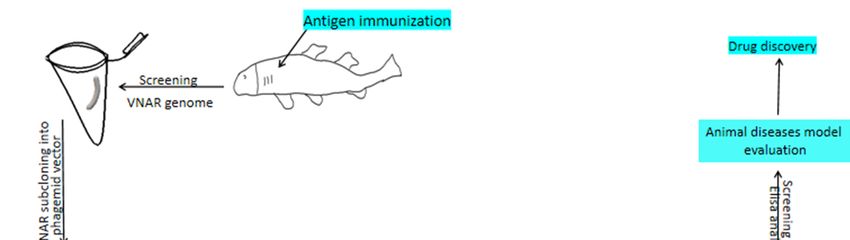
Cells 2021, 10, 1140                                                                                         8 of 13

                       tures of VNAR may be useful for drug discovery; the isolated VNAR called V13 produced
                       using phage display to select against a human recombinant vascular endothelial growth
                       factor (VEGF165) cytokine was isolated from an immunized Heterodontus francisci shark.
                       They founded this V13 VNAR penetrated the cornea without the need for an injection
                       and without causing ocular surface abrasions or signs of discomfort in an animal model
                       (probably due to its small size and long CDR3 of 27 amino acids). Their findings demon-
                       strated the potential applicability of VNAR V13 as a new drug candidate for vascular eye
                       diseases [40].
                             Another VNAR produced by immunizing nurse shark with human-induced costim-
                       ulatory ligand (ICOSL), reformatted by using fragments crystallization (Fc) region of a
                       human antibody. When anti-mouse ICOSL Fc introduced to uveitis mouse model of (IRBP)
                       inter-photoreceptor retinoid-binding protein-induced uveitis tested in vivo, eventually
                       decreases the inflammation compared to untreated mouse [18,29].
                             Other findings showed the functional activity of isolated VNAR using a semi-synthetic
                       phage display library bounded to a panel of BAFF (B cell-activating factor) with low nM
                       potency. All receptors (BR3, TACI, and BCMA) selected in the study blocked by anti-
                       BAFF VNARs, suggesting that the bio-specific antibodies with added functionality may be
                       effective in treating complex autoimmune diseases [41].
                             Arthritis (tenderness and swelling joints) has become a serious human disease in
                       elderly populations, especially in women. Tumor necrosis factor-alpha (TNF-α) is a cy-
                       tokine involved in systemic inflammation [12,42] and is identified to be a good target
                       to treat arthritis [13]. The structure of anti-TNF-α VNAR-human TNF-α complexes has
                       been solved and novel epitopes have been discovered by crystallization analysis where
                       VNAR interacts with two adjacent TNF-α protomers [13,30]. Another VNAR isolated from
                       sharks has shown promising features to alleviate the progress of arthritis in animal models,
                       and average in vivo arthritis inhibition and histopathology scores are 88% and 86% using
                       Quad-XTM at doses of 0.5 and 30 mg/kg, respectively [13], demonstrating the valuable
                       application of IgNAR in drug discovery again.

                       7. Viability of VNAR in Relation with Immunogenicity
                             Therapeutic antibodies in humans could potentially cause anti-drug antibodies (ADAs)
                       that lead to unfavorable outcomes of the medication [43]. The desired immunogenicity in
                       humans is exhibited by lowering the differences between the natural IgG and bispecific
                       antibody format [17,43–45]. The engineered full-length mAbs give rise to reduction into
                       single-chain Fv fragments (scFvs) that retain the binding specificity of the parent anti-
                       body [46–48], thus leading to low immunogenicity [49] as well as a potentially unique
                       molecule to be used especially in cancer treatment [46].
                             Sharks possess low overall sequence homology (≈30%) to human VH/VL sequences [44],
                       but the VNAR domain observed by crystallization data of their framework regions in a similar
                       manner to human immunoglobulin variable domains, [17,44,50]. Thus, the similarities in
                       the arrangement of framework regions are advantageous to therapeutic developments due
                       to the direction to humanized VNAR binders. Here, undesired immunogenicity of shark
                       antibody can be minimized via humanizing VNAR by replacing amino acid residues in the
                       framework of the VH domain [43] and utilized as a tiny molecule domain delivery vehicle. It
                       was reported that VNAR can be combined with monospecific antibodies to develop bispecific
                       agents [41]. Individual VNARs have been converted into VNAR-Fc fusions [29]. ScFv (single-
                       chain fragment variable) antibodies have been constructed mainly from B lymphocytes in
                       humans [46].
                             The tightly packed VNAR allows penetration into the active sites of different targets.
                       VNAR antibody is desirable in the drug development field due to its small peculiarity.
                       The combined molecules of VNAR with other proteins were reported to be effective to
                       imitate adaptations of the parental VNAR without losing its efficiency. The stability of the
                       VNAR domain through its disulfide bond can significantly improve the stability of human
                       antibodies when transferred to the variable region of those [14].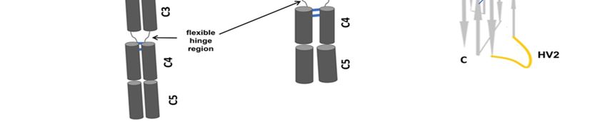
Cells 2021, 10, 1140                                                                                           9 of 13

                             The first VNAR designated as E06 was isolated from immunization of dogfish (Squalus
                       acanthias) shark species, based on binding epitopes recognized by VNAR binds specifically
                       and with high affinity to humans, this study did not base on determining anti-drug anti-
                       bodies (ADAs,) but involving in non-human primates (NHP) but showed no evidence of
                       anti-VNAR antibody production [51]. Later, scientists humanized VNAR by converting
                       more than half of their CDRs to those of a human germline Vk 1 sequence, DPK9 [17]. The
                       specificity and affinity of antigen-binding of the parental VNAR (E06) were retained by
                       humanized VNAR (huE06 v1.1) upon binding with human serum albumin (HSA) [17].
                       Therefore, this study gives a foundation for further design and humanization of shark
                       IgNARs [17]. John Steven and colleagues extended the study of E06 VNAR by using hu-
                       manized VNAR (huE06v1.10) as a template to isolate domains with improved biophysical
                       properties and reduced antigenicity [50]. In their study, the immunogenicity of lead clones
                       was assessed in a T-cell proliferation assay using ProImmune Ltd. REVEAL® Immuno-
                       genicity System DC–T cell assay. The prediction of immunogenicity in silico modeling of
                       VNAR domains was lower, both the wild-type E06 and humanized variants had a very
                       low response index (RI) compared to positive controls, thus a low level of immunogenicity
                       with similar values for those seen for a human Fc region was predicted [50].
                             Cotton et al. reports about oncofetal protein receptor tyrosine kinase ROR1 that
                       overexpressed on solid tumor. The cross-reactivity revealed by the constructed VNAR-
                       drug conjugates targeted ROR1, thus even the highly similar family member (ROR2) could
                       not bind. However, the study is in progress to assess the in vivo efficiency, but the complex
                       structure of VNAR was successfully combined with sdAbs and scFv which were directed
                       to other cell-surface protein targets [52].
                             Delta-like ligand 4 (DLL4) is prone to overgrowing tumors, which is a good target for
                       pancreatic cancer. VNAR against Delta-like ligand 4 (DLL4) was conjugated to therapeutic
                       nanoparticle (NPs) poly(lactic-co-glycolic) acid PEGylated by using surface maleimide
                       functional groups. This study result also showed the specificity binding ability of these
                       nanoconjugates [53].
                             Blood-brain barrier (BBB) penetration is a major challenge in therapeutic develop-
                       ment. The large size of conventional antibodies is a restriction to this penetration [54], but
                       the small size of VNAR is a good weapon, allowing them to reach the buried epitopes,
                       facilitating the discovery of mouse–human cross-species reactive sDAbs. This is a feature
                       not always accessible with conventional IgGs [54]. The study published in July 2017 by
                       Frank S. Walsh revealed the penetration ability of VNAR fusion with IgG. The combination
                       achieved by merged a single-chain variable fragment (scFv) of shark domain with terminus
                       end (N- or C-) of the heavy and light chain of an IgG. The bispecific antibody formats
                       produced retained antibody-dependent cell-mediated cytotoxicity [43,55].
                             The report from the previous study revealed the low inherent immunogenicity of the
                       VNAR. Impact of anti-drug antibodies (ADAs) detected on preclinical in vivo efficacy using
                       non-immunoglobulin VNAR fusion anti-hTNF-α biologics (Quad-X™ and D1-NDure™-C4)
                       and Humira® , a brand of adalimumab [30], demonstrating the promising application of
                       VNAR in biomedical industries.

                       8. IgVNAR Potential in Immunoassays
                             The isolation of single-domain antibodies has been successful using a naïve nurse
                       shark VNAR library with PCR extension assembly and self-ligation (EASeL). Based on this
                       technique, glypican3, human epidermal growth factor receptor 2 (HER2), and programmed
                       cell death-1 (PD1) as well as the viral antigens middle east respiratory syndrome (MERS)
                       and spike proteins [22] have been tested. Non-immunized adult spiny dogfish (Squalus
                       acanthias) and smooth dogfish (Mustelus canis) sharks have also been used to construct
                       libraries using Luminex100 and traditional ELISA assays. Shark VNAR sdAb libraries
                       can be used to specifically demonstrate their binding ability to different antigens, as well
                       as identifying agents that have been suggested as new venues to be used for pathogen
                       detection [56].
Cells 2021, 10, 1140 10 of 13
The high thermal stability of the variable region of the receptor VNAR from sharks is
important for the diagnosis of different diseases. Monoclonal antibodies in malaria rapid di-
agnostic tests (RDTs) using VNAR shark binders have been studied, with splenocytes used
to construct a single domain antibody (sdAb). Three recombinant malaria biomarker pro-
teins for Plasmodium falciparum (PfHRP2-histidine-rich protein 2, PfpLDH-plasmodium lac-
tate dehydrogenase, and fructose 1, 6-biphosphate aldolase) from immunized wobbegong
sharks (Orectolobus ornatus) were successfully used to isolate target-specific bacteriophage
VNARs using phage display technology to identify antigen binders [57].
Consideration of the variable domain of sharks as therapeutic agents is caused by the
peculiarity of this antibody’s structure from different discoveries: short α-helix structure
of constant domain (C1) [15], salt bridge, and hydrophobic core are thought to contribute
to the stability of constant domains [10]. Compared with murine mAbs and scFvs, shark
VNAR sdAbs molecules are more sensitive and thermally stable to viral nucleoprotein
(NP) of ZEBOV [58]. The variable domain of the shark antibody can be modified into
a high number of formats and fused to various molecules and produced a satisfactory
outcome. The advantage of VNAR stability in extreme pHs, for example, incubation with
acid produced by gastric glands [23,59]. The two distinct clones of VNAR reported to
have a good binding ability and stability to P. gingivalis KgP [58]. All these findings are
important to consider IgNAR as a satisfactory therapeutic agent.
9. Strength of VNAR Domain over Traditional mAbs
The production of recombinant VNAR is easier due to no post-translational modifi-
cations are required and expressions of VNAR are performed well using E. coli than in
mammalian cells [60], high solubility is achieved because the hydrophilic residues were
presented within VNAR surfaces [61], therefore it is not expensive compared to the pro-
duction of traditional mAbs [60,61]. After injection of a specific antigen, it takes around
4–6 months to get the desired antibody because the IgNAR response of sharks is slower
than the process observed in mammals [5,61].
There are many residues in IgG which are not available in VNAR, and thus making
it the smallest antibody fragment [48,61]. The long CDR3 gives the VNAR uniqueness
to be able to target a small epitope that can be easily reached by conventional IgG. The
binding-specific activity of VNAR can be attained even after exposure to temperature up
to 95 ◦ C [61]. Besides, VNAR exhibited high physicochemical stability [48]. This feature
is desirable for applications of VNAR than traditional antibodies in a therapeutic and
biotechnological setup. During the comparison between commercial reagents derived from
conventional polyclonal sera and monovalent VNAR clone to P. falciparum AMA1, the
data showed that heat stability of purified recombinant VNAR was superior to that of
conventional mAbs, and even the refolding property of VNAR was retained when the
temperature increased to 80 ◦ C. Therefore, the usage of VNAR as a binder to malaria is
preferable [62].
10. Conclusions
IgNAR’s structures endow the molecule with specific functions; for example, its small
size and thermal stability are advantageous for penetration into the epitopes of tumors;
these features have made IgNAR a focus for diverse applicable conditions. We summarized
the progress on IgNAR discoveries as valuable conjugate and highlighted its structural
features and potential applications for drug discovery with pathogen detection as well as
immunogenicity to show the value of IgNAR as a biomedical agent. Despite the uniqueness
of structural features of IgNAR and its characteristic in the broadening drugs target to
undefeated diseases, extensive efforts are needed to overcome challenges in the time-
consuming process of raising and immunizing sharks during the production of IgNAR and
on the establishment of dynamic monitoring methods for IgNAR in sharks. More studies
are still required to study the categories of IgNAR clusters in various ways than being
consistent with extra cysteine residues only. Additionally, novel technologies should beCells 2021, 10, 1140 11 of 13
considered to make sure that VNAR molecules are delivered to their targets at the right
place in the body to treat diseases such as tumors (a task that remains challenging).
Author Contributions: S.N.J.: literature searching and writing. X.G., S.H., J.S. and Z.L.: writing,
review and editing, use of software. G.C. and L.L.: validation, project administration, and revision of
the manuscript. All authors have read and agreed to the published version of the manuscript.
Funding: This research was funded by the Natural Science Foundation of Zhejiang Province
under Grant No. LQ20H300005 and the Public Welfare Research Project of Zhejiang Province
(LGF19H140002), as well as a startup grant from Zhejiang SCITECH University (11612932618290).
Institutional Review Board Statement: Not applicable.
Informed Consent Statement: Not applicable.
Data Availability Statement: All data is included in the manuscript.
Conflicts of Interest: The authors declare that they have no competing interests.
References
1. Matz, H.; Dooley, H. Shark IgNAR-derived binding domains as potential diagnostic and therapeutic agents. Dev. Comp. Immunol.
2019, 90, 100–107. [CrossRef] [PubMed]
2. Reader, R.H.; Workman, R.G.; Maddison, B.C.; Gough, K.C. Advances in the Production and Batch Reformatting of Phage
Antibody Libraries. Mol. Biotechnol. 2019, 61, 801–815. [CrossRef]
3. Schroeder, H.W., Jr.; Cavacini, L. Structure and function of immunoglobulins. J. Allergy Clin. Immunol. 2010, 125, S41–S52.
[CrossRef]
4. Bates, A.; Power, C.A. David vs. Goliath: The Structure, Function, and Clinical Prospects of Antibody Fragments. Antibodies 2019,
8, 28. [CrossRef] [PubMed]
5. Cabanillas-Bernal, O.; Duenas, S.; Ayala-Avila, M.; Rucavado, A.; Escalante, T.; Licea-Navarro, A.F. Synthetic libraries of shark
vNAR domains with different cysteine numbers within the CDR3. PLoS ONE 2019, 14, e0213394. [CrossRef]
6. Goldman, E.R.; Liu, J.L.; Zabetakis, D.; Anderson, G.P. Enhancing Stability of Camelid and Shark Single Domain Antibodies: An
Overview. Front. Immunol. 2017, 8, 865. [CrossRef]
7. Hamers-Casterman, C.; Atarhouch, T.; Muyldermans, S.; Robinson, G.; Hamers, C.; Songa, E.B.; Bendahman, N.; Hamers, R.
Naturally occurring antibodies devoid of light chains. Nature 1993, 363, 446–448. [CrossRef]
8. Greenberg, A.S.; Avila, D.; Hughes, M.; Hughes, A.; McKinney, E.C.; Flajnik, M.F. A new antigen receptor gene family that
undergoes rearrangement and extensive somatic diversification in sharks. Nature 1995, 374, 168–173. [CrossRef]
9. Zhou, H.; Liu, S.; Yin, X.; Li, Z.; Yang, Z.; Zhou, R. Molecular Origin of the Stability Difference in Four Shark IgNAR Constant
Domains. Biophys. J. 2019, 116, 1907–1917. [CrossRef]
10. Feige, M.J.; Grawert, M.A.; Marcinowski, M.; Hennig, J.; Behnke, J.; Auslander, D.; Herold, E.M.; Peschek, J.; Castro, C.D.; Flajnik,
M.; et al. The structural analysis of shark IgNAR antibodies reveals evolutionary principles of immunoglobulins. Proc. Natl. Acad.
Sci. USA 2014, 111, 8155–8160. [CrossRef]
11. Wesolowski, J.; Alzogaray, V.; Reyelt, J.; Unger, M.; Juarez, K.; Urrutia, M.; Cauerhff, A.; Danquah, W.; Rissiek, B.; Scheuplein,
F.; et al. Single domain antibodies: Promising experimental and therapeutic tools in infection and immunity. Med. Microbiol.
Immunol. 2009, 198, 157–174. [CrossRef]
12. Cheong, W.S.; Leow, C.Y.; Abdul Majeed, A.B.; Leow, C.H. Diagnostic and therapeutic potential of shark variable new antigen
receptor (VNAR) single domain antibody. Int. J. Biol. Macromol. 2020, 147, 369–375. [CrossRef]
13. Ubah, O.C.; Steven, J.; Porter, A.J.; Barelle, C.J. An Anti-hTNF-alpha Variable New Antigen Receptor Format Demonstrates
Superior in vivo Preclinical Efficacy to Humira(R) in a Transgenic Mouse Autoimmune Polyarthritis Disease Model. Front.
Immunol. 2019, 10, 526. [CrossRef]
14. Zielonka, S.; Empting, M.; Grzeschik, J.; Konning, D.; Barelle, C.J.; Kolmar, H. Structural insights and biomedical potential of
IgNAR scaffolds from sharks. MAbs 2015, 7, 15–25. [CrossRef]
15. Zhang, W.; Qin, L.; Cai, X.; Juma, S.N.; Xu, R.; Wei, L.; Wu, Y.; Cui, X.; Chen, G.; Liu, L.; et al. Sequence structure character
of IgNAR Sec in whitespotted bamboo shark (Chiloscyllium plagiosum). Fish Shellfish Immunol. 2020, 102, 140–144. [CrossRef]
[PubMed]
16. Stanfield, R.L.; Dooley, H.; Flajnik, M.F.; Wilson, I.A. Crystal structure of a shark single-domain antibody V region in complex
with lysozyme. Science 2004, 305, 1770–1773. [CrossRef]
17. Kovalenko, O.V.; Olland, A.; Piche-Nicholas, N.; Godbole, A.; King, D.; Svenson, K.; Calabro, V.; Muller, M.R.; Barelle, C.J.;
Somers, W.; et al. Atypical antigen recognition mode of a shark immunoglobulin new antigen receptor (IgNAR) variable domain
characterized by humanization and structural analysis. J. Biol. Chem. 2013, 288, 17408–17419. [CrossRef]
18. English, H.; Hong, J.; Ho, M. Ancient species offers contemporary therapeutics: An update on shark VNAR single domain
antibody sequences, phage libraries and potential clinical applications. Antib. Ther. 2020, 3, 1–9. [CrossRef] [PubMed]Cells 2021, 10, 1140 12 of 13
19. Zielonka, S.; Weber, N.; Becker, S.; Doerner, A.; Christmann, A.; Christmann, C.; Uth, C.; Fritz, J.; Schafer, E.; Steinmann, B.;
et al. Shark Attack: High affinity binding proteins derived from shark vNAR domains by stepwise in vitro affinity maturation.
J. Biotechnol. 2014, 191, 236–245. [CrossRef]
20. Zielonka, S.; Empting, M.; Konning, D.; Grzeschik, J.; Krah, S.; Becker, S.; Dickgiesser, S.; Kolmar, H. The Shark Strikes Twice:
Hypervariable Loop 2 of Shark IgNAR Antibody Variable Domains and Its Potential to Function as an Autonomous Paratope.
Mar. Biotechnol. 2015, 17, 386–392. [CrossRef] [PubMed]
21. Gonzalez-Sapienza, G.; Rossotti, M.A.; Tabares-da Rosa, S. Single-Domain Antibodies as Versatile Affinity Reagents for Analytical
and Diagnostic Applications. Front. Immunol. 2017, 8, 977. [CrossRef] [PubMed]
22. Feng, M.; Bian, H.; Wu, X.; Fu, T.; Fu, Y.; Hong, J.; Fleming, B.D.; Flajnik, M.F.; Ho, M. Construction and next-generation
sequencing analysis of a large phage-displayed VNAR single-domain antibody library from six naive nurse sharks. Antib. Ther.
2019, 2, 1–11. [CrossRef]
23. Kovaleva, M.; Ferguson, L.; Steven, J.; Porter, A.; Barelle, C. Shark variable new antigen receptor biologics - a novel technology
platform for therapeutic drug development. Expert Opin. Biol. Ther. 2014, 14, 1527–1539. [CrossRef]
24. De Silva, D.P.N.; Tan, E.; Mizuno, N.; Hosoya, S.; Reza, M.S.; Watabe, S.; Kinoshita, S.; Asakawa, S. Transcriptomic analysis of
immunoglobulin novel antigen receptor (IgNAR) heavy chain constant domains of brownbanded bamboo shark (Chiloscyllium
punctatum). Fish Shellfish Immunol. 2019, 84, 370–376. [CrossRef] [PubMed]
25. Ohtani, M.; Hikima, J.; Jung, T.S.; Kondo, H.; Hirono, I.; Aoki, T. Construction of an artificially randomized IgNAR phage display
library: Screening of variable regions that bind to hen egg white lysozyme. Mar. Biotechnol. 2013, 15, 56–62. [CrossRef] [PubMed]
26. Liu, X.; Chen, Q. Progress in shark single-domain antibody. Sheng Wu Gong Cheng Xue Bao 2020, 36, 1069–1082. [CrossRef]
27. Healer, J.; Triglia, T.; Hodder, A.N.; Gemmill, A.W.; Cowman, A.F. Functional analysis of Plasmodium falciparum apical membrane
antigen 1 utilizing interspecies domains. Infect. Immun. 2005, 73, 2444–2451. [CrossRef]
28. Camacho-Villegas, T.; Mata-Gonzalez, T.; Paniagua-Solis, J.; Sanchez, E.; Licea, A. Human TNF cytokine neutralization with a
vNAR from Heterodontus francisci shark: A potential therapeutic use. MAbs 2013, 5, 80–85. [CrossRef] [PubMed]
29. Kovaleva, M.; Johnson, K.; Steven, J.; Barelle, C.J.; Porter, A. Therapeutic Potential of Shark Anti-ICOSL VNAR Domains is
Exemplified in a Murine Model of Autoimmune Non-Infectious Uveitis. Front. Immunol. 2017, 8, 1121. [CrossRef]
30. Ubah, O.C.; Porter, A.J.; Barelle, C.J. In Vitro ELISA and Cell-Based Assays Confirm the Low Immunogenicity of VNAR
Therapeutic Constructs in a Mouse Model of Human RA: An Encouraging Milestone to Further Clinical Drug Development.
J. Immunol. Res. 2020, 2020, 7283239. [CrossRef]
31. Crouch, K.; Smith, L.E.; Williams, R.; Cao, W.; Lee, M.; Jensen, A.; Dooley, H. Humoral immune response of the small-spotted
catshark, Scyliorhinus canicula. Fish Shellfish Immunol. 2013, 34, 1158–1169. [CrossRef]
32. Sabir, J.S.; Atef, A.; El-Domyati, F.M.; Edris, S.; Hajrah, N.; Alzohairy, A.M.; Bahieldin, A. Construction of naive camelids VHH
repertoire in phage display-based library. C. R. Biol. 2014, 337, 244–249. [CrossRef] [PubMed]
33. Chan, S.K.; Rahumatullah, A.; Lai, J.Y.; Lim, T.S. Naive Human Antibody Libraries for Infectious Diseases. Adv. Exp. Med. Biol.
2017, 1053, 35–59. [CrossRef] [PubMed]
34. Moon, S.A.; Ki, M.K.; Lee, S.; Hong, M.L.; Kim, M.; Kim, S.; Chung, J.; Rhee, S.G.; Shim, H. Antibodies against non-immunizing
antigens derived from a large immune scFv library. Mol. Cells 2011, 31, 509–513. [CrossRef]
35. Grzeschik, J.; Konning, D.; Hinz, S.C.; Krah, S.; Schroter, C.; Empting, M.; Kolmar, H.; Zielonka, S. Generation of Semi-Synthetic
Shark IgNAR Single-Domain Antibody Libraries. Methods Mol. Biol. 2018, 1701, 147–167. [CrossRef] [PubMed]
36. Van Wyngaardt, W.; Malatji, T.; Mashau, C.; Fehrsen, J.; Jordaan, F.; Miltiadou, D.; Du Plessis, D.H. A large semi-synthetic
single-chain Fv phage display library based on chicken immunoglobulin genes. BMC Biotechnol. 2014, 4, 6. [CrossRef]
37. Dooley, H.; Flajnik, M.F.; Porter, A.J. Selection and characterization of naturally occurring single-domain (IgNAR) antibody
fragments from immunized sharks by phage display. Mol. Immunol. 2003, 40, 25–33. [CrossRef]
38. Loureiro, L.R.; Carrascal, M.A.; Barbas, A.; Ramalho, J.S.; Novo, C.; Delannoy, P.; Videira, P.A. Challenges in Antibody
Development against Tn and Sialyl-Tn Antigens. Biomolecules 2015, 5, 1783–1809. [CrossRef] [PubMed]
39. Solemani Zadeh, A.; Grasser, A.; Dinter, H.; Hermes, M.; Schindowski, K. Efficient Construction and Effective Screening of
Synthetic Domain Antibody Libraries. Methods Protoc. 2019, 2, 17. [CrossRef] [PubMed]
40. Camacho-Villegas, T.A.; Mata-Gonzalez, M.T.; Garcia-Ubbelohd, W.; Nunez-Garcia, L.; Elosua, C.; Paniagua-Solis, J.F.; Licea-
Navarro, A.F. Intraocular Penetration of a vNAR: In Vivo and In Vitro VEGF165 Neutralization. Mar. Drugs 2018, 16, 113.
[CrossRef] [PubMed]
41. Hasler, J.; Flajnik, M.F.; Williams, G.; Walsh, F.S.; Rutkowski, J.L. VNAR single-domain antibodies specific for BAFF inhibit B cell
development by molecular mimicry. Mol. Immunol. 2016, 75, 28–37. [CrossRef] [PubMed]
42. Ciebiera, M.; Wlodarczyk, M.; Zgliczynska, M.; Lukaszuk, K.; Meczekalski, B.; Kobierzycki, C.; Lozinski, T.; Jakiel, G. The Role
of Tumor Necrosis Factor alpha in the Biology of Uterine Fibroids and the Related Symptoms. Int. J. Mol. Sci. 2018, 19, 869.
[CrossRef] [PubMed]
43. Husain, B.; Ellerman, D. Expanding the Boundaries of Biotherapeutics with Bispecific Antibodies. BioDrugs 2018, 32, 441–464.
[CrossRef]
44. Ubah, O.C.; Buschhaus, M.J.; Ferguson, L.; Kovaleva, M.; Steven, J.; Porter, A.J.; Barelle, C.J. Next-generation flexible formats of
VNAR domains expand the drug platform’s utility and developability. Biochem. Soc. Trans. 2018, 46, 1559–1565. [CrossRef]Cells 2021, 10, 1140 13 of 13
45. Barelle, C.; Porter, A. VNARs: An Ancient and Unique Repertoire of Molecules That Deliver Small, Soluble, Stable and High
Affinity Binders of Proteins. Antibodies 2015, 4, 240–258. [CrossRef]
46. Ahmad, Z.A.; Yeap, S.K.; Ali, A.M.; Ho, W.Y.; Alitheen, N.B.; Hamid, M. scFv antibody: Principles and clinical application. Clin.
Dev. Immunol. 2012, 2012, 980250. [CrossRef]
47. Monnier, P.; Vigouroux, R.; Tassew, N. In Vivo Applications of Single Chain Fv (Variable Domain) (scFv) Fragments. Antibodies
2013, 2, 193–208. [CrossRef]
48. Konning, D.; Zielonka, S.; Kaempffe, A.; Jager, S.; Kolmar, H.; Schroter, C. Selection and Characterization of Anti-idiotypic Shark
Antibody Domains. Methods Mol. Biol. 2020, 2070, 191–209. [CrossRef]
49. Satheeshkumar, P.K. Expression of Single Chain Variable Fragment (scFv) Molecules in Plants: A Comprehensive Update. Mol.
Biotechnol. 2020, 62, 151–167. [CrossRef] [PubMed]
50. Steven, J.; Muller, M.R.; Carvalho, M.F.; Ubah, O.C.; Kovaleva, M.; Donohoe, G.; Baddeley, T.; Cornock, D.; Saunders, K.; Porter,
A.J.; et al. In Vitro Maturation of a Humanized Shark VNAR Domain to Improve Its Biophysical Properties to Facilitate Clinical
Development. Front. Immunol. 2017, 8, 1361. [CrossRef]
51. Muller, M.R.; Saunders, K.; Grace, C.; Jin, M.; Piche-Nicholas, N.; Steven, J.; O’Dwyer, R.; Wu, L.; Khetemenee, L.; Vugmeyster,
Y.; et al. Improving the pharmacokinetic properties of biologics by fusion to an anti-HSA shark VNAR domain. MAbs 2012,
4, 673–685. [CrossRef]
52. Cotton, G.; Thom, J.; Trumper, P.; Kamenski, A.; Bell, S.; Wappett, M.; Barelle, C.; Kovaleva, M.; Campion, A.; Persiani, E.; et al.
Abstract 538: Exploiting the properties of VNAR domains for the development of novel efficacious protein drug conjugates
targeting the oncofetal protein ROR1. Cancer Res. 2020, 80, 538. [CrossRef]
53. Leach, A.; Smyth, P.; Ferguson, L.; Steven, J.; Greene, M.K.; Branco, C.M.; McCann, A.P.; Porter, A.; Barelle, C.J.; Scott, C.J.
Anti-DLL4 VNAR targeted nanoparticles for targeting of both tumour and tumour associated vasculature. Nanoscale 2020,
12, 14751–14763. [CrossRef]
54. Pothin, E.; Lesuisse, D.; Lafaye, P. Brain Delivery of Single-Domain Antibodies: A Focus on VHH and VNAR. Pharmaceutics 2020,
12, 937. [CrossRef] [PubMed]
55. Walsh, F.S.; Wicher, K.; Szary, J.; Stocki, P.; Demydchuk, M.; Rutkowski, L. Abstract 3631: Delivery of a CD20 transferrin receptor
VNAR bispecific antibody to the brain for CNS lymphoma. Cancer Res. 2017, 77, 3631. [CrossRef]
56. Liu, J.L.; Anderson, G.P.; Delehanty, J.B.; Baumann, R.; Hayhurst, A.; Goldman, E.R. Selection of cholera toxin specific IgNAR
single-domain antibodies from a naive shark library. Mol. Immunol. 2007, 44, 1775–1783. [CrossRef]
57. Leow, C.H.; Fischer, K.; Leow, C.Y.; Braet, K.; Cheng, Q.; McCarthy, J. Isolation and characterization of malaria PfHRP2 specific
VNAR antibody fragments from immunized shark phage display library. Malar. J. 2018, 17, 383. [CrossRef]
58. Leow, C.H.; Cheng, Q.; Fischer, K.; McCarthy, J. The Development of Single Domain Antibodies for Diagnostic and Therapeutic
Applications. Antib Eng. 2018. [CrossRef]
59. Griffiths, K.; Dolezal, O.; Parisi, K.; Angerosa, J.; Dogovski, C.; Barraclough, M.; Sanalla, A.; Casey, J.; González, I.; Perugini, M.;
et al. Shark Variable New Antigen Receptor (VNAR) Single Domain Antibody Fragments: Stability and Diagnostic Applications.
Antibodies 2013, 2, 66–81. [CrossRef]
60. Konning, D.; Kolmar, H. Beyond antibody engineering: Directed evolution of alternative binding scaffolds and enzymes using
yeast surface display. Microb Cell Fact. 2018, 17, 32. [CrossRef]
61. Leow, C.H.; Fischer, K.; Leow, C.Y.; Cheng, Q.; Chuah, C.; McCarthy, J. Single Domain Antibodies as New Biomarker Detectors.
Diagnostics 2017, 7, 52. [CrossRef] [PubMed]
62. Henderson, K.A.; Streltsov, V.A.; Coley, A.M.; Dolezal, O.; Hudson, P.J.; Batchelor, A.H.; Gupta, A.; Bai, T.; Murphy, V.J.; Anders,
R.F.; et al. Structure of an IgNAR-AMA1 complex: Targeting a conserved hydrophobic cleft broadens malarial strain recognition.
Structure 2007, 15, 1452–1466. [CrossRef] [PubMed]You can also read