SINGLE-CELL ANALYSIS OF MAMMALIAN CARDIOGENESIS ELUCIDATING AN ESSENTIAL ROLE OF OUTFLOW TRACT PROGENITORS
←
→
Page content transcription
If your browser does not render page correctly, please read the page content below
Thesis for doctoral degree (Ph.D.) 2021

        From the Department of Cell and Molecular Biology,
                Karolinska Institutet, Stockholm, Sweden

               SINGLE-CELL ANALYSIS OF
            MAMMALIAN CARDIOGENESIS
  ELUCIDATING AN ESSENTIAL ROLE OF
         OUTFLOW TRACT PROGENITORS

                                          Chikai Zhou

                                            周驰凯

                                          Stockholm 2021
All previously published papers were reproduced with permission from the publisher.
Published by Karolinska Institutet.
Printed by Universitetsservice US-AB, 2021
© CHIKAI ZHOU, 2021
ISBN: 978-91-8016-296-8
Single-cell analysis of mammalian cardiogenesis
elucidating an essential role of outflow tract progenitors

THESIS FOR DOCTORAL DEGREE (Ph.D.)

By

Chikai Zhou

The thesis will be defended in public at Louis, Widerströmska huset (Solna), Tomtebodavägen
18a, Karolinska Institutet, Stockholm,

Monday, the 4th of October, 2021 at 14:00

Zoom Meeting ID: 665 6794 9990
Passcode: 183692

Principal Supervisor:                         Opponent:

Professor Kenneth R Chien                     Dr. Stéphane Zaffran
Karolinska Institutet                         Aix-Marseille University, France
Department of Cell and Molecular Biology      Marseille Medical Genetics

Co-supervisor(s):                             Examination Board:

Dr. Makoto Sahara                             Asscociate Professor Qiaolin Deng
Karolinska Institutet                         Karolinska Institutet
Department of Cell and Molecular Biology      Department of Physiology and Pharmacology

Dr. Emil Hansson                              Professor Gergana Dobreva
Karolinska Institutet                         Heidelberg University
Department of Medicine                        Department of Medical Faculty Mannheim

Assistant Professor Olaf Bergmann             Professor Johan Ericson
Karolinska Institutet                         Karolinska Institutet
Department of Cell and Molecular Biology,     Department of Cell and Molecular Biology
Karolinska Institutet
Dedicated to my beloved family
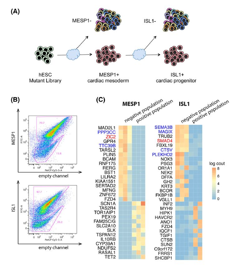
ABSTRACT
The heart is the first organ to form and start to function during mammalian embryogenesis. This
complex organ system is constructed by a diverse set of cell types, involving mesodermal
precursors and heart progenitors, at the earliest embryonic stages. These progenitor cells
contribute to the formation of the distinct heart regions, such as atria, right and left ventricle,
and outflow tract (OFT). However, it is still controversial and undefined which specific
progenitors and paracrine molecular cues are responsible to form each of the distinct heart
regions (e.g., OFT), requiring the rigorous analysis at higher resolution to identify the detailed
cellular and molecular pathways on developing hearts.

To tackle this problem, we applied a wide variety of the state-of-the-art biotechnologies and
assays, including the in vitro cardiac differentiation system of human embryonic stem cells
(hESCs), handling of the murine and human embryonic hearts, CRISPR/Cas9 gene editing,
single-cell RNA sequencing (RNA-seq), and a mouse lineage tracing approach. We provide a
comprehensive gene expression resource, characterizing the transcriptional dynamics of cardiac
lineage specification and identifying novel markers of developing cardiac derivatives from
multipotent progenitors to mature cardiac cells. Importantly, we have discovered the uniquely
stage- and region-specific mesodermal precursors and/or heart progenitors that are essential on
mammalian cardiogenesis.

In Paper I, to determine the key regulators for cardiac linage specification and commitment,
we first established a genome-wide CRISPR/Cas9 knockout screen platform using the in vitro
hESC differentiation where we monitored the two distinct stage markers, an early cardiac
mesodermal marker MESP1 and a heart progenitor marker ISL1. From the screen output, we
compiled a list of 15 candidate genes and finally identified ZIC2 as an essential gene for early
cardiac mesoderm formation. Interestingly, RNA-seq profiles of the ZIC2-mutant cells revealed
that the mutants switched their cell fate alternatively to the noncardiac cell lineage. Further,
single-cell RNA-seq analysis showed the ZIC2 mutants affected the apelin receptor-related
signaling pathway during mesoderm formation. Our results provide a new link between ZIC2
and human cardiogenesis and document the potential power of a genome-wide unbiased
CRISPR-knockout screen to identify the key steps during the in vitro hESC cardiogenesis.

In Paper II, through population and single-cell analysis of the in vitro hESC cardiac
differentiation and the in vivo human embryonic/fetal hearts, we chart the developmental
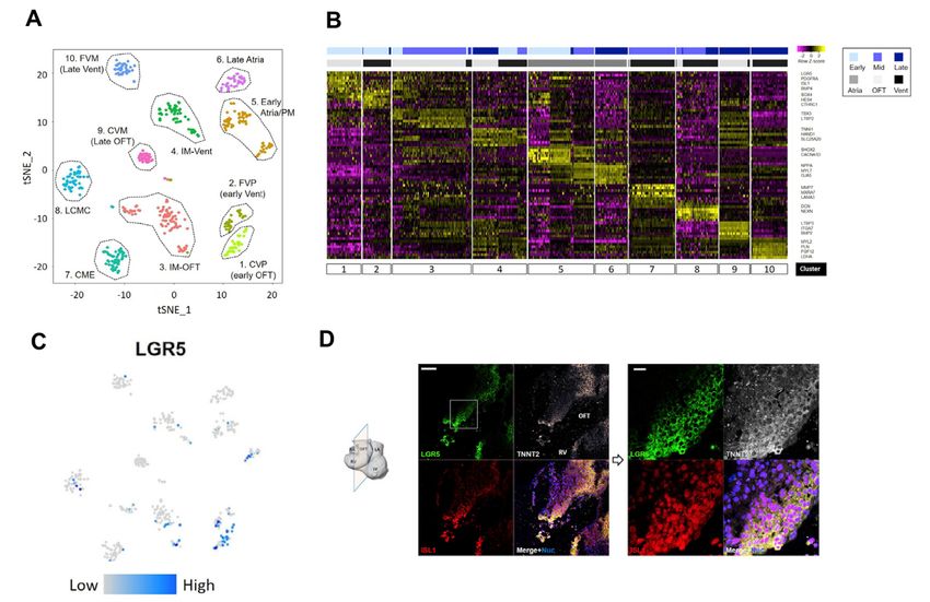
landscape of human cardiac formation at the cellular and molecular basis. Importantly, we have
discovered a uniquely human subset of the OFT region-specific heart progenitors, marked by
LGR5. The LGR5+ progenitors emerge specifically in the proximal OFT of human embryonic
hearts (4 to 5 weeks of fetal age) and likely contribute to the OFT formation and alignment.
Our results provide a deeper understanding of human cardiogenesis, which may uncover the
putative origins of certain human congenital cardiac malformations.

In Paper III, to obtain a whole picture of transcriptional and epigenetic regulation in the
mesoderm lineage on the developing hearts, we first established Mesp1+ mesodermal lineage
tracing mice. Mesp1 gene encodes a transcription factor of the b-HLH family, which is
expressed broadly in the mesodermal cells and critical for the cardiovascular development in
mammals. Using the CRISPR/Cas9 system and an IRES2-Cre cassette, we generated a Mesp1-
IRES2-Cre knock-in mouse line and cross-bred them with reporter mice (Rosa26-tdTomato).
On the Mesp1+ lineage tracing mice (Mesp1Cre/+; Rosa26tdTomato), we observed that more than
95% of the atria and ventricular cells in the hearts on an embryonic day 10.5 are the Mesp1+
mesodermal lineage. Interestingly, less percentage (
LIST OF SCIENTIFIC PAPERS
1. Jiejia Xu, CHIKAI ZHOU, Kylie S. Foo, Ran Yang, Yao Xiao, Kristine Bylund, Makoto
Sahara, Kenneth R. Chien#.

Genome-wide CRISPR screen identifies ZIC2 as an essential gene that controls the cell
fate of early mesodermal precursors to human heart progenitors. Stem cells, 38, no. 6
(2020): 741-755.

2. Sahara, Makoto*#, Federica Santoro*, Jesper Sohlmér, CHIKAI ZHOU, Nevin Witman,
Chuen Yan Leung, Mimmi Mononen, Kristine Bylund, Peter Gruber, Kenneth R. Chien#.

Population and Single-Cell analysis of human cardiogenesis reveals unique LGR5
ventricular progenitors in embryonic outflow tract. Developmental cell, 48, no. 4 (2019):
475-490.

3. CHIKAI ZHOU, Ran Yang, Kenneth R Chien, Makoto Sahara#.

Single-cell analysis of Mesp1 mesodermal precursors for embryonic outflow tract
formation. Manuscript.

* These authors contributed equally to this work.
# Corresponding author.
LIST OF PAPERS NOT INCLUDED IN THIS THESIS
1. Randolph, Lauren N., Xiaoping Bao, Chikai Zhou, and Xiaojun Lian.

An all-in-one, Tet-On 3G inducible PiggyBac system for human pluripotent stem cells and
derivatives. Scientific reports 7, no. 1 (2017): 1-8.

2. Kylie S Foo*, Miia L Lehtinen*, Chuen Yan Leung*, Xiaojun Lian, Jiejia Xu, Wendy Keung,
Lin Geng, Terje R S Kolstad, Sebastian Thams, Andy On-Tik Wong, Nicodemus Wong,
Kristine Bylund, Chikai Zhou, Xiaobing He, Shao-Bo Jin, Jonathan Clarke, Urban Lendahl,
Ronald A Li, William E Louch, Kenneth R Chien#.

Human ISL1+ ventricular progenitors self-assemble into an in vivo functional heart patch
and preserve cardiac function post infarction. Molecular Therapy 26, no. 7 (2018): 1644-
1659.

3. Witman, Nevin, Chikai Zhou, Niels Grote Beverborg, Makoto Sahara#, Kenneth R. Chien#.

Cardiac progenitors and paracrine mediators in cardiogenesis and heart regeneration.
In Seminars in cell & developmental biology, vol. 100, pp. 29-51. Academic Press, 2020.
(Review)

4. Ran Yang*, Alexander Goedel*, Yu Kang*, Chenyang Si, Chu Chu, Yi Zheng, Zhenzhen
Chen, PeterJ. Gruber, Yao Xiao, Chikai Zhou, Nevin Witman, Elif Eroglu, ChuenYan Leung,
Yongchang Chen, Jianping Fu, Weizhi Ji#, Fredrik Lanner#, Yuyu Niu#, Kenneth Chien#.

Amnion signals are essential for mesoderm formation in primates. Nature
communications, 12.1 (2021): 1-14.

* These authors contributed equally to this work.
# Corresponding author.
CONTENTS
1   Literature review………………………………………………………………………...1
1.1 Introduction……………………………………………………………………………...1
1.2 Murine heart development………………………………………………………………1
1.3 Important progenitors’ markers during mammalian heart development………………..2
1.3.1 Mesp1………………………………………………………………………………..2
1.3.2 Isl1…………………………………………………………………………………...2
1.3.3 Nkx2-5……………………………………………………………………………….3
1.3.4 Mef2c………………………………………………………………………………...4
1.4 Epigenetic mechanisms in cardiac development and disease……………………………4
1.5 Long Noncoding RNA in cardiac development and disease…………………………….5
1.6 Cardiogenic signaling pathways in cardiogenesis and congenital heart disease………...6
1.6.1 Wnt signaling pathway………………………………………………………………6
1.6.2 TGF-β superfamily signaling pathway………………………………………………6
1.6.3 FGF signaling pathway………………………………………………………………7
1.6.4 Retinoic acid signaling pathway……………………………………………………..8
1.7 Heart regeneration………………………………………………………………………..8
1.8 Novel therapeutic strategies for heart disease in humans ………………………………10
1.9 Outflow tract formation and diseases …………………………………………………..12
1.9.1   Persistent truncus arteriosus (PTA)…………………………………………………12
1.9.2 Double outlet right ventricle (DORV)……………………………………………….12
1.9.3 Overriding aorta (OA)………………………………………………………………..12
1.9.4 Ventricular septal defect (VSD)……………………………………………………...12
1.9.5 Tetralogy of Fallot (ToF)……………………………………………………………..13
1.10 Single-cell RNA and chromatin sequencing technology……………………………….13
1.10.1 Single-cell RNA sequencing (scRNA-seq)…………………………………………..13
1.10.2 ATAC sequencing (ATAC-seq)……………………………………………………...14
1.11 Murine lineage tracing approach………………………………………………………..15
2   Methodology……………………………………………………………………………..17
2.1 Human embryonic stem cell (hESC) culture……………………………………………..17
2.2 Cardiomyocyte differentiation in vitro…………………………………………………...17
2.3 CRISPR/Cas9-mediated gene editing and screening…………………………………….19
2.4 Flow cytometry analysis and cell sorting by FACS……………………………………...22
2.5 Single-cell RNA and ATAC sequencing: Sample preparation and data processing…..23
2.5.1 Single-cell RNA sequencing (scRNA-seq) ……………………………………….23
2.5.2 Single-cell ATAC sequencing (scATAC-seq) ……………………………………24
2.6 Generation of gene-edited mouse lines via CRISPR/Cas9-mediated technology…….25
2.7 Murine genetic lineage tracing………………………………………………………...26
2.8 Light-sheet fluorescence microscopy………………………………………………….26
2.9 Immunostaining………………………………………………………………………..27
2.10 Ethical considerations………………………………………………………………...27
3 Aims……………………………………………………………………………………...29
4 Results……………………………………………………………………………………31
4.1 STUDY 1……………………………………………………………………………..31
4.2 STUDY 2……………………………………………………………………………..35
4.3 STUDY 3……………………………………………………………………………..40
5 General discussion and outlook………………………………………………………….45
6 Acknowledgements……………………………………………………………………...47
7 Reference………………………………………………………………………………...49
LIST OF ABBREVIATIONS

AMI: Acute myocardial infarction
BMPs: Bone morphogenetic proteins
Cas9: CRISPR associated protein 9
CC: Cardiac crescent
CHD: Congenital Heart Defects
CM: Cardiomyocytes
CPCs: Cardiac progenitor cells
CRISPR: Clustered regularly interspaced short palindromic repeats
CVDs: Cardiovascular diseases
DCTs: Destination cell types (DCTs)
DORV: Double outlet right ventricle
DPC: Days post conception
FACS: Fluorescence-activated cell sorting
FGFs: Fibroblast growth factors
FHF: First heart field
HDR: Homology-directed recombination
HT: Heart tube
hESC: Human embryonic stem cell
hiPSC: Human induced pluripotent stem cell
hPSC: Human pluripotent stem cell
HR: Homologous recombination
IRES2: Internal Ribosome Entry Sites2
ISL1: ISL LIM Homeobox 1 transcription factor
IWP: Inhibitor of Wnt ligand production
LEM: Lateral extraembryonic mesoderm
LGR5: Leucine rich repeat containing G protein-coupled receptor 5
lncRNA: Long noncoding RNA
LPM: Lateral plate mesoderm
LSFM: Light-sheet fluorescence microscopy
LV: Left ventricle
mESCs: Mouse embryonic stem cells
MI: myocardial ischemia
NHEJ: Non-homologous end joining
NHP: Non-human primate
OA: Overriding aorta
OFT: Outflow tract
PA: Pulmonary artery
PAM: Protospacer adjacent motif
RV: Right ventricle
SHF: Second heart field
TFs: Transcription factors
TGA: Transposition of great arteries
TGF-β: Transforming growth factor-β
ToF: Tetralogy of Fallot
t-SNE: T-distributed stochastic neighbor embedding
UMAP: Uniform manifold approximation and projection
VEGF: Vascular endothelial growth factor
VSD: Ventricular septal defect
The word cloud shows the key genes for heart development. Created by wordart.com
1. LITERATURE REVIEW

1.1 Introduction

Cardiovascular diseases (CVDs) are the most common diseases worldwide, especially in
Western countries. According to the reports from WHO (https://www.who.int), approximately
17.9 million people died from CVDs all over the world in 2019. Congenital heart defects (CHDs)
are one major subtype of CVD in children and exhibit malformation in heart and/or great vessel
structure at birth. Gene mutations are one of the main causes of CHD. Therefore, exploring the
cellular and molecular mechanisms underlying the formation of the heart is essential to
understand cardiac development more deeply and can help with developing novel therapies
against CVDs.

1.2 Murine heart development

The heart is the first functional organ formed during mammalian embryogenesis (Ivanovitch et
al., 2017). Two heart progenitor populations build the entire heart during mammalian cardiac
development. One is called the first heart field (FHF), and the other is the second heart field
(SHF) (Später et al., 2013). The FHF contributes to the development of the left ventricle (LV)
and part of the atria, while the SHF gives rise to the rest of the heart, including the right ventricle
(RV), outflow tract (OFT) and part of the atria (Vincent and Buckingham, 2010). Mice are a
common and good model for investigating mammalian heart development. In a murine model,
the FHF precursors form the cardiac crescent (CC) at embryonic day 7.5 (E7.5) (Tyser et al.,
2016). Then, the CC fuses and transforms into a heart tube at E8.0, which further develops into
the LV and most of the atria after looping (Zaffran and Frasch, 2002). At the CC stage, the SHF
are positioned posteromedially and immediately adjacent to the CC, and then progressively
contributes new cardiac progenitors that form the RV and OFT at the arterial pole, and part of
the inflow tract and atria inflow tract at the venous pole (Cai et al., 2003a) (Figure 1).

                                                                                                     1
Figure 1: Lineage of cardiac cell types. FHF and SHF cells and their lineages are shown in red
and green, respectively. Taken from Laugwitz, et al. Development (2008) (Laugwitz et al., 2008).
1.3 Important progenitor markers during mammalian heart development
1.3.1 Mesp1
Mesp1 is a basic helix-loop-helix transcription factor and is initially expressed in the primitive
streak in mammals. (Saga et al., 1999, 2000). Previous studies have shown that Mesp1+
progenitor cells give rise to multiple mesoderm lineages and contribute to cranial skeletal
myogenesis, hematopoiesis, vasculogenesis and especially cardiogenesis (Saga et al., 1999;
Lindsley et al., 2008; Bondue and Blanpain, 2010; Chan et al., 2013; Lescroart et al., 2014a,
2018a). Mesp1 is one of the earliest marker genes in cardiovascular development. Quantitative
analysis has shown that approximately 250 Mesp1 progenitors contribute to cardiogenesis
(Chabab et al., 2016). The Mesp1 lineage tracing study showed that Mesp1+ progenitor cells at
E6.5 label only the LV in the heart, whereas Mesp1+ progenitor cells at E7.25 label the rest of
the heart (Lescroart et al., 2014b). Of interest, the single-cell RNA sequencing (scRNA-seq)
study showed that Mesp1+ progenitor cells at E6.75 and E7.25 give rise to the five distinct
destination cell types (DCTs) (Lescroart et al., 2018a). In this study, the DCT1 cells contribute
to the endothelial or endocardial lineage, and the DCT2 cells contribute to cardiomyocytes
(Lescroart et al., 2018a). Separately, both late extraembryonic mesoderm (LEM) and lateral
plate mesoderm (LPM) progenitor cells also contribute to cardiomyocytes (Zhang et al., 2021).
1.3.2 Isl1
Isl1 gene encodes a LIM homeodomain transcription factor. The Isl1 gene is expressed in many
cell types, such as adult islet cells (Karlsson et al., 1990), nerve cells (Pfaff et al., 1996) and
2heart progenitor cells(Cai et al., 2003b). Previous studies have shown that the FHF and SHF
lineages are two independent developmental fields (Lescroart et al., 2014b). Several studies
using murine lineage tracing have shown that Isl1 is a typical SHF lineage marker (Cai et al.,
2003a; Bu et al., 2009). When the murine Isl1 gene was knocked out, the OFT and RV failed
to be constructed in the embryonic heart, highlighting the essential role of Isl1 to promote the
proliferation and differentiation of the SHF progenitors (Cai et al., 2003a). However, recent
studies have shown that Isl1 is also transiently expressed in the FHF lineage (Yuan and
Schoenwolf, 2000; Prall et al., 2007; Li et al., 2018). Therefore, Isl1 should be considered as
not only a SHF marker but a pan-cardiac progenitor marker. A recent report revealed LIM
Domain-Binding Protein 1 (Ldb1) binds the Isl1 protein and represses its degradation (Caputo
et al., 2015). Isl1 also interacts with the Brg1-Baf60c complex to modify the epigenetic
landscape of cardiac progenitor cells (CPCs) for progression of the cardiomyocyte lineage (Gao
et al., 2019). Of note, previous studies showed that a few Isl1-positive cells are still present in
the postnatal rat, mouse and human myocardium (Laugwitz et al., 2005), and that the Isl1-
positive cells in the inflow tract are mainly derived from the neural crest lineage (Hatzistergos
et al., 2020). Isl1 also plays a key role during pacemaker development (Liang et al., 2015).
1.3.3 Nkx2-5
Nkx2-5 gene encodes a NK2 homebox5-containing transcription factor. It is also one of the
earliest marker genes for heart progenitors. Nkx2-5 is detected in the early CC at E7.5 and
expressed in both the FHF and SHF. In murine Nkx2-5 knockout embryos, the heart stopped to
be developed after looping of the heart tube with poorly developed blood vessels (Tanaka et al.,
1999a). Of interest, in the heart of adult chimeric mice generated from Nkx2-5 null murine
embryonic stem cells (mESCs), there were almost no Nkx2-5-deficient mESC-derived
cardiomyocytes, while there were substantial contributions of Nkx2-5-deficient cells in other
organs’ development (Tanaka et al., 1999b). An elegant murine study demonstrated that if 60%
of Nkx2-5+ cardiac progenitor cells were ablated at E7.5, the embryos could not survive;
however, ablation to a similar degree at E9.0 could be fully rescued with no obvious cardiac
deficit in adult mice (Sturzu et al., 2015). In mESC differentiation, overexpression of Nkx2-5
delayed cardiac lineage specification and inhibited Isl1 expression (Dorn et al., 2015). Nkx2-5
binds to an Isl1 enhancer to regulate and repress the transcriptional activity of Isl1 (Dorn et al.,
2015). Some in vivo study showed that Nkx2-5A118S homozygous mice generated by a single
point mutation have a thin apical myocardial wall at E13.5 (Gifford et al., 2019).
31.3.4 Mef2c
The Mef2c gene is known to be the direct downstream target of the transcription factors Isl1
and Nkx2-5 (Tanaka et al., 1999c; Dodou et al., 2004). The lineage tracing study using the
Mef2c-Cre transgene showed that the Mef2c+ lineage contributes to the anterior heart field and
its derivatives(Verzi et al., 2005). Deletion of the Mef2c gene in mice causes cardiac defects
(Verzi et al., 2005) and embryonic lethality at approximately E10.5 (Lin et al., 1997).
Furthermore, it has been reported that in Mef2c-AHF-CreERT2:mTmG mice, the early wave
of the labeled cells (at E7.5) is mainly in the aorta (Ao), and the second wave (from E8.5 until
E11.5) is mainly in the pulmonary artery (PA) (Jin et al., 2019).
1.4 Epigenetic mechanisms in cardiac development and disease
Epigenetics is an academic field to investigate heritable phenotype changes that occur with the
same DNA sequence (Holliday, 2006). Epigenetic modifications cause turning on and/or off
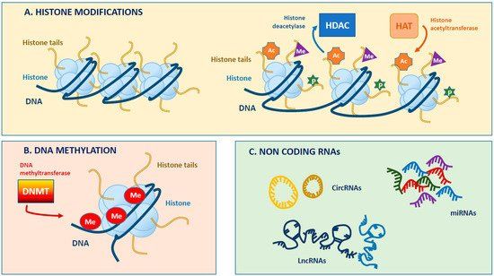
the genes. There are many epigenetic modifications, such as DNA methylation, histone
modifications and noncoding RNA (Figure 2). During mammalian heart development, cardiac
cell specification is regulated by specific transcription factors (TFs) at an epigenetic level
(Martinez et al., 2015). Overexpression of the Tet3 gene that is an epigenetic factor to regulate
DNA methylation can induce the expression of the ectoderm marker gene Fgf5 and can repress
the expression of the cardiac lineage marker genes Myh6, Nkx2-5, Tnnt2 and Myh7 (Li et al.,
2016). The function of Tet3 is a key factor that can direct mESCs toward cardiac mesoderm
cell fate. In Tet2 conditional knock-out mice, neonatal cardiomyocytes can proliferate.
However, compared to wild-type cells, Tet2 knockout cells showed less BrdU and pH3 staining,
indicating their lower proliferative capabilities. Furthermore, many cell cycle-related genes
were downregulated in the Tet2 knockout cells (Greco et al., 2016). Kabuki syndrome is caused
by mutation in the H3K4 methyltransferase Kmt2d. Sixty percent of patients with Kabuki
syndrome are diagnosed as CHDs, suggesting a critical role for Kmt2d in heart development
(Matsumoto and Niikawa, 2003; Adam and Hudgins, 2005; Yuan, 2013; Ang et al., 2016).
Jarid2 is a member of the Jmj family that functions as a histone lysine demethylase. Some
studies have shown that Jarid2 knockout mice have cardiac defects (Barth et al., 2010;
Mysliwiec et al., 2011).
4Figure 2: Schematic illustration of epigenetic modifications. Taken from Coco, et al. International journal of
molecular sciences (2019) (Coco et al., 2019).
1.5 Long Noncoding RNA in cardiac development and disease
Long noncoding RNA (lncRNA) is longer than 200 nucleotides and not translated into proteins,
as contrasted with messenger RNA (Wilusz et al., 2009; Kung et al., 2013). Multiple studies
have shown that lncRNAs have important roles during mammalian heart development. The
lncRNA Braveheart is required for cardiovascular lineage commitment and progression in a
report investigating mESC differentiation (Klattenhoff et al., 2013). In this paper, they showed
that Braveheart lncRNA is necessary to activate the Mesp1 gene. They also showed that
Braveheart lncRNA interacts with a zinc finger gene SUZ12 and mediates the epigenetic
regulation of cardiovascular lineage commitment (Klattenhoff et al., 2013). Another human-
specific lncRNA, Heart Brake lncRNA 1 (HBL1), is reported to regulate cardiomyocyte
differentiation in vitro (Liu et al., 2017). HBL1 interacts with MIR1 in an AGO2 complex. A
previous study has showed that overexpression of HBL1 did repress cardiomyocyte
differentiation from human inducible pluripotent stem cells (hiPSCs), while knockdown and
knockout of HBL1 could increase efficiency of cardiomyocyte differentiation from hiPSCs (Liu
et al., 2017). In mice, deletion of lncRNA Hand2os1 causes congenital heart defects (Han et al.,
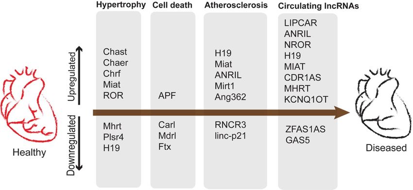
2019). In general, lncRNAs can serve as biomarkers of heart diseases (Figure 3). Patients with
heart failure have high expression levels of the lncRNA LIPCAR (Kumarswamy et al., 2014)
and low expression levels of the lncRNA GAS5 in the blood plasma (Yin et al., 2017). Likewise,
5patients with acute myocardial infarction (AMI) have high expression levels of lncRNA
CDR1AS and low expression levels of the lncRNA ZFAS1. (Zhang et al., 2016).
Figure 3: Potential lncRNA biomarkers in human heart diseases. Taken from Sweta, et al. Frontiers in cell and
developmental biology (2019), (Sweta et al., 2019).
1.6 Cardiogenic signaling pathways in cardiogenesis and congenital heart disease
1.6.1 Wnt signaling pathway
The Wnt family is divided into two major classes based on the primary functions of the
members, including 19 types of Wnt proteins and 10 types of Frizzled receptors (MacDonald
et al., 2009). Early in vitro differentiation studies showed that Wnt/β-catenin signaling
promotes mesoderm and endoderm formation (Lindsley et al., 2006; Ueno et al., 2007).
Overexpression of Wnt11 did promote cardiac lineage specification and differentiation in the
mESC differentiation system (Ueno et al., 2007). Furthermore, in cardiac organoid cells, the
addition of Wnt3A protein did increase expression of the SHF marker genes but decrease
expression of the FHF marker genes (Andersen et al., 2018). In an in vivo study, β-catenin
knockout mice showed a phenotype of right ventricular and outflow tract hypoplasia, but
overexpression of β-catenin led to right ventricular enlargement and hyperplasia (Ai et al., 2007;
Qyang et al., 2007). Wnt11 and Wnt5a knockout mice exhibited outflow tract defects and
impaired pharyngeal artery patterning, respectively (Pandur et al., 2002; Palpant et al., 2007).
1.6.2 TGF-β superfamily signaling pathway
6The signaling pathways involving the transforming growth factor-β (TGF-β) superfamily play
an important role in the regulation of cell growth, differentiation, and development. There are
more than 30 growth factors in the TGF-β superfamily, including bone morphogenetic proteins
(BMPs), TGF-βs, activin and nodal (Gu and Feng, 2018). Mice with conditional knockout of
the Bmp4 gene or Bmpr1a gene in the heart showed abnormal cardiac morphogenesis(Jiao et
al., 2003; Liu et al., 2004). Interestingly, the scRNA-seq analysis study showed that Bmp4
specifically labeled the murine cardiomyocyte population at E7.25 (Lescroart et al., 2018b).
The genome-wide CRISPR/Cas9 screening study showed that deletion of the Smad4 gene in
human ESCs (hESCs) caused a complete loss of cardiomyocyte induction (Xu et al., 2019).
Several cases of mutation in the TGF-β superfamily genes were detected in CHD patients.
(Massagué et al., 2000; Gordon and Blobe, 2008). When the TGF-β2 gene was knocked out in
mice, the mice showed significant morphological disorders in the heart, including the double-
outlet right ventricle, double-inlet left ventricle and ventricular septal defect (Sanford et al.,
1997; Molin et al., 2002). Loeys-Dietz syndrome is caused by mutations in the TGF-β gene.
The disorder is characterized by the aortic aneurysms that are often caused in children. The
weakened layers of the aortic wall in this syndrome can undergo a sudden dissection and rapture.
There are five types of this syndrome, labeled types I through V, which are distinguished by
their genetic subtypes. Type 1 to Type 5 are caused by mutations in TGFBR1, TGFBR2,
SMAD3, TGFB2, and TGFB3, respectively (Loeys et al., 2005; Van De Laar et al., 2011;
Bertoli-Avella et al., 2015).
1.6.3 FGF signaling pathway
There are more than 20 ligands and 4 transmembrane receptor tyrosine kinases in the fibroblast
growth factors (FGFs) signaling pathway (Itoh and Ornitz, 2004; Bertoli-Avella et al., 2015).
Many studies have shown that this pathway also plays important roles in heart development
and disease. Fgf8 knockout mice lacked mesoderm-derived structures and died at the
gastrulation stage (Sun et al., 1999). Conditional knockout of the Fgf8 gene using Tbx1-Cre
knockin mice led to impaired outflow tract morphologies (Frank et al., 2002). Fgf9 knockout
cardiomyocytes (CMs) lost their proliferative capability (Lavine et al., 2005). Overexpression
of the Fgf10 gene promotes mouse CM proliferation. However, when the Fgf10 gene was
7knocked out in mice, the RV of the embryonic heart showed an abnormal morphology (Rochais
et al., 2014).
1.6.4 Retinoic acid signaling pathway
The retinoic acid (RA) signaling pathway also plays an important role in heart development.
Vitamin A (retinol) is metabolized and altered to RA. The cargo protein, retinol binding protein
transports retinol into the target cells through a membrane protein STRA6 (Lara-Ramírez et al.,
2013) . In humans, the STRA6 gene mutation causes Matthew-Wood syndrome, which has
malformation in various organs, including heart (e.g., atrial and ventricular septal defects), eye
(anophthalmia), lung, and diaphragm (Golzio et al., 2007). When retinol enters into the
cytoplasm of cells, the retinaldehyde dehydrogenase 2 (Raldh2; Aldh1a2) enzyme converts it
to RA (Lara-Ramírez et al., 2013; Nakajima, 2019). Importantly, the RA signaling is associated
with the patterning and proliferation of the SHF and its derivatives (Sirbu et al., 2008;
Stefanovic et al., 2021a). In mice, the Raldh2 knockout embryos showed left-right looping
malformation at E9.5 with downregulated expression of Tbx1 and Fgf8/10 (SHF marker genes)
and died at E10.5 (Niederreither et al., 1999, 2001), indicating that the RA signaling is
indispensable for normal cardiac OFT development. The previous studies have also shown that
both the overactivated and decreased RA signaling could cause OFT defects in mice,
respectively (Sakabe et al., 2012; Rydeen and Waxman, 2016). Further, interestingly, the
Keller’s group showed that the enhanced RA signals could transfer the phenotype of ventricular
CMs to that of atrial CMs (Lee et al., 2017). In addition, the Hu’ group showed that RA could
promote the maturation of hESCs-derived CMs (Miao et al., 2020).
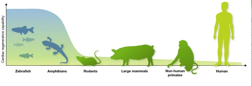
1.7 Heart regeneration
A fundamental biological question is whether the heart organ can regenerate by itself. For
organisms, regenerative capabilities are relatively a common feature. For example, plants have
the ability to regenerate fully in general. After leaves of the most plants fall down, newly
generated leaves can grow up again. Many plants can regenerate themselves after their roots,
branches, flowers or other parts are damaged even seriously. Among animals, lower animals
such as flatworms, annelids, and echinoderms have strong regenerative capabilities, by which
any part of their bodies can be regenerated in principle (Wagner et al., 2011; Zhao et al., 2016).
Amphibians and reptiles are slightly higher, taxonomically speaking, and their regenerative
abilities are not as strong as ones of the above lower animals (Zhao et al., 2016; Alibardi, 2018).
8Only one or some parts of the body can be regenerated in amphibians and reptiles. Even higher
animals such as birds and mammals do have much less ability to regenerate their organs and/or
bodies (Figure 4). However, the reasons explaining these limited regenerative capabilities in
higher animals are still unclear. In general, in the long and complicated trajectories of biological
evolution, the higher the taxonomic level of the animal is, the lower the regenerative ability is,
suggesting that the regenerative ability in animals has been gradually eliminated (Zhao et al.,
2016).
Figure 4: Schematic representation indicating the cardiac regenerative capabilities of various model organisms.
Taken from Sahara Makoto, et al. The EMBO Journal (2015) (Sahara et al., 2015).
The hearts have the least regenerative capability among tissues and organs, especially in
mammals, and therefore, a bunch of studies have been and is still focusing on heart regeneration
(Laflamme and Murry, 2011). The hearts in several fish and newt species have the abilities to
completely regenerate themselves after injury (Becker et al., 1974; Poss et al., 2002). A
previous study showed that in zebrafish, the earlier cardiac progenitor cells were the main
source of newly generated cardiomyocytes after injury (Lepilina et al., 2006). However, in 2010,
two groups using the Cmlc2-CreER transgenic zebrafish have shown that pre-existing
cardiomyocytes were the main source for cardiomyocyte regeneration after injury (Jopling et
al., 2010; Kikuchi et al., 2010). Newts are also good models for investigating organ regeneration,
including heart regeneration. Notably, it has been reported that even after ablation of up to 20%
of gross volume in newt’s hearts, they could regenerate completely (Godwin et al., 2017).
Godwin’s group also reported that after removing macrophage cells, the newt’s hearts could
not regenerate after injury any more (Godwin et al., 2017). Unlike zebrafish and newts, the
hearts of adult mammals such as humans, primates and rodents have very limited capabilities
to regenerate after injury (Sadek and Olson, 2020). In mice, the neonatal hearts have
regenerative potential, but this ability is rapidly lost by postnatal day 7 (P7) (Porrello et al.,
92011). With RNA and chromatin sequencing analyses of murine neonatal hearts, the Olson’s
laboratory showed that the epicardial regulator Rspo1 plays an important role during murine
neonatal heart regeneration through angiogenesis (Wang et al., 2020). Interestingly, another
laboratory showed that when the Rspo1’s receptor Lrp6 was knocked down, murine neonatal
cardiomyocytes could proliferate again until the juvenile stage (P7 to P14) (Wu et al., 2020).
Separately, Bergman et al. showed that only 0.5%-1% of human cardiomyocytes renew
annually (Bergmann et al., 2009). The Deepak’s laboratory showed that overexpression of 4
cell cycle-related genes (CDK1, CDK4, cyclin B1, and cyclin D1) could efficiently induce
cardiomyocyte proliferation in mice, rats, and humans in vitro and in mice in vivo (Mohamed
et al., 2018).
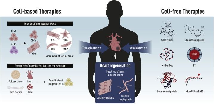
1.8 Novel therapeutic strategies for heart disease in humans
Heart disease is a leading cause of mortality in the developed countries with insufficient
therapeutic options and poor prognosis (Machaj et al., 2019). In recent years, many of novel
treatment strategies for heart disease in humans have been developed and reported. In general,
these strategies can be divided into the two classes, cell-based and cell-free therapies (Figure
5).
Figure 5: Schematic of novel therapeutic strategies for heart disease. Taken from Witman, Nevin, et al. Seminars
in cell & developmental biology (2020), (Witman et al., 2020).
10In 2014, the Murry’s laboratory showed that transplantation approach using clinically scalable
hESC-derived cardiomyocytes (CMs) was feasible (Chong et al., 2014). In fact, they
transplanted 1 billion of hESC-CMs into the hearts of non-human primates (NHPs) after
myocardial ischemia (MI) injury. The transplanted cells gave rise to muscle grafts within 2
weeks. In addition, these transplanted cells showed regular calcium transients, indicating they
were working functionally in the recipient’s hearts. Later, the Murry’s laboratory also showed
that the transplanted hESC-CMs could improve heart function after MI injury in NHP models
(Liu et al., 2018). To address the problem of immune rejection after chimeric transplantation,
another team from Japan established iPSC lines from MHC haplotype (HT4) homozygous
primates. Then, they transplanted this genetically modified primate-derived iPSC-CMs into the
injured heart in primates. They found that no immune rejection was caused after transplantation,
and the transplanted cells could significantly improve the damaged heart’s function (Shiba et
al., 2016). Similar to the previous studies, the Chien’s laboratory generated and transplanted an
hESC-derived Isl1+ cardiac progenitor cell (CPC) population in murine normal and injured
hearts (Foo et al., 2018). These cells could assemble into functionally mature ventricular muscle
grafts. In 2018, a group from France reported that they transplanted hESC-derived
CD15+Isl1+ CPCs into the hearts of patients with severe heart failure (Menasché et al., 2018).
This was the first clinical trial using the hESC-derived CPCs for transplantation against heart
disease in humans. Another well-known CPCs are the c-kit+ CPCs (Zhou and Wu, 2018). Since
2001, the Anversa’s and Bolli’s teams reported that transplanted c-kit+ cells harvested from
bone marrow rescued damaged hearts and improved heart function in patients with ischemic
heart disease, undergoing coronary artery bypass grafting (Orlic et al., 2001). However, many
other research groups claimed they could not reproduce the findings reported by the Anversa’s
laboratory. Ultimately, the Brigham and Women’s Hospital announced that 31 papers from the
Anversa’s laboratory would be retracted (Chien et al., 2019).
The other novel treatment strategy for heart disease is a cell-free approach. In 2014, the
Mercola’s laboratory screened a microRNA library and found miR25 as an anti-heart protective
factor (Wahlquist et al., 2014). In 2019, the Giacca’s laboratory from Italy reported that
overexpression of human microRNA-199a through an adeno-associated viral vector (AAV) in
porcine infarcted hearts could stimulate cardiomyocyte proliferation (Gabisonia et al., 2019).
More recently, modified mRNA (modRNA) technology has been widely used for gene transfer
(Warren et al., 2010; Mandal and Rossi, 2013; Sultana et al., 2017). Two groups showed that
injection of modRNA of vascular endothelial growth factor A (VEGFA) could improve cardiac
function in injured hearts of mice and pigs (Zangi et al., 2013; Carlsson et al., 2018).
111.9 Outflow tract formation and diseases
Cardiac outflow tract (OFT) defects are the most frequent malformations among CHDs. The
embryonic OFT is a transient structure that gives rise to the aorta and pulmonary arteries (Verzi
et al., 2005; Stefanovic et al., 2021b). Three cell lineages can contribute to the OFT: myocardial
progenitors, endocardial cushions and cardiac neural crest cells (Stefanovic et al., 2021b).
Abnormal OFT development causes CHDs, such as persistent truncus arteriosus (PTA), double
outlet right ventricle (DORV), overriding aorta (OA), ventricular septal defect (VSD), and
Tetralogy of Fallot (ToF) as below.
1.9.1 Persistent truncus arteriosus (PTA)
Persistent truncus arteriosus (PTA) is also called truncus arteriosus. In this disorder, during
heart development, the pulmonary artery and the aorta fail to properly divide(Barata, 2013).
Genetic mutations and many environmental factors can cause this disease phenotype.
1.9.2 Double outlet right ventricle (DORV)
Normally, the aorta is connected to the LV, and the pulmonary trunk is connected to the RV.
However, in DORV patients, both the aorta and pulmonary trunk are connected to the RV
(Anderson et al., 2001).
1.9.3 Overriding aorta (OA)
If the aorta is positioned directly over a ventricular septal defect (VSD) but not over the LV,
then some blood from the RV flows into the aorta (Apitz et al., 2009). We call this type of CHD
as overriding aorta (OA).
1.9.4 Ventricular septal defect (VSD)
12Ventricular septal defect (VSD) is a common heart defect. In these patients, there is a hole in
the septum of the ventricles, which allows the blood to flow from the LV to the RV (Penny and
Vick, 2011). Usually, this defect is usually repaired surgically before 1 year old.
1.9.5 Tetralogy of Fallot (ToF)
Tetralogy of Fallot (ToF) is the most common CHD. As suggested by the name, patients have
four congenital abnormalities at the same time: (1) RV outflow tract obstruction (RVOTO); (2)
VSD; (3) OA and (4) RV hypertrophy (Apitz et al., 2009).
1.10 Single-cell RNA and chromatin sequencing technology
Recent advanced biotechnologies such as RNA and chromatin sequencing at the single-cell
levels using next-generation sequencers (NGSs) have opened a new high-throughput way to
deconstruct cellular heterogeneity in vitro and in vivo and to identify cellular hierarchies and
molecular signatures in various types of mixed cell populations, organoids, tissues, and organs
(Picelli et al., 2013; Treutlein et al., 2014b; Petropoulos et al., 2016).
1.10.1 Single-cell RNA sequencing (scRNA-seq):
Through the single-cell RNA sequencing (scRNA-seq) technology, we can obtain and analyze
the transcriptome data representing gene expression patterns in various biological samples,
including developmental materials. Then, we can identify which genes are upregulated or
downregulated in any of cell types and when those happen. Combined with the genes’ loss-of-
function or gain-of-function settings, it is possible to further gain deep insights into the
transcriptional regulation and dynamisms in biological processes through performing clustering
analysis, differential expression genes’ analysis, cell trajectory analysis, etc (Townes et al.,
2019; van den Berge et al., 2020). Compared to the conventional population RNA-seq analysis
using bulk materials, the scRNA-seq analysis can help obtaining cellular and molecular atlases
at much higher resolution (Saliba et al., 2014). The first study using the scRNA-seq approach
was from the Surani’s laboratory at Cambridge University, and they used this method to analyze
the transcriptome of single murine blastomeres (Tang et al., 2009). Among heart studies, the
first study using the scRNA-seq method revealed differences between in vitro and in vivo
13cardiac development in 2015 (Kokkinopoulos et al., 2015). More recently, the scRNA-seq
approach using murine and human developing hearts has provided novel insights on
cardiogenesis at higher resolution than before, and identified previously unrecognized heart
progenitors and/or molecules that would spatiotemporally play certain roles in cardiac
development (DeLaughter et al., 2016; Cui et al., 2019; Sahara et al., 2019).
1.10.2 ATAC sequencing (ATAC-seq):
The activation or inactivation of a gene is controlled by epigenetic factors, such as DNA
modification, transcription factors (TFs)’ binding, and noncoding RNAs (Nakao, 2001). In gene
enhancer and promoter regions, there are commonly many TF binding sites. These sites are
critical for development. Usually, these binding sites are closed. When TFs trigger expression
of some of the target genes, TFs bind to the opened promoter region of the target genes.
Therefore, the epigenetic analysis to identify the state (opened or closed) of these binding sites
by sequencing the opened genome chromatin fragments with NGSs would help to determine
whether and when the TFs can trigger know and/or unknown target genes (Zambelli et al.,
2013). Among several chromatin sequencing technologies, recently, the assay for transposase-
accessible chromatin sequencing (ATAC-seq) approach is relatively widely used to
investigate the genome-wide chromatin accessibility status (Buenrostro et al., 2013a). The
first paper to use this technique was published in 2013 (Buenrostro et al., 2013b). The principle
of the ATAC-seq is to use the Tn5 enzyme to cut the open chromatin sites and add sequencing
primers to the ends of the fragments. Then, these DNA fragments can be sequenced using NGSs
(Figure 10). Currently, using the 10x Genomics Chromium platform
(http://support.10xgenomics.com/single-cell-atac), the single-cell ATAC-seq analysis can be
performed (Cusanovich et al., 2018; Stuart et al., 2019).
14Figure 6: The workflow of ATAC-seq. Created by BioRender.
1.11 Murine lineage tracing approach
The genetic lineage tracing is a classical technology to explore the function of a gene in
developmental biology. It applies the Cre-LoxP, Dre-RoxP and Flpe-FRT systems, among
which the Cre-LoxP system is the much most widely used (Anastassiadis et al., 2009; He et al.,
2017). Murine genetic lineage tracing experiments require two genetically modified mouse
lines. One of them carries the Cre gene whose expression is driven by a promoter region of the
gene of interest. Cre is a 38kD recombinase from the P1 bacteriophage (Sauer and Henderson,
1990). The other is the reporter line, which has a LoxP-flanked STOP cassette preventing
transcription of a CAG promoter-driven fluorescent protein (Muzumdar et al., 2007; Sousa et
al., 2009; Madisen et al., 2010). The Cre recombinase catalyzes and cuts the LoxP-flanked sites
out of the gene. After removal of the LoxP-flanked STOP cassette by the Cre-mediated
recombination, the reporter fluorescence starts to be expressed and then used as a tracer of the
specific cell lineage. In recent years, the Bin’s group has developed a new genetic lineage
tracing system using dual recombinases, such as Cre and Dre (He et al., 2017, 2021; Han et al.,
2021). This new system appears to improve the precision of the conventional Cre–LoxP-
mediated lineage tracing and to be able to trace three different cell populations at the same time
(Figure 7) (Liu et al., 2020).
15Figure 7: Schematic illustration of the results after the Dre-RoxP and Cre-LoxP dual recombination. Taken from
Liu, et al. Journal of Biological Chemistry (2020).
162. METHODOLOGY
2.1 Human embryonic stem cell (hESC) culture
hESCs are derived from the inner cell mass (ICM) of human blastocysts. They have two
properties. One is that they can maintain their self-renewal capabilities, and the other is that
they can differentiate into a diverse set of cell types comprising the body in vitro and in vivo.
Therefore, they serve as a promising tool in cell therapy, tissue engineering and other basic
research. In 1998, the Thomson’s laboratory established the first hESC line(Thomson, 1998),
and thereafter, many of the hESC lines have been generated by various laboratories(Guhr et al.,
2006). The traditional protocols for hESC culture involve an inactivated mouse embryonic
fibroblast (MEF) feeder or human feeder layer-conditioned medium. However, many groups
have established defined, feeder-free medium for hESC culture (Xu et al., 2001; Ludwig et al.,
2006a, 2006b). Briefly, hESCs are cultured on Matrigel-coated plates in mTeSR (StemCell
technologies) or E8 medium (Thermo Fisher). Once the undifferentiated hESCs exhibit 70%-
80% confluence on the plates, the cells are dissociated using Accutase (Thermo Fisher) and
passaged onto new plates with fresh media containing a ROCK inhibitor (Tocris). The medium
is changed every day.
In this thesis, we used two hESC lines, ES03 and H9. Both of the two lines are derived
from human female blastocysts and purchased from WiCell (USA). The use of the H9 line is
approved by the NIH, while the ES03 is not approved by the NIH. The H9 cells were cultured
on Matrigel-coated plates in mTeSR medium, while the ES03 cells were cultured on
Vitronectin-coated plates in E8 medium.
2.2 Cardiomyocyte differentiation in vitro
Human pluripotent stem cells (hPSCs), such as hESCs and hiPSCs, are widely used in
biomedical research and are a good model for the in vitro cardiogenesis research. There are two
major protocols for cardiac differentiation: embryonic body (EB)-based methods (Kehat et al.,
2001) and chemically induced two-dimensional methods (Lian et al., 2012, 2013). Although
the EB-based approaches were first developed, it appears to be difficult to reproduce the results
using this method, which usually exhibits low efficiency for induction of cardiac cells (Osafune
et al., 2008). Therefore, the EB-based approaches may not be suitable for the purpose of
17generating clinically scalable cardiac cells in clinical research. In recent years, an increasing
number of groups have used chemically induced two-dimensional approaches for cardiac
differentiation (Figure 8). This protocol involves two important chemicals: CHIR99021, a Wnt
signaling enhancer by inhibiting glycogen synthase kinase 3b (GSK3b); and IWP2, a Wnt
inhibitor. On the first day (day 0) of differentiation, we add 12 µM of CHIR99021 in medium.
Then, the cells begin to express early mesodermal transcription factors Brachyury on day 1 and
MESP1 on day 3. CHIR99021 is removed after 24 h on day 1, while IWP2 is added in medium
on day 3 and kept for 48 h. Then, the cells express a cardiac progenitor marker ISL1 on day 6,
and cardiomyocyte markers NKX2-5 and TNNT2 on day 7 (Lian et al., 2013, 2015; Burridge
et al., 2014). Using this protocol, we can easily obtain a large number of beating cardiomyocytes
on day 10-12 onward. In this protocol, there are two major time points: day 3 (multipotent
cardiac progenitors) and day 6 (Isl1+ cardiac progenitors). Generally, the generated
cardiomyocytes are mixed populations. The main cells generated on day 10-12 onward are
ventricular cardiomyocytes, but atrial cardiomyocytes and endothelial cells are also present.
Cardiomyocytes and non-cardiomyocytes have different metabolic pathways, respectively
(Fisher et al., 1981; Werner and Sicard, 1987; Tohyama et al., 2013). To obtain the pure
ventricular cardiomyocytes, the Fukuda group used a metabolic selection method to purify
cardiomyocytes (Tohyama et al., 2013). After Day 14, this group replaced the cell culture media
into glucose minus and sodium DL-lactate plus PRMI media (Tohyama et al., 2013).
Figure 8: Schematic of the chemically induced cardiomyocyte differentiation protocol. Created by Biorender.
18In this thesis, we mainly used the GiWi protocol for in vitro cardiomyocyte differentiation
((Lian et al., 2012, 2013, 2015)) (Figure 8). In particular, we focused on the two time points,
day 3 and 6.
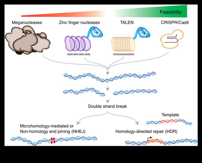
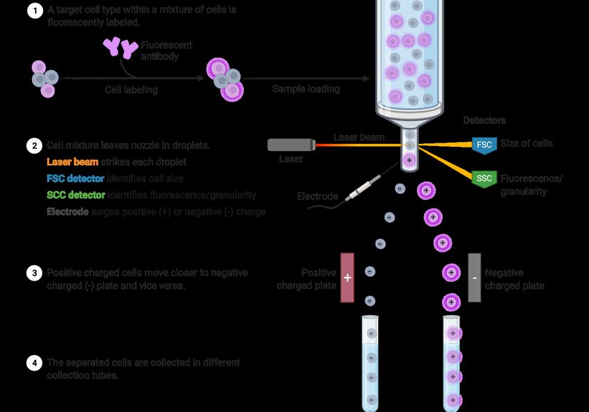
2.3 CRISPR/Cas9-mediated gene editing and screening
To date, there are four different gene editing tools: meganucleases, zinc-finger nucleases,
transcription activator-like effector nucleases and the CRISPR/Cas9 system (Figure 9) (Gordon
et al., 1980; Kim et al., 1996; Hockemeyer et al., 2011; Cong et al., 2013).
Figure 9: Schematic of the use of four genome editing tools for genome editing. Taken from Mazhar Adli. Nature
communications (2018) (Adli, 2018).
We can use these tools to generate the gene knockout and knock-in cells and animals for
cardiogenesis research. Especially with development of the CRISPR/Cas9 technology, we can
easily generate genetically modified cells and animals. CRISPR stands for The Clustered
Regularly Interspaced Short Palindromic Repeats. This system was first discovered in bacteria
(Bhaya et al., 2011; Wiedenheft et al., 2012). The system is based on a primitive immune system
19You can also read