Role of Cell Death in Cellular Processes During Odontogenesis - Frontiers
←
→
Page content transcription
If your browser does not render page correctly, please read the page content below
REVIEW
                                                                                                                                             published: 18 June 2021
                                                                                                                                      doi: 10.3389/fcell.2021.671475

                                            Role of Cell Death in Cellular
                                            Processes During Odontogenesis
                                            John Abramyan 1 , Poongodi Geetha-Loganathan 2 , Marie Šulcová 3,4 and
                                            Marcela Buchtová 3,4*
                                            1
                                             Department of Natural Sciences, University of Michigan–Dearborn, Dearborn, MI, United States, 2 Department of Biological
                                            Sciences, SUNY Oswego, Oswego, NY, United States, 3 Department of Experimental Biology, Faculty of Science, Masaryk
                                            University, Brno, Czechia, 4 Laboratory of Molecular Morphogenesis, Institute of Animal Physiology and Genetics, Czech
                                            Academy of Sciences, Brno, Czechia

                                            The development of a tooth germ in a precise size, shape, and position in the
                                            jaw, involves meticulous regulation of cell proliferation and cell death. Apoptosis, as
                                            the most common type of programmed cell death during embryonic development,
                                            plays a number of key roles during odontogenesis, ranging from the budding of
                                            the oral epithelium during tooth initiation, to later tooth germ morphogenesis and
                                            removal of enamel knot signaling center. Here, we summarize recent knowledge
                                            about the distribution and function of apoptotic cells during odontogenesis in several
                                            vertebrate lineages, with a special focus on amniotes (mammals and reptiles). We
                       Edited by:           discuss the regulatory roles that apoptosis plays on various cellular processes during
                     Nai Yang Fu,
          Duke-NUS Medical School,          odontogenesis. We also review apoptosis-associated molecular signaling during tooth
                        Singapore           development, including its relationship with the autophagic pathway. Lastly, we cover
                      Reviewed by:          apoptotic pathway disruption, and alterations in apoptotic cell distribution in transgenic
               Aaron R. H. LeBlanc,
       University of Alberta, Canada
                                            mouse models. These studies foster a deeper understanding how apoptotic cells affect
                       Xiao-Jing Zhu,       cellular processes during normal odontogenesis, and how they contribute to dental
  Hangzhou Normal University, China         disorders, which could lead to new avenues of treatment in the future.
                  *Correspondence:
                    Marcela Buchtová        Keywords: teeth, dental lamina, apoptosis, odontogenesis, morphogenesis
                    buchtova@iach.cz

                    Specialty section:     INTRODUCTION
         This article was submitted to
             Cell Growth and Division,     Over the past several decades, the contribution of apoptosis to vertebrate odontogenesis has
                a section of the journal   received considerable attention, promoted by technical advancement and increased availability of
  Frontiers in Cell and Developmental      diverse laboratory models (Nozue, 1971; Moe and Jessen, 1972; Kindaichi, 1980; Nishikawa and
                                Biology    Sasaki, 1995; Shibata et al., 1995; Lesot et al., 1996; Vaahtokari et al., 1996). More recent studies
        Received: 23 February 2021         have revealed that apoptosis is not just a silent mechanism of cell removal during embryonic
           Accepted: 24 May 2021           development. Rather, apoptotic cells produce numerous signaling molecules that affect the behavior
          Published: 18 June 2021
                                           of surrounding cells, inducing morphogenesis, cell migration, and alteration of cell fate. Here, we
                              Citation:    review these relationships and propose a broader contribution of apoptosis to the cellular processes
                          Abramyan J,      of odontogenesis than was previously thought. First, we briefly summarize the distribution of
Geetha-Loganathan P, Šulcová M and
                                           apoptotic cells during mammalian odontogenesis since mammals represent the most common
Buchtová M (2021) Role of Cell Death
         in Cellular Processes During
                                           models for the study of their localization, distribution, and function during odontogenesis. Next,
                      Odontogenesis.       we review the available literature on non-mammalian groups such as reptiles and fishes, which are
      Front. Cell Dev. Biol. 9:671475.     becoming increasingly common models for the study of odontogenesis due to their unique dental
     doi: 10.3389/fcell.2021.671475        characteristics. Subsequently, we discuss cellular processes directed by the effects of apoptosis on

Frontiers in Cell and Developmental Biology | www.frontiersin.org                 1                                             June 2021 | Volume 9 | Article 671475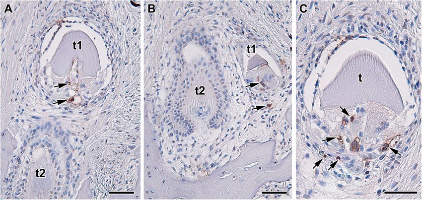
Abramyan et al.                                                                                                          Cell Death in Odontogenesis

surrounding non-apoptotic cells, as well as the indirect                    (Nair, 2010). In later developmental stages during root formation,
roles of apoptosis in individual steps of tooth development                 mesenchyme exhibits apoptosis linked with a proportion of the
and morphogenesis.                                                          Hertwig’s epithelial root sheath (HERS) cells, as was shown in
                                                                            the rat upper molar (Kaneko et al., 1999). Remaining sheath cells
                                                                            aggregated in the periodontal area and form the epithelial rests
DISTRIBUTION OF APOPTOTIC CELLS                                             of Malassez (ERM) (Hamamoto et al., 1989), which also undergo
DURING ODONTOGENESIS IN                                                     apoptosis later in development as part of a normal mechanism of
VERTEBRATES                                                                 turnover or remodeling (Cerri and Katchburian, 2005; Lee et al.,
                                                                            2012).
Tooth development is characterized by complex, reciprocal                      Apoptotic cells, identified as osteoclasts, are also located on
interactions between the stomodeal epithelium and the                       the surfaces of the developing alveolar bone around developing
underlying cranial neural crest-derived mesenchyme (Thesleff,               molars (Vaahtokari et al., 1996). Growing teeth have previously
2003; Balic, 2019). This interaction drives tooth morphogenesis,            been shown to be associated with osteoclast activity and
including differentiation of individual components at the                   resorption of the surrounding alveolar bone in embryonic mice
molecular level (Thesleff, 2003; Balic and Thesleff, 2015). In spite        (Reponen et al., 1994), or the remodeling of bone due to
of differences in final size and shape, teeth undergo consecutive           compressive forces produced by the occlusion and eruption of
developmental stages common to all vertebrates, including                   ever-growing rodent incisor (Irie and Ozawa, 1990). Apoptosis
epithelial thickening, bud, cap, and bell stages (Thesleff, 2003).          may serve to remove these osteoclasts after they have completed
The distribution of apoptotic cells during odontogenesis strongly           their function of creating space/facilitating interaction between
correlates with specific morphogenetic events and associated                the tooth and surrounding bone. In support of this theory,
tissues. For example, apoptosis is usually confined to epithelial           the elimination of compressive forces by the incisors has been
cells undergoing folding, while very few dying cells are found in           found to lead to the inactivation of osteoclasts (Irie and Ozawa,
the surrounding mesenchyme. We first review the localization of             1990). Otherwise, there are surprisingly few apoptotic cells
apoptotic cells in mammalian dentition, focusing on the mouse               located in the odontogenic mesenchyme, and those present are
as the most common model organism, and then compare their                   without any discernable pattern (Stock et al., 1997). Why are
distribution to non-mammalian groups.                                       there so few apoptotic cells in the mesenchyme? One possible
                                                                            explanation is the considerable plasticity of the mesenchyme
Mammalia                                                                    during odontogenesis, where cells can be easily relocated in the
At early developmental stages, apoptotic cells are located in               loose tissue architecture without the necessity of eliminating cells
the budding epithelium of the molars, specifically in the cells             through death. Moreover, cells spread throughout the dental
facing the oral cavity (Peterková et al., 1998). After epithelial           papilla and dental follicle, with cell signaling forming gradients
invagination, a streak of apoptotic cells extends to the tip of the         without any local concentration or the presence of distinct
developing molar bud (Nair, 2010). At the cap stage, clusters               signaling centers.
of apoptotic cells in the inner enamel epithelium localize to the              It should be noted that in rodents, the above-described
primary enamel knots (PEKs), as was shown by studies in the                 patterns only apply to molar development, with the incisors
murine molar (Lesot et al., 1996; Jernvall et al., 1998). Later, at         exhibiting a different pattern of apoptosis. In mice, the epithelial
the bell stage of molar development, apoptosis can be detected              thickenings that initiate incisor development demonstrate low
in the secondary enamel knots (SEKs) and surrounding cells,                 numbers of apoptotic cells (Kieffer et al., 1999). Later, apoptotic
including the stratum intermedium and adjacent mesenchyme                   cells can be found in the superficial part of the dental lamina
(Vaahtokari et al., 1996). After the disappearance of the enamel            connecting the enamel organ with the oral epithelium (Lesot
knots, apoptotic cells appear in the superficial part of the dental         et al., 1996; Kieffer et al., 1999), as well as in the inner
lamina (Lungová et al., 2011), which develops in mouse as                   dental epithelium close to the epithelio-mesenchymal junction
just a short epithelial connection between the tooth germ and               at the future incisor ridge (Kieffer et al., 1999). Interestingly,
the oral epithelium, sometimes called the dental stalk or the               an accumulation of apoptotic cells is also found in the
gubernaculum (Dosedelova et al., 2015; Chaudhry and Sobti,                  mesenchyme surrounding the labial cervical loop, in the area
2020). After the molar is fully formed, apoptotic cells are also            where development of the cervical loop was more pronounced
involved in the tooth eruption stage, exhibiting concentrations             compared to the lingual side (Kieffer et al., 1999).
in the oral epithelium above the erupting teeth as well as the
superficial part of the dental lamina (Moriguchi et al., 2010;              Reptilia
Dosedelova et al., 2015; Figure 1). Apoptotic cells are also situated       The presence of apoptotic cells in the context of tooth
in the anterior-most portion of the first molar epithelium (Lesot           development has been previously described in a several species
et al., 1996), which in rodents abuts an edentulous diastema.               of crocodilians and squamates (snakes and lizards). A key reason
Interestingly, the mandibular diastema is devoid of apoptosis,              for the use of reptiles as models for odontogenesis is the fact
whereas the maxilla displays apoptotic signal in the diastema               that most species are polyphyodont and exhibit lifelong tooth
region, attributed to the presence of transitory tooth buds in              replacement, with a smaller subset being monophyodont and
the maxillary diastema (Vaahtokari et al., 1996). At the bud                developing a single generation of teeth that fuse to the jaws
stage, there are no apoptotic cells seen in the mesenchyme                  and are never replaced (Edmund, 1960; DeMar, 1972). During

Frontiers in Cell and Developmental Biology | www.frontiersin.org       2                                       June 2021 | Volume 9 | Article 671475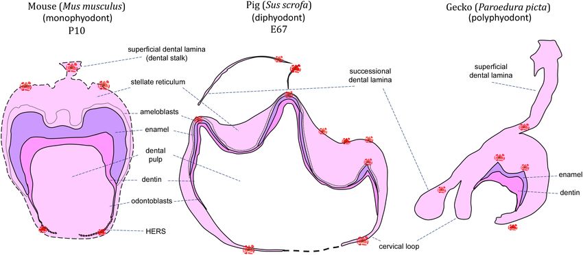
Abramyan et al.                                                                                                                             Cell Death in Odontogenesis

  FIGURE 1 | Schematic of apoptotic cells distribution through mineralization stage of the tooth germ. Clusters of apoptotic bodies are labeled in red and label areas
  of possible future interest in monophyodont mouse (A), diphyodont pig (B), and polyphyodont gecko (C).

reptile odontogenesis, apoptosis is associated with the enamel                          of superficial (vestigial) teeth (Handrigan and Richman, 2010).
knot region (homologous to the mammalian enamel knots)                                  When the developing tooth germ was exposed to cyclopamine,
at early developmental stages, and the formation of complex                             a proven SHH inhibitor, an increased number of apoptotic cells
tooth morphology at later mineralization stages. Additionally,                          appeared in the stellate reticulum (Handrigan and Richman,
apoptosis may be found during successional dental lamina                                2010) using a TUNEL (Terminal deoxynucleotidyl transferase
development/disruption, egg tooth formation or venom canal                              dUTP nick end labeling) assay, confirming the role of SHH in
formation in viperid snakes (Figures 1, 2).                                             cell maintenance. TUNEL assays are one of the most common
    At early – cap and bell – stages, apoptotic bodies were detected                    methods for the detection of apoptotic cells by targeting DNA
in the enamel organ of several reptile species including veiled                         fragmentation during programmed cell death (Gavrieli et al.,
chameleon (Chamaeleo calyptratus), ocelot gecko (Paroedura                              1992). Nevertheless, the possibility of toxicity from cyclopamine
picta), bearded dragon (Pogona vitticeps), African rock python                          should be considered in this case. In the African rock python,
(Python sebae), and Siamese crocodile (Crocodylus siamensis)                            apoptosis was also detected during the early stages of tooth
(Buchtová et al., 2007; Handrigan and Richman, 2010; Landova                            development, localized in the stellate reticulum of the enamel
Sulcova et al., 2020). The veiled chameleon possesses heterodont                        organ. However, an obvious morphological appearance of enamel
teeth, with the rostral-most teeth being nearly conical with one                        knot-like structure was not observed in this species (Buchtová
rounded tip, whereas more caudal teeth are multiple-cusped, with                        et al., 2007). The absence of a morphologically distinct enamel
a dominant central cusp flanked by accessory cusps (Landova                             knot is usually associated with the development of a very small
Sulcova et al., 2020). Additionally, the central cusp is divided into                   enamel organ and reduced stellate reticulum. However, the
labial and lingual crests, separated by a shallow groove. In the                        existence of an apoptotic cell cluster in the same area as observed
chameleon (Figure 2), the pattern of apoptotic cell distribution                        in other reptile species may indicate an identical function for
is similar to what was described in the mammalian tooth, with                           apoptosis during odontogenesis in python.
apoptotic cells located in an enamel knot-like cluster at the cap                          It is important to note that while some reptile species
stage (Landova Sulcova et al., 2020), revealing the existence of                        exhibit characteristics of mammalian enamel knots such as
a similar signaling center as was described in mammals. The                             reduced proliferation, specific expression of SHH, FGF, and
ocelot gecko is a homodont species that has small, peg-shaped                           BMP signaling molecules, and apoptosis (Jernvall et al., 1998),
teeth with labial and lingual enamel crests at the tips, similar                        many do not. For example, thickened dental epithelium has
to the central cusp of the chameleon tooth (Landova Sulcova                             previously been described for the American alligator (Alligator
et al., 2020). Developing gecko teeth also exhibit recognizable                         mississippiensis) (Westergaard and Ferguson, 1987), veiled
enamel knot-like structures with few apoptotic cells located in the                     chameleon (Buchtová et al., 2013) as well as the leopard
inner enamel epithelium or in the stellate reticulum just above it                      gecko (Eublepharis macularius) (Handrigan and Richman, 2011).
(Landova Sulcova et al., 2020). Later, at the bell stage, apoptotic                     Meanwhile histological studies of ball python (Python regius) and
cells are situated at the tip of the inner enamel epithelium where                      the bearded dragon revealed a distinct lack of any such tissue
morphogenesis occurs, and where the future cusp will form.                              swelling (Handrigan and Richman, 2011). Furthermore, within
    In bearded dragon, which has broad, triangular and single-                          those with an “enamel knot” homolog such as the chameleon and
cusped teeth (Handrigan and Richman, 2011), apoptosis was                               ocelot gecko, some of the classic enamel knot characteristics such
found in the dental papilla, odontoblasts, and pre-ameloblasts                          as reduced proliferation, SHH expression, and even apoptosis

Frontiers in Cell and Developmental Biology | www.frontiersin.org                   3                                              June 2021 | Volume 9 | Article 671475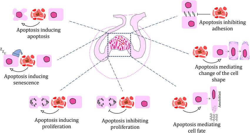
Abramyan et al. Cell Death in Odontogenesis
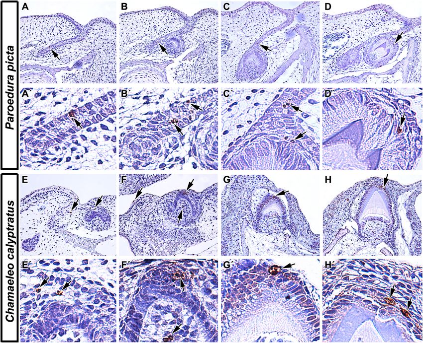
FIGURE 2 | Localization of apoptotic cells in squamate teeth. (A–D) Detection of apoptotic cells in developing tooth germs of Ocelot gecko (Paroedura picta).
(A,A0 ,B,B0 ) TUNEL-positive cells were found both in the epithelium of interdental dental lamina and in successional dental lamina, indicating their role not only in the
growth of tooth germs but also in continuous tooth replacement (black arrows). (C,C0 ) Apoptotic cells were also situated in the stellate reticulum of the developing
tooth and in the EK-like cluster of cells (black arrows). (D,D0 ) Later in development, TUNEL-positive cells were mostly situated above the enamel ridge (black arrow).
(E–H) Presence of TUNEL positive cells during different stages of odontogenesis in Veiled chameleon (Chamaeleo calyptratus). (E) Apoptotic cells appear in the
presumptive stellate reticulum of early cap stage tooth germ (E0 ) and in the labially situated, developing salivary glands. (F,F0 ) Later, clustering of TUNEL positive cells
takes place in the EK-like area of the cap stage tooth germ, with few apoptotic cells found in the adjacent mesenchyme (F). (G,G0 ,H,H0 ) Once odontogenesis
proceeds and hard tissue production has begun, there are a few TUNEL-positive cells located at the top of the forming ridge, delimiting the margins of developing
enamel grooves. TUNEL-positive cells (brown, DAB), TUNEL-negative cells (blue, Hematoxylin).
were observed (Landova Sulcova et al., 2020), while reptiles with of apoptotic cells depending on the length of time for dental
simple, unicuspid teeth such as snakes, exhibit no apoptosis in lamina persistence. In polyphyodont species, where an extensive
the inner enamel epithelium but a cluster of TUNEL-positive dental lamina connects several tooth generations, a successional
cells were located in the stellate reticulum just above (Buchtová lamina is retained as an extension of the dental lamina off the
et al., 2008). Therefore, there does appear to be a signaling center newest forming tooth and facilitates continuous replacement of
similar to the mammalian enamel knot in some reptile lineages, teeth (Whitlock and Richman, 2013). In diphyodont species, the
but its development seems to be associated with the general dental lamina degenerates during the initiation of the second
complexity of tooth crown morphology. tooth generation (Whitlock and Richman, 2013). In the veiled
An additional key difference of reptilian odontogenesis is chameleon, a monophyodont species, a successional lamina is
their enhanced tooth replacement. The fact that polyphyodont initiated during embryonic development but later regresses.
and monophyodont species have been studied, and that the Surprisingly, even the degenerating dental lamina exhibited very
retention of a successional lamina is the key difference between few apoptotic cells, with removal attributed to different cellular
the two groups, we can now obtain a better appreciation for mechanisms (Buchtová et al., 2013). In the bearded dragon,
how apoptosis separates the two categories of reptiles. Apoptotic another monophyodont species, apoptosis was located in the
cells have been identified in association with the dental lamina degrading dental lamina in association with decreased WNT
in reptiles regardless of tooth generation number. However, pathway activity (Richman and Handrigan, 2011). However, a
there are some differences in their distribution and amount subsequent study found just a few TUNEL-positive cells under
Frontiers in Cell and Developmental Biology | www.frontiersin.org 4 June 2021 | Volume 9 | Article 671475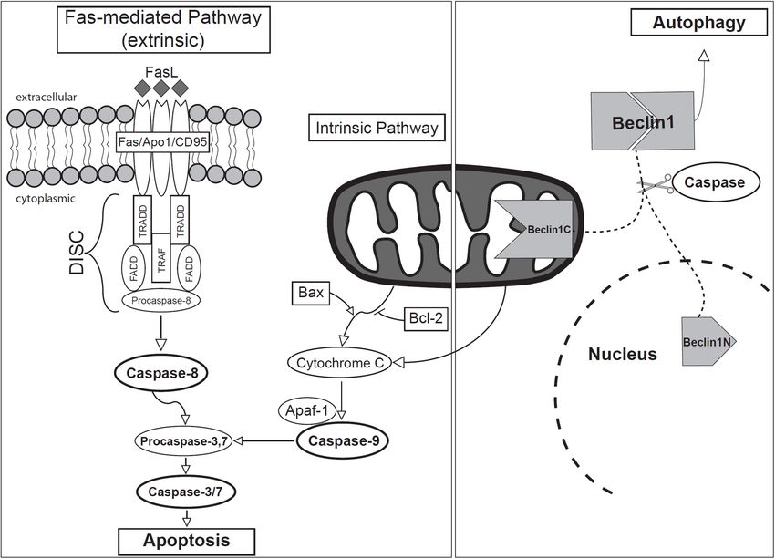
Abramyan et al.                                                                                                         Cell Death in Odontogenesis

normal physiological conditions, and those were restricted to              earlier study from the same group identified degeneration of the
the mesenchyme surrounding the successional dental lamina                  egg tooth enamel organ shortly before hatching also in the grass
(Salomies et al., 2019). Since lamina morphology differs along             snake (Natrix natrix) (Hermyt et al., 2017).
the jaw in this species, this discrepancy in apoptotic cell numbers            Several groups of reptiles develop embryonic (i.e., vestigial or
could be explained by local differences between morphology and             superficial) teeth (Westergaard and Ferguson, 1986; Handrigan
role of the dental lamina along the rostral-caudal axis of the jaw.        and Richman, 2010; Zahradnicek et al., 2012), the function of
In the polyphyodont ocelot gecko (Figure 2), a few TUNEL-                  which remains a mystery. Superficial teeth of the American
positive cells were located in the dental lamina but not in its            alligator (Alligator mississippiensis) develop from elevations along
tip, but rather in a more superficial area at the edge of the              the oral epithelium, almost perpendicular to the jaw surface,
enamel organ of the associated tooth (Landova Sulcova et al.,              before the formation of the dental lamina, and they are described
2020). In the African rock python, another polyphyodont species,           as being poorly differentiated and lacking enamel (Westergaard
numerous TUNEL-positive cells were found in the dental lamina              and Ferguson, 1986). As embryonic development progresses and
connecting the tooth to the oral epithelium, but once again not in         functional teeth start to form, the dental cells of the embryonic
the tip of the successional lamina (Buchtová et al., 2007).                teeth begin to degenerate; leaving a dentine matrix that is either
    In reptiles, later mineralization stages of odontogenesis reveal       resorbed or shed. Westergaard and Ferguson hypothesized that
apoptosis associated with the formation of an enamel groove. In            there may be “death factors” at play here, however, they do not
veiled chameleon and ocelot gecko (Figure 2), both of which                specifically show any evidence of apoptotic cells in their studies
exhibit enamel ridges at the tooth tip with two crests and a               (Westergaard and Ferguson, 1986). In the bearded dragon, on the
central groove, identical distribution of TUNEL-positive cells was         other hand, Handrigan and Richman performed TUNEL analysis
described. Once cell differentiation advanced and mineralization           and detected apoptosis in the dental papilla and pre-ameloblast
of enamel progressed, two distinct clusters of apoptotic bodies            cells as the vestigial tooth generation reached the cap stage of
were detected at the margins of the developing enamel grooves. In          development (Handrigan and Richman, 2010).
contrast, the relatively simple, conical teeth of Siamese crocodiles
possessed only one distinct apoptotic area at the very tip of the          Amphibia
single cusp (Landova Sulcova et al., 2020).                                Living amphibians are classified into three Orders: frogs and
    Apoptosis is also crucial for the formation of the venom               toads (Order: Anura), salamanders and newts (Order: Caudata),
canal in the fangs of viperid snakes. In the white-lipped pit              and caecilians (Order: Gymnophiona). Despite being one of the
viper (Trimeresurus albolabris), apoptotic cells are located in the        most charismatic groups of vertebrates, there is little in the way
central area of the first developing fang, where they contribute           of recent scientific literature focusing on the cellular processes
to cell removal in the formation of an empty venom transport               during their odontogenesis. Early studies indicate that some form
canal (Zahradnicek et al., 2008). Interestingly, apoptotic cells           of cell death does take place during amphibian odontogenesis,
were not found during the early stages of canal invagination               with reference to “autophagy,” “necrosis,” and “degeneration” of
and do not seem to be responsible for the loss of inner enamel             cells, some of which are likely to be apoptotic events that have not
epithelium in the shaft area prior to the cell removal process.            been recognized as such.
Besides concentration in the central canal, apoptotic cells are also          Anurans are polyphyodont and develop simple, conical,
situated in the tip of the fangs in two symmetrical lateral zones.         bicuspid teeth during metamorphosis (Gillette, 1955; Shaw,
It was proposed that these cells contribute to the clearance of            1979). Prior to odontogenesis, anuran larvae (tadpoles) possess
space for the later emergence of the tooth from the oral mucosa,           keratinized mouthparts that function in the place of true teeth
but the possible role in morphogenesis and cell arrangement in             and that are broken down through autolysis at metamorphosis
the inner enamel epithelium may also be a valid prediction here            (Kaung, 1975; Davit-Beal et al., 2007). While there is little
(Zahradnicek et al., 2008).                                                in the way of evidence for apoptosis during odontogenesis in
    Apoptosis was also identified in the egg tooth of the brown            anurans, several authors mention “degeneration” of cells at the
anole (Anolis sagrei) (Hermyt et al., 2020). Since most reptiles           tip of the enamel organ immediately prior to eruption (Shaw,
are oviparous, they have evolved “egg teeth” that assist in their          1979; Huysseune and Sire, 1998). In Xenopus laevis, an aquatic
hatching from the amniotic egg. In some groups, a caruncle                 species from the family Pipidae, odontoblasts at the tips of the
develops in the form of a modified scale (e.g., birds and turtles)         developing teeth were shown to flatten, change orientation, and
(Clark, 1961), while squamates develop a structure that is for             nuclei become pyknotic just when metamorphosis is complete
all intents and purposes, a tooth (Fons et al., 2020). In the              (Shaw, 1979); where pyknosis is now recognized as a telltale
brown anole, degeneration of the enamel organ was observed in              sign of apoptosis or necrosis in a cell (Burgoyne, 1999). The
the egg tooth shortly before its eruption and after subsequent             degenerative process was described as continuing into the basal
hatching, and this process was attributed to apoptosis based               part of the teeth until no active odontoblasts were visible
on morphological characteristics (Hermyt et al., 2020). Authors            (Shaw, 1979). In the leopard frog (Rana pipiens), a terrestrial
theorized that loosening of the intercellular junctions in that            species from the family Ranidae, Zaki, and MacRae identified
epithelium, as opposed to simply shedding it, would allow for              autophagic vacuoles in ameloblasts, concomitant with the loss
penetration of inflammatory cells and tissue exudate, making it            of some cells at the transitional stage of amelogenesis (Zaki
a first-line of defense against pathogenic infections, similar to          and MacRae, 1977). These vacuoles were observed to contain
the junction epithelium in mammals (Hermyt et al., 2020). An               debris of membranous organelles and were associated with

Frontiers in Cell and Developmental Biology | www.frontiersin.org      5                                       June 2021 | Volume 9 | Article 671475
Abramyan et al. Cell Death in Odontogenesis
lysosome-like structures, and therefore, were assumed to be not seem to contribute to dental papilla reduction and other
autophagic. In a later study comparing secretory and non- cellular mechanisms that cooperate to augment shape changes
secretory ameloblasts, they specify that the autophagic vacuoles after SHH inhibition.
were only found in secretory ameloblasts (Zaki and MacRae, In non-model fish, there is also little evidence for apoptosis
1978). However, the authors ultimately concluded that these during odontogenesis. In gar embryos, which develop single-
structures are involved in reorganization rather than replacement cusped, conical oral dentition, the expression of protein p63 was
of ameloblasts (Zaki and MacRae, 1977). detected in almost the entire dental epithelium (Rostampour
Salamanders and newts also undergo metamorphosis, albeit et al., 2019). Activation of p63 protein in so-called TA form
involving a less drastic morphological change than anurans. As (bearing a transcription activation domain) usually leads to
such, odontogenesis of relatively simple, bicuspid teeth begins transcription of genes resulting in cell cycle arrest or apoptosis
at larval stages, with a general transition from monocuspid (Little and Jochemsen, 2002). However, the association of p63-
to bicuspid teeth at metamorphosis (Davit-Beal et al., 2007). positive cells to apoptosis remains to be investigated.
Since all amphibians are polyphyodont (Tucker and Fraser,
2014), much of the literature on cellular breakdown is focused Chondrichthyes
on tooth replacement. In the Iberian ribbed newt (Pleurodeles In the catshark (Scyliorhinus canicula), which possesses teeth
waltl), during resorption of the first-generation teeth, necrosis with a long central cusp and various numbers of lateral smaller
was reported in several cell populations in the pulp cavity and cups, TUNEL analysis failed to uncover apoptotic cells during
HERS, in conjunction with osteoclasts (or odontoclasts) and early odontogenesis, or later during tooth shaping, leading to the
macrophages (Davit-Beal et al., 2007). In the axolotl (Ambystoma conclusion that there is no true enamel knot in Chondrichthyes
mexicanum), the degeneration of odontoblasts and ameloblasts (Debiais-Thibaud et al., 2015). The murine enamel knot is
was described after ankylosis of the tooth to the jaw (Wistuba generally defined as a non-proliferative, tightly packed group of
et al., 2002), although once again there is no specification of this cells that express SHH, FGF, and BMP signaling molecules, that
being an apoptotic event. finally meet an apoptotic fate (Jernvall et al., 1998). However,
despite some differences in histological appearance, studies have
Actinopterygii identified homologous regulatory pathways (SHH, BMP and
The zebrafish is the preeminent fish model in the field of FGF) (Debiais-Thibaud et al., 2015; Rasch et al., 2016), as well
odontogenesis, despite the lack of oral dentition and formation as a distinct lack of proliferation signaling in dental epithelium
of only relatively simple, conical pharyngeal teeth associated with (Rasch et al., 2016), suggestive of a homologous signaling center if
their rear branchial arches (pharyngeal jaws) (Wautier et al., not a true enamel knot (Rasch et al., 2016). The lack of an enamel
2001). Regardless of their simplicity and small size, pharyngeal knot may also be associated with the fact that sharks do not form
teeth exhibit continuous replacement, as well as position- true enamel, instead producing an enamel-like, mineralized tissue
dependent differences in tooth length, height, neck–crown angle, called enameloid (Gillis and Donoghue, 2007; Manzanares et al.,
cusp depth, and crown curvature, making them a useful model for 2016). Such findings in non-mammalian research models will
odontogenesis in vertebrates (Wautier et al., 2001). However in likely redefine exactly what the field refers to as an “enamel knot”
zebrafish, apoptosis is not involved in odontogenesis to the extent and whether the apoptotic end of the mammalian enamel knot
as was observed in tetrapods. At early stages, no apoptotic cells should be considered a defining characteristic.
were found in the dental epithelium and only a few Caspase3-
positive cells were detected in the mesenchyme adjacent to
the tooth germ, indicative of apoptosis (Yu et al., 2015). At ROLES OF APOPTOSIS IN
later developmental stages, apoptotic cells were located in the
distal part of the dental epithelium at 72 hours post-fertilization
MORPHOGENETIC PROCESSES
(hpf) in the area where the tip of the tooth is formed. This DURING ODONTOGENESIS
distribution of apoptotic cells is similar to reptile species with very
Previous studies have proposed a number of roles for apoptosis
small and simple-shaped tooth crowns. The question of whether
during odontogenesis, including the shaping of embryonic
there is a larger involvement of apoptotic cells in the dental
structures through selective deletion of specific cells or cell
epithelium of more complex teeth in different fish species will be
populations. During odontogenesis, clusters of apoptotic cells
interesting to pursue.
may be found in key positions that contribute to the morphology
Even when the odontogenic pathway is disrupted, little
of the tissue. Here, we review different morphogenetic processes
apoptosis is induced in zebrafish. For example, when the
where apoptosis is involved.
SHH signaling pathway was perturbed by CyA treatment,
morphological changes in the tooth germ were observed,
specifically associated with the reduction of the dental papilla (Yu Apoptosis in Epithelial Invagination
et al., 2015). Numerous Caspase3-positive cells were identified During Early Odontogenesis
in the dental epithelium and mesenchyme surrounding tooth While there are no apoptotic cells in the epithelial thickening
germ at early and later stages, which is indicative of apoptosis. of the oral epithelium, later in development (E12–E13 mouse
Surprisingly, there were no apoptotic cells found inside the dental embryo), during epithelial budding, apoptotic cells are localized
papilla, which was strongly affected. Therefore, apoptosis does to the oral surface in the central area of the tooth bud, as well
Frontiers in Cell and Developmental Biology | www.frontiersin.org 6 June 2021 | Volume 9 | Article 671475Abramyan et al. Cell Death in Odontogenesis
as in the budding epithelium just beneath the oral ectoderm smaller number of adult animals with supernumerary teeth
(Peterková et al., 1996; Vaahtokari et al., 1996). This localization (Lagronova-Churava et al., 2013).
of apoptotic cells suggests possible involvement of cell death
in budding morphogenesis in the epithelium. One possible Contribution of Apoptosis to the
mechanism for this process may be the induction of epithelial
Decision of Final Tooth Generation
bending through cytoskeletal rearrangement and generation of
Apoptosis can contribute to the reduction of early dental germs
an apicobasal pulling force, which induces deformation of the
as well as the decrease of the final number of tooth generations
surrounding cells and the epithelial surface (Monier et al., 2015).
in some mammals. This condition was found in the Asian house
In mouse, phalloidin staining has confirmed uneven distribution
shrew (Suncus murinus), which is a monophyodont species where
of actin filaments in epithelial thickenings during early molar
the primary tooth generation is initiated up to the early bell stage
development (Li et al., 2016). Cytoskeleton rearrangement
but degenerates prematurely, with functional teeth developing
associated with apoptotic cell appearance has also been previously
from the second generation (Shigehara, 1980; Yamanaka et al.,
observed at later stages of odontogenesis, during inner enamel
2010). Apoptosis is the likely mode of breakdown since TUNEL-
epithelium folding and enamel ridge formation in chameleon
positive cells are found in the primary tooth germ, located on
(Landova Sulcova et al., 2020). Therefore, it is probable that
the buccal side of secondary teeth (Sasaki et al., 2001). The first
clusters of apoptotic cells observed during epithelial budding
generation of teeth is also aborted in the common shrew (Sorex
produce a pulling force generated by their accumulation in a
araneus) through enhanced apoptosis. In this species, however,
small, localized area and thereby, contribute to the folding of
it is proposed that the replacement tooth initiates suppression
surface epithelium during invagination.
of the first generation (Järvinen et al., 2008), with unknown
molecular mechanisms contributing to the slowdown of the first-
Inhibition of Tooth Development in generation’s growth. The induction of increased apoptosis is,
however, observed during growth progression in the second tooth
Edentulous Areas of the Jaw
generation and tooth germ enlargement. It is interesting that a
In addition to dental epithelium invagination, early apoptosis
similar induction of apoptosis is not initiated in the mouse, where
can be also involved in the prevention of tooth development
the opposite condition occurs as the first generation progresses in
in specific regions of the jaw. A primary example of such
development while the second generation remain undeveloped,
an area is the diastema in rodents, which is an edentulous
but without significant apoptosis in rudimentary tooth anlage
section of the jaw located between the incisors and the first
(Dosedelova et al., 2015; Popa et al., 2019).
molars. In the mouse, both maxillary as well as mandibular
In reptiles, it is unclear if apoptosis contributes to the removal
diastema reveal several rudimentary tooth germs mesial
of non-functional, early tooth generations. In the ocelot gecko,
to the first molar, thought to be remnants of ancestral
bearded dragon and the American alligator, a null generation
premolars (Peterková et al., 1996, 2002; Turecková et al.,
of non-functional tooth germs is initiated (Westergaard and
1996; Viriot et al., 2000). However, they stop development
Ferguson, 1986; Handrigan and Richman, 2010; Zahradnicek
at the epithelial thickening or bud stages and are eliminated
et al., 2012). These tooth germs are located superficially and
through apoptosis (Peterková et al., 1998, 2000; Yamamoto
either protrude from the oral epithelium or develop deeper in
et al., 2005). A similar mechanism of early stage tooth germ
the mesenchyme. The bearded dragon is the only species where
elimination was observed in the diastema of the vole (Setkova
apoptosis was tested and detected in the vestigial tooth generation
et al., 2006). These tooth germs were proposed to be remnants
(Handrigan and Richman, 2010). In the gecko, apoptosis was
of ancestral premolars, which are absent in mouse. Moreover,
not tested in “null generation” teeth, however, their position and
this rudimentary tooth germ seems to be involved in the
developmental stage ultimately affects their fate where some teeth
initiation of the sequential development in mouse molars,
are expelled from the oral cavity, others are incorporated into the
and therefore plays a key role in tooth patterning (Prochazka
functional teeth, while some are absorbed into surrounding tissue
et al., 2010). Interestingly, the development of rudimentary
(Zahradnicek et al., 2012). Apoptosis can be involved in all of
tooth germs in the mandibular diastema can be rescued by
these processes, which will necessitate further evaluation. In the
the alteration of FGF signaling (Peterková et al., 2009; Li
alligator, the authors inferred apoptosis as the underlying cause of
et al., 2011). Exogenous FGF8 ligand applied to the mouse
their disappearance, but also did not test any aspect of apoptosis
embryonic diastema using protein-soaked beads rescued
specifically (Westergaard and Ferguson, 1986).
vestigial tooth development (Li et al., 2011). Downregulation
of FGF antagonists, using transgenic animals, exhibited a
similar effect on diastemal tooth initiation and their growth Regulation of Final Tooth Size Through
progression. In Spry2−/− embryos, supernumerary teeth formed Apoptosis
in the diastemal region because of decreased apoptosis in the The size of individual teeth must be precisely controlled to limit
vestigial primordium, in association with increased proliferation or prevent its expansion into the area of neighboring enamel
(Klein et al., 2006; Peterková et al., 2009). Supernumerary organs. If overlap occurs, teeth may fuse together, inducing
tooth development was also initiated in Spry4−/− embryos. malfunction of teeth and/or disrupting the eruption process.
However, a large number of supernumerary tooth germs Apoptosis in the outer enamel epithelium and dental lamina
underwent degeneration during development, resulting in a are thought to prevent mesial and vertical overgrowth of the
Frontiers in Cell and Developmental Biology | www.frontiersin.org 7 June 2021 | Volume 9 | Article 671475Abramyan et al. Cell Death in Odontogenesis
tooth germ; thereby representing a key cellular process regulating Jernvall et al., 1998; Shigemura et al., 2001; Nadiri et al., 2004,
the final tooth size by the limitation of size expansion after 2006; Svandova et al., 2018), with Bmp4 expression being
certain size of tooth germ was reached. Indeed, a large number dependent on Msx2 (Bei et al., 2004). Interestingly, despite
of apoptotic cells can be observed in the outer enamel epithelium the increase in apoptotic cell numbers as the enamel knot is
of large tooth germs in pig embryos (Buchtová et al., 2012), while eliminated, the region of the tooth does not exhibit reduction
few apoptotic cells are observed in the relatively narrow tooth in cell mass, presumably due to rapid replacement by highly
germs of polyphyodont groups such as snakes or geckos, where proliferating cells that surround the enamel knot (Matalova et al.,
distances between individual teeth are extensive compared to 2004). Furthermore, studies have suggested that the PEK may
tooth size (Edmund, 1960; DeMar, 1972). have cellular continuity with the SEK (Coin et al., 1999), which
On the other hand, it is necessary to mention that there would necessitate that some cells of the PEK escape apoptosis.
are species such as the monophyodont veiled chameleon where
the fusion of enamel organs between adjacent teeth is part of
the normal developmental process (Buchtová et al., 2013). In The Effect of Apoptosis on the Tooth
this group, there are almost no apoptotic cells located in the Crown Shaping
outer enamel epithelium, even in very late stages when the The folding of the inner enamel epithelium contributes to
enamel organ is large and stellate reticulum is expanded (Landova enamel cusp/ridge formation in mammals. The enamel knot itself
Sulcova et al., 2020), similar to pig embryos. Nevertheless, the size is proposed to drive epithelial bending (Jernvall et al., 1994;
of the tooth germ needs to be regulated in this case as well and Vaahtokari et al., 1996). As was mentioned above, SEKs appear
proper fusion of individual layers initiated. What prevents tooth quite late in development, during tooth germ transition from late
germ overgrowth and controls tooth germ size in chameleon is cap to early bell stages, when future cusps distribution is set up
still unknown and will be interesting to follow up in the future. (Jernvall et al., 1994; Thesleff et al., 2001). The suspected role of
SEKs in tooth cusp formation was confirmed in the Tabby mutant
mouse, where SEKs appear to fuse together in the molar, leading
Silencing of the Enamel Knot Signaling to a fewer number of tooth cusps in comparison to wild-type
Center Through Apoptosis animals (Pispa et al., 1999).
The enamel knot is a transient, non-proliferating signaling Species-specific cusp positions are determined by signaling
center essential for cusp patterning during tooth development from the enamel knots as well (Jernvall et al., 2000), with
(Jernvall et al., 1994), likely involved in the evolution of various differences in the apoptotic cell distribution observed in teeth
tooth morphologies in different vertebrate species (Vaahtokari with dissimilar morphologies. In mice, there are a large number
et al., 1996). More than 50 genes, including some common of apoptotic cells located in the inner enamel epithelium of the
developmental genes such as Shh, Bmp-2, -4, -7, and Fgf-4, PEK, with only a few situated above this area, in the stratum
have been identified as actively transcribed in the enamel knots intermedium (Vaahtokari et al., 1996; Li et al., 2016). In gerbils,
(Vaahtokari et al., 1996; Jernvall et al., 2000). In the mouse which possess lophodont molars characterized by long ridges
model, the single-cusped incisors form a single enamel knot running between the buccal-lingual cusps, most of the apoptotic
generation (Kieffer et al., 1999), while the molars, which are cells were found in deeper enamel organ area including the
multi-cusped, produce multiple generations of enamel knots stratum intermedium, while almost no apoptotic cells were
(Kettunen and Thesleff, 1998). During the bud to cap stage located in the inner enamel epithelium (Li et al., 2016). However,
transition, PEKs develop in molars (Jernvall et al., 1998; Cho it is important to mention that the aforementioned study only
et al., 2007), while SEKs develop at the bell stage and are thought analyzed early developmental stages and therefore SEKs were
to determine the cusp position, their final number, and promote not fully formed yet, which should be more important for tooth
their growth by creating folds in the dental epithelium (Jernvall morphogenesis (Li et al., 2016).
and Thesleff, 2000). Tertiary enamel knots (TEKs) appear next Odontogenesis was also analyzed in voles, which exhibit long
to the enamel free areas at the cusp tips and are thought to enamel ridges and diagonal cusp pattern similarly to gerbils.
play a role in controlling the process of enamel deposition This is in contrast to mice, where crests were lost during
(Luukko et al., 2003). evolution (Jernvall et al., 2000). Apoptotic cells in voles also
After fulfilling their signaling roles, enamel knots are display different distribution pattern in comparison to mice,
eliminated. In the incisors, enamel knots disappear through with the increased presence of apoptotic cells in the stellate
histological reorganization (Kieffer et al., 1999; Lesot et al., 2002), reticulum, especially above the enamel knots (Setkova et al.,
with only few apoptotic cells found in the knots themselves. 2006). However, again no later developmental stages with SEKs
Apoptosis does appear at the tip of the forming incisor, but have been analyzed yet, and therefore their involvement in
prior to the histological arrangement of the enamel knot specific cusp patterning cannot be confirmed or ruled out.
(Matalova et al., 2004). In developing molars, on the other hand, A specific distribution of apoptotic cells was also found during
apoptosis mediates the disappearance of the PEKs at the cap the folding of the inner enamel epithelium in reptiles, where
stage and SEKs at the bell stage (Vaahtokari et al., 1996; Lesot distinct structures such as enamel ridges and enamel grooves
et al., 2002). In studies of murine odontogenesis, induction of arise. In veiled chameleon and ocelot gecko (Figure 2), apoptotic
the apoptotic pathway in the enamel knots involves epithelial cells are located in the stellate reticulum cells individually or
expression of Bmp4, Bmpr1a, and Bmpr2 (Vaahtokari et al., 1996; in small clusters immediately above the enamel ridge area
Frontiers in Cell and Developmental Biology | www.frontiersin.org 8 June 2021 | Volume 9 | Article 671475Abramyan et al. Cell Death in Odontogenesis
(Landova Sulcova et al., 2020). In the distal teeth of chameleons, Regardless of the initiating mechanism, a proportion of HERS
two enamel ridges are formed with two distinct clusters of cells are thought to undergo apoptosis (Kaneko et al., 1999), while
TUNEL-positive cells found above each enamel ridge and others transdifferentiate into cementoblasts (Sonoyama et al.,
central groove area between them devoid of apoptotic cells. 2007), and others still, emigrate into the periodontal ligament and
Non-apoptotic cells adjacent to those undergoing apoptosis form epithelial rests of Malassez (ERM) (Hamamoto et al., 1989;
demonstrate altered morphology with their long axes pointing Kaneko et al., 1999). The process of HERS cell disintegration
in the opposite direction (Landova Sulcova et al., 2020). Similar is accompanied with a number of cellular processes including
folding and shape alterations have been observed in cells apoptosis (Gonçalves et al., 2008), however, the fragmentation
located near apoptotic cells during epithelial morphogenesis in of the sheath is not caused by apoptosis directly (Suzuki et al.,
Drosophila, where surface bending was induced by localized 2002). Programmed cell death rather helps to clear out the rest
deformation of the epithelium (Monier et al., 2015), as described of the HERS cells which didn’t migrate to adjacent periodontal
in section “Apoptosis in Epithelial Invagination During Early ligament or differentiate into the cementoblasts (Kaneko et al.,
Odontogenesis.” In the chameleon, similar apico-basal forces 1999). Even though HERS is present throughout the basal
associated with the rearrangement of cytoskeleton were proposed vertebrates and reptiles (Luan et al., 2006; LeBlanc et al., 2021)
to be the driving mechanism contributing to the final tooth crown no sign of its disintegration has been described (with the
shape (Landova Sulcova et al., 2020). Moreover, intercellular exception of crocodilians); therefore the presence of apoptotic
spaces were found to widen around apoptotic cells, especially in cells is not expected.
the folding areas (Landova Sulcova et al., 2020), which indicates Epithelial rests of Malassez are a cluster of cells, found
possible disruptions in cell adhesion molecules and loosening of predominantly in the cervical and furcation part of the tooth
their connections with neighboring cells. Alteration of protein root, and which undergo apoptosis in order to slowly deplenish
expression in components of adherens junction molecules such themselves (Cerri et al., 2000). The exact function of ERMs
as E-cadherin, α-catenin, and β-catenin are associated with remains unknown, and theories range from a role in cementum
dying cells and to contribute to the surface deformations of repair (Hasegawa et al., 2003), to prevention of ankylosis of
epithelial cells in Drosophila (Monier et al., 2015). In chameleons, the tooth to the adjacent bone (Lindskog et al., 1988; Cerri
a similar downregulation was observed in case of Na+ /K+ - and Katchburian, 2005). Irrespective of their specific role,
ATPase, acting as a signal transducer (Garcia et al., 2018), there appears to be a degree of ERM cell turnover with both
during enamel ridge formation (Landova Sulcova et al., 2020), proliferative and apoptotic signals having been detected in these
indicating the involvement of apoptotic cells in the modifications cells (Cerri and Katchburian, 2005; Lee et al., 2012). Ultimately,
of morphogenesis through disruption of cell–cell interactions. the number of Malassez’s rests decreases with age in both rodents
(Wesselink and Beertsen, 1993) and humans (Simpson, 1965).
Apoptosis in Hertwig’s Epithelial Root
Sheath and Epithelial Rests of Malassez The Association of Apoptosis With
Root formation is another key developmental step necessary Tissue Differentiation
for proper tooth attachment to underlying bone. In mammals Apoptotic pathways have been shown to regulate not only
and crocodilians, the tooth root is tightly connected to the cell death but also cell differentiation, based on Caspase
alveolar bone by periodontal ligaments, which ensure its stable targeting and activation of substrates or cofactors (Fernando
and flexible anchorage to the jaw, called gomphosis (McIntosh and Megeney, 2007). In most vertebrates, teeth are capped
et al., 2002). Tooth root development is characterized by the with enamel; a unique substance that is secreted by ameloblast
appearance of a structure called Hertwig’s epithelial root sheath cells as an organic matrix and then matures into an inorganic,
(HERS), along which the tooth root will form (Luan et al., 2006). mineralized tissue (Lacruz et al., 2017). The development of
During root formation, the outer and inner enamel epithelium ameloblasts includes several stages: a proliferation phase where
first proliferate and fuse to form HERS at the cervical loop of the ameloblasts differentiate into presecretory ameloblasts and begin
developing tooth (Kumakami-Sakano et al., 2014). Interestingly, to synthesize the enamel matrix (Karcher-Djuricic et al., 1985;
this two layer-thick protrusion of the inner and outer enamel Ruch, 1985), a secretory phase where enamel matrix is actively
epithelium was firstly described in amphibians. Nevertheless, in secreted (Woltgens et al., 1987; Josephsen et al., 1990), and finally,
mammals and crocodilians, unlike in other vertebrate species, a maturation phase when cells participate in the maturation and
HERS begins disintegrating from the very beginning of root mineralization of the enamel matrix (Kondo et al., 2001).
elongation (Luan et al., 2006). Between the end of the secretory phase and the beginning of
The HERS is thought to play an inductive role in the formation enamel matrix maturation, ameloblasts enter a transition stage
of root dentin (Bosshardt and Selvig, 1997). After dentinogenesis, (Warshawsky et al., 1981; Smith and Nanci, 1995; Robinson,
ectomesenchyme cells from the dental follicle migrate through 2014). Apoptosis is associated with this stage of development,
the HERS as the sheath structure is disrupted (Cho and Garant, when ameloblast height is decreased and the disappearance of the
1988). There is still some debate about exactly how HERS stratum intermedium is accompanied with hypertrophy of the
disruption occurs, whether ectomesenchymal cells play a role papillary layer (Reith, 1970; Moe and Jessen, 1972; Kallenbach,
in this process or, HERS cells disintegrate themselves and then 1974; Smith and Warshawsky, 1977; Nishikawa and Sasaki,
dental follicle cells migrate through (Yamamoto et al., 2014). 1995; Bronckers et al., 1996; Liu et al., 2015). About 25% of
Frontiers in Cell and Developmental Biology | www.frontiersin.org 9 June 2021 | Volume 9 | Article 671475Abramyan et al. Cell Death in Odontogenesis
the ameloblasts undergo apoptosis at the transition stage and superficial region of the dental lamina undergoes degeneration,
another 25% later, during the early maturation stage (Smith not only involving apoptosis but also fenestrations of connective
and Warshawsky, 1977), when water and protein are removed tissue (Dosedelova et al., 2015). However, apoptotic cells are
from the mineralizing matrix (Nanci, 2008). Macrophages and already dispersed through the superficial lamina at P0, before
adjacent surviving ameloblasts remove the cell debris of dying the first signs of degradation are visible (Lungová et al., 2011).
cells (Nishikawa and Sasaki, 1995). The pattern of apoptotic Since apoptosis is known to contribute to the alteration of cell
cell distribution in murine ameloblasts is generally similar in adhesions through caspases activation (Kwon et al., 2015), it is
incisors and in the first molar tooth germs during this period. likely that these early apoptotic cells initiate the disruption of
However, apoptotic cells are also located near the enamel-free epithelial integrity in the disappearing superficial dental lamina.
area in the mouse molar cusps (Bronckers et al., 1996). In In developing mouse molars, apoptotic cells were also
general, apoptosis is proposed to eliminate unneeded epithelial observed in more superficial areas of the dental lamina in close
cells and regulates the number of cells entering the differentiation proximity to the oral epithelium, where the lamina merges
during amelogenesis. Moreover, it contributes to the removal of with the dental gingiva (Dosedelova et al., 2015). However, this
shortened and inactive ameloblasts at the end of their existence population of apoptotic cells was not equally distributed through
as enamel matures and becomes essentially inorganic. the tooth and most of them were observed in the lingual area,
In the surrounding mesenchyme, apoptosis was also identified while tissue above the erupting tooth was shed into the oral cavity.
in odontoblasts, sub-odontoblastic regions, central pulp
fibroblasts, and perivascular endothelial cells (Bronckers et al.,
1996; Vermelin et al., 1996; Franquin et al., 1998). Odontoblasts
Involvement of Apoptosis in the
are primarily tasked with the formation of dentin in the Disruption and Removal of the Dental
vertebrate tooth (Kawashima and Okiji, 2016). A surprisingly Lamina
small number of odontoblasts are thought to undergo apoptosis. The dental lamina develops as an epithelial protrusion growing
A possible scenario to explain this phenomenon is that apoptosis from the oral epithelium into the mesenchyme as a continuous
does take place, however, rapid phagocytosis by neighboring structure along the jaw, including interdental sections. Deep
cells contributes to the observation of the low numbers of outgrowth is especially visible in diphyodont and polyphyodont
apoptotic cells and therefore underestimation of their actual species since teeth are initiated from this structure. In
role in dentinogenesis (Franquin et al., 1998). On the other polyphyodont species with lifelong tooth replacement (most
hand, apoptosis-related molecules such as Bcl2 can affect the reptiles, amphibians, and fishes), the dental lamina connects
differentiation of odontoblasts (Zhang et al., 2010). The proposed the tooth to the oral epithelium and is retained throughout life
role of apoptosis in odontoblasts is based on the necessity to (Buchtová et al., 2008; Zahradnicek et al., 2012), and therefore
retain a certain level of odontoblast turnover, which removes exhibits only a few apoptotic cells located in its superficial areas
aged or damaged cells and possibly stimulates progenitor (Buchtová et al., 2007). An exception to this pattern of dental
differentiation into mature odontoblasts in order to maintain a lamina lies in crocodilians, which are also polyphyodont. In the
pool of fully functional cells (Zhang et al., 2010). American alligator, juvenile and adult dental laminae do not
have a connection to the oral epithelium, while they do show
Role of Apoptosis in Tooth Eruption continuity across tooth families despite the families themselves
Tooth eruption is a coordinated complex of cellular and being separated by dentary bone (Wu et al., 2013). Conversely,
molecular process that leads to tooth relocation through its a study of the Nile crocodile (Crocodylus niloticus), which are
eruptive path. Here, cell death contributes to tissue remodeling considered “pseudoheterodont” and have different tooth types in
and eliminates supernumerary cell populations (Kondo et al., the jaw, revealed thinning and even physical breaks in the dental
2001; Moriguchi et al., 2010). Five different phases can be lamina between tooth types along the jaw (Kieser et al., 1993).
recognized during tooth eruption: pre-eruptive movement, intra- As to whether any of the abovementioned crocodilian lamina
osseous eruption, mucosal penetration, pre-occlusal eruption, patterns are due to apoptosis remains to be seen.
and post-occlusal eruption (Marks and Schroeder, 1996). During On the other hand, in monophyodont or diphyodont species
the mucosal penetration stage, in order to establish an eruptive (most mammals and some reptiles), the dental lamina produces
pathway, the connective tissue underlying the gingiva undergoes either one or two generations of teeth, respectively, and
structural changes that are dependent on apoptosis and alteration subsequently degenerates (Vaahtokari et al., 1996; Buchtová
of the vasculature (Marks and Schroeder, 1996; de Pizzol Júnior et al., 2012; Dosedelova et al., 2015). In monophyodont species,
et al., 2015). The aforementioned ameloblast apoptosis occurs apoptotic cells in the dental rudiment were few and sporadic
slightly later, during the eruption stage (Shibata et al., 1995; both in mice and rats (Khaejornbut et al., 1991; Dosedelova
Kaneko et al., 1997). et al., 2015), as well as in veiled chameleon and bearded dragon
At the initiation of tooth eruption, the epithelium of the (Buchtová et al., 2013; Salomies et al., 2019). While previously
enamel organ fuses with the oral epithelium. In the mouse published studies proposed that apoptosis directly contributes
embryo, the eruption of incisors takes place around postnatal to the regression of the successional dental lamina or the
day (P)10 (Greenham and Greenham, 1977), while molars erupt degradation of the superficial dental lamina connected to the
around P16 (Lungová et al., 2011; Dosedelova et al., 2015). oral epithelium (Vaahtokari et al., 1996), only a few apoptotic
During the postnatal stages of odontogenesis in mice, the cells are observed in the successional dental lamina throughout
Frontiers in Cell and Developmental Biology | www.frontiersin.org 10 June 2021 | Volume 9 | Article 671475You can also read