Spectral sensitive phonon wipeout due to a fluctuating spin state in a Fe2+ coordination polymer
←
→
Page content transcription
If your browser does not render page correctly, please read the page content below

Spectral sensitive phonon wipeout due to a fluctuating spin
                     state in a Fe2+ coordination polymer
      V. Gnezdilov1, P. Lemmens2*, P. Scheib2, M. Ghosh2, Yu. G. Pashkevich3,
          H. Paulsen4, V. Schünemann5, J. A. Wolny5, G. Agustí6, J. A. Real6

1
  B.I. Verkin Inst. for Low Temperature Physics and Engineering, NASU, 61103 Kharkov, Ukraine;
2
  Inst. for Condensed Matter Physics, TU Braunschweig, D-38106 Braunschweig, Germany;
3
  A.A. Galkin Donetsk Phystech NASU, 83114 Donetsk, Ukraine;
4
  Inst. für Physik, Univ. zu Lübeck, Ratzeburger Allee 160, 23538 Lübeck, Germany;
5
  Fachbereich Physik, TU Kaiserslautern, E. Schrödinger Str., D-67663 Kaiserslautern, Germany;
6
  Instituto de Ciencia Molecular/Departamento de Química Inorgánica Edifício de Institutos de
Paterna, Apartado de correos 22085, 46071 Valencia, Spain

     Abstract:
     Raman scattering in the spin-crossover system [Fe(pmd)(H2O){Au(CN)2}2]·H2O reveals a
     complex three-phase spin-state transition in contrast to earlier observations in magnetization
     measurements. We observe different spin state phases as function of temperature and
     electromagnetic radiation in the visible spectral range. There exists a fluctuating spin state
     phase with an unexpected wipeout of the low frequency phonon scattering intensity.
     Furthermore we observe one phase with reduced symmetry that is attributed to a cooperative
     Jahn-Teller effect. Pronounced electron-phonon interaction manifests itself as a strong Fano-
     resonance of phonons related to {FeN6} and {FeN4O2} coordination octahedra. Density
     functional theory supports this interpretation.

PACS numbers: 78.30.-j, 78.90.+t, 75.30.Wx

*: Author for correspondence.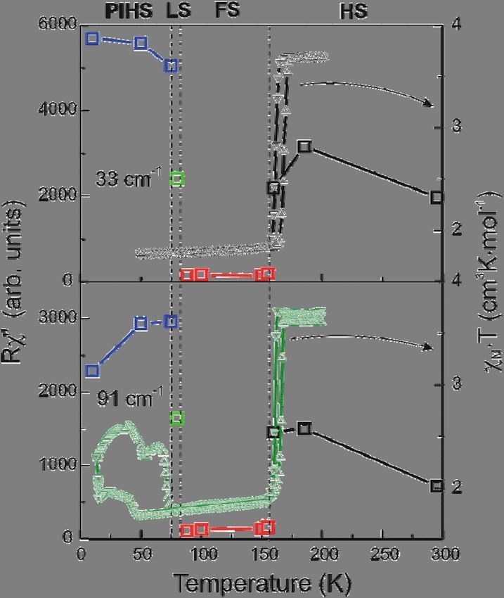
Introduction
Functional materials with switching properties gained considerable interest in view of their
potential technological applications.1,2 Despite numerous reports on coordination polymers with
specific topologies 3, the incorporation of electronically and/or optically active building blocks as
essential structural components of such functional materials has scarcely been explored.4 In this
regard, the use of spin-crossover building blocks has been shown to be a suitable strategy since they
change reversibly their magnetic, structural, dielectric and optical properties in response to stimuli
such as a variation of temperature or pressure and light irradiation.5-8 In particular, six-coordinate
Fe2+ compounds, with 3d6 electron configuration, represent an important class of switchable
molecular systems. In pseudo-octahedral symmetry, they change reversibly its ground state from an
1
A1 (t2g6), i.e. low-spin (LS) state to a 5T2 (t2g4 eg2), i.e. high-spin (HS) state.
The light induced excited spin-state trapping (LIESST) effect 9,10 describes a light induced transition
of the 1A1 (LS) → 5T2 (HS) states in Fe2+ spin-crossover compounds at temperatures far below the
thermal spin state transition temperature Tc. According to a two-step mechanism, postulated by
Decurtins et al.10, irradiation into the LS d-d bands, i.e. 1A1 → 1T1/1T2, leads to the population of
the 5T2 HS state via the 3T1/3T2 state. The relation between photo-induced phase transitions and
thermally induced ones, however, has only recently been highlighted. Based on the similarity of
absorption spectra, magnetic properties 11, and reflection spectra 12,13
it has been argued the photo-
induced phase at low temperatures is the same state as the equilibrium phase at high temperatures.
Recent experiments have shown that this assumtion has to be revised. Using resonant Raman
spectroscopy, it was found by Tayagaki et al.14 that at low temperatures the photo-induced phase
has a broken symmetry and differs from the thermally-induced high-symmetry phase. This was
illustrated more recently in the spin-crossover field by Real et al.15 who demonstrated the
occurrence of light-induced polymorphism on single crystals at low temperature. Similar
conclusions have been made recently based on pico-second time-resolved crystallography 16.
According to the thermal evolution of the fraction of Fe2+ centers in the HS state, HS(T), the
spin change can be classified as (a) “gradual”, covering a wide temperature range; (b) “stepwise” or
“abrupt”, with changes from one state to another within only a few Kelvin; (c) showing a hysteresis
effect. The former transitions are characterized by continuous evolution of the X-ray pattern
indicating that no distinguishable phases exist, whereas in the latter transitions (b) and (c) at least
two independent crystallographic phases, HS and LS, can be detected. The hysteretic behavior
stems from cooperative changes in the crystal and confers to these materials a memory function.
Cooperativeness can be enhanced improving communication between the active spin-crossover
building blocks, i.e. integrating them in rigid frameworks. For instance, the possibility to induce
polymerization through suitable coordinated anions, i.e. cyanide ligands, has been considered only
recently as a suitable strategy. Cyanide-bridged homo- and hetero-metallic coordination polymers
have been shown to exhibit a remarkable diversity of structural types with interesting magnetic,
electrochemical, magneto-optical, thermo-mechanical, and zeolitic properties.17 In particular,
Hofmann-like clathrate compounds18 containing Fe(II) ions have opened up the opportunity to build
highly cooperative thermo-, piezo-, and photo-switchable two- and three-dimensional coordination
polymers 19-22.
The compound [Fe(pmd)(H2O){Au(CN)2}2]·H2O (1Au), pmd = pyrimidine, represents a
singular coordination polymer due to its structural and electronic properties. It is made up of three
identical and independent three-dimensional interlocked frameworks (Fig. 1) with two
crystallographically independent {Fe(1)N6} and {Fe(2)N4O2} sites. Both iron sites lie at inversion
centers and correspond to an elongated and a compressed coordination octahedron, respectively.
Interestingly, the three frameworks collapse at elevated temperatures (ca 400 K) due to a
cooperative topochemical ligand substitution which changes the local coordination of the Fe(2) site
23
to form a new 3D framework (2Au) . Magnetic susceptibility measurements show that (1Au)
undergoes a sharp cooperative spin-crossover with critical temperatures Tcdown = 163 K and Tcup =
171 K for cooling and warming, respectively, leading to a hysteresis of Tc = 8 K. Crystallographic
data demonstrates that only site Fe(1) undergoes the spin-crossover. It is worth noting that the spin-
crossover is accompanied by a drastic color change from pale yellow (HS) to deep red (LS). 23
Raman spectroscopy is a well-established technique to probe spin states of spin-crossover
14,24,-27
materials . Earlier Raman spectroscopy investigations have mainly been devoted to the
differentiation between high- and low-spin Fe2+ complexes or to the estimation of the respective
entropy contributions. More attention has been given recently to Raman scattering (RS) as a
convenient and easily accessible probe of light induced spin transitions28 for temperatures far below
and above the spin state transition temperature. Unfortunately, much less is known to spin
conversion phenomena at temperatures close to Tc where a complex competition of temperature and
light induced effects is expected. In addition, especially complex scenarios are possible in spin-
crossover systems with more than one Fe2+ site on nonequivalent lattice sites as given in the present
coordination polymer.
Here, we report on a Raman scattering (RS) study of the two site Fe2+ coordination polymer
[Fe(pmd)(H2O){Au(CN)2}2]·H2O to establish a new phase diagram and demonstrate a more
dynamic evolution of the spin state as function of temperature and light irradiation than expected
from earlier magnetization data 23. A novel phonon “wipe-out” effect is demonstrated proposing a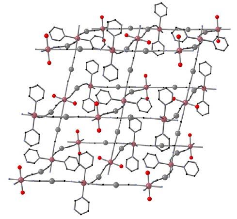
fluctuating spin (FS) state phase in the phase diagram. Our phonon assignment is supported by
density functional calculations (DFT).

                                                                         Au(1)

                                                                 Fe(1)

                                           water

                              Au(2)
                                             Fe(2)

Fig. 1. Fragment of the coordination polymer [Fe(pmd)(H2O){Au(CN)2}2]·H2O displaying the
asymmetric unit (top), the expanded version of the archetypal network structure of CdSO4 (middle)
and the triple interpenetration of identical networks (bottom). Color code: O, red; N, blue; C black;
Au, grey; Fe, pink.
Experimental details
     The studied compound has been synthesized and characterized according to Ref. 23. An Ar/Kr
ion laser was used for Raman excitation at 514.5 nm (2.41 eV) and 647 nm (1.92 eV). The laser
output power was kept below 0.5 mW on a focus of approximately 100μm diameter to protect the
sample from possible heating effects. All previous Raman experiments on spin-crossover systems
that are known to us have been performed with at least 10 times large laser intensities. The scattered
light was collected in quasi-backscattering configuration and dispersed by a triple monochromator
DILOR XY on a liquid-nitrogen-cooled CCD detector. Measurements were performed in the
parallel polarization configuration. Temperature dependencies were measured in a continuous
helium flow cryostat from 10 K to room temperature. Experiments at elevated temperatures up to
370 K on the 2Au dehydrated modification were carried out using a helium gas filled heating stage.
The variable temperature magnetic susceptibility measurements were carried out in SQUID
apparatus operating at 1 T and at temperatures 16 – 200 K. An externally placed frequency doubled
Nd:YAG-laser (λ = 532.1 nm) and optical fiber were used for light-irradiation under magnetic
measurements.
                                                                                                      29
     The Raman and IR spectra were calculated with use of GAUSSIAN03 programme system
for HS and LS isomers of a trinuclear model of (1Au) with Ci symmetry described later. Becke’s
                                                   30                                                 31
three-parameter hybrid functional B3LYP,                which contains Becke’s exchange functional
                                                                                  32
together with the local spin density correlation functional III of Vosko et al.        and the non-local
                                           33,34
correlation functional LYP of Lee et al.           was used for geometry optimization. As a basis set
                     35-37
CEP-31G was used.

Results and discussion
     For the monoclinic (space group P21/c, Z = 4) structure, the factor group analysis yields 156
Raman-active modes (78Ag + 78Bg) for the zone center vibrations. Due to the large number of
allowed vibrations and the transparency of the irregular shaped samples a symmetry analysis of the
observed modes in our Raman experiment has been omitted.
     The observed excitation (Fig. 2) can be divided into three groups of Raman frequencies.
Following earlier assignments38-42 in other compounds with compositional similarity, these modes
can be attributed mostly to internal modes of pyrimidine (600 - 1600 cm-1), internal and external
modes of {Fe(1)N6} and {Fe(2)N4O2} cores (below 600 cm-1), and C-N stretching-like vibrations
(above 2150 cm-1). This assignment is confirmed by the normal co-ordinate analysis performed
within the framework of DFT, described further below.
Fig. 2. Room-temperature Raman spectrum of a [Fe(pmd)(H2O){Au(CN)2}2]·H2O (1Au) single
crystal. The inset shows a sketch of the spin state diagram including a light induced excitation
with a 514nm laser line.
As we do not expect changes in the frequency region of the pyrimidine vibration, we will
focus our attention to the high-frequency and to the low-frequency regions where remarkable
changes are observed as function of temperature. We note that the relative change of the high-
frequency modes with spin state is expected to be markedly smaller and that they have a smaller
contribution to the evolution of entropy43. Representative Raman spectra of
[Fe(pmd)(H2O){Au(CN)2}2]·H2O at different temperatures are presented in Figs. 3 and 4. A
summary of the Raman spectral changes vs. temperature is presented in Table 1. It is clearly seen
from Fig. 3 that a three-step transition takes place with reducing temperatures and that the effects
involve both frequency as well as intensity of the modes in the low to intermediate frequency
regime. Therefore these effects cannot be attributed to a change of optical constants, e.g. the optical
penetration depth.Fig. 3. Temperature dependent Raman spectra of the [Fe(pmd)(H2O){Au(CN)2}2]·H2O
measured in the frequency regime of 0 – 550 cm-1 and 2150 – 2200 cm-1. The inset shows
details of the spectra in the FS state phase.
Fig. 4. Temperature dependent Raman spectra in the frequency region of internal modes of the
pyrimidine.Frequency regimes
-1
10-600cm 800-1700cm-1 2150 cm-1 and higher
Temperature Internal and external Internal modes of C-N stretching like
regimes (K) modes of Fe(1)N6 Pyrimidine vibrations
and spin state phase and Fe(2)N4O2
370 < T < 300 K - - Structural change at
345 K
300 < T < 160 K No significant No significant All bands shift
high spin (HS) change in position change in position monotonically to high
frequency
160 < T < 88 K Abrupt decrease in No significant Few new modes arises
fluctuating intensity with 514.5 change in intensity and band position
spin phase (FS) nm, not with 647 nm with 514.5 nm and shifts
laser excitation 647 nm lasers
88 < T < 80 K Intensity and line Intensity pattern Intensity pattern
low spin (LS) number recover, similar to HS similar to HS
similar to HS
T < 80 K New bands appear Intensity increases No change
photo-induced HS
(PIHS)
Table 1. Summary of Raman spectral changes for different frequency regimes in the Fe2+
coordination polymer [Fe(pmd)(H2O){Au(CN)2}2]·H2O as function of temperature and laser
excitation. The structural change at 345 K is related to the dehydration of the compound from
(1Au) to (2Au) 23.
The first transition happens at temperature below 160 K. This is the critical temperature, Tc,
where one of the two Fe2+ sites in (1Au) undergoes a thermally induced first-order spin-crossover
transition from the HS- to the LS-state 23. In the following we will denote the corresponding phases
according to the spin state of this Fe(1) ion site. Given that the HS and the LS phases of (1Au) have
the same crystal symmetry, it is reasonable to expect no significant differences in their vibrational
spectra. Instead just below Tc we observe dramatic changes in the form of an abrupt loss of the
Raman intensity in the low-frequency part of the spectra. Due to the magnitude of this intensity
reduction it is even difficult to extract their intensity from the background noise.Fig. 5. Temperature dependence of integrated Raman intensities Rχ” for selected low frequency
phonon lines (33 and 91 cm-1, open squares in upper and lower plot, respectively) and the
susceptibility χM·T without light irradiation 23 (triangles, upper right scale) and under laser (λ = 540
nm) irradiation (triangles, lower right scale).
Figure 5 shows the behavior of the integrated intensity of selected low-frequency lines versus
temperature. Usually, Raman spectra in the HS and LS states have approximately the same
intensity14,25,43 or the intensity of the LS-spectra is even higher 41. In our case the intensity of low-
frequency part of the spectra in the new phase is less than 5% from the intensity in of the HS-phase.
Besides that, we observe changes in the number of lines as well as in their frequency (see Fig. 6).
We note that the modes in the middle-frequency region (800 - 1700 cm-1) have comparable
intensities in both phases, while in the high frequency Raman signal the new phase shows up as two
times smaller scattering intensity as in the HS phase. These spectral changes were found reversible
and reproducible over several measurement cycles.PIHS LS FS HS PIHS LS FS HS 2Au
120
1Au 2185
100
2180
Raman shift (cm-1)
80
2175
60 2170
40 2165
20 2160
0 50 100 150 200 250 300 0 50 100 150 200 250 300 350
Temperature (K)
Fig. 6. Temperature dependence of the phonon line positions in the frequency regions of the
{Fe(1)N6} and {Fe(2)N4O2} octahedrons (left) and C-N stretching-like vibrations (right).
How to explain this unexpected result, namely a “wiping out” and sudden decrease of the RS
intensity in the low frequency region? Checks for reproducibility with 514.5 nm excitation
wavelength on different single crystals and the simultaneous investigation of low and intermediate
frequency regime convinced us in its intrinsic nature. A review of recent results on spin-crossover
systems shows that photo excitation and relaxation 44,45 processes are evident for some systems and
complexities may develop if a competition between thermo- and light-driven processes exists on a
molecular scale. The last aspect is most relevant in the temperature range where the zero-point
energy difference between the LS and HS energy surfaces, HL , is small (see inset in Fig.2).
0
Dynamical spin state switching can be then be induced by any type of lattice distortions47 as well as
light irradiation. The resulting spin state – phonon interaction is strong and could even have non-
46
adiabatic contributions as the time scale of interconversion (τinter 80fs) of the excited singlet to
the quintet state in Fe(II) complexes matches roughly with the period of typical phonons of the
respective coordinations (400 cm-1). As the evidence for a pure and static LS-state of the Fe(1) ions
at temperatures below Tc is based only on magnetization data without light irradiation 23 we will inthe following assume a dynamic competition between temperature- and light-irradiation-driven
processes in the Raman scattering experiments.
In the temperature range of 88 K < T < 160 K we consider a dynamically fluctuating spin (FS)
state with an incoherent switching between HS and LS states on different Fe(1) sites. The resulting
incoherent variations of metal-ligand distances of the order of 0.1 Å are expected to lead to an
anomalous damping of internal and external modes of {Fe(1)N6} core. As to the Fe(2)N4O2
complexes, the influence of the surrounding medium, namely fluctuating internal pressure, can lead
to a damping of phonon spectra, too. Just as to the spin state of Fe(2), the influence of the
surrounding medium is not necessarily very dramatic 44. Our aim should be to test this scenario by
further experiments.
A further open issue concerns effects arising from a distortion of the octahedral symmetry.
Most spin-crossover complexes have symmetry lower than octahedral and if the ligand field
deviates strongly from octahedral symmetry even an intermediate spin state (triplet) can become the
48
ground state . A complex spin state transformation with the appearance of an intermediate spin-
state of Co3+ ions (3d6-configuration) in a strongly distorted octahedral cage has been found
recently in the rare-earth layered cobaltites RBaCo2O5.5 by muon relaxation experiment49. We do
not think that a deviation from the octahedral symmetry due to the lattice strain is considerably
strong for the Fe(2)N4O2 octahedrons but this as well as the effect of irradiation on Fe(2) spin state
has to be clarified. Therefore, further structural investigations should be performed.
The mixed character of spin states displays itself in the high-frequency region of Raman
spectra. It can be seen from Fig. 3 that going from the HS-state phase to the mixed fluctuating spin
(FS) state phase leads to significant changes in the frequency region of C-N stretching-like
vibrations. Besides the frequency shift, the lines at 2162 and 2172 cm-1 split into two lines. The
frequency behavior of high-frequency phonon lines is plotted in Fig. 6. A similar manifestation of
mixed HS and LS states in the C-N stretching region has been observed earlier in an IR study in the
spin-crossover system 50 [Fe(phen)2(NCSe)2].
We performed further Raman scattering experiments with a red-647nm laser line excitation.
This energy is smaller than the 1A1 → 1T1/1T transition. Therefore light-induced processes should
be diminished. Figure 7 shows at three selected temperatures that the Raman scattering intensity
does not drop through the thermally induced spin state transition.1500
647 nm laser
Intensity (arb. un.)
1000 110 K
155 K
500
185 K
0
25 50 75 100 125 150
-1
Raman shift (cm )
Fig. 7. Raman spectra of the [Fe(pmd)(H2O){Au(CN)2}2]·H2O excited by a red-647 nm laser line.
To our knowledge, this is the first observation of a “wiping out” effect in a spin-crossover
system. Similar effects, anomalous damping and collapsing of phonon modes associated with
51
dynamic phase fluctuation, have been observed earlier in La0.7Ca0.3MnO3 . However, our
coordination polymer has a different, more complex spectrum of low frequency modes compared to
the manganite. This could provide several competing relaxation paths leading to a glass like
behavior. Analyzing recent efforts to investigate photo-excitation processes in spin-crossover
compounds, we want to underline that all experiments reveal a complex picture of light irradiation
action inside a thermal spin conversion, even at low temperatures 52. Unfortunately, corresponding
experiments at elevated temperatures on systems with a thermal hysteresis have not been fully
45
successful. Still, unexpected and unexplained effects have been observed . In this context, we
highlight two interesting and unusual photoswitching phenomena recently observed in
[Fe(pyrazine){Pt(CN)4}], namely i) both the LS→HS and the HS→LS transitions were triggered
by the same irradiation wavelength and ii) a mixed spin state was observed in a wide temperature
53
range of the thermal hysteresis . We also highlight that the excitation dependence and resonantnature of the “wiping out” effect in (1Au) is different from the resonant effects that have been
observed in the spin-crossover system [Fe(pic)3]Cl2EtOH 54. In the latter system an enhancement of
only the ligand molecule phonon lines has been observed.
In the following we discuss the implications of the sequence of transitions and the related
dynamics of the spin state for our understanding of the phonon wipeout: The second transition
happens at temperature below 88 K. The Raman intensity increases again and the spectra become
quite similar in number of lines, frequencies and intensities to those of the HS state. Therefore we
assign this transition to a thermally induced LS-state. X-ray diffraction study did not reveal any
23
crystal structure change in a wide temperature range , so the differences in the Raman spectra
comparing the HS- and the LS-state are not expected and indeed are not observed, in the low
frequency as well as in the high frequency region. The thermally-induced LS state is located in a
very narrow temperature range of ~10 K.
The third and last transition occurs in the temperature region between 80 K and 75 K. While
the high-frequency Raman spectra do not show (except small increases of frequency) any
transformation under this spin state transition, a drastic change is observed in the low-frequency
regime. A number of additional lines appear below 600 cm-1 which are not observed at higher
temperatures. Experiments on several samples with varying laser spot position and intensity show
that this is an intrinsic and reproducible process. We attribute this transition to the light-induced 1A1
(LS) → 5T2 (HS) transition due to the LIESST effect. Our assignment is confirmed by SQUID
measurements – a light-induced thermal hysteresis loop is observed at temperatures below 75 K
(see Fig. 4, lower panel). The spectral change strongly indicates that a symmetry lowering takes
place in the photo-induced phase. According to the model of Tayagaki et al. 14, the formation of the
photo-induced HS-state occurs in the initial LS-state phase. Irradiation leads to the population of the
5
T2 (HS) metastable state (threefold orbital degenerate) via one or two intersystem crossing steps.
14
The photo-induced phase is stabilized by a cooperative Jahn-Teller transformation , i.e. the
symmetry lowering. As a result, formerly only IR-active modes appear in the Raman spectra of the
low-temperature high-spin photo-induced phase.Fig. 8. Fano resonance in the PIHS phase of [Fe(pmd)(H2O){Au(CN)2}2]·H2O.
The presence of strong electron-phonon interaction manifests itself also directly in the Raman
spectra (Fig. 8). A broad structured band centered at ~650 cm-1 probably due to scattering on
electronic excitations appears in the low-temperature spectra. Electron-phonon coupling leads to a
very strong Fano resonance at 650 and 690 cm-1, frequencies attributed to Jahn-Teller-like and
breathing-like phonon modes of FeN6 octahedra, respectively, with pyrimidine in-phase bending
coupled to Fe-N stretching. To our knowledge, this it is the first observation of a Fano resonance in
the light-induced phase of spin-crossover systems.370 K
360 K
1500 350 K
340 K
330 K
320 K
Intensity (arb. un.)
Au2
1000
Au1
500
2150 2160 2170 2180 2190 2200
-1
Raman shift (cm )
Fig. 9. High-temperature Raman spectra of [Fe(pmd)(H2O){Au(CN)2}2]·H2O single crystal in
the frequency region of C-N stretching-like vibrations.
Finally we have studied Raman spectra under heating the coordination polymer. In the
temperature range 323-382 K the set of interpenetrating and independent 3D networks in (1Au) is
transformed to the single 3D network (2Au) by losing the coordinated and nonbonded water
molecules23. This transformation leads to significant structural changes: the (pmd) ligand now
bridges directly the Fe(1) and Fe(2) sites defining a system of infinite chains {-Fe(1)-pmd-Fe(2)-}
running parallel to the a axis. The [Au(CN)2] groups of one chain link with the equatorial positions
of the iron centers, connecting adjacent chains and defining a single 3D network. Raman spectra at
selected temperatures in the frequency region of 2150 – 2200 cm-1 are presented in Fig. 9. The
measurements were performed after first heating the sample to T = 370 K and then cooling it downto the given temperatures. Various measurements were done during heating and cooling of the
sample to proof reproducibility of the dehydration effect. Within the described temperature protocol
also no hysteresis was found. The structural changes at ~345 K lead to a jump-like shift of CN
stretching modes at 2160 and 2179 cm-1 to the positions of 2163 and 2180 cm-1, while the 2167 cm-1
mode shifts quite a lot to the position of 2186 cm-1. Such different behavior point to the
nonequivalent positions of CN groups in the crystal structure.
DFT Calculations of a spin state coordination
The DFT calculations have been performed in order to allow a decisive assignment of the
relevant observed Raman bands. The DFT approach was recently shown to be quite an effective
55-58
method to predict the vibrational properties of spin-crossover systems . For the system under
study, because of its polymeric nature, the calculations shall in principle involve the complete cell
under periodic boundary conditions. However with the available software allowing for periodic
boundary conditions, the calculations for equally large and complex systems are hardly feasible.
Therefore we decided to use the Gaussian programme that offers high-end functionals and basis sets
for a model system, which is as relevant as possibly to the (1Au) complex. As we are mainly
interested in vibrations that are dependent on the spin state transition, we chosed a model that
reproduces the environment on the spin-crossover Fe(II) (Fe(1)) in the best possible way. On the
other hand, in order to keep the calculations feasible we had to limit the size of the molecule.
Therefore the calculations have been performed for a trinculear unit of Ci symmetry encompassing
a Fe(pmd)2([Au(CN)2]4 fragment with two Au(CN)2 bridging to the two terminal
Fe([Au(CN)2]4(H2O)2. Therefore an anionic unit with stoichiometry
Fe3[Au(CN)2]10(H2O)4(C4H4N2)2 and a charge of -4 was used for calculations. The system looks
like the fragment shown in Fig. 1 (top) with one more FeN4O2 fragment centered on Fe(1) attached,
so that the Fe(2) atom lies in the symmetry center.An initial test of the quality of the calculation results may be the comparison of the obtained metal-
ligand bond lengths for the spin-crossover iron centre. These values are listed in the Table 2.
Calculated (DFT) bond lengths (Å) Bond lengths from X-ray data for (1Au) (Å)
Fe-N(pyrimidine) 2.215 HS 2.200 HS
2.024 LS 1.986 LS
Fe-N (NC – bridg.) 2.182 HS 2.149/2.164 HS
1.930/1.948 LS
1.970 LS
Fe-N (CN-nonbridg.) 2.142 HS ------
1.967 LS
Au-C 2.016 HS 1.992/1.989 HS
2.016 LS 1.976/1.981 LS
Table 2. Calculated (model coordination) and experimental metal-ligand bond-lenghts23 around the
spin-crossover iron centre of the model complex.
The above data reveal that the calculated in vacuo values show a general bias towards longer
bond lengths compared to the solid state data. However the observed differences are less than 0.04Å
and therefore reasonable vibrational frequencies may be obtained in the following normal co-
ordinate analysis.
The normal coordinate analysis yields 249 normal vibrations that are of Ag (Raman active)
and Au (IR active) type. Those relevant to the above presented spectra are listed in table below.
Mode type LS (cm-1) HS (cm-1)
Au-C-N bending 305-360 306-355
Fe-N-Au-C (no CN stretching) stretching around Fe(1) and Fe(2) 523 489
(with Fe(1)-N4 breathing)
Fe(2)-N –Au-C (no CN stretching) stretching 492 499
pyrimidine in-phase bending + Fe(1)-N(pyrimidine) stretching 644 632
pyrimidine in-phase bending + Fe(1)-N(pyrimidine) stretching 690 678
pyrimidine ring bending + Fe-N stretching 997 985
pyrimidine ring bending 1066 1065
pyrimidine C-H in-plane bending 1085 1074
pyrimidine C-H in-plane bending 1144 1141
pyrimidine C-H in-plane bending 1221 1215
pyrimidine C-H in-plane bending 1237 1230pyrimidine ring stretching 1603 1593
N-C-Au stretching around Fe(2) 2179 2168
Fe(1)-N-C-Au(axial) (CN stretching) stretching 2185 2174
Fe(1)-N-C-Au(axial) (CN stretching) stretching 2196 2183
Table 3. The calculated Raman active Ag vibrations, relevant to the observed Raman spectra.
Generally, the calculations reveal a pattern that is in line with the observed spectra. On the other
hand they shed light on the problem why the changes in the observed spectral pattern on LS-HS
59
transition are so subtle. It’s generally accepted that a change of a spin state in Fe(II) nitrogen
complexes results in a shift of metal-ligand stretching vibrations by 100-200 cm-1. Yet in our case
no Raman shift is observed in the 200-500 cm-1 regime and the calculated Fe-N(CN) vibrations
show a limited shift of a few tenths of a wavenumber. This may be due to the fact that every Fe-
N(CN) stretching is also a Au-C (stretching) and therefore its frequency is primarily dependent on a
more covalent component. On the other hand, although the calculated Fe-N(pyrimidine) stretching
modes show frequency shifts on the LS-HS transition (HS Au: 186, 204, 242; LS Au: 402, 492 ) the
modes are of ungerade symmetry and are therefore not observable in Raman spectra. They might
become Raman active if the centre of symmetry of the molecule is cancelled. For the polymeric
system (1Au) this may happen when two different spin states of Fe(1) are present, i.e. if the
cooperative Jahn-Teller transformation occurs.
Conclusion
Using Raman spectroscopy we have demonstrated that the spin state conversion of the Fe(1)
site in the cyanide-based bimetallic coordination polymer [Fe(pmd)(H2O){Au(CN)2}2]·H2O follows
a complex sequence of high spin, fluctuating spin, low spin and photo-induced high spin states. A
novel “wiping out” effect was observed in the temperature range of 88 K < T < 160 K. We attribute
this effect to a competition of thermo- and light-driven processes in the temperature range with
small HL and a matching of the interconversion time scale with the period of the involved
0
vibrations. This competition leads to the creation of a complex spin-state fluctuating phase and, as a
result, to the anomalous damping of internal and external modes of the {Fe(1)N6} complexes. Our
experimental and theoretical data are fully consistent with an intrinsic origin of the fluctuating spin
state. This is in contrast with recent findings in a Prussian Blue analog with a stoichiometry
distribution60. At low temperatures the PIHS phase in the coordination polymer appears due to theLIESST effect. The additional lines which are neither observed in the HS nor in the LS phases indicate that the vibrational selection rules are modified in the photo-induced phase. Acknowledgments: We acknowledge important discussions with P. Gütlich, J. J. McGarvey, and G. G. Levchenko. This work has been supported by the DFG Priority Program SPP1137 on Molecular Magnetism and the network of the European Science Foundation Highly Frustrated Magnetism. V. G. and Yu. G. P. acknowledge the support of the Ukrainian-Russian grant 2008-8. V.S. and J.A.W. acknowledge the support of the BMBF under 05KS7UK2
References:
  1. M.D. Hollingsworth, Science 295, 2410 (2002).
  2. C. Janiak, J. Chem. Soc., Dalton Trans. 2781 (2003).
  3. S. R. Batten, R. Robson, Angew. Chem. Int. Ed., 37, 1460 (1998); R. Robson, J. Chem.
     Soc., Dalton Trans., 3735 (2000); M. J. Zaworotko, Chem. Comm., 1 (2001); O. M. Yaghi,
     H. Li, C. Davis, D. Richardson, T. L. Groy, Acc. Chem. Res. 31, 474 (1998); M. Eddaoudi,
     D. B. Moler, H. Li, B. Chen, T. M. Reineke, M. O’Keeffe, O. M. Yaghi, Acc. Chem. Res.
     34, 319 (2001).
  4. D. Maspoch, S. R. Molina, J. Veciana, Chem. Soc. Rev., 36, 770 (2007).
  5. P. Gütlich, H. A. Goodwin, Spin Crossover in Transition Metal Compounds, Top. Curr.
     Chem., Vols. 233, 234, 235 (2004).
  6. J. A. Real, A. B. Gaspar, M. C. Muñoz, Dalton Trans., 2062 (2005).
  7. J. A. Real, A. B. Gaspar, V. Niel, M. C. Muñoz, Coord. Chem. Rev., 236, 121 (2003).
  8. P. Gütlich, A. Hauser, H. Spiering, Angew. Chem. Int. Ed. Engl., 33, 2024 (1994).
  9. S. Decurtins, P. Gütlich, C.P. Köhler, H. Spiering, and A. Hauser, Chem. Phys. Lett. 105, 1
     (1984).
  10. S. Decurtins, P. Gütlich, K. M. Hasselbach, H. Spiering, and A. Hauser, Inorg. Chem. 24,
     2174 (1985).
  11. Y. Ogawa, S. Koshihara, K. Koshino, T. Ogawa, C. Urano, and H. Takagi, Phys. Rev. Lett.
     84, 3181 (2000).
  12. S. Koshihara, Y. Takura, K. Takeda, and T. Koda, Phys. Rev. Lett. 68, 1148 (1992).
  13. S. Koshihara, Y. Takura, T. Mitani, G. Saito, and T. Koda, Phys. Rev. B 42, 6853 (1990).
  14. T. Tayagaki and K. Tanaka, Phys. Rev. Lett. 86, 2886 (2001).
  15. A. L. Thompson, A. E. Goeta, J. A. Real, A Galet, M. C. Muñoz, Chem. Commun., 1390
     (2004).
  16. E. Collet, M.-H. Lemée-Cailleau, M. Buron-Le Cointe, H. Cailleau, S. Techert, M. Wulff, T.
     Luty, S. Koshihara, M. Mayer, L. Toupet, and P. Rabiller, Science 300, 612 (2003).
  17. K. R. Dunbar, R. A. Heintz, Prog. Inorg. Chem., 45, 283 (1997); M. Verdaguer, A. Bleuzen,
     V. Marvaud, J. Vaissermann, G. M. Seuleiman, C. Desplanches, A. Scuiller, C. Train, R.
     Garde, G. Gelly, C. Lomenech, I. Rosenman, P. Veillet, C. Cartier, F. Villain, Coord. Chem.
     Rev., 190–192, 1023 (1999); M. Ohba, H.Okawa, Coord. Chem. Rev., 198, 313 (2000).
  18. T. Iwamoto in Inclusion Compounds, Vol. 5 (Eds.: J.L. Atwood, J.E.D. Davies, D.D.
     MacNicol), Oxford University Press, London, UK. p. 177 (1991).
19. T. Kitazawa, Y. Gomi, M. Takahashi, M. Takeda, A. Enomoto, T. Miyazaki, T. Enoki, J. Mater. Chem. 6, 119 (1996). 20. V. Niel, J. M. Martines-Agudo, M. C. Muñoz, A. B. Gaspar, J. A. Real, Inorg. Chem. 40, 3838 (2001). 21. V. Niel, M.C. Muñoz, A.B. Gaspar, A. Galet, G. Levchenko, J. A. Real, Chem. Eur. J. 8, 2446 (2002). 22. V. Niel, A. Galet, A. B. Gaspar, M. C. Muñoz, J. A. Real, Chem. Commun., 1248 (2003). 23. V. Niel, A. Thompson, M. C. Muñoz, A. Galet, A. Goeta, and J.A. Real, Angew. Chem. Int. Ed. 42, 3760 (2003). 24. A. Bousseksou, G. Molnar, and G. Matouzenko, Eur. J. Inorg. Chem., 4353 (2004). 25. N. Moussa, G. Molnár, S. Bonhommeau, A. Zwick, S. Mouri, K. Tanaka, J. A. Real, and A. Bousseksou, Phys. Rev. Lett. 94, 107205 (2005). 26. N. Moussa, G. Gábor, X. Ducros, A. Zwick, T. Tayagaki, K. Tanaka, A. Bousseksou, Chem. Phys. Lett. 402, 503 (2005). 27. G. Brehm, M. Reiher, and S. Schneider, J. Phys. Chem A 106, 12024 (2002). 28. N. Suemura, M. Ohama, and S. Kaizaki, Chem. Commun., 1538 (2001). 29. M. J. Frisch, G. W. Trucks, H. B. Schlegel, G. E. Scuseria, M. A. Robb, J. R. Cheeseman, J. A. Montgomery, Jr., T. Vreven, K. N. Kudin, J. C. Burant, J. M. Millam, S. S. Iyengar, J. Tomasi, V. Barone, B. Mennucci, M. Cossi, G. Scalmani, N. Rega, G. A. Petersson, H. Nakatsuji, M. Hada, M. Ehara, K. Toyota, R. Fukuda, J. Hasegawa, M. Ishida, T. Nakajima, Y. Honda, O. Kitao, H. Nakai, M. Klene, X. Li, J. E. Knox, H. P. Hratchian, J. B. Cross, C. Adamo, J. Jaramillo, R. Gomperts, R. E. Stratmann, O. Yazyev, A. J. Austin, R. Cammi, C. Pomelli, J. W. Ochterski, P. Y. Ayala, K. Morokuma, G. A. Voth, P. Salvador, J. J. Dannenberg, V. G. Zakrzewski, S. Dapprich, A. D. Daniels, M. C. Strain, O. Farkas, D. K. Malick, A. D. Rabuck, K. Raghavachari, J. B. Foresman, J. V. Ortiz, Q. Cui, A. G. Baboul, S. Clifford, J. Cioslowski, B. B. Stefanov, G. Liu, A. Liashenko, P. Piskorz, I. Komaromi, R. L. Martin, D. J. Fox, T. Keith, M. A. Al-Laham, C. Y. Peng, A. Nanayakkara, M. Challacombe, P. M. W. Gill, B. Johnson, W. Chen, M. W. Wong, C. Gonzalez and J. A. Pople, Gaussian 03, Revision B.1 Gaussian, Inc., Pittsburgh PA, 2003. 30. A. D. Becke, J. Chem. Phys. 98, 5648 (1993). 31. A. D. Becke, Phys. Rev. A 38, 3098 (1988). 32. S. H. Vosko, L. Wilk and M. Nuisar, Can. J. Phys. 58, 1200 (1980). 33. C. Lee, W. Yang and R. G. Parr, Phys. Rev. B 37, 785 (1988). 34. B. Miehlich, A. Savin, H. Stoll and H. Preuss, Chem. Phys. Lett. 157, 200 (1989).
35. W. J. Stevens, H. Basch and J. Krauss, J. Chem. Phys., 81, 6026 (1984).
36. W. J. Stevens, M. Krauss, H. Basch and P. G. Jasien, Can. J. Chem. 70, 612 (1992).
37. T. R. Cundari and W. J. Stevens, J. Chem. Phys. 98, 5555 (1993).
38. G. Molnár, T. Kitazawa, L. Dubrovinsky, J. J. McGarvey, and A. Bousseksou, J. Phys.:
   Condens. Matter 16, S1129 (2004).
39. K. Hosoya, T. Kitazawa, M. Takahashi, M. Takeda, J.-F. Meunier, G. Molnár, and A.
   Bousseksou, Phys. Chem. Chem. Phys. 5, 1682 (2003).
40. N. Moliner. L. Salmon, L. Capes, M. C. Muñoz, J.-F. Létard, A. Bousseksou, J.-P.
   Tuchagues, J. J. McGarvey, A. C. Dennis, M. Castro; R. Burriel, and J. A. Real, J. Phys.
   Chem. B 106, 4276 (2002).
41. G. Molnár, V. Niel, A. B. Gaspar, J. A. Real, A. Zwick, A. Bousseksou, and J. J. McGarvey,
   J. Phys Chem. B 106, 9701 (2002).
42. T. Taugaki, A. Galet, G. Molnár, M. C. Muñoz, A. Zwick, K. Tanaka, J. A. Real, and A.
   Bousseksou, J. Phys. Chem. B 109, 14859 (2005).
43. A. Bousseksou, J. J. McGarvey, F. Varret, J.A. Real, J.-P. Tuchaguues, A. C. Dennis, M. L.
   Boilott, Chem. Phys. Lett. 318, 409 (2000).
44. A. Hauser, Top Curr. Chem. 234, 115 (2004) and A. Hauser, J. Jeftić, H. Romstedt, R.
   Hinek, and H. Spiering, Coord. Chem. Rev. 190-192, 471 (1999).
45. F. Varret, K. Boukheddaden, E. Codjovi, C. Enachescu, J. Linarès, Top Curr. Chem. 234,
   199 (2004).
46. C. Brady, J.J. Mc Garvey, JK. McCusker, H.Toftlund, D.N. Hendrickson, in Spin Transition
   in Metal Compunds, P. Gütlich and H.A. Goodwin, Eds, Top. Curr. Chem. 235, 1 (2004)
   and references therein.
47. E.S. Zhitlukhina, K.V. Lamonova, S.M. Orel, P. Lemmens, and Yu.G. Pashkevich, Journ.
   Phys.: Cond. Matter 19, 156216 (2007).
48. H. Toftlund, Coord. Chem. Rev. 94, 67 (1989).
49. H. Luetkens, M. Stingaciu, Yu. G. Pashkevich, K. Conder, E. Pomjakushina, A. A. Gusev,
   K. V. Lamonova, P. Lemmens, and H.-H. Klauss, Phys. Rev. Lett. 101, 017601 (2008).
50. M. Sorai, and S. Seki, J. Phys. Chem. Solids 35, 555 (1974).
51. J. Zhang, P. Dai, J. A. Fermandez-Baca, E. W. Plummer, Y. Tomioka, and Y. Tokura, Phys.
   Rev. Lett. 86, 3823 (2001).
52. R. Hinek, H. Spiering, P. Gütlich, A. Hauser, Chem. Eur. J. 2, 1427 (1996); R. Hinek, H.
   Spiering, D. Scholmeyer, P. Gütlich, A. Hauser, Chem. Eur. J. 2, 1435 (1996).
53. S. Bonhommeau, G. Molnár, A. Galet, A. Zwick, J. A. Real, J. J. McGarvey, and A.
   Bousseksou, Angew. Chem. Int. Ed. 44, 4069 (2005).
54. T. Tayagaki, K. Tanaka, H. Okamura, Phys. Rev. B 69, 064104 (2004).
55. K. L. Ronayne, H. Paulsen, A. Höfer, A. C. Dennis, J. A. Wolny, A. I. Chumakov, V.
   Schünemann, H. Winkler, H. Spiering, A. Bousseksou, P. Gütlich, A. X. Trautwein, and J. J.
   McGarvey, J.. Phys. Chem. Chem. Phys., 2006, 8, 4685.
56. L. H. Böttger , A. I. Chumakov, C. M. Grunert, P. Gütlich, J. Kusz, H. Paulsen, U. Ponkratz,
   V. Rusanov, A. X. Trautwein and J. A. Wolny, Chem. Phys. Lett. 429, 189–193 (2006).
57. Y. Garcia, H. Paulsen, V. Schünemann, A. X. Trautwein and J. A. Wolny, Phys. Chem.
   Chem. Phys. 9, 1194-1201 (2007).
58. M. M. N. Wolf, R. Groß, C. Schumann, J.A. Wolny, V. Schünemann, A. Døssing, H.
   Paulsen, J. J. McGarvey and R. Diller, Phys. Chem. Chem. Phys. 10, 4264 (2008).
59. J.-P Tuchagues, A. Bousseksou, G. Molnar, J.J. McGarvey and F. Varret, in Spin Transition
   in Metal Compounds, P. Gütlich and H.A. Goodwin, Eds, Top. Curr. Chem. 235, 85 (2004)
   and references therein.
60. E. J. M. Vertelman, T. T. A. Lummen, A. Meetsma, M. W. Bouwkamp, G. Molnar, Paul H.
   M. van Loosdrecht, and P. J. van Koningsbruggen, Chem. Mater. 20(4), 1236 (2008).
You can also read