Standardized MRI Protocol for Brain Tumor Clinical Trials - Benjamin M. Ellingson, Ph.D. Assistant Professor of Radiology at UCLA - QIBA Wiki
←
→
Page content transcription
If your browser does not render page correctly, please read the page content below
Standardized MRI Protocol for
  Brain Tumor Clinical Trials
          Benjamin M. Ellingson, Ph.D.
    Assistant Professor of Radiology at UCLA
Standardized MRI Protocol for Therapeutic Studies

• FDA Meeting in January 2014 highlighted the need to
  standardize MRI acquisition protocol
   – Needed to increase the FDA confidence in using imaging
     response as a surrogate for drug efficacy in brain tumors

   – Most clinical MRI sequences are T1 or T2 “weighted”
      • Lesion contrast is highly dependent on sequence parameters
      • Lesion size is subjective due to ability for reader (or
        algorithm) to generalize across levels of image quality
   – Comparisons and pooling across studies, drugs, etc.
      • Ethically important to limit number of patients on ineffective
        drugs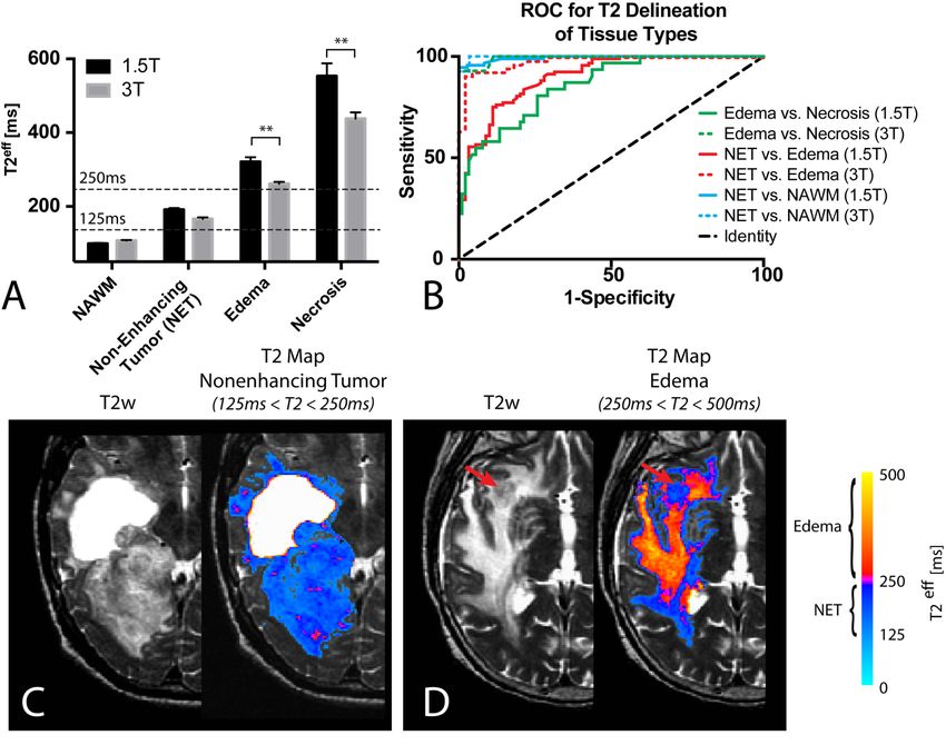
Why do we need Image Standardization?
• Reduce measurement variability due to protocol differences
   – Minor differences in hardware or sequence timing (e.g. TE/TR) can
     result in significant changes in image contrast

  Bidim=7.53                                                    Bidim=7.05

                TE=13ms; TR=560ms         TE=3ms; TR=10ms
                2D Fast Spin Echo         3D IR Gradient Echo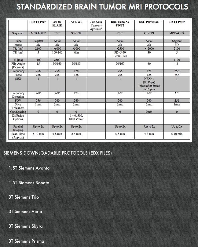
Why do we need Image Standardization?
• Reduce variability due to contrast timing
   – Time between injection and imaging affects contrast enhancement

       • Contrast agent type, dose, and timing (4-8 min after admin is optimal (Akeson, Acta Radiol, 1997b))

                                                                          Time
Why do we need Image Standardization?
• Automated Volumetric Segmentation & Feature Extraction
   – Difficulty in defining the exact margins and identifying the largest diameter or
     perpendicular diameter (Fornage, Radiology, 1993)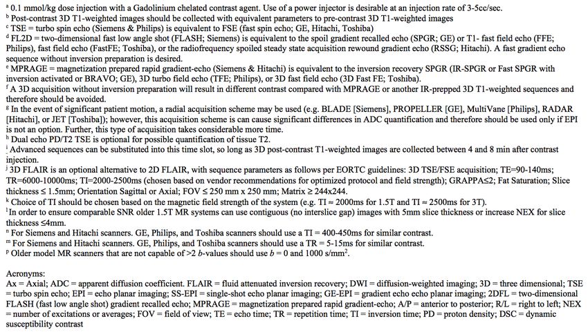
Why do we need Image Standardization?
• Automated Volumetric Segmentation & Feature Extraction
   – Low Reproducibility in 1D/2D Measurements                 (Hopper, 1996; Lavin, 1980; Quiox, 1988;
     Thiesse, 1997; Warr, 1993)
   – High Reproducibility in Volume Measurements
       •   Kaus, Radiology, 2001 – Interobserver COV = 2% (automated segmentation) and 13.6% (manual
           segmentation)
       •   Salman, J Biomed Sci Eng, 2009 – Interobserver COV = 2.5%-10% (automated)
       •   Shah, Neuro Oncol, 2006 – 99.4% intraobserver correlation and 88.9% interobserver correlation
           (N=50 patients)
Why do we need Image Standardization?
• Automated Volumetric Segmentation & Feature Extraction
   – Imaging Genomics & Atlas-Based Approaches for Response Characterization

                                                                                 Leu, Cancer Imaging, 2014

                                     Ellingson, Curr Neurol Neurosci Rep, 2015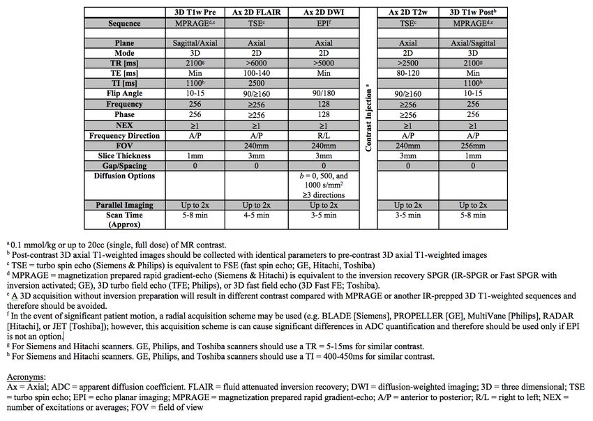
Standardized MRI Protocol for Therapeutic Studies

•    Large variety of imaging capabilities for large clinical trials:
      – Small Outpatient Clinics & Imaging Centers – Minimal Capabilities
      – Community Medical Centers – Basic Capabilities
      – Academic Medical Centers – Advanced Capabilities
•    Need for 3 different & synergistic imaging protocols:
      – Minimal Standardized MRI Protocol
         • Designed for the small outpatient clinics and community imaging facilities
         • Large throughput, fast protocol, minimal chance for error (< 30 min)
      – Basic Standardized MRI Protocol
         • Designed to work for most community medical centers and most sites
         • Standard throughput, typical protocol (< 30 min)
      – Advanced Standardized MRI Protocol + Optional “Modules”
         • Designed for academic centers with expertise in advanced imaging
         • Optional “modules” allow for flexibility depending on needs for the trial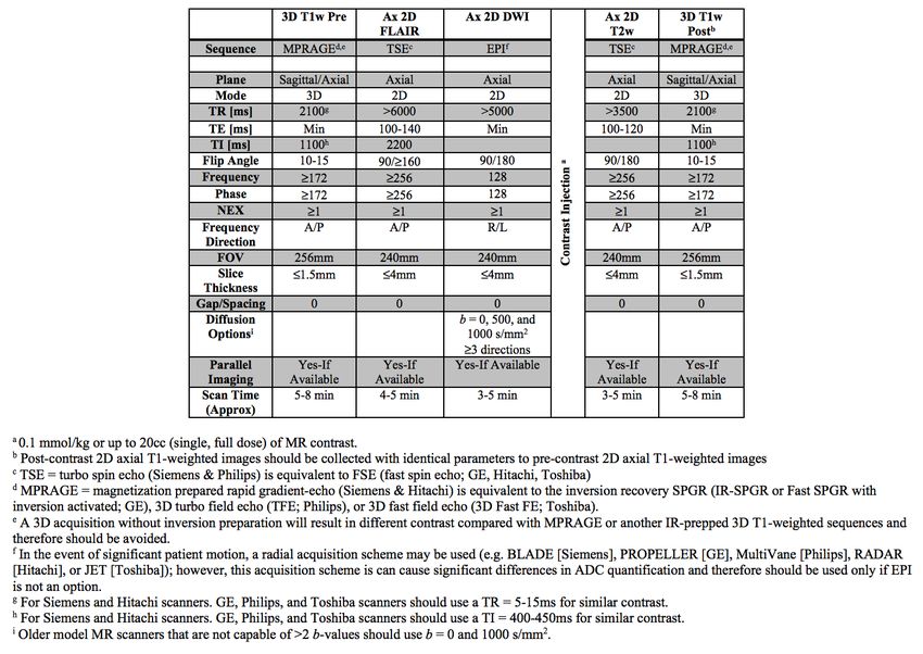
Standardized MRI Protocol for Therapeutic Studies

•    Designed to be aligned with the EORTC, ACRIN, Alliance, and ABTC

•    Designed to work with almost all community medical centers and most
     sites in ACRIN, EORTC, and the Alliance

•    Standard throughput, similar to “basic” EORTC protocol
      – Under 1 hour set up to take down (
Balance Between Maximizing Compliance & Data
Quality
Philosophical Questions Regarding Goals of MRI
Standardization in Brain Tumor Clinical Trials
Maximizing Compliance Maximizing Data Quality
Parameters reflect the range Parameters reflect the range
of values that 100% of centers use of values that >80% of centers useMinimum Standard 1.5T & 3T MRI Protocol
Minimum Standard 1.5T & 3T MRI Protocol

                           • MPRAGE Pre- and Post

                           • 1-1.5mm isotropic

                           • Can be reformatted to 3mm
                             slices (axial, sagittal, or coronal)

                           • Can be used for RANO

                           • Allows for T1 subtraction

                           • Allows for longitudinal registration

                           • Available from all 3 major
                             vendors as part of ADNI

                                    T1+C              T1 Subt.
T1 Subtraction
• Suto, Comput Assist Tomogr, 1989 – Subtracted synthetic images on Gd-DTPA
enhanced MRI
• Lloyd, Br J Radiol, 1993 – Subtraction Gd-enhanced MR for head/neck imaging
• Lee, AJR, 1996 – Digital subtraction for brain lesions or hemorrhage
• Gaul, AJNR, 1996 – Enhancing brain lesions vs. hemorrhage
• Melhem, JMRI, 1999 – Enhancing brain lesions
Pre-Contrast T1 T1+C T1 Subtraction
Ring Enhancing Lesion
59 y.o. Female with Thyroid Carcinoma + Headaches
Adjacent to VentricleT1 Subtraction
• Extent of Resection
Pre-Contrast T1w Post-Contrast T1w T1 Subtraction MapT1 Subtraction
• Phase II, Multicenter Trial of Bev vs. Bev+CPT11 in Recurrent GBM (BRAIN Trial)
(Ellingson, Radiology, 2014)
Post-Bev
FLAIR T1w T1+C CE-ΔT1w MapT1 Subtraction • Phase II, Multicenter Trial of Bev vs. Bev+CPT11 in Recurrent GBM (BRAIN Trial) (Ellingson, Radiology, 2014)
T1 Subtraction
T1 Subtraction
• BRAIN Trial (Ellingson, Radiology, 2013)

                                         Cox Regression,   Cox Regression,
                                         P = 0.589         P = 0.303

                                                           Cox Regression,
                                         Cox Regression,
                                                           P = 0.053
                                         P = 0.004
T1 Subtraction
• BRAIN Trial (Ellingson, Radiology, 2013)
• Further improved by “confirmatory scan”
1.00
not responder at 25%
Responder
0.75
25% Decrease + < 25% Increase,
0.50
0.25
0.00 Cox Regression, P = 0.018
0 3 6 9 12 15 18 21 24 27 30
IRF [Month from Post-Tx]
Number at risk
non-responder 73 64 45 29 21 17 11 9 9 6 2
responder 69 66 57 41 32 23 17 17 13 12 6
p=0.018 by log rank testT1 Subtraction
• Phase III, Multicenter Trial of TMZ+RT+Bev vs. TMZ+RT in Newly Diagnosed GBM
(AVAglio Trial)
Pre-Contrast T1 T1+C T1 SubtractionT1 Subtraction
• Phase III, Multicenter Trial of TMZ+RT+Bev vs. TMZ+RT in Newly Diagnosed GBM
(AVAglio Trial)
Pre-Contrast T1 T1+C T1 SubtractionT1 Subtraction
• Phase III, Newly Diagnosed GBM with DC Vaccination
Post-Surgery Post-RT Mid-DC Post-DC
(Day -38) (Day -5) (Day +11) (Day +98)
Post-Contrast
T1w
T1 Subtraction
MapsMinimum Standard 1.5T & 3T MRI Protocol
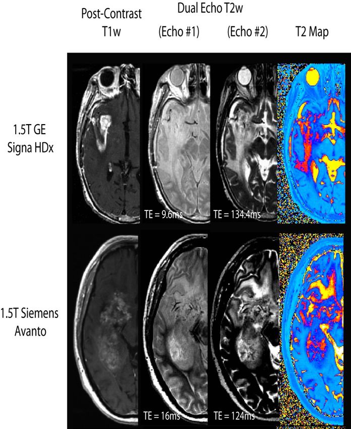
• 2D T2w TSE (Dual Echo
PD/T2 TSE Recommended)
• Can be used for current
RANO evaluations
• Available on all scanners as
part of ADNI
• Part of ACR scanner accred.
• Allows for quantification of T2
within clinically feasible scan
times
T2w T2eff MapDual Echo Turbo Spin Echo MRI
3-5% Variability Across Scanner Measurements of T 2effDual Echo Turbo Spin Echo MRI
125ms < T2eff < 250ms has 60-70% sensitivity and 80-90% specificity for containing NET
N = 50 PatientsDual Echo Turbo Spin Echo MRI 125ms < T2eff < 250ms has 60-70% sensitivity and 80-90% specificity for containing NET
Dual Echo Turbo Spin Echo MRI
  T2eff –defined NET volume is predictive of PFS and OS After Radiation Therapy (new GBM),
Radiation Therapy and Concurrent Temozolomide (new GBM), and Bevacizumab (recurrent GBM)
Minimum Standard 1.5T & 3T MRI Protocol
                            • Timing of Contrast & T2

                            • Timing between pre- and
                              post-contrast T1w images is
                              critical to ensure
                              extravasation
Minimum Standard 1.5T & 3T MRI Protocol
• T2w FLAIR
• Used for RANO evaluations
• Similar to ACRIN, EORTC,
and Alliance Protocols
• 3D FLAIR is optionalFLAIR and T2 TSE @ 3mm for 1.5T & 3T
GE 1.5T Signa
Siemens 1.5T Sonata Siemens 3T Trio
3mm no skip
3mm no skip 3mm no skip
FLAIR
T2wFLAIR and T2 TSE @ 3mm for 1.5T & 3T
GE 1.5T Signa Siemens 1.5T Avanto Siemens 3T Trio
3mm no skip 3mm no skip 3mm no skip
FLAIR
T2wFLAIR and T2 TSE @ 3mm for 1.5T & 3T
GE 1.5T Signa Siemens 1.5T Sonata Siemens 3T Trio
3mm no skip 3mm no skip 3mm no skip
FLAIR
T2wMinimum Standard 1.5T & 3T MRI Protocol
• Diffusion Weighted Imaging
• Uses recommendations by
the ISMRM/NCI Diffusion
Consensus Mtg. 2008
• 3 b-values (0, 500, 1000
s/mm2) are recommendedMinimum Standard 1.5T & 3T MRI Protocol
Minimum Standard 1.5T & 3T MRI Protocol
Recommended 3T Protocol
Recommended 1.5T Protocol
Examples of Compatible Protocols
          Standard Protocol + DCE
Examples of Compatible Protocols
Standard Protocol + DTI + DSC PerfusionExamples of Compatible Protocols
Standard Protocol + Site Specific SequencesSiemens Versions Available for Download http://www.ellingsonbiomedical.com/Ellingson_Biomedical/MRI_PROTOCOLS.html
You can also read