Technology advancements in spinal endoscopy for staged management of painful spine conditions
←
→
Page content transcription
If your browser does not render page correctly, please read the page content below
Editorial

Technology advancements in spinal endoscopy for staged
management of painful spine conditions
Friedrich Tieber1, Kai-Uwe Lewandrowski2,3
1
Medical Technologies Consulting, Augsburg, Germany; 2Center for Advanced Spine Care of Southern Arizona and the Surgical Institute of Tucson,
Tucson, AZ, USA; 3Department of Orthopaedics, Fundación Universitaria Sanitas, Bogotá, D.C., Colombia.
Correspondence to: Kai-Uwe Lewandrowski. Staff Orthopaedic Spine Surgeon, Center for Advanced Spine Care of Southern Arizona and the
Surgical Institute of Tucson; Visiting Professor, Department of Orthopaedics, Fundación Universitaria Sanitas, Bogotá, D.C., Colombia.
Email: business@tucsonspine.com.

Submitted Sep 20, 2019. Accepted for publication Oct 09, 2019.
doi: 10.21037/jss.2019.10.02
View this article at: http://dx.doi.org/10.21037/jss.2019.10.02

The history                                                                 electricity, optics, and precision mechanics.
                                                                               In 1973, Parvis Kambin introduced the concept of a
The development of endoscopy, in general, reflects the
                                                                            transforaminal approach with the use of percutaneously
medical history of the last 200 years in an extraordinary
                                                                            placed Craig’s cannula’s through which he performed
way. It is so extraordinary because endoscopy expanded
                                                                            microdiscectomy in a non-visualized fashion (3-5). The
into almost all fields of medicine, including ENT, urology,
                                                                            introduction of a specially modified arthroscope into
gynecology, orthopedics, and neurosurgery. Success in these
                                                                            the intervertebral disc, and, thus, the first visualized
subspecialties inspired the development of new fields of
                                                                            microdiscectomy, was first reported by Forst and Hausman
operative techniques and clinical subspecialties. Modern
                                                                            in 1983 (6). Coaxial endoscopes were developed because
endoscopy incorporates many technical innovations
that initially were not developed for medical purpose                       they offered the option to visualize and remove the
but stemmed from military developments or consumer                          painful pathology. The addition of a motorized shaver was
products. The desire to look inside the human body cavities,                described by Onik in 1985 which led to the coining of the
to establish a diagnosis and to treat disease is at least 6,000             term “Automated Percutaneous Nucleotomy” (7). Kambin
years old. However, the first product of an endoscopic                      published his first “discoscopic views” from within the disc
diagnostics system consisting a light source, optics, and                   in 1988 and later emphasized the importance of epidural
a housing assembly was the brainchild of the Frankfurter                    visualization as well (8). One year later, Schreiber described
physician Philipp Bozzini, who also developed specula                       the injection of indigo carmine dye into the disc to stain
shaped instruments to create access to a body cavity (1).                   abnormal nucleus pulposus and annular fissures (9). Kambin
   The crucial achievement of Philipp Bozzini was to divide                 also first described the “safe” or “working” zone in 1990 as
the tube in half (2). This principle is valid until today.                  the triangle bordered by the exiting nerve root, the inferior
Through one channel, the light is directed to the object                    endplate and the superior articular process of the inferior
and then viewed through the other by the reflected image                    vertebra, and medially by the traversing nerve root (10).
through a lens system. The Bozzini light guide was equipped
with a lens, an eyepiece, and a candle serving as a light
                                                                            The paradigm shift
source. Due to the insufficient illumination by candlelight,
the invention had nearly no practical value. With his idea,                 With the development of miniature cameras (1980) such as
however, he inspired the following generations of engineers                 the tube color camera (Lemke/Storz), the operating area
to continue development of endoscopy into its current state                 was no longer viewed directly through the endoscope but
of the art. Additional developments and related inventions                  on a monitor (11,12). This changed both the handling of
were driven by substantial knowledge gains in the fields of                 endoscopes and the ergonomic and organizational processes

© Journal of Spine Surgery. All rights reserved.                  J Spine Surg 2020;6(Suppl 1):S19-S28 | http://dx.doi.org/10.21037/jss.2019.10.02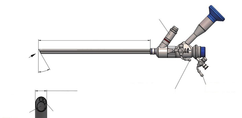
S20                                                                            Tieber and Lewandrowski. Technology in spinal endoscopy

                                                                          in Figure 1. It combines ergonomic advantage with good
                                                                          image reproduction and durability in routine use in frequent
                                                                          sterilization cycles at the senior authors surgery center.

                                                                          Illumination problems in early spinal endoscopes

                                                                          The ability to illuminate cavities inside the human
                                                                          body was the first significant milestone in the history of
                                                                          endoscopy. The use of a platinum glow wire at the tip of
                                                                          an endoscope is attributed to many inventors. Max Nitze
                                                                          and the instrument maker Josef Leiter developed a useful
                                                                          instrument in 1879 and published the application and
                                                                          results worldwide (2). In 1887, the platinum glow wire
                                                                          was replaced by a miniaturized light bulb developed by
                                                                          Alva Edison. Josef Leiter was awarded the first patent for an
                                                                          endoscope following the standards of high-quality design
                                                                          and inventive level. In 1906, Prof. Dr. Ringleb, the Zeiss
                                                                          physicist von Rohr and Georg Wolf, significantly improved
                                                                          the optical system by calculating the glass path and lenses
                                                                          as well as the use of the Amici prism (2). Prof. Dr. Ringleb
Figure 1 OEM Lumbar foraminoscope with working cannula                    contributed the geometric data for the mechanics, which
made by asap endoscopic disc systems GmbH. This ergonomic                 Georg Wolf transformed into a product that set the quality
45-degree design has a fixed and rotatable luer lock for irrigation       standard for the next 60 years (1).
and suction channel. The 30-degree viewing angle is preferred by             In the early 1990ies, multichannel endoscopes with
the senior author because if facilitates visualization in the hidden      optics, illumination, a working channel, and flushing
zone of Macnab between the exiting and traversing nerve root, and         function appeared on the market. Yeung designed the
during bony foraminal decompression.                                      first usable multichannel endoscope in cooperation with
                                                                          Wolf. Other products were simultaneously developed
                                                                          by Schreiber and Leu (Zurich) (9) in cooperation with
of endoscopic examinations and treatments. The first
                                                                          Karl Storz Endoskopie, Tuttlingen, and by Hal Matthews,
minimally invasive procedures using a visualization system
                                                                          who introduced the first fiber-optics endoscope in
were performed laparoscopically. Professor Kurt Semm
                                                                          cooperation with Danek Inc., Memphis (14). Since the
developed insufflators, surgical instruments, and pelvic                  endoscope was directed via the posterolateral approach
trainers for many years. The connection with a miniature                  into the lumbar neuroforamen, the surgical technique was
camera system provided all the components that formed                     called “Foraminoscopy”. The Danek foraminoscope was
the basis for minimally invasive procedures (13). At first                produced by Citation Medical Corp., Reno NV, and was
controversial, the minimally invasive endoscopic surgery                  introduced for the first time at the Laser Conference 1991
conquered almost all medical subspecialities within a few                 by Hal Matthews in San Francisco. Danek, Inc. held 10%
years. Nowadays, it is an integral part of routine diagnostic             Shares and had a world-wide exclusive licensing agreement.
and therapeutic protocols in spinal surgery as well. In                   The Danek foraminoscope used fiber bundles for image
1967, when no more than 1% of all medical procedures                      transmission and therefore offered only a viewing angle of
were performed with the help of an endoscope, currently,                  0°. The working length was 210 mm with a 3.5 mm Ø inner
more than 50% are employing an endoscope worldwide.                       working channel which allowed the use of instruments
Companies, such as Karl Storz or Richard Wolf, Smith                      up to that size. The outer Ø was 6.5 mm. The concept
and Nephew, Stryker and Olympus, who have recognized                      behind this design was to use the Danek foraminoscope
this trend early. Now, new OEM companies are joining                      for median and paramedian herniations intraforaminal
in, and continue as prolific innovators. An example of the                herniations. Unfortunately, this concept design failed for
senior authors preferred OEM spinal endoscope is shown                    several reasons. In most cases, the foraminoscope did not

© Journal of Spine Surgery. All rights reserved.                J Spine Surg 2020;6(Suppl 1):S19-S28 | http://dx.doi.org/10.21037/jss.2019.10.02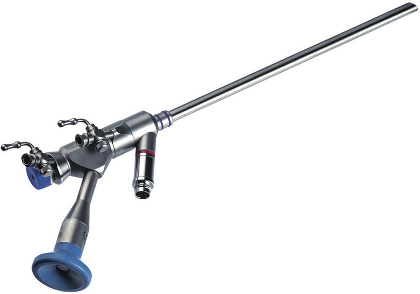
Journal of Spine Surgery, Vol 6, Suppl 1 January 2020                                                                                S21

fit through the foramen, and the 0° optic was not suitable          advantages with a brilliant image, and ample number of
for visualization of herniations located in the central canal.      instruments required to do the endoscopic discectomy
Moreover, the fiber optical system delivered a poor image           surgery. It could be sterilized and therefore was suitable
quality and the single-use endoscope raised the case cost           multi-use. Not needing disposable endoscopes lowered the
above reimbursement level for the procedure. Subsequently,          case cost during routine clinical operation significantly.
the product was removed from the market in 1994.                    Moreover, large capital purchases for proprietary
    Illumination and lens diameter, however, traditionally          videoendoscopic tower equipment were not necessary since
has been the limiting factor dictating design constraints for       the Leu-designed and Storz produced foraminoscope easily
spinal endoscopes as a minimum diameter for the lens was            connected to existing Video Towers. The foraminoscope
required to transport the image of the intraspinal anatomy          designed by Hans-Jörg Leu was offered by Karl Storz until
to perform surgery adequately. Hence, the size of the               2012 when it was replaced by more contemporary products
working and irrigation channels in some designs had to be           after a 21-year run.
reduced to accommodate an appropriate lens system. Newer               Endoscopes with a wider optical viewing angle
designs employing advanced illumination technologies                were introduced by Schreiber in 1989 allowing dorsal
have overcome these problems in part. Advances in the               vision around an annular tear (9). Kambin and Zhou
spinal endoscopy platform demanded by expert surgeons               demonstrated the use of a 30-degree endoscope
who are attempting to perform more complex endoscopic               recognizing that lateral recess stenosis can hamper
decompression and reconstructive surgeries will likely              the effectiveness of the procedure (17). Foley (18),
continue to push the limits of contemporary design                  M a t h e w s ( 1 4 ) , a n d D i t s w o r t h (19) furthered the
constraints and stimulate the quest for workarounds based           field of endoscopic spinal surgery by popularizing the
on new technologies.                                                transforaminal approach in their clinical studies published
                                                                    between 1998 and 1999. In 1997, Yeung introduced
                                                                    The Yeung Endoscopic Spine System (YESS) using
Advanced optics & next generation spinal
                                                                    a multichannel, wide-angled endoscope produced by
endoscopes
                                                                    Richard Wolf (20). The System consisted of a multi-
At the end of the 50ies, the physicist Harald H. Hopkins            channel, oval-shaped endoscope with a 207 mm working
developed rod lenses for endoscopy, which were based                channel of 2.7 mm Ø for use of 2.5 mm surgical
on the inversion principle. The proportion of glass in              instruments, an irrigation channel, and a rod lens. It was
the endoscope was increased significantly, and the air              designed for intradiscal decompression for protrusions,
chambers were substantially reduced. The result was a               for foraminal and extraforaminal herniations and for
higher contrast, and a brighter and sharper image. Only             foraminoplasty with lasers and radiofrequency. The YESS
Karl Storz recognized the meaning of the invention in the           system set itself apart from its competitor’s systems as a leap
middle of 1960 and executed an exclusive contract with              forward in quality with excellent visualization consisting
H. H. Hopkins. Through the use of glass fibers and halogen          of high-quality optics, camera, cold light projector and
lamps, Karl Storz redefined the illumination of body cavities       monitor (21).
under the name “cold-light source.” He was awarded
numerous patents for his inventions. By rod lenses and cold
                                                                    Endoscopy as minimally invasive spine surgery
light source, the quality of the intracorporeal observation
of body cavities reached new quality standard that were             At the beginning of the 1900s, physicians tried to create
previously considered impossible (15).                              artificial access to body cavities in addition to the natural
   The Leu foraminoscope was designed for treatment of              entry ways to body cavities in several subspecialties,
foraminal and extraforaminal herniations. It was produced           including ENT, gynecology, urology, and thoracic surgery.
by Karl Storz and its clinical use was demonstrated by              Laparoscopy is an examples of this development. In the
Dr. Leu in 1991 (16). The Storz-built endoscope used a              1970ies, the introduction of chip technology enabled the
Hopkins rod lens system with a 6° viewing angle which was           production of miniature camera systems and electronic
acceptable for the chosen indications. The working length           CO 2 insufflation and flushing devices. From 1962 and
was 145 mm and the inner working channel allowed the                onward, Kurt Semm, a physician and engineer, developed
use of instruments up to 3.0 mm in diameter. It had several         CO 2-insufflation devices and surgical instruments that

© Journal of Spine Surgery. All rights reserved.          J Spine Surg 2020;6(Suppl 1):S19-S28 | http://dx.doi.org/10.21037/jss.2019.10.02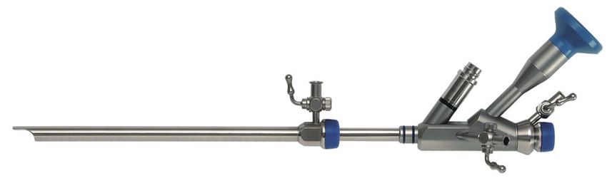
S22                                                                     Tieber and Lewandrowski. Technology in spinal endoscopy

were capable of performing laparoscopic surgery. Norbert           with working channel in many different lengths, diameters
Lemke produced cameras for Prof. Wolfgang Mauermeyer               of the working channel and outer diameter. In 2001, Dr.
since the early 1970s (1). In conjunction with endoscopes,         Hoogland became the co-founder of joimax ® . At the
these cameras enabled the video transfer of live surgeries         time, he brought his wealth of clinical knowledge of some
to seminar rooms (11). Karl Storz recognized at an early           5,000 endoscopic surgeries during the development of the
stage the potential of the development of video visualization      THESSYS method to the table. THESSYS is still used by
systems, consisting of an endoscope, cold light source,            many spine surgeons around the world. In 2004, Hoogland
miniaturized color camera, video monitor, and a recording          separated from joimax ® and founded his own company
system. An exclusive contract between Storz and Lemke was          maxmore® Hoogland Spine Products Inc in 2006. In 2001,
signed in 1980. The first mobile video towers were created,        Knight et al. showed that endoscopic foraminoplasty with a
consisting of these components (11).                               side-firing Ho: YAG laser can be useful in neural element
   In 1992, Dr. Thomas Hoogland was one of few surgeons,           decompression (23). The advent of lasers also stimulated
who bought the Danek System when its European                      electrothermal annuloplasty for low back pain which was
headquarter was established in Germany. He quickly                 also described by Tsou and Yeung in 2002 (24). Soon
realized that the Danek foraminoscope had significant              thereafter, more contemporary systems were introduced by
shortcomings and was unsuitable to treat intracanal,               Antony Yeung in 2003 with new custom instruments and
foraminal, extraforaminal herniations, and herniations with        cannula configurations following the lauch of the Yeung
free fragments in lumbar spine. In 1994, he concluded              Endoscopic Spine System (YESS™) in 1998, which was
that a foraminoplasty was necessary to treat the variety           designed around the transforaminal endoscopic approach
of disc herniations frequently encountered in clinical             for intradiscal and epiduroscopic procedures (25).
practice. He went on to develop a system of reamers, and
drills which was deployable over special guidance system,
                                                                   Image quality drives clinical protocols
called “Tom Shidi”. This system was produced in 1994
by an OEM manufacturer according to Hoogland’s exact               Image quality in endoscopy is perceived by magnification,
dimensions. At the time, he had the experience of several          depth of field, resolution, color truth, and high image
thousand of minimally invasive spine surgeries. But none           contrast as well as low distortion and homogeneous
of the endoscopes commercially available at the time               illumination up to the edge of the image. The image
were specifically produced for removal of herniated disc.          quality is visible to the viewer on the screen. The direct
It required to break away from traditional time-proven             view through the endoscope is only rarely used. The image
concepts from neurosurgical and urological product lines.          is transported from its place of origin inside the human
New optical designs based on unproven calculations                 body over a large number of individual components to
were required. Eventually, Hoogland also found and                 the screen. Only if all individual parts of the visualization
a manufacturer capable of producing a foraminoscope                system are perfectly coordinated with each other, a high-
allowing him reach intracanal herniations by a target-             quality image is produced. With the introduction of HD
oriented approach which is also known as the outside-in            flat screens and HD video cameras starting 2005 and direct
technique (22).                                                    marketing to consumers, even the layman was able to
   In 1998 Dr. Hoogland found a German OEM producer                detect a significant improvement in image quality. The
who was willing to invest in a new endoscope design. The           endoscope manufacturers were essentially forced by the
OEM calculated the rod lens optical system thoroughly              appearance of this invention on the consumer market to
and developed also all mechanical parts specially for this         develop endoscopes that provided optimized image quality
purpose. The result was a multichannel endoscope with              for the HD Video chain. Some of these technological
a length of 180 mm, a working channel of Ø 3.6 mm for              advancements in the 2000 coincided with introduction
instruments up to Ø 3.5 mm, an irrigation channel, a fiber         of endoscope into transforaminal spinal surgery. Future
glass illumination system with 0° and 30° optic variants, and      advances in clinical protocols will likely be driven by
an outer diameter of 6.3 mm. The OEM was able to realize           higher image quality standards that may provide the basis
these specifications by the use of 1.9 mm Ø rod-lens system        for artificial intelligence applications in image recognition,
without having to compromise on image quality. To date,            robotics, integration and automatization of surgical
this OEM offers still the widest range of coaxial endoscopes       processes.

© Journal of Spine Surgery. All rights reserved.         J Spine Surg 2020;6(Suppl 1):S19-S28 | http://dx.doi.org/10.21037/jss.2019.10.02
Journal of Spine Surgery, Vol 6, Suppl 1 January 2020 S23
Rigid endoscopes as the backbone of modern instruments and to effectively remove the debris they create
spine surgery from the surgical site. Surgeons who are expanding clinical
indications to replace traditional translaminar surgeries
At the distal end of the spinal endoscope is the lens. The
with further miniaturized versions of them are the impetus
image that a lens produces from an object is a so-called real
for implementing technology advances into the build of a
image. From the lens, the image must be transported to the
spinal endoscope which requires better illumination and
other end, to the eyepiece. The installation of rod lenses
image quality. While such support infrastructure advances
does this. Rod lenses are glass rods with optically processed
may be fueled by technology transfers from the space-,
ends, the spaces between the rod lenses are referred to as
military- or consumer sector developments in the area
air lenses. Each rod lens generates an intermediate image
of illumination, image quality, and high-definition video
that creates another intermediate image from the air lens
quality, development of more and stress-resistant spinal
and the next rod lens. According to the desired length of
endoscopes capable of carrying out the endoscopic spine
an endoscope as many rod lenses must be used until the
surgeries of the future will likely hinge on continued expert
image plane of the eyepiece is reached to allow viewing or surgeon input.
forwarding of the image to the camera. The quality of the
optical system depends on the type of glass selected for the
rod lens. The calculation of the glass path, which is the path Aberrations & image quality
the light beam travels from the lens to the eyepiece and is Spinal endoscopes suffer from limitations inherent to any
worked out by a physicist (7). The glass surface of the rod optical system. When transporting the image over several
lens is typically tempered. By refraction of the light in a lens systems, aberrations occur. The aberrations can be
converging lens, the propagation direction changes. Due to detected in the context of a geometric optics (4,5). They can
the calculated surface shape of the lens, the incident light be divided into four groups:
rays are collected at the focal point and thus produce an (I) Image sharpness error (spherical aberration,
intermediate image. astigmatism, field curvature, coma);
While this rod-lens design is time-proven for simple (II) Image scale error (distortion);
discectomies, newer designs of spinal endoscopes will (III) Chromatic aberration;
likely address some of the common problems encountered (IV) Image illumination error (vignetting, scattered
during contemporary spinal stenosis decompression- and light, and reflections).
reconstructive fusion procedures. For example, the lens is To deal with these types of aberrations, one needs to
near the surgical decompression area in the spine and is consider how a beam emanating from a particular object
subject to abuse not only by surgical instruments, including point behaves after passing through the lens system. Ideally,
power burs but also to the thermal stress of repetitive the rays intersect at one point. Due to the aberrations, there
sterilization cycles. Failure of the lens seal resulting in is instead a narrow constriction of the beam, which may
leakage is a common mode of failure in spinal endoscopes also be in the wrong place (distortion or field curvature).
with acute image deterioration during surgery. Technology In the case of aberrations, the beam behaves indifferently.
advancements in that area by replacing epoxy-based glues For example, in spherical aberration, the rays do not meet
with gold soldering have improved the durability of spinal in one point, which leads to blurring. Astigmatism, on the
endoscopes substantially. Another problem arises from other hand, occurs when objects are outside the optical axis
the use of glass rod lenses. Modern power drills, burs, and and, hence, are blurred. The cause is the different focal
aggressive decompression tools including chisels, and large lengths. The coma arises when light enters the system
rongeurs may put the structural integrity of the glass rod obliquely to the optical axis beam. Aplanatic lenses correct
system at risk due to vibration or hammering when these both the spherical aberration and the coma. If an image is
instruments are employed through the inner working created on a curved surface instead of a flat surface, field
channel of the spinal endoscope. Common failure modes curvature is created. Image sharpness decreases towards
include scratching of the lens or breakage of the rod lenses the edge. Additional aberrations may arise from chromatic
or failure of the seal between them. Several manufacturers aberrations resulting in color errors. The refractive index
have now endoscopes on the market with large irrigation of optical glass depends on the wavelength of the incident
and working channels to accommodate these motorized light. This phenomenon is called dispersion. It is the cause
© Journal of Spine Surgery. All rights reserved. J Spine Surg 2020;6(Suppl 1):S19-S28 | http://dx.doi.org/10.21037/jss.2019.10.02S24 Tieber and Lewandrowski. Technology in spinal endoscopy
of the chromatic aberration. The Gaussian error occurs standard and developed with Anthony Yeung the YESS™—
when colors spread at different speeds depending on the instrumentation. The design of the working cannula was
frequency of the light and split at optical interfaces. The tricky because the end was shaped in a slope, so the nerve
dependence of a physical parameter on the frequency of a and exiting root could be retracted and protected. Because
wave is the definition of dispersion. Such decomposition of all the indications Anthony Yeung had in mind, he
of white light into its spectral colors occurs as it passes designed a wide range of instruments which he showcased
through a prism, where the light rays diverge outward. The routinely during IITTS training courses at his Phoenix
dispersion of the optical lenses causes a variation of the Squaw Peak Surgical Facility through a mentorship
remaining aberrations with the wavelength. Correcting for program.
these aberrations is of critical importance when attempting
to construct a high-quality spinal endoscope. Aberrations
Optimized spinal endoscopes
are corrected in endoscopy by combining different types
of glass and partially using aspherical surfaces. It is obvious Extra-low-dispersion glass (ED glass) produces remarkably
that this a cost factor and determines the overall quality sharp, high-contrast images. ED glass is a fluoride-rich glass
and longevity of the spinal endoscope in routine clinical melt (FL glass) from which apochromatically corrected
use where endoscopes should be usable for 200 to 250 lenses are manufactured. ED glass can focus the entire color
sterilization cycles (surgeries). spectrum more precisely and almost eliminates the color
fringing caused by chromatic aberration. Barrel and pillow-
shaped distortions are avoided.
The mechanics of spinal endoscopy
An objective lens consists of several individual lenses. For
A modern spinal endoscope consists of a large number a lateral viewing angle, a prism is used, this is not necessary
of mechanical parts such as the cladding tube, the body, at 0° viewing direction. Additional components consist of
spacers, fixation screws, snap rings and many other items— distal end glass (sapphire glass), entrance pupil, aperture,
all in all approximately 150 parts. These mechanical parts and the objective lens. In the camera lens area, the focal
assemble the optical system in a way that prevents shifting length and the depth of field are determined. The aperture
of the optical components. A shift in these components influences the image brightness. All individual components
would result in a significant reduction in image quality and influence each other and lead to aberrations. The optimized
negate the benefits of optical enhancements to HD quality. production of rod lenses is based on physical calculation,
These mechanical parts have to be designed, manufactured testing with the latest measuring equipment and production
with the highest possible precision, and assembled and on the latest production machines. Also necessary are the
adjusted by long-term experienced technicians (26). The adhesives used and the surface finish used. An eyepiece
effort required and the impact on the overall quality of consists of a lens system, which is coupled via a PEEK ring
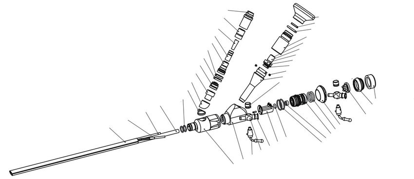
an endoscope are often underestimated. An example of a to the camera head. The PEEK ring prevents the ingress of
mechanical parts of high-end modern spinal endoscope is stray light. An eyepiece consists of field diaphragm, lenses,
shown in Figure 2. The use of CNC multi-axis machines filter thread, and socket. Due to the different arrangement
enables the production of high-precision mechanical parts, of lenses, there are a variety of eyepiece types. The
even in the micro range. The high-resolution, high-contrast characteristics of an eyepiece are defined by focal length,
color reproduction and sharpness display screen have exit pupil, and field of view (Figure 1). The same rules apply
prompted manufacturers of endoscopes and visualization to HD optimization as to rod lenses.
systems to optimize all components such as endoscope, The image quality of an endoscope can be optimized by
camera, processor, and display for HD image quality. The measuring the following parameters: the intensity of the
improved detail recognition, the greater depth of focus, and optical fibers, light intensity, and quality of the lens system,
color truth help to assess images more accurately, which will color truth, sharpness and focus, viewing angle, the field
likely form the basis for integration of artificial intelligence of view (visible surface). For a detailed evaluation of an
and robotics in endoscopic spine surgery. The use of CNC- optical system, it is not enough, only to consider individual
Machines also had a significant influence on the production aberrations. To adequately describe the performance of the
of precise surgical instruments. Richard Wolf offered the optics, consideration of the modulation transfer function is
opportunity to produce surgical instruments of the highest essential. Excellent image quality is characterized by the fact
© Journal of Spine Surgery. All rights reserved. J Spine Surg 2020;6(Suppl 1):S19-S28 | http://dx.doi.org/10.21037/jss.2019.10.02Journal of Spine Surgery, Vol 6, Suppl 1 January 2020 S25
Storz-connector - 16
Wolf-connector - 15 17 - Eyepiece
18 - Sapphire glas
Glass cone - 14 19 - Sapphire glas
Glass cone - 13 20 - Sealing bush
ACMI connector upper part - 12 21 - Achromatic lens
Indicator ring support - 11 22 - Occular diaphragm
23 - Clamp ring
Ring black - 10 24 - Threaded pin
ACMI - lower part - 9 25 - Clamp bushing
26 - Threaded pin
Strain relief - 8
27 - Occular connecting piece
Angle piece - 7 28 - Spring screw/cap
Transition piece - 6
O-ring - 5 43 - Sealing cap
WC-tube - 4 42 - WC-con-
Inner tube - 3 necting cone
41 - Corss - slit valve
WC.tube - 2
40 - Bushing
Outer tube - 1 39 - O-ring
35 - O-ring 38 - Connector WC
34 - Mirror mount 37 - O-ring
33 - Cylindrical pin 36 - O-ring
32 - Flush - stopcock
31 - Stop - cock
30 - Main body bipartite,p2
29 - Main body bipartite,p1
Light plug
177
Woring length
Z
30°
Fixed luer lock Rotatable luer lock
ANSICHT Z Ø6.9/Ø6.1(oval) (suction/irrigation channel) (working channel)
M 2:1
Suction/irrigation channel
Working channel ID=1.8mm
ID=4.1mm
Figure 2 Technical drawings and specifications of a rigid OEM lumbar endoscope made by asap endoscopic disc systems GmbH showing
the assembly of the outer and inner tube with an oval inner working channel, the light source and eye piece assembly. The oval working
channel allows for rapid lavage of debris encountered during bony and soft tissue decompression.
that individual delicate structures are reproduced not only processor (CPU). The clamping ring at the proximal end
separately (resolution), but also with a sufficient difference of the camera head establishes the connection between
between light and dark (contrast). the eyepiece and the camera sensor. The optical image
is converted into electrical signals. Behind it is the focus
ring; it is used to focus the image. Once set, the zoom
Full HD playback
ring can be used to zoom the image without the need for
For the HD optimization of the endoscopic image during refocusing (parfocal zoom). With the optical zoom, the
spinal endoscopy, light generation and transmission to the high image quality is maintained because the image section
spinal anatomy is crucial. Xenon light sources with 300 is mechanically enlarged, and thus the high resolution is not
watts or light sources with LED and laser light technology changed. Digital zoom will extrapolate an image section, but
achieve the best possible illumination for full HD image details may be lost. C-MOS sensors have proven themselves
reproduction. A HD camera consists of camera head and for HD image transmission. C-MOS sensors convert the
© Journal of Spine Surgery. All rights reserved. J Spine Surg 2020;6(Suppl 1):S19-S28 | http://dx.doi.org/10.21037/jss.2019.10.02S26 Tieber and Lewandrowski. Technology in spinal endoscopy
incident light into voltage. The conversion of the light is OLEDs can emit light in different colors, while
done by transistors, which are located directly on the pixel. LCDs require additional color filters;
Specific data such as photosensitivity, pixel number, readout The energy consumption of OLED screens is lower;
speed, dynamic range, and chip size achieve outstanding Shorter electro-optical response times.
values. The image noise is mostly suppressed. The disadvantage of OLED technology is currently
a much shorter life cycle. The OLED technology is
subject to further improvements and may eventually find
HD screen
implementation in the spinal endoscopy infrastructure
The surgeons are highly aware of the technology prompting surgeons to identify and validate new clinical
advancements introduced by industry into the consumer protocols.
market since the ongoing improvements in high quality of
flat panel displays has become affordable for home audio-
Information technology & integration
video applications. The terms HD, Full HD, 4K, and 8K
refer to the number of pixels that are spread across the Modern information technology (IT) is the basis for
screen and relate to the resolution. Current flat screens advanced OR integration. First systems enabled recording
offer more than high sharpness and a clear picture. and storage of still images and simple video sequences
Behind a wealth of terms hide technological innovations for electronic documentation. Another step up was the
that significantly affect the image quality. The earliest development of software programs for storing image data
technology was offered as LCD (Liquid Chrystal Display). and video sequences with interfaces for hospital PACS
As liquid crystals only modulate the light but do not systems (8). These systems, in combination, provide the
emit light themselves, the backlight is placed behind the foundation for the paperless electronic health records
display for televisions, whereby the image information is (EHR). With EHR, large amounts of data can be stored
displayed in transmitted light. LED TV are televisions that in a small space and while providing ready access to work
use light-emitting diodes (LEDs) for backlighting. More stations over the healthcare organization. The purpose of
appropriately, such devices may have been called “LED the OR Integration concept is to bring together actions
background Illuminated LCD TVs”. HDR stands for High that can be optimized through electronic recording
Dynamic Range Image and denotes a high-contrast image. and control processes to enable faster action and safer
In principle, this technology approaches the eyesight of operations (9). Software programs create an easy-to-use
the human eye by using high bit values to achieve a large interface that enables sterile control of electronic devices
number of brightness levels. Image files are generated, through a touch screen, as well as the retrieval and storage
which better capture natural brightness. HRD Video uses of intraoperative data by the surgeon, assistant or nurse, and
a color depth of 10 bits and thus achieves a high dynamic is also developed for specialized disciplines and common
range. By adding three subpixels of the primary colors red, types of surgery. Complex workplaces, which contain all
green and blue, colors appear in the television picture. the equipment required for the intervention, are recorded,
Above all, HDR improves the appearance of saturated light controlled and their use monitored by data management.
colors and very dark colors. The HDR technique also avoids Other data sources of intraoperative modalities such as
over- and under-exposure. OLED (organic light-emitting Navigation, Endoscopy, C-arm, and CT are also connected
diode)—an organic light-emitting diode is a luminous thin- via interfaces, and the image, text, and vital data are
film component made of organic semiconducting materials. displayed on large monitors. The surgeon is key to creating
The difference to the LCD technology is that every single integration OR systems that assist in the clinical problem
pixel itself shines. This results in a significant improvement solving.
of the image contrast because each pixel assumes arbitrary
brightness levels and produces a much-improved color
Key future technologies
fidelity. OLED screens offer several advantages:
OLED thin-film components are inexpensive to Nanotechnology is used in semiconductor and surface
manufacture; physics combined with chemical processes. Nanotechnology
OLEDs do not require backlighting as they emit is also used in sub-areas of mechanical engineering and food
light themselves; production. Nanotechnology will likely reach significance
© Journal of Spine Surgery. All rights reserved. J Spine Surg 2020;6(Suppl 1):S19-S28 | http://dx.doi.org/10.21037/jss.2019.10.02Journal of Spine Surgery, Vol 6, Suppl 1 January 2020 S27
in endoscopy by in coating processes. The coating of to declare.
endoscope cavities, of endoscope lenses but also screens
(nanotubes) is conceivable. Photonics describes the technical Ethical Statement: The authors are accountable for all
mastery and use of light in every form. Why photons aspects of the work in ensuring that questions related
play such a unique role in science and industry is due to to the accuracy or integrity of any part of the work are
the extraordinary properties of light that benefit humans. appropriately investigated and resolved.
Photonics can be found in all critical areas of modern
society and economy. Examples can be found in medicine,
References
industrial production with laser systems or energy-efficient
illumination with LEDs and OLEDs. Photonics offers 1. Reuter MA. Geschichte der Endoskopie: Handbuch und
excellent potential to deliver innovative solutions for the Atlas (German Edition) (German) Hardcover. Reuter M,
markets and challenges of tomorrow. Volume 1-4, 1998.
Artificial Intelligence (AI) will likely play a crucial 2. Reuter MA. Geschichte der Endoskopie: Handbuch und
role in the development of health technology, including Atlas (German Edition) (German) Hardcover. Reuter M,
endoscopy (10). If the current trend continues an increasing Volume 5-7, 1998.
amount of healthcare data will be processed without 3. Kambin P, Sampson S. Posterolateral percutaneous
the need for intervention or processing by clinical staff. suction-excision of herniated lumbar intervertebral
The shortage of highly trained clinical personnel and discs. Report of interim results. Clin Orthop Relat Res
increasing pressure for cost-cutting are additional drivers 1986;(207):37-43.
of this trend. AI could take over routine tasks that do not 4. Kambin P, Brager MD. Percutaneous posterolateral
require the specific expertise of a professional (11). It discectomy. Anatomy and mechanism. Clin Orthop Relat
may aid during delicate portions of the spinal endoscopy Res 1987;(223):145-54.
procedure. Examples imaged based recognition of 5. Kambin P, Schaffer JL. Percutaneous lumbar discectomy.
anatomical structures or defining the three-dimensional Review of 100 patients and current practice. Clin Orthop
extent of interventional or surgical procedures such as the Relat Res 1989;(238):24-34.
bony decompression procedure. Additional benefits may 6. Forst R, Hausmann B. Nucleoscopy--a new examination
play out in decreasing operative times, or improved quality technique. Arch Orthop Trauma Surg 1983;101:219-21.
assurance, and standardization. Very likely a combination 7. Onik G, Helms CA, Ginsberg L, et al. Percutaneous
of AI and robotics will appear in clinical applications in one lumbar diskectomy using a new aspiration probe: porcine
form or another. and cadaver model. Radiology 1985;155:251-2.
8. Kambin P, Nixon JE, Chait A, et al. Annular protrusion:
pathophysiology and roentgenographic appearance. Spine
Conclusions
(Phila Pa 1976) 1988;13:671-5.
The spine surgeon users expect technological improvements 9. Schreiber A, Suezawa Y, Leu H. Does percutaneous
of spinal endoscopes and their supporting video equipment nucleotomy with discoscopy replace conventional
to improve clinical outcomes and expand the indications discectomy? Eight years of experience and results in
for the procedure. Transfer of innovative key technologies treatment of herniated lumbar disc. Clin Orthop Relat Res
from other industries and the consumer market will likely 1989;(238):35-42.
continue leading to improvements in the efficacy and safety 10. Kambin P, Zhou L. History and current status of
of spinal endoscopy. percutaneous arthroscopic disc surgery. Spine (Phila Pa
1976) 1996;21:57S-61S.
11. Birkfellner W. Applied Medical Image Processing: A Basic
Acknowledgments
Course. Second Edition ed. Vienna 2014.
None. 12. Diethelm V. Physik Mittelstufe: Optik, Magnetismus,
Elektrizitätslehre, Atomphysik (Mentor Lernhilfen Physik)
Taschenbuch. Langenscheidt Fachv; 1998.
Footnote
13. Kramme R. Medizintechnik. Verfahren - Systeme -
Conflicts of Interest: The authors have no conflicts of interest Informationsverarbeitung: Springer Reference Technik
© Journal of Spine Surgery. All rights reserved. J Spine Surg 2020;6(Suppl 1):S19-S28 | http://dx.doi.org/10.21037/jss.2019.10.02S28 Tieber and Lewandrowski. Technology in spinal endoscopy
2017. Springer-Verlag Berlin Heidelberg, 2017. endoscopy and discectomy: state of the art. Mt Sinai J Med
14. Mathews HH. Transforaminal endoscopic 2000;67:327-32.
microdiscectomy. Neurosurg Clin N Am 1996;7:59-63. 22. Hoogland T, van den Brekel-Dijkstra K, Schubert M, et al.
15. Pedrotti FL. Introduction to Optics. Pearson; 3rd ed. Endoscopic transforaminal discectomy for recurrent lumbar
16. Leu HJ, Hauser R. Die perkutan posterolaterale disc herniation: a prospective, cohort evaluation of 262
Foraminoskopie: Prinzip, Technik und Erfahrungen seit consecutive cases. Spine (Phila Pa 1976) 2008;33:973-8.
1991. Arthroskopie 1996;9:26-31. 23. Knight MT, Goswami A, Patko JT. Cervical percutaneous
17. Kambin P, Zhou L. Arthroscopic discectomy of the lumbar laser disc decompression: preliminary results of an ongoing
spine. Clin Orthop Relat Res 1997;(337):49-57. prospective outcome study. J Clin Laser Med Surg
18. Foley KT, Smith MM, Rampersaud YR. Microendoscopic 2001;19:3-8.
approach to far-lateral lumbar disc herniation. Neurosurg 24. Yeung AT, Tsou PM. Posterolateral endoscopic excision
Focus 1999;7:e5. for lumbar disc herniation: Surgical technique, outcome,
19. Ditsworth DA. Endoscopic transforaminal lumbar and complications in 307 consecutive cases. Spine (Phila
discectomy and reconfiguration: a postero-lateral approach Pa 1976) 2002;27:722-31.
into the spinal canal. Surg Neurol 1998;49:588-97; 25. Yeung AT, Yeung CA. Advances in endoscopic disc and
discussion 597-8. spine surgery: foraminal approach. Surg Technol Int
20. Yeung AT. Minimally Invasive Disc Surgery with the 2003;11:255-63.
Yeung Endoscopic Spine System (YESS). Surg Technol Int 26. Krause W. Konstruktionsmerkmale der Feinmechanik.
1999;8:267-77. Werner Krause. 4th Edition ed.
21. Yeung AT. The evolution of percutaneous spinal
Cite this article as: Tieber F, Lewandrowski KU. Technology
advancements in spinal endoscopy for staged management of
painful spine conditions. J Spine Surg 2020;6(Suppl 1):S19-S28.
doi: 10.21037/jss.2019.10.02
© Journal of Spine Surgery. All rights reserved. J Spine Surg 2020;6(Suppl 1):S19-S28 | http://dx.doi.org/10.21037/jss.2019.10.02You can also read


