The Easter Seal Guide To Children's Orthopaedics - Prevention, Screening, And Problem Solving
←
→
Page content transcription
If your browser does not render page correctly, please read the page content below
The Easter Seal Guide To
Children’s Orthopaedics

     Prevention, Screening,
      And Problem Solving
The
      Easter Seal
      guide to
      children's
      orthopaedics

Prevention, Screening and Problem Solving

     © 1982, The Easter Seal Society, Ontario
CONTENTS

Chapter I                                                         Page 4
    GENERAL FEATURES

    A.   Diagnosis
    B.   Explanations
    C.   Psychological Traits in Mothers
    D.   Referral

Chapter II                                                    Page 14

    THE ROLE OF THE PRIMARY CARE PHYSICIAN

    A.      Prevention — Spina Bifida
                       — Cerebral Palsy
                       — Genetic Disease
                       — Injury
                       — Child Abuse
    B.      Screening — Congenital Dislocation of the Hip
                      — Scoliosis

    C.      Advisor on Lifestyle
    D.      Problem Solving — injury
                             — limp
                             — pain
                             — hip
                             — knee
                             — tibia
                             — spine
                             — the painful swollen joint
                             — osteomyelitis
                             — tumours

Chapter III                                                       Page 47
    THE PRIMARY CARE PHYSICIAN'S ROLE IN PARTICULAR AREAS
    1.      Newborn Nursery — congenital dislocation of the hip
                              — feet
                              — torticollis
                              — obstetric paralysis
                              — spina bifida
                              — arthrogryposis
                               — congenital amputations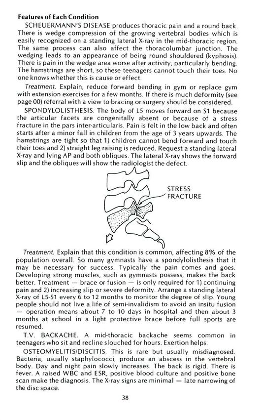
2.    Toddlers and Preschoolers — torsional problems
                                   — angular deformities
                                   - flat feet
                                   — trigger fingers
                                   — curly toes

   3.    Adolescents — spinal screening
                       foot problems

Chapter IV                                                 Page 69

    SHORT TOPICS

    1.   Sports Medicine
    2.   Chronically Handicapped
    3.   Record Keeping

Chapter V                                                  Page 79
    HANDOUTS TO PHOTOCOPY AND GIVE TO PARENTS

Chapter VI                                                 Page 91
    SKILLTEST

    A note about The Easter Seal Society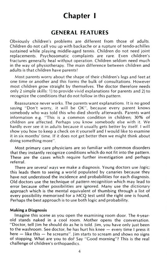
Chapter I

                      GENERAL FEATURES

Obviously children's problems are different from those of adults.
Children do not call you up with backache or a rupture of tendo-achilles
sustained while playing middle-aged tennis. Children do not need joint
replacements. Psychosomatic complaints are rare. Even children's
fractures generally heal with.out operation. Children seldom need much
in the way of physiotherapy. The main difference between children and
adults is that children have parents!

  Most parents worry about the shape of their children's legs and feet at
one time or another and this forms the bulk of consultations. However
most children grow straight by themselves. The doctor therefore needs
only 2 simple skills: 1) to provide vivid explanations for parents and 2) to
recognize the conditions that do not follow in this pattern.

  Reassurance never works. The parents want explanations. It is no good
saying "Don't worry,it will be OK", because every parent knows
somebody who was told this who died shortly afterwards. Parents want
information e.g.   "This   is   a common   condition   in   children;   30%   of
children are affected. Perhaps you know somebody else with it. We
hardly ever see it in adults because it usually gets better by itself, I will
show you how to keep a check on it yourself and I would like to examine
it in six months' time. If it does not get better then we might think about
doing something more".

   Most primary care physicians are so familiar with common disorders
that they instantly recognize conditions which do not fit into the pattern.
These are the cases which require further investigation and perhaps
referral.
  There are several ways we make a diagnosis. Young doctors use logic;
this leads them to seeing a world populated by canaries because they
have not understood the incidence and probabilities for each diagnosis.
Old doctors use the technique of pattern recognition which may lead to
error because other possibilities are ignored. Many use the dictionary
approach which is the mental equivalent of thumbing through a list of
every possibility memorised for a MCQ test until the right one is found.
Perhaps the best approach is to use both logic and probability.

Making a Diagnosis
   Imagine this scene as you open the examining room door. The 4-year-
old stands naked in a cool room. Mother opens the conversation.
"Doctor, tell Jim he should do as he is told. Jim, you have only just been
to the washroom. See doctor, he has hurt his knee — every time I press it
here — like this — he screams". Jim starts to scream and shows no signs
of stopping. What are you to do? Say "Good morning"? This is the real
challenge of children's orthopaedics.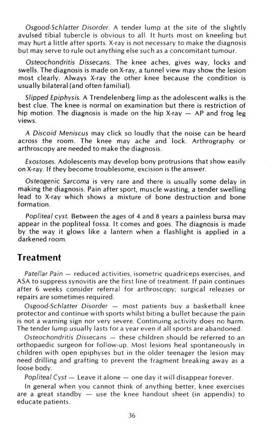
When a child is screaming on its mother's lap, it is hard to take a
history, accumulate evidence, impart information and remain looking
cool. Both children and their parents are easily frightened and upset.
Subterfuge is necessary to make a diagnosis. Initial examination may
easily get out of hand.

The Consultation
  Have toys around and enough space to see a child walk about — the
corridor outside can often be used.

Establish Rapport
   It is always good to try to make friends with your patients. You don't
know when you may need it. Use body language — get close to talk —
don't stand talking down to them or sit behind the desk. Look them in the
eye. Smile. Talk to the parents so that the children have time to get used
to you and then gradually involve the child in the conversation.
   Use their names, don't call them "mother" or "princess". Sit down to
talk, don't stand — people think you are about to walk away.

History Taking
  Discover the reason for the visit.
    1.   Is it real pain or disability?
    2. Is it a desire to catch a problem early?
    3. Were they sent by Granny or a worrying neighbour?
    4. Is there a family history of a problem?
    5. Do they not know what to expect and want a guarantee that the
       child is normal?

  When you understand the reason for the visit you will appreciate what
they are expecting from you. What ever else you may feel the situation
demands you will deal with this first.
  Wise parents don't like to make negative remarks about their children
within earshot. At least some of the interview should take place without
the child being present to allow for these exchanges. Either send the child
out into the waiting area to play with some toys, ask the receptionist to
entertain him or leave him getting dressed while the parents go into
another room with you.
  Always enquire about a family history and their knowledge of the
condition to avoid the scenario like this:
Doctor — "He will grow out of it".
Mother — "Are you sure"?
Doctor — "Well, as a matter of fact, I listened to a three hour lecture on
just this subject last week. Experts from Japan and New York agreed that
children grow out of this".
Mother, pulling up her skirt — "Then, how do you explain these scars
doctor? My legs were just like his and I needed operations".
  Now all you can do is wriggle, wondering whether you meant that 99%
get better and that she was just one of the unlucky 1 % or did you mean
that the operation was unnecessary. "If only I had taken a family history I
would have been a little more accurate in what I said".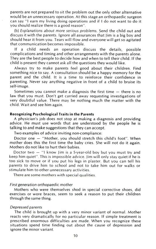
Examination
  Quieten infants with a bottle when you examine them. A music box is
wonderful. Small clinging children should be examined on their parent's
knees to avoid the cutaneo-lachrimal reflex — when the skin touches the
examining couch the tears begin to flow. If there is a tender place leave
this until the last and then feel it gently, only once. A thorough
examination should be carried out. The thirty-second examination of one
small part lacks conviction. Undress the children sufficiently.
ROUTINE
                                   EXAMINATION
             2
                                   IN 18 STEPS
  'WALK'     'RUN'
  ? LIMP     ? COORDINATION

                      'WALK ON YOUR HEELS'
                       ? SHORT HEELCORDS
'JUMP ON 1 LEG'
? STRENCTH OF EACH
LEG
                                             WALK ON TIPTOES
                                             ? STRENGTH

             'HOLD YOUR HANDS
            TOGETHER AND BEND
             FORWARDS'
             ? SCOLIOSIS

                                       ? SHOULDERS LEVEL
           ? TORTICOLLIS
                                       ? SPINAL DIMPLES
           ? CARRYING ANGLE
                                       ? SPINAL HAIRY PATCH
           ? LEG LENGTHS EQUAL
           ? KNOCK KNEE, BOW
           LEG
           ? FOOT ARCHES
           ? IN-TOEING, OUT-
           TOEING

                               7
'SIT DOWN, NOW

? KYPHOSIS                                           STAND UP'

? LORDOSIS                                           SHOWS ARMS USED TO
                                                     PUSH UP.
                                                     A POSITIVE GOWER'S
                                                     SIGN FOR MUSCULAR
                                                     DYSTROPHY.
           11
           'NOW LIE DOWN'
           ? EFFUSION
           ? WASTING

                                                13
                                                'DO THE SPLITS.'
                                                NEWBORNS SHOULD
                                                GET KNEES TOUCHING
 12                                             THE TABLE.
 'BEND UP.'                                     ? ABDUCTION
 ? STIFFNESS                                    ? HIP DISEASE
 ? CLICKING MENISCI

      14
                                                        16
      'AGAIN.'               15
                                                        TEST EXTERNAL
      ? ABDUCTION            'LIE FACE DOWN'.
                                                        ROTATION.
      ? HIP DISEASE          ? POPLITEAL CYST
                                                        ? HIP DISEASE
                                                        ? TORSION

                 17                                   18
                                                      'CAN YOU MAKE YOUR
                 TEST INTERNAL
                 ROTATION.                            THUMB TOUCH YOUR
                                                      FOREARM?'
                 ? HIP DISEASE
                 ? TORSION                            ? JOINT LAXITY
Investigations
  Order an X-ray with as much deliberation as you would an expensive
bottle of wine. If you want one bottle, do not ask for the whole cellar.
Here are some guides:
Hips:      request an AP and frog leg of the pelvis. It is important to
           compare one side with the other. An AP alone will miss half
           the pathology
Spine:     scoliosis can only be detected on standing films. A single 3'
           standing P.A. spine is sufficient. The PA projection irradiates
           the breast less than an AP projection. Do not order bending
           films. The fewer films the better for screening.
          backpain. An AP, lateral and spot lateral of L5.S1 are a good
          choice. A spondylolisthesis can be recognized as easily on the
          spot lateral as on obliques.
Knee:      An AP and lateral is a good start. When there is patellar pain
           ask for an axial view with the knee flexed 45 degrees and the
           quadriceps relaxed. This will show subluxation. A tunnel view
           is required for osteochondritis. To assess knock-knees request
           a standing AP.
Feet:      Standing films always (unless fracture is likely.)
Legs:      If you want the whole of the legs or you want to compare leg
           lengths request a 3 foot orthoroentgenogram.
  Always put in your provisional diagnosis if you want to get a helpful
report.

  Radiologists lead lonely lives sitting in the dark reporting films. They
enjoy talking. So if you don't know what to order or what the film means
ask the radiologist. They always know what to do next.
  Many parents will quiz you on the hazards of radiation — a difficult
subject. Give them a photocopy of the handout in the appendix.

Explanations
  "Relieving anxiety is a large part of every physician's stock-in-trade"
Victor Fuchs
  (a) Minor problems. The more trivial the complaint the greater the
psychological content. Careful explanations are required that will be
understood and which will carry conviction when repeated to Granny.
Unfortunately, some parents blank out as soon as you start to speak One
answer to this is the distribution of written handouts. This saves time and
the chance of misunderstanding. If people look puzzled after a simple
explanation you should not launch into a more elaborate explanation.
Instead ask them to tell you whatyou just said.
  A large part of an explanation is the art of putting problems into
perspective. Some people see minor problems as major ones and regard
normal variants as mysterious, rare diseases.
   If simple analysis does not smooth the mother's furrowed brow it may
help if you describe how you or your friends survived when your own
children had the same problem. In desperation you may point out that if
                                     9
parents are not prepared to sit the problem out the only other alternative
would be an unnecessary operation. At this stage an orthopaedic surgeon
can say "I earn my living doing operations and if I do not want to do it
you should realize there is a good reason".
(b) Explanations about more serious problems. Send the child out and
discuss it with the parents. Ignore all assurances that Jim is a big boy and
should hear it from you. Tears will flow and everyone will get so agitated
that communication becomes impossible.
If a child needs an operation discuss the details, possible
complications and timing and other arrangements with the parents alone.
They are the best people to decide how and when to tell their child. If the
child is present they cannot ask all the questions they would like.
Always try to make parents feel good about their children. Find
something nice to say. A consultation should be a happy memory for the
parent and the child. It is a time to reinforce their confidence in
parenting. Never say anything negative in front of a child to bruise its
self-image.
Sometimes you cannot make a diagnosis the first time — there is no
law that you must. Don't get carried away requesting investigations of
very doubtful value. There may be nothing much the matter with the
child. Wait and see him again.
Recognizing Psychological Traits in the Parents
A physician's job does not stop at making a diagnosis and providing
advice. He must use words that are understood by the people he is
talking to and make suggestions that they can accept.
Two examples of advice inviting non-compliance:
Doctor one — "Mother, you should stretch this child's foot". When
mother does this the first time the baby cries. She will not do it again.
Mothers do not like to hurt their babies.
Doctor two — "1 know Jim is a 5-year-old boy but you must try and
keep him quiet". This is impossible advice. Jim will only stay quiet if he is
too sick to move or if you put his legs in plaster. But you can tell his
parents to drive him to school and not to take him out for walks or
stimulate him to other unnecessary activities.
There are some mothers with special qualities.
First generation orthopaedic mother
Mothers who were themselves shod in special corrective shoes, did
exercises or wore braces, seem to seek a reason to put their children
through the same thing.
Depressed parents
The child is brought up with a very minor variant of normal. Mother
reacts very dramatically for no particular reason. If simple treatment is
prescribed enormous difficulties are made. When you recognize these
situations spend time finding out about the cause of depression and
ignore the minor variant
10Little League father
This is well known.
The faddists
They may be into health shoes, gymnastics or vitamins Fundamentally
they want you to endorse the view they intend to impose on their children.
Fastidious mothers
Some mothers of very young children, particularly first children, have
such an overriding impression of a beautiful bouncing baby that they get
quite shaky at the knees if there is talk of putting casts on feet or Pavlik
harness for the hip. Non-compliance is the rule for a rich variety of
reasons. Each time the child is brought to the office looking beautiful
with very lovely clothes but without any treatment being continued You
will either have to accept frequent removal of apparatus or if it is a
serious problem, admission to hospital.
Mothers with mothers
Some Crannies seem to wield power in a family by undermining the
self-image of the younger members. A frequent target is the shape of the
grandchild's foot or leg. Sometimes, of course, Cranny is right. If you
suspect the situation ask "Are you worried about this or is it someone
else in the family?" If the answer is "Granny" tell mother what to say to
Granny.
Mothers with daughters
Mothers and teenage daughters sometimes choose to fight in the
office.
"It began last week".
"No, it was the week before".
"It is worse after exercise".
"No, it is worse at night" and so on.
The discussion usually settles on the fact that mother doesn't like her
daughter standing with round shoulders and wants a course of exercise
for her. The daughter may be shy about her image but is angered by
mother's criticism of her body. Take the daughter to one side and tell her
she is good enough to be a cheer leader. Take the mother on one side
secretly and tell her that being a cheer leader will be the best possible
posture training she could have.
Tricky parents
Some parents will quiz you and can easily lead you into saying things
which give the wrong impression and which you regret.
Doctor — "I think that he will grow out of it, but may I see him again
in a year just to see that it is going along as expected"
Parent — "I guess you want to see him again because there is the
chance that it won't get better".
Doctor, not rising to the bait — "No, it is just routine".
Parent — "And if it doesn't get better; what would you do then".
Doctor — "Well a small proportion require operation at the age of
18".
11Parent — "Doctor, one minute you say it is nothing, the next minute
you tell me he may need an operation. I guess you are going to
tell me next that the operation may not work?"
The answer to this is to keep cool, explain it again, and don't lose the
initiative.
Parents burdened by responsibility
Parent — "I know it's very slight, but I think he will blame me when
he gets older for not getting it fixed when he was small".
Doctor — "The operation is the same whether he is young or old. Why
not leave it until he is old enough to decide for himself?"
Immigrant parents
They don't understand but keep saying "Yes". They have rehearsed the
history so that speech is better than comprehension In addition to
language problems there is a cultural difference.
Talking loudly, though frequently tested, is not enough. You need an
interpreter. "Do you have a friend who speaks your language whom we
could telephone now?"
Follow-up
Should you ask patients to come back so that they can give you a
progress report? Skill is required to strike the right note
"If it doesn't get better, come back". This says that you are
disinterested.
"Make an appointment to come back in two weeks. But if it is better
by then you should cancel it." You are saying that you are interested.
"Come back in six weeks so that you can tell me how it feels". You are
interested but you are encouraging a pre-occupation with symptoms.
"Come back in six weeks for a check. If it is no better the next step
would be " Not only are you interested but you are the judge of
progress. Frivolous complaints are discouraged by describing the next
step.
"There is no need to make another appointment. I am sure it will settle
in a week. But if it doesn't I would like to be the first person to know".
When you are 99% certain of cure this strikes the right note.
When should Patients be Referred?
It is hard to lose points when making a referral, unless you send
patients to the wrong person. Paediatricians believe, of course, that "The
natural enemy of a child is the orthopaedic surgeon" When you do make
a referral send a letter stating the problem you want solved. Only a line or
two is necessary Send the X-rays; an X-ray report is valueless. Describe
the past and social history to provide some continuity. Referrals may be
for:
1, Unfamiliar problems. When a patient comes with an unfamiliar
problem; you can either read it up or refer them, which is much less
trouble There may be some diseases you do not like, and this is a good
reason for referral.
VI2. Diseases which need help you cannot provide. For example, a child
with multiple problems requiring services of a team, such as a child with
cerebral palsy.
3. Parents who need your opinion reinforced because they have doubts.
As soon as a patient is thinking about a second opinion, it is wise to
arrange one Do not create a scene if you want to keep them as patients.
When the second opinion reinforces your advice they will be more
inclined to believe you in the future. When patients shop for advice
unassisted they become very confused and disbelieving.
4. If you sense a potentially litiginous situation.
13Chapter II
THE ROLE OF THE PRIMARY CARE PHYSICIAN
The primary care physician has four roles.
1. Prevention of musculoskeletal disorder — for which he is in a
unique position.
2. Screening — recognizing problems before parents do.
3. Advisor on lifestyle.
4. Problem solving — diagnosis and management of patients with
complaints.
PREVENTION
The worst diseases lend themselves to the preventive approach. The
scourges of the past, e.g. polio, rickets and tuberculosis, have been
eradicated by this approach. With help from primary care physicians,
children's orthopaedic surgeons (who are now regarded as strong as a
dinosaur and twice as smart) may in the future become just as extinct.
The incidence of diseases such as cerebral palsy, spina bifida and
muscular dystrophy — the heavy weights — can be reduced through
prevention. These are heart-breaking diseases. Many of the children
require institutional care. Each child may cost a million dollars in
medical, welfare and residential costs. There are strong human and
economic arguments for prevention.
But prevention is always controversial. The evidence is often soft and
people will say that their rights are violated. "If I want to have a baby at
home without a doctor I am going to" Physicians will object to being
directed and will talk about unforeseen effects. Even the success of a
program is hard to assess in the early stages. Although prevention seems
like a good idea, programs are slow to catch on.
Complete prevention of some diseases is already within grasp but little
implementation has been undertaken.
Spina Bifida
Currently maternal serum AFP (alphafetoprotein) screening at about
the fourth month of pregnancy will identify twins, open neural tube
defects (anencephaly and spina bifida) and a few other even rarer
disorders. Ultrasound, amniocentesis and fetoscopy will distinguish the
cause of a raised AFP and make it possible to offer therapeutic abortion
for the pathological foetus. The technique identifies 98% of affected
foetuses and the proponents claim to have avoided false positives. The
Spina Bifida Parents' Association has endorsed this campaign, but
unfortunately the test is still not yet generally available.
In one study the incidence of spina bifida was reduced by a factor of
10 by nothing more than mother consuming extra vitamins before
conception and during the first trimester.
14Cerebral Palsy
At least 40% of cerebral palsy is preventable by the provision of good
perinatal care. The leading causes of cerebral palsy are 1) anoxic brain
damage due to foetal distress and 2) lack of exemplary care for the very
vulnerable brain of the premature. Trauma has little to do with it.
The primary care physician is in the strongest position to ensure
excellent care. Some aspects depend on mother — such as avoiding
smoking and drinking during pregnancy. But the physician should
identify a high-risk pregnancy using a scoring system such as the Ontario
antenatal record. A history of a previous major obstetric problem or still
birth, for example, may demand referral to a high-risk centre. Seventy
percent of problems encountered during labour can be forecast in
advance. A mother in premature labour should be referred to a high-risk
centre so that the premature baby has an intensive care unit at hand
when it is delivered. The uterus is still safer transportation than an
incubator. But if a premature must be transported to a neonatal intensive
care unit this should be done quickly and with care to keep the baby
warm, oxygenated and hydrated. Foetal monitoring offers the best
prospect for preventing intrapartum anoxia. Everywhere there is much
greater willingness to perform a caesarian section for any suggestion of
foetal distress.
All this effort may seem misplaced when the incidence of cerebral
palsy is only 2 to 3 per thousand - almost a lifetime's obstetric practice
for a family physician. But these efforts have halved perinatal mortality
in 10 years, may reduce the incidence of cerebral palsy and mental
retardation and will perhaps people the world with geniuses.
Genetic Counselling
Several neuromuscular diseases are genetic, e.g. Duchenne's
dystrophy, Friedreich's ataxia, and peroneal muscular atrophy. They
should be referred for genetic counselling.
Counselling is not just a question of saying "Don't have more
children". For example, Duchenne carriers can be recognized by an
elevated serum CPK level (creatine phosphokinase). These female
carriers can be told that each of their sons has a 50% chance of having
muscular dystrophy and each of their daughters has a 50% chance of
being a carrier. However amniocentesis will distinguish the male foetus;
a blood sample from the foetus can be tested for the CPK level. When the
CPK level is high a termination of pregnancy may be advised.
Injury
While most injuries are trivial and leave no permanent ill effects,
injury remains a major cause of death and disability. More than half the
deaths in childhood are due to accidents. Many are preventable, e.g.,
drownings, burns, falls from highrise apartments, falls from 'untended'
baby's changing tables, diving accidents, hydro-electrical accidents.
Children need car seats. Older children need to be taught bicycle safety.
The primary care physician can do as much to save a child's life by
counselling accident prevention as he can with a stethoscope. The Easter
15Seal Society of Ontario has appointed a prevention coordinator from
whom literature can be obtained to distribute from your waiting room.
Child Abuse
Child abuse can probably be reduced by organizing child rearing
classes in school so that the parents of the future will cope better. Much
is being done to assist the recognition of the prospective abuser in the
first few days of a child's life so that help can be offered. In some ways, it
is too late to offer help when the child has already been abused, though it
is better than nothing. Parents Anonymous group fill this role.
Highway Accidents
Much has been written about car seats for children, seat belts and the
teaching of road safety by policemen at school. It has been shown that if
a primary care physician talks to mothers about the importance of
automobile restraints that parents are more likely to use them. Restraints
are the best way to reduce highway deaths.
Drownings
Drownings are common in private pools and physicians should suggest
that parents who have a pool should adopt a few inviolable safety rules.
Diving Quadriplegia
Diving quadriplegia is another disaster resulting from somebody diving
into a pool with insufficient water or hitting an obstruction under the
water. Everyone should follow the rule "jump in before you dive". Never
let a child dive off a parent's shoulders into shallow water.
SCREENING
Screening means testing a group of apparent normals for a disorder. It
is only worthwhile if early treatment is available and if it is better than
late treatment.
Screening is controversial because it may worry more people than it
helps. Most people are happy to be screened and found normal, but the
false positives who require investigation to prove they are, in fact,
normal become disenchanted Hip and spine screening are well
established and should be a part of everyone's repertoire Congenital
dislocation of the hip and genetic scoliosis are well worth catching early
because treatment is so much easier Surgery can be avoided in the early
stages.
Other diseases may be prevented by screening — such as Volkmann's
ischaemia after injury, spastic dislocation of the hip in young, chairfast,
retarded children or those with cerebral palsy.
16ONTARIO
ANTENATAL RECORD 2
DOCTOR
PATIENTS
NAME
ADDITIONAL COMMENTS: AT EACH ANTENATAL VISIT INSERT THE CURRENT
^ PREGNANCY RISK GRADE-A.B or C. USE THIS GRADE
|_ FOR CONTINUING PREGNANCY MANAGEMENT.
Eq: C Consider Tr^nsier.
B Consider Consultation.
TWO OR MORE MINOR RISK PROBLEMS CAN COMBINE
TO PRODUCE A MAJOR PHEGNANCY RISK.
MEASURE SVMPMYSISFUNDUS I
Tee C rymcnvW Bj too o< funoui 90 HEIGHT AT EACH ANTEN/
NUTRITION
VISIT AND RECORD ON GF
i 75 0F»POSITE. DIET
I 11 < 50 REST
mw
20 wk. ULTRASOUND TRAVEL
n ZONE ,' 2!i PRENATAL CLASSES
I, *" 10 BREAST CARE
LASORATORY RESULTS
LAI Of FOR OAT 2. *•* 1 TYPE OF OELIVERY
FATHER AT OELIVERY
%
OR TW nt
BREAST FEEDING
ROOM-IN
EARLY DISCHARGE
/ ->> CIRCUMCISION
,/ MA m BIRTH CONTROL
ANAES MED.
PAEDIATRICIAN
A
18 2022 24 2628303234363840
OESTAT1ONAL AOE (WEEKS) PHYSICIAN'S SIGNATURE
17A GUIDE TO PREGNANCY RISK GRADING
At each antenatal visil please give your assessment of pregnancy (fetal plus maternal) risk accord-.ng to the
following grading system. The risk factors or problems listed below are intended as examples only. Addi
tional space is provided for other risk producing problems which you have identified. This risk grading
system is intended as a basis tor planning the ongoing management of the pregnancy.
Pregnancy at no predictable risk
! NO PRIOR PERINATAL MORTALITY OR LOW BIRTHWEIGHT INFANT
ID NO SIGNIFICANT MEDICAL DISEASE
I D NO PREGNANCY COMPLICATIONS NOW OR IN THE PAST (Bleeding, Hypertension, Premature Labour)
□ FETAL GROWTH SEEMS ADEQUATE
Pregnancy at risk
The fetus and/or mother are definitely at risk and consultation should be obtained with a specialist obstetri
cian in your area. In addition, consultation with an appropriate internist may be necessary. These patients
may be managed by continuing collaborative care and delivery in an obstetrical unit with intermediate level
nursing facilities OR they may be returned to the care of the referring physician with a suggested plan of
management for the remainder of the pregnancy
DIABETES, CLASS A (GESTATIONAL} OR RENAL DISEASE WITHOUT HYPERTENSION
CLASS B MILD TOXAEMIA
HYPERTENSION WITHOUT TOXAEMIA CONTROLLED PREMATURE LABOUR
APH, CEASED AND IN HOSPITAL MULTIPLE PREGNANCY
CERVICAL INCOMPETENCE BREECH PRESENTATION
HYDRAMNIOS
PRIMIGRAVIDA (age 35 *■)
POST • DATE PREGNANCY (42 weeks + )
HISTORY OF GENETIC DISEASE IN FAMILY
HISTORY OF PRIOR STILL BIRTH OR
(Genetic Amniocentesis or Counselling required)
NEONATAL DEATH
MATERNAL OBESITY ANAEMIC NOT RESPONDING TO IRON (A page for parents
MAKE A CHILD'S WORLD SAFE
Pre-school children do not know how to prevent accidents. You can
help by making their world as safe as possible. The commonest accidents
which bring children to hospital are:
Falls from furniture — beds, changing tables, chairs and chesterfields.
You can help by never leaving a baby alone on a table; it takes only a few
seconds to fall off. Don't allow toddlers to stand on chairs.
Falls on stairs — Never leave a child in a walker or stroller near the stairs.
Close the door to the cellar steps. Put gates at the top and bottom of the
stairs.
Cuts — Bare feet are cut by glass. Children should not go outdoors in
bare feet. Knives and needles should be kept in a safe place.
Pulled elbow — A sudden jerk to a child's hand or wrist can stretch the
elbow. Don't lift your toddler by one arm.
Crushed fingers — Don't play near drawers, windows and doors.
Scalds and burns — Coffee, tea, hot water and irons. Keep hot things in
the middle of the table. Turn pot handles towards the back of the stove.
Keep kettle cords out of reach. Run cold bath water first and then warm
it up. Ask Hydro to turn down the hot water thermostat to 70°C. Don't
leave a hot iron and a child alone in a room together.
Car accidents — Don't let your toddler play on the road or ride a trike on
the road. Backing out is a dangerous time; be sure you can see your
toddler before moving an inch. Child passengers should always wear seat
belts. They are safest in the back seat.
Playground falls — Falls from slides are common. Prevent pushing and
showing off.
Falls from buildings — Balconies and windows of high rises are notorious.
Make sure they are childproof.
Then there is poisoning by drugs and medicines. Keep them out of sight
and out of reach. Don't take pills with children watching.
And poisoning by chemicals. Keep cleaners, polishes, shampoos,
turpentine, bug spray and so on out of reach. Empty the cupboard under
the sink and put the stuff on a high shelf. Don't put chemicals in soft
drink bottles
And drowning. Teach children to swim early. Don't take your eyes off a
child in the water.
Make safety a habit
Modified from
"Make my world safe"
The Hospital for Sick Children, Toronto
19ADVISOR ON LIFESTYLE
With the medicalization of everyday life some parents expect their
physician to advise on exercises, sports, shoes and so on.
A handout on Normal Development for parents is included in the
Appendix. You can photocopy it and give it to them. Neither you nor they
need read it, of course; but both can feel good because advice has been
sought and given.
PROBLEM SOLVING
Believe parents. If they say a child limps accept this and try to find the
cause. Do not, on the basis of a quick examination, laugh them off. It
may be wise to request an X-ray examination just to be on the safe side.
Injury
A child falls and mother wants to know if he has a fracture. An X-ray is
the only way to detect an undisplaced fracture. Buckle fractures and
epiphyseal separations produce little swelling; X-ray all limbs with
persisting pain. You will never be thanked for not taking an X-ray.
Making a Good Start
Concern, not disbelief, is your role. Assume for a moment that there is
a fracture; the best first move is not to press on it hard or try to displace it
by moving the arm.
Instead, ask the child to point to the painful place. Test sensation and
active (not passive) movements of the digits. Look for a wound Check
the circulation. Apply a splint and arrange for an X-ray. After the X-ray
has been taken further examination may be indicated.
When the diagnosis has been made spend a few minutes talking to the
parents. Explain what treatment will be needed.
Scenario I - An example of a lucid explanation.
"He has a fracture of the clavicle as you thought. He will need to
wear a brace for 3 weeks while it heals."
"After removal of the brace he should be off contact sports and
gym to avoid risk of refracture while the bone regains strength."
"I will write a note for his school."
"There is usually a large bump at the site of the fracture formed by
the new bone joining the ends together. The bump disappears
within a year."
"Do you have any questions?"
Scenario II
"It looks like the kind of fracture that will require setting under an
anaesthetic by an orthopaedic surgeon. When did he last have
anything to eat or drink?"
"I will phone the orthopaedic surgeon about this and make
arrangements for you to see him at the hospital. Don't let him have
anything to eat or drink from now on because of the anaesthetic —
20at least 4 to 6 hours after eating and drinking must pass before it is
safe to give an anaesthetic and this often causes a few delays".
Ordering X-rays
If you don't make a habit of looking at children's X-rays you may have
trouble distinguishing an epiphyseal line from a fracture. Every child is
bilaterally symmetrical to help you out of this problem. Always X-ray the
other side if you are uncertain
When you suspect a shaft fracture include joints above and below the
fracture. This will help you to know where things are and will prevent you
from missing a dislocation
When you suspect an injury to the knee joint or ankle joint order three
views (AP, Lateral and Oblique) so that you do not miss subtle injury.
Occasionally, X-rays should be repeated after two weeks because some
injuries, such as a fracture of the scaphoid, become apparent only after a
delay.
Child Abuse
Every fracture in a child under the age of 18 months should be
assumed to be due to abuse or neglect until proved otherwise. Even if
nothing suggestive emerges from the history and examination a home
visit by a public health nurse should be arranged.
Diseases that may be mistaken for child abuse include leukaemia,
osteogenesis imperfecta and congenital pseudarthrosis of the tibia.
SOME COMMON CHILDREN'S INJURIES
1. Complete fracture. Often requires general anaesthetic, reduction
and 6 weeks in a cast. Femoral shaft fractures may require traction in
hospital.
All these fractures have a great capacity for shortening and angulation
during healing and require skilled supervision to avoid disappointment
when the cast is removed.
OOPS
2. A Greenstick fracture. If it is angulated reduce under anaesthetic.
Over correct the fracture to take the spring out of it. Cast for about 4
weeks. X-ray at one and two weeks because some displace in the cast.
Beware greenstick fractures of the proximal tibia even when there is no
suggestion of displacement; this fracture is a common source of
litigation because of the frequency of a knock knee deformity
afterwards. It should be referred promptly.
213. A Buckle fracture. There is little swelling. Apply a cast for 2 weeks to
relieve pain.
4. Bend. A rare injury but requires general anaesthetic to straighten.
Epiphyseal separation
Undisplaced. Common. X-ray normal. Swollen and
tender exactly over GP Apply cast for 3 weeks to
eliminate pain while it heals.
■4 OUCH I
Displaced epiphyseal separation. Closed reduction
under anaesthetic. Cast immobilisation for 3 weeks is
sufficient.
Fracture through epiphysis. An operation
is usually required whether they are
displaced or not. Internal fixation will
maintain growth potential and a smooth
joint surface
22Dislocation
Dislocation and ligament injuries are unusual in children — but the
following joints may dislocate.
1. Radial head, with an ulnar fracture. (Monteggia fracture). Closed
reduction using anaesthesia is generally successful.
2. Patella. In adolescents the patella may momentarily dislocate
laterally and then reduce spontaneously making diagnosis difficult. The
medial border is usually tender when the tissues have been stretched or
torn. The knee is swollen. Attempts to redislocate the patella by pushing
it laterally produce apprehension. X-ray may show an osteochondral
fragment lying in the joint,
Treatment: A padded bandage reinforced by a backslab for three
weeks is popular. A quadriceps strengthening program follows. One in six
adolescents proceeds to recurrent dislocation and will need surgical
repair.
3. Metacarpophalangeal joints. Closed reduction under anaesthetic is
desirable.
4. Hip. Urgent reduction prevents damage to the circulation of the
femoral head.
5. Pulled elbow. When unwilling small children are yanked along by
parents the carrying angle may be straightened out. The radial head is
believed to be pulled distally and gets jammed within the annular
ligament. The child screams and won't move the elbow or use the arm
(pseudoparalysis). X-rays are normal.
Treatment is easy — supinate the forearm and extend the elbow until
you are looking at the palm. There will be a click and within a few
minutes the child will start using the arm. Warn the parents not to yank
the arm again.
"No Bony Injury" Injuries
If all the signs point to a fracture (injury followed by bony tenderness
and an unwillingness to use the limb) but X-ray is normal, believe the
signs and putona cast for a couple of weeks.
Which fractures need reduction?
Any fracture with displaced fragments should probably be reduced.
Open operative reduction is indicated for fractures which cross the
epiphysis and for all fractures entering the joint.
Plaster Care
Ask parents to let you know immediately if the limb is painful or
circulation and sensation become impaired. A cast should make a limb
more comfortable — not worse. Pain within hours of application means
either that the cast is tight or that there is a compartment syndrome. Split
the cast to the skin and if the pain does not go away refer as an
emergency as a possible Volkmann's ischaemia. Late pain may mean
23they have pushed a pencil inside the case; window the cast. Keep casts
dry. Bath, if your patients must, with a polyethylene bag taped to the
limb.
Wait 36 to 48 hours before putting weight on a walking cast.
Above elbow casts should be used in preference to below elbow casts
in children because 1) they do not come off and 2) they relieve pain
otherwise produced by forearm rotation.
All casts should be padded. Infants' casts should be glued on with
Friar's balsam (Tincture of Benzoin).
When the cast comes off. The limb is hairy, thin and often stiff. Unlike
adults, most children start to use the limb without help in their own time.
Physiotherapy should only be prescribed if no sign of recovery is seen
after 2 weeks. All children limp for 4 to 6 weeks after a leg cast is
removed because the calf muscle becomes weak and thin. The leg may
swell for about 2 weeks.
Will remodelling help?
Remodelling. Because children's fractures heal quickly and remodel
there is seldom a need for open reduction for fractures of the shafts of
long bones. But it means that the X-ray may look unsatisfactory when the
cast is removed. Remodelling helps:
1) children with 2 years growth ahead
2) fractures near the ends of bones
3) deformity in the plane of movement of the joint.
The deformity disappears in the course of a year as remodelling
straightens the bone and the muscle builds up again.
24Remodelling will not help:
1) displaced intra-articularfractures
2) midshaft fractures that are grossly shortened or angulated
3) varus or valgus
These problems should be corrected before healing. When noticed late
surgical correction may still be helpful.
All children's fractures round off on the X-ray but this does not help
malalignment.
Major growth problems as a result of fracture through the epiphyseal
plate are unusual in well treated injuries. Trivial growth problems are
common. Parents should be warned of these possibilities before
treatment begins.
Should you look after fractures? Consistently good results are the best
protection against litigation. The answer is "Yes", if you have training
and a good relationship with a local orthopaedist who will provide
continuing care when you get out of your depth.
LIMP
Limp means difficulty walking — a word so simple that you can be
sure that no ophthalmologist had a hand in its construction. Limp is a
diagnostic challenge in young children because they have difficulty
locating pain. Adults come to the point "I think it's my cartilage again
doctor. It's making me limp". Young children just limp and require a
veterinary approach to discover the cause.
The challenge is to find where and what the problem is with the
smallest number of unnecessary tests.
Questions to ask:
"Does it hurt anywhere"? The answer may quickly lead you to the
region affected.
"How long has he limped"? "Is it getting worse"? The longer it has
been present the more chance of clinical and radiological signs if there is
any significant cause Acute rapidly worsening pain suggests infection,
injury, a slipped epiphysis (in a teenager) or synovitis of the hip (in
younger children).
The approach requires skill: It is easy to start badly, e.g.
Dr. X - "Walk John". John hurtles past.
Dr. X — "There doesn't seem to be anything the matter".
Affronted Mother — "He is not limping now. Can't you see I'm worried
doctor"
You should have known better. As the child hurtled past you should
have said "Is he limping now"? Mother would have said "He only does it
when he is tired" and you would have passed into another phase of
examination
25Where? Localizing the Cause By Examination 1) There is no harm asking where it hurts and asking mother if she has noticed a tender place. 2) Next look at the child walking — a small room is useless — a hallway is best. Which side is being favoured? Are both legs taking as much weight? Then look at one part at a time, 1) the trunk — is there excessive side-to-side sway (a Trendelenberg gait) suggesting guarding of one hip, 2) the hip — is it moving, 3) the knee — does it extend fully and flex, 4) the foot — does the heel touch the ground first? 3) Ask the child to run — a hemiplegic or other neurological cause becomes more obvious. 4) "Now jump on the right leg and now on the left. Stand still — bend down and touch your toes" — this might reveal a stiff back. The best way to get a toddler to bend down is to drop a toy at his feet and ask him to pick it up. 5) Lie the child down. Look for signs of swelling, or wasting in the legs. Test movements at the hip, knee, ankle and foot. Check reflexes. Palpate for tender places. Turn the patient over remembering that half the leg is at the back. Palpate the abdomen for enlargement of the liver and spleen. Test the iliac fossae for tenderness. Check for enlarged lymph glands. Take the temperature. If you still have no leads you may decide that there is nothing significant the matter and recheck the child in a week. If you feel you must do some tests, in order to be credible, request a hip AP and frog leg view, a sedimentation rate and a WBC. Normal values are reassuring. When you have noted some signs consider the various pathological possibilities and arrive at a provisional diagnosis. Order X-rays and lab. tests to confirm the diagnosis. It is usually possible to come to some conclusion. Though many of the diseases require referral it is satisfying to send them on with the diagnosis made.
SOME CAUSES OF LIMP

                                 Anatomical Causes

                                 Cerebral Palsy

                                 C1-2 instability in dwarfs and        Down's
                                 Syndrome

                                 Back    -   spondylolisthesis,     spinal
                                 osteomyelitis, neurological lesion

                                 Hip - CDH, synovitis,     Perthes,    Slipped
                                 epiphysis, snapping hip

                                 Knee - osteochondritis, tumour

                                 Toddler's   fracture,   stress       fracture,
                                 pathologic fracture, bone cyst

                                 Kohler's Disease. Tarsal coalition, stone
                                 in shoe, tight shoes

                                 Short Leg

General Causes
  muscle disease - Duchenne dystrophy

Disease of Bone
  leukemia
  rickets: renal failure
  infection; osteomyelitis, Brodie's abscess
  tumours

Joint Diseases
  rheumatoid arthritis
  septic arthritis
Hysteria (Rare)

Obvious things
  old polio- clubfoot
  old injuries, etc.
  recent injuries

                                    27
PAIN I
A List of Common Causes in Children
Ch ildren do not complain much of pain —
they reduce their level of activity first.
HIP PAIN Synovitis
—
Perthes
—
Slipped epiphysis
—
Septic arthritis
— Pathologic fracture through cyst
KNEE PAIN - Slipped epiphysis- referred from hip
— Osgood-Schlatter
—
Chondromalacia patellae
—
Osteochondritis dissecans
— Patellar dislocation/subluxation
TIBIA Toddler's fracture
—
Stress fracture
— Crowing pains
FOOT Heel pad pain
— Tarsal coalition
SPINE Scheuermann's
—
Osteomyelitis
— Spondylolisthesis
ELBOW Pulled elbow
ANYWHERE — The swollen painful joint — rheumatoid arthritis,
septic arthritis and haemarthrosis.
— Osteomyelitis
—
Tumours and tumour like conditions
Trauma
28The Hip
Problems at the hip produce pain, limp, stiffness or clicking. While the
diagnosis can often be made easily over the phone considering nothing
more than the child's age, the basic cause of these problems is obscure.
Although dislocation does not produce pain, the condition is best
considered here.
(1)
Congenital dislocation of the hip. It
is seen in the newborn and up until
the age of about 4 years. At first the
femoral head moves in and out of
the acetabulum. In the new born
nursery this goes easily unnoticed.
The problem is only discovered by
screening using the Ortolani Test,
which is described on page 48. X-ray
is usually normal. Do not be misled
As time goes by the femoral head
stays out of the acetabulum all the
time producing a limp, a short leg
and a diminished range of
abduction. The diagnosis is obvious
on X-ray.
(2)
Synovitis - Age 4 - 8. The pathogenesis remains a
mystery. The child goes to bed with a little ache in
the groin and wakes up in the morning with a stiff
hip and groin pain. He walks with a limp. He is
usually afebrile [ruling out septic arthritis), has no
liver or spleen or other joint involvement (ruling
out rheumatoid arthritis and Leukemia), but the
hip has movement reduced by pain. Investigations
should include an X-ray, white blood cell count,
E.S.R. and in negroes a Sickle Cell Test. If any of
these are abnormal another disease should be
mental concept
considered.
inflamed synovium
29(3)
Perthes' Disease or Avascular
Necrosis. This disease was described
in the year Captain Peary reached
the North Pole with a dog team.
Although remarkable advances have
1. Head smaller than
occurred in space travel since that
the other side
time, we are no nearer
understanding the cause of this
condition. Apparently, the
circulation of the femoral head is
cut off leading to death of the
femoral head. The circulation
creeps back in the course of 6
2. Lateral X-ray shows
months to 1 year. With a return of
subchondral fracture
the circulation, a pathological
fracture develops in the head and
this leads to the first complaint of
pain and limp; until then the disease
has been silent. Untreated the head
squashes flat over the course of
months and leads to arthritis later
on. 3. Bone resorbs 4. Head crushes
Metaphyseal cyst
The AP X-ray is usually
normal or almost normal, a
V!
lateral X-ray is needed for
diagnosis. An honours
graduate may notice that a
straight line drawn along
the neck does not cross the
head.
(4)
Slipped Epiphyses. Unlike most
epiphyses, which are held on by
modified periosteum, the femoral
epiphysis is held only by articular
cartilage. In very heavy adolescents
or very active people the plate may
give way and allow the epiphyses to
slip off posteriorly. It usually occurs
slowly producing a little limp and a
little aching, but is occasionally as
dramatic as a fracture of the Lateral X-ray shows
femoral neck. posterior displacement
(5) Septic Arthritis. See Page 39.
30HIP EXAMINATION 1TTAICES MUCH PRACTICE
TO DO IT WELL &c IT IS
(D INCOMPLETE
PA1W ? LIMP? (JUITHOUT
HOCO LOM6 ? AN X. R.AV
0N5ET?
ASK
CHILD
r'K
I \\\
TO WALXL ,V«
\ I n
ARE BOTH LEGS THE .SAME LENGTH 7
FEEL ANT. 5UP. ILIAC SPINES '
EVFBALL LEVEL-
TVPICAL ® HIP
LIMP : MEAD ii,
TRUNK 5HIFT
TO®
UUHAT POSITION DO LEGS LIE fSj
NATUP.ALLV ? IS ONE HIP
FLEXED OR. ADDUCTED?
MEASURE THI6H Olf^TH AS
A GUIDE TO HIP MUSCLE WASTING.
THOMAS The looked
like TH13 TEST
FOP. FIXED FLEXION
(6VHEN TEST ABDUCTION -60HEN OND/ CWE
V^HIP HASL'MITEP A£DN IT 15 EASILV M15SED.
FLAT SPINE
30° FIXEt) FLEXION
NOR.MAL ©LIMITED SAME +
CHEATIM©
(7)OKTOLAW1 TEST (iN IMFANJTS ONLV 5£CT/on.
test kotation (see inToeme secTion).
31Investigations
Any child complaining of hip pain should have an AP and frog X-ray of
both hips. History and examination are good diagnostic tools in the
hands of the experienced butX-rays are betterfor the inexperienced.
Hip Problems in Summary
Septic
CDH Synovitis Perthes SUFE Arthritis
Age 0-4 4-8 4-10 8-15 Any
Limp + + + + Won't walk
Pain — + + + + + +
Limited
movt. Abd Abd& IR Abd& IR Abd& IR All
X-ray Dis Normal Subchondral AP may be Normal
location fracture normal.
Dense head Frog shows
Pebble stone slip
epiphysis
Treatment
CDH: requires referral.
0-6 months — Pavlik harness
8-18 months — Reduction (closed or open) and cast
18 months — open reduction and osteotomy
SYNOVITIS: The cause of this common condition is unknown. The
main distinction is from septic arthritis. The child will walk, there is no
fever and ESR and WBC (which should be measured) is normal
Advise bed rest at home: in hospital only if: 1) diagnosis in doubt, 2)
mother cannot control at home and 3) distance problem.
The pain usually goes after a night's rest but a full painless range of
movement takes 4 to 7 days to return. When movement is full gentle
activities may be resumed. The wise physician obtains an X-ray — 4 to 6
months later because 3% of these children develop Perthes Disease.
PERTHES' DISEASE: The diagnosis is usually obvious on X-ray, and
children should be referred for treatment as emergencies. The aim of
treatment is to prevent the femoral head from becoming flat. There are
several methods for producing a good result — bracing, short periods of
rest, soft tissue releases and osteotomy. The choice of treatment depends
more on the surgeon's training than the details of the patient's disease.
Supervision is required for at least a year or two.
SLIPPED EPIPHYSIS: Any adolescent with a limp or with knee pain
should have an AP and frog X-ray of both hips. Otherwise cases of slipped
epiphysis will be overlooked. Once the diagnosis is made the patient
should be taken off weight-bearing and referred for treatment as an
emergency.
Treatment: Minimally displaced epiphyses are pinned in situ. When
there is acute displacement gentle reduction in traction precedes
pinning. Remodelling after pinning takes care of most hips pinned in
marked displacement.
32Complications are not infrequent and may be serious such as fracture,
avascular necrosis and chondrolysis which are all present with pain.
Other Problems
SNAPPING HIP: The tensor fasciae femoris snaps over the greater
trochanter in adolescent girls. They say that the hip is dislocating. The
diagnosis is obvious when they stand up waggling their hips. The advice
— desist and itwill go away.
BONE CYST: The diagnosis is obvious in X-ray. Injection with steroid
usually produces healing.
THE PAINFUL KNEE
One of the commonest problems is a teenager with pain in one or both
knees. Unlike the adult the symptoms seldom arise from the ligaments or
menisci. Knee pain is unusual in younger children.
Causes
Referred pain from the hip.
Esp. slipped epiphysis 5%
Tumour such as an osteogenic
sarcoma 0.5%
Osteochondritis dissecans 20%
Discoid meniscus 1%
Osgood Schlatter 35%
Patellar pain 35%
etc. 96.5%
Questions to Ask
The history is often not helpful apart from asking the patient to point
to the place that hurts with one finger. Adolescents usually say "yes" to
most questions.
"Have you had an injury?" "Yes".
"How long have you had it and how did it start?" Patient and parent
disagree violently about the right answer to this question.
"Does it swell? Is it swollen now?" "Yes, yes", comes the answer in the
face of a knee that does not look the least swollen.
All teenagers have knees that lock and give way.
Ask them about pain after sitting still in a cinema and on climbing and
descending stairs — pain points to patella pain.
"Has the knee cap ever jumped out of place?"
Then ask about gym to guide you about the advisability of stopping
gym.
33THE KNEE
WASTING?
MEASURE aDiagnosis
History, examination, X-ray, special X-rays, trial of exercises and
reduced activity, will solve 90% of patient's problems. When symptoms
continue or doubt remains arthroscopy is indicated.
Common Causes
Patella pain may arise from recurrent subluxation, suprapatellar
synovitis or chondromalacia. Subluxation is commonest in girls. They
may complain that they feel the knee cap slipping out of place. When
they sit in a chair the patella faces upwards and outwards. They have a
positive apprehension sign. They often have knock-knees, recurvatum,
loose joints and small quadriceps. An axial X-ray may show the degree of
subluxation. Suprapatellar synovitis and chondromalacia can only be
distinguished on arthroscopy. Both have pain on stairs and after sitting in
the cinema. Both have pain and crepitus when the patella is pushed down.
: • ■:■:■;
THE LEFT PATELLA IS THE APPREHENSION
HIGH AND SQUINTS TEST. HE IS TRYING TO
OUTWARDS. THIS IS DISLOCATE THE
ONE OF THE BEST SIGNS PATELLAR LATERALLY.
OF A PATELLA AT RISK THE CHILD DOES NOT
OF DISLOCATION. LIKE WHAT HE FEELS.
ONLY CHILDREN AT
RISK OF DISLOCATION
COMPLAIN.
I'AA JUST PUSHING
KNEECAP
DOWNWARDS.
A SIMPLE TEST FOR PATELLAR PAIN
35Osgood-Schlatter Disorder. A tender lump at the site of the slightly
avulsed tibial tubercle is obvious to all. It hurts most on kneeling but
may hurt a little after sports. X-ray is not necessary to make the diagnosis
but may serve to rule out anything else such as a concomitant tumour.
Osteochondritis Dissecans. The knee aches, gives way, locks and
swells. The diagnosis is made on X-ray, a tunnel view may show the lesion
most clearly. Always X-ray the other knee because the condition is
usually bilateral (and often familial).
Slipped Epiphysis. A Trendelenberg limp as the adolescent walks is the
best clue. The knee is normal on examination but there is restriction of
hip motion. The diagnosis is made on the hip X-ray — AP and frog leg
views.
A Discoid Meniscus may click so loudly that the noise can be heard
across the room. The knee may ache and lock. Arthrography or
arthroscopy are needed to make the diagnosis.
Exostoses. Adolescents may develop bony protrusions that show easily
on X-ray. If they become troublesome, excision is the answer.
Osteogenic Sarcoma is very rare and there is usually some delay in
making the diagnosis. Pain after sport, muscle wasting, a tender swelling
lead to X-ray which shows a mixture of bone destruction and bone
formation.
Popliteal cyst. Between the ages of 4 and 8 years a painless bursa may
appear in the popliteal fossa. It comes and goes. The diagnosis is made
by the way it glows like a lantern when a flashlight is applied in a
darkened room.
Treatment
Patellar Pain — reduced activities, isometric quadriceps exercises, and
ASA to suppress synovitis are the first line of treatment. If pain continues
after 6 weeks consider referral for arthroscopy; surgical releases or
repairs are sometimes required.
Osgood-Schlatter Disorder — most patients buy a basketball knee
protector and continue with sports whilst biting a bullet because the pain
is not a warning sign nor very severe. Continuing activity does no harm.
The tender lump usually lasts for a year even if all sports are abandoned.
Osteochondritis Dissecans — these children should be referred to an
orthopaedic surgeon for follow-up. Most lesions heal spontaneously in
children with open epiphyses but in the older teenager the lesion may
need drilling and grafting to prevent the fragment breaking away as a
loose body.
Popliteal Cyst — Leave it alone — one day it will disappear forever.
In general when you cannot think of anything better, knee exercises
are a great standby — use the knee handout sheet (in appendix) to
educate patients.
36You can also read