The Effects of Whole-Body Vibration Exercise on Anticipatory Delay of Core Muscles in Patients with Nonspecific Low Back Pain
←
→
Page content transcription
If your browser does not render page correctly, please read the page content below
Hindawi
Pain Research and Management
Volume 2021, Article ID 9274964, 10 pages
https://doi.org/10.1155/2021/9274964

Research Article
The Effects of Whole-Body Vibration Exercise on Anticipatory
Delay of Core Muscles in Patients with Nonspecific Low Back Pain

          Yi-Li Zheng ,1 Hao-Yu Hu ,1,2 Xiao-Chen Liu ,3 Xuan Su ,1 Pei-Jie Chen ,1
          and Xue-Qiang Wang 1,2
          1
            Department of Sport Rehabilitation, Shanghai University of Sport, Shanghai, China
          2
            Department of Rehabilitation Medicine, Shanghai Shangti Orthopaedic Hospital, Shanghai, China
          3
            Department of Rehabilitation Medicine, Shanghai East Hospital, Shanghai, China

          Correspondence should be addressed to Pei-Jie Chen; chenpeijie@sus.edu.cn and Xue-Qiang Wang; wangxueqiang@sus.edu.cn

          Received 17 April 2021; Accepted 26 July 2021; Published 4 August 2021

          Academic Editor: Shoji Yabuki

          Copyright © 2021 Yi-Li Zheng et al. This is an open access article distributed under the Creative Commons Attribution License,
          which permits unrestricted use, distribution, and reproduction in any medium, provided the original work is properly cited.
          Objective. The objective of this study is to determine the effect of whole-body vibration (WBV) exercise on the anticipatory delay of
          core muscles in nonspecific low back pain (NSLBP) patients. Methods. Forty participants with NSLBP were randomly divided into
          the WBV group and the control group. The sEMG signals of deltoid, erector spines (ES), multifidus (MF), rectus abdominis (RA),
          and transversus abdominus/internal oblique muscles (TrA/IO) were recorded before and after the intervention in the weight-
          shifting task. The relative activation time of each muscle was calculated. Results. In the WBV group, the relative activation time of
          bilateral MF and bilateral TrA/IO was significantly reduced on shoulder flexion (right MF: P � 0.014; left MF: P � 0.011; right
          TrA/IO: P � 0.008; left TrA/IO: P � 0.026). As for shoulder abduction, except for the left TrA/IO and the left RA, the relative
          activation time of other muscles was significantly reduced (right ES: P � 0.001; left ES: P < 0.001; right MF: P � 0.001; left MF:
          P � 0.009; right TrA/IO: P < 0.001; right RA: P � 0.001). In the control group, there was no significant difference in the relative
          activation time of each muscle before and after the intervention (P > 0.05). Conclusions. WBV exercise can effectively alleviate the
          anticipatory delay of core muscles in NSLBP patients, but the long-term effects still need further study. This trial is registered
          with ChiCTR-TRC-13003708.

1. Introduction                                                          reported delayed feedforward activation of deep abdominal
                                                                         muscles [7]. In addition, the alteration of lumbar paraspinal
Nonspecific low back pain (NSLBP) is the most frequent                    muscle activity occurring in patients with low back pain
form of low back pain. The proportion of NSLBP in low back               gives rise to changes in not only the nervous system in-
pain accounts for up to 90% [1]. Because NSLBP has an                    cluding reflex inhibition and muscle’s nerve supply loss but
unknown pathoanatomical cause, treatment concentrates on                 also supraspinal changes [9, 10]. Surface electromyography
reducing pain and its consequences [2]. There is plenty of               (sEMG) is a clinical tool recording electric activities of
evidence demonstrating that NSLBP prominently impacts                    lumbar muscles in both static and dynamic postures [11].
on postural control [3–5], hypothesizing that altered pos-               The relative activation time of muscles in response to ex-
tural control may overload the passive tissues of the spine,             pected perturbations, as a measure of APAs, has been de-
contributing to low back pain symptoms [2, 6].                           veloped to be an attempt to explore and expand the clinical
    Anticipatory postural adjustments (APAs), happening                  utility of sEMG in the field of NSLBP. Studies have shown
ahead of voluntary functional movements, are the essential               that the altered activity of the lumbar spinal muscles, for
aspects of postural control [7] and seen as a key role to                example, erector spinae (ES) and lumbar multifidus (MF), is
maintaining lumbopelvic stability [8]. Before the onset of               thought to cause NSLBP or may be secondary to an episode
predictable postural movement, patients with low back pain               of low back pain [12, 13]. Researchers have shown an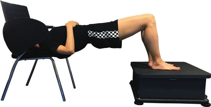
2 Pain Research and Management
anticipatory delay of MF in those with a history of NSLBP 2.1. Sample Size. Use GPower 3.1.9.2 to count power cal-
[14] and in those with experimentally induced low back pain culation. Previous studies reported that the effect size of the
[15]. This indicates that the recruitment of lumbar spinal transversus abdominus/internal oblique (TrA/IO) was 0.957
muscles is altered in low back pain patients, furthermore, after 4 weeks of ordinary physical therapy for low back pain
potentially reducing its effectiveness during rapid arm lift- [34]. Therefore, to conduct a paired-samples t-test, with an
ing. Also, it has been established in the experiments that low alpha value of 0.05 (2-tailed), power of 0.95, and effect size of
back pain has shown anticipatory delay in stabilizing 0.957, the estimated sample size was 17 participants; that is,
muscles such as the ES [16] and transverse abdominis (TrA) the sample size required for the study was 17 participants.
[17]. Sadeghi et al. [18] investigated the timing of the ac-
tivation of lumbar muscles, including transverse abdomi-
nus/internal oblique (TrA/IO), ES, and rectus abdominis 2.2. Participants. The participants were recruited through
(RA) and demonstrated that TrA/IO has a significant onset the Internet and posters placed at Shanghai Shangti Or-
delay during unilateral rapid arm movements. Hodges et al. thopaedic Hospital. A total of 40 individuals participated in
[16] assessed the EMG activity in the superficial and deep this study and were randomly allocated to the control group
fibers of the MF during functional tasks and observed and WBV group (Figure 1). All subjects underwent X-ray or
delayed muscular activation during induced pain. MRI to eliminate specific low back pain, and the clinician
Among our previous studies, a cross-sectional study performed lumbar function tests assessing lumbar rotation,
demonstrated a negative correlation between lumbar neu- flexion, and extension. Inclusion criteria were as follows:
romuscular function and pain in NSLBP patients [19] and a 18–35 years of age, low back pain persisting for approxi-
meta-analysis verified that compared with general exercise, mately 12 weeks or longer, and at least 3 episodes. Exclusion
core stability exercise is more effective in alleviating pain and criteria were as follows: taking analgesic and/or anti-in-
increasing the lumbar muscular functional status in patients flammatory agent, previous major trauma and/or surgery of
with low back pain in the short term [20]. As a new type of the spine, serious spinal pathology (vertebral fracture, in-
core stability exercise in pain relief [21], whole-body vi- flammatory arthropathy, spondylolisthesis, rheumatic dis-
bration (WBV) exercise requires the individual to perform eases, cauda equina syndrome, tumor or cancer),
static or steadily controlled exercises on an oscillating cardiovascular and/or neurological disorders, insufficiently
platform [22, 23] and becomes a credible procedure for treated hypertension, acute inflammation of the musculo-
enhancing muscular performance [24–27]. A number of skeletal system, and pregnancy. Participants were asked not
vibration-related research studies have suggested that these to change their daily lifestyle and/or to perform additional
positive acute effects are attributed to neural adaptation, physical therapy during the study period.
containing increased facilitated stretch reflex and muscle
activation [23, 28, 29]. It is worth noting that the parameters 2.3. Procedure. After the collection of participant’s basic
used in WBV could influence the nervous system’s neuro- information, the surface electrodes were placed on their
muscular responses. High vibration levels for prolonged bilateral erector spinae (ES), bilateral multifidus (MF), bi-
periods of time increase the risk of low back pain [30, 31], lateral transversus abdominus/internal oblique (TrA/IO),
but it has been documented that frequencies below 20 Hz bilateral rectus abdominis (RA), right deltoid anterior, and
plus exercise intervention enhanced NSLBP patients’ lumbar right deltoid middle. Forty individuals were randomly al-
segmental stabilization [32] and proprioception [33]. located to the control group and WBV group. The WBV
However, less is known about whether the delayed activation group performed a 3-minute warm-up, 15-minute WBV
of lumbar muscles is altered after WBV exercise during the training, and 3-minute cool-down exercise. The control
weight-shifting task which is induced by upper extremity group only performed the 3-minute warm-up and 3-minute
lifting. cool-down exercise with a 15-minute break. Before and after
A better understanding of how WBV exercise affects the intervention, sEMG signals of each muscle were recorded
NSLBP patients’ lumbar muscles APAs during functional during right shoulder flexion and abduction in the standing
tasks may help study the neuromuscular disfunction com- position for 3 times, and the relative activation time of each
monly encountered clinically. As such, we conducted this muscle was calculated.
study as an extension of our previous study [33] to further
evaluate the acute effects of WBV exercise on anticipatory
delay of core muscles in NSLBP patients. 2.4. Intervention. In the WBV group, all exercises were
performed on a vertical vibration instrument (AV009;
2. Materials and Methods BODYGREEN, Taiwan, China). Participants were asked to
take off their shoes to avoid slowing vibrations on the human
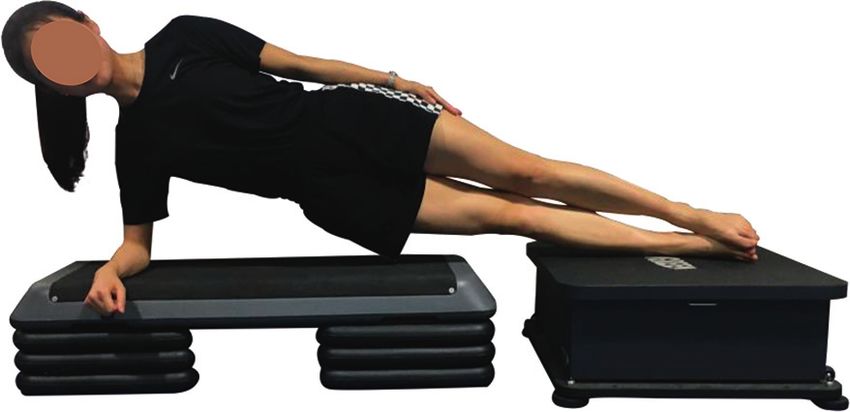
The study was a single-blind randomized controlled study body. WBV exercise contains six exercise postures: bridge,
and approved by the Ethics Committee of the Shanghai bridge with leg lift, side plank, plank, inverse bridge, and
University of Sport, China, and by the Chinese Clinical Trial balancing table pose. Postures were maintained in two
Registry (registry number ChiCTR-TRC-13003708). All modes (no WBV and WBV) for 20 s and repeated twice with
participants signed written informed consent. As such, we 15 s of rest. The vibration frequency was 20 Hz, and the
conducted this study as an extension of our previous study amplitude was 2 mm. In clinical practice, these postures are
[33] to further evaluate. widely used and are safe for patients with LBP. Figure 2 and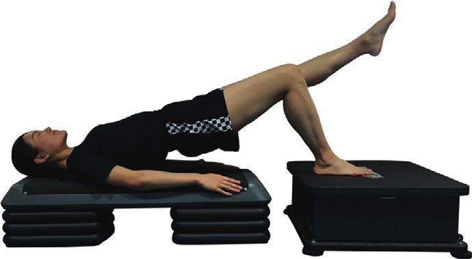
Pain Research and Management                                                                                                                    3

                     Enrollment                             Assessed for eligibility (n = 43)

                                                                                      Excluded (n = 3)
                                                                                       (i) Not meeting inclusion criteria (n = 2)
                                                                                      (ii) Other reasons (n = 1)

                                                                    Randomized (n = 40)

                                                                         Allocation

              Allocated to WBV group (n = 20)                                             Allocated to control group (n = 20)
               (i) Received allocated intervention (n = 20)                                (i) Received allocated intervention (n = 20)
              (ii) Did not receive allocated intervention (n = 0)                         (ii) Did not receive allocated intervention (n = 0)

                                                                          Analysis
                                 Analyzed (n = 20)                                                        Analyzed (n = 20)

                                         Figure 1: Flowchart of the study. WBV, whole-body vibration.

                           (a)                                              (b)                                               (c)

                          (d)                                               (e)                                               (f )

Figure 2: Training program for whole-body vibration exercise. Training program included (a) bridge, (b) bridge with leg lift, (c) side plank,
(d) plank, (e) inverse bridge, and (f ) balancing table pose.

Table 1 display more detailed information about the WBV                                Right deltoid anterior: The upper Ag/AgCl surface
exercise protocol. All exercises were completed under the                              electrode is placed approximately 3 cm below the right
supervision of registered physical therapists.                                         clavicle bone, and then, follow the muscle fibers, the
    In the control group, participants also took off their                              lower electrode goes laterally at approximately a 25-
shoes, rested for 15 minutes after completing the 3-minute                             degree angle from vertical.
warm-up exercise on the same exercise plane and then                                   Right deltoid middle: The two electrodes, 2 cm apart,
performed the 3-minute cool-down exercise. During the                                  are placed on the lateral aspect of the right upper arm
break, the subject is provided health education by the re-                             and approximately 3 cm below the acromion, and run
habilitation therapist.                                                                parallel to the muscle fibers.
                                                                                       ES: Place the first electrode piece 2 cm laterally from the
                                                                                       spinous process of L1 and the other piece upward.
2.5. sEMG Recording. Fine sandpaper and alcohol swab are
used to abrade and clean the skin. After the skin being dry,                           MF: Connect the posterior superior iliac spine to the
pairs of Ag/AgCl surface electrodes were placed to the                                 center of L1/L2 vertebrae, place the electrodes at the
following sites:                                                                       intersection of horizontal line along the L5 vertebra [35].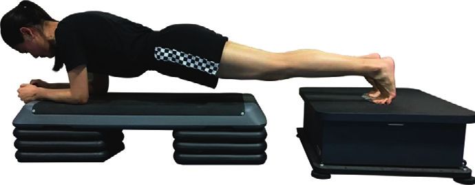
4                                                                                              Pain Research and Management

                        Table 1: Parameters and intensity of whole-body vibration exercise in WBV group.
Exercise program                      No vibration                   Vibration (20 Hz, 2 mm)                  Repetitions (times)
Bridge                              20 s, interval 15 s                 20 s, interval 15 s
Bridge with leg lift                20 s, interval 15 s                 20 s, interval 15 s
Side plank                          20 s, interval 15 s                 20 s, interval 15 s
                                                                                                                      2
Plank                               20 s, interval 15 s                 20 s, interval 15 s
Inverse bridge                      20 s, interval 15 s                 20 s, interval 15 s
Balancing table pose                20 s, interval 15 s                 20 s, interval 15 s

     TrA/IO: Place electrodes about 2 cm inferior and me-         2.6. Data Processing. The sEMG data were collected by
     dial to the anterior superior iliac spine. This area is      Noraxon TeleMyo 2400 DTS system (Noraxon, Inc., USA)
     bounded inferiorly by the inguinal ligament and RA,          and processed by MATLAB 2016a (The Mathworks, USA).
     and is below the external oblique fibers.                     Raw sEMG signals sampled at 1500 Hz performed band-pass
     RA: Electrodes placed 2 cm lateral to the mid-line and       filtered between 10 and 500 Hz. Subsequently, proceed to
     3 cm upward to the umbilicus [36].                           full-wave rectification. Then, there are three steps to process
                                                                  data for reflecting muscles’ temporal firing pattern. First, a
    The weight-shifting task: the participant stood naturally     threshold value was calculated by two standard deviations
with their feet shoulder width apart and arms naturally           from the mean value of first 400 frames of each sEMG
drooping, a 10-pound [37] dumbbell in their right hand and        channel. Second, determine the onset moment of muscle
5-pound in their left hand to stabilize the trunk. When the       activity. That moment, named muscle onset time, was de-
EMG signal of each muscle were observed to be stable, the         fined as the time when the sEMG signal beyond its threshold
participant was given a verbal cue to make their right            for a period of 50 ms [38, 39]. Third, the relative differences
shoulder flexion to 170° or make their right shoulder ab-          in the muscle onset times between the prime mover (i.e., the
duction to 170° as quickly as possible. The participant should    deltoid) and each trunk muscles (i.e., the ES, MF, RA and
try to avoid trunk rotation and shrug during the right-arm        TrA/IO) were calculated [40]. The onset time difference
movement. Before and after intervention, the standing             between the prime mover and each muscle was calculated by
shoulder flexion and abduction test was repeated 3 times.          the following equation:
Furthermore, to minimize the impact on participant an-
ticipation of the verbal cue, a random time interval between
verbal cues was set up.

             target muscle relative onset time(ms) � target muscle onset time(ms)-prime mover onset time(ms).                (1)

    Correspondingly, a negative value represented that the        independent-samples t-test was used to compare the dif-
target muscle activated before the prime mover, and vice          ference of relative activation time between two groups.
versa. In this study, the prime mover for the right shoulder      Significance level was set as P < 0.05.
flexion is deltoid anterior, and for right shoulder abduction
is deltoid middle. Each onset time not only processed in
MATLAB 2016a but also was checked visually to verify that         3. Results
sEMG activation was not ambiguous or misinterpreted by
movement artefact.                                                3.1. Demographics Data. 20 NSLBP patients average aged
                                                                  23.6 years old in the WBV group and 20 NSLBP patients
                                                                  average aged 24.2 in the control group voluntarily partici-
2.7. Statistical Analysis. SPSS 20.0 and Microsoft Excel 2016     pated in this study. Other baseline demographic and clinical
were used for data logging and statistical analysis. Demo-        characteristics of participants are shown in Table 2. No
graphic data were collected for descriptive statistics, which     adverse events were observed by physical therapists or re-
are described as mean ± standard deviation (SD). The data         ported by NSLBP patients during and after the intervention.
were tested for normality using the Shapiro–Wilk test. The
independent-samples t-test was used to compare the de-
mographic data of the WBV group and control group. Each           3.2. Comparison of Relative Activation Time between/within
subject was required to complete 3 times right shoulder           Groups on Shoulder Flexion. At the baseline, when flexing
flexion and 3 times right shoulder abduction before and after      the shoulder, the bilateral ES, bilateral MF, bilateral TrA/IO,
the intervention. The relative onset time of each muscle was      and bilateral RA in two groups are activated after the prime
calculated and averaged, named relative activation time.          mover muscles (delta anterior muscle). There was no sig-
The paired-samples t-test was used to compare relative            nificant difference in the relative activation time of each
activation time before and after intervention, and                muscle among two groups (P > 0.05) (Table 3).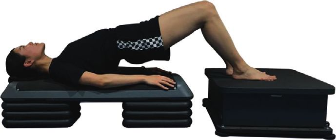
Pain Research and Management                                                                                                                               5

                                        Table 2: Demographic and clinical characteristics of participants.
                                                             WBV group (n � 20)               Control group (n � 20)             t value         P value†
Age (y)                                                          23.6 ± 3.3                         24.2 ± 2.4                   −0.721           0.475
Height (cm)                                                     168.8 ± 7.7                        169.1 ± 9.5                   −0.110           0.913
Weight (kg)                                                    64.83 ± 13.18                      63.88 ± 13.24                   0.227           0.821
BMI (kg/m2)                                                     22.53 ± 3.10                       22.11 ± 2.82                   0.454           0.652
Time since first experience with NSLBP (mo)                      50.8 ± 45.0                        28.9 ± 24.5                    1.911           0.064
VAS max                                                         4.40 ± 1.57                        4.75 ± 1.55                   −0.709           0.483
VAS mean                                                        2.65 ± 0.81                        2.75 ± 0.97                   −0.354           0.725
WBV, whole-body vibration; BMI, body mass index (calculated as weight in kilograms divided by height in meters squared); NSLBP, nonspecific low back
pain; VAS, visual analogue scale. Values are expressed as mean ± SD.† Analyzed by the independent-samples t-test.

               Table 3: Comparison of relative activation time between/within groups on shoulder flexion (x ± s, unit: ms).
                                         WBV group (n � 20)              Control group (n � 20)            WBV – Control (95% CI)                P† value
                        Pre-test            41.5 ± 156.5                      61.8 ± 125.5                  −20.3 (−111.1 to 70.5)                0.653
Right ES
                        Post-test          46.5 ± 139.8                        71.5 ± 94.6                  −25.1 (−101.4 to 51.3)                0.511
                        P‡ value               0.875                              0.564
                        Pre-test            34.0 ± 94.5                        21.7 ± 95.2                     12.2 (−48.5 to 72.9)                0.686
Left ES
                        Post-test            5.6 ± 71.9                       −1.6 ± 68.7                      7.2 (−37.8 to 52.2)                 0.748
                        P‡ value               0.089                              0.083
                        Pre-test            85.2 ± 94.4                       54.6 ± 103.7                     30.6 (−32.9 to 94.1)                0.335
Right MF
                        Post-test           48.6 ± 78.7                        71.1 ± 98.2                    −23.1 (−80.0 to 33.9)                0.418
                        P‡ value               0.014∗                             0.281
                        Pre-test             68.3 ± 81.9                       52.3 ± 99.0                     16.0 (−42.1 to 74.2)                0.580
Left MF
                        Post-test            30.2 ± 81.1                       56.9 ± 95.1                    −26.7 (−83.3 to 29.9)                0.346
                        P‡ value               0.011∗                             0.710
                        Pre-test           147.2 ± 79.9                       202.2 ± 94.0                   −55.1 (−110.9 to 0.8)                0.053
Right TrA/IO
                        Post-test          103.4 ± 96.4                      205.9 ± 125.1                  −102.5 (−174.0 to −31.0)              0.006∗
                        P‡ value               0.008∗                             0.860
                        Pre-test           160.3 ± 119.3                     222.8 ± 168.2                   −62.5 (−155.9 to 30.8)                0.183
Left TrA/IO
                        Post-test          97.2 ± 159.0                      195.2 ± 147.9                   −98.0 (−196.3 to 0.3)                 0.051
                        P‡ value               0.026∗                             0.167
                        Pre-test           259.4 ± 137.1                     236.1 ± 162.2                    23.3 (−72.9 to 119.4)                0.627
Right RA
                        Post-test          234.0 ± 118.1                     218.2 ± 156.7                    15.8 (−73.0 to 104.6)                0.721
                        P‡ value               0.337                              0.346
                        Pre-test           231.4 ± 155.0                     267.7 ± 132.3                   −36.4 (−128.6 to 55.9)               0.430
Left RA
                        Post-test          174.3 ± 147.8                     272.0 ± 141.7                   −97.8 (−190.4 to −5.1)               0.039∗
                        P‡ value               0.062                              0.772
Relative activation time � muscle onset time-prime mover onset time (ms), a negative value indicated that the target muscle fired before the prime mover, and
vice versa. WBV, whole-body vibration; ES, erector spinae; MF, multifidus; TrA/IO, transversus abdominus/internal oblique; RA, rectus abdominis; CI,
confidence interval. Values are expressed as mean ± SD.† Analyzed by the independent-samples t-test;‡ analyzed by the paired-samples t-test;∗ significant at
P < 0.05.

    Using the independent-samples t-test to compare the                         difference compared with baseline (t � −0.159, P � 0.875). In
posttest data, it was found that after intervention, the relative               control group, there was no significant difference in the
activation time of the right TrA/IO and the left RA in the                      relative activation time of each muscle before and after the
WBV group was significantly less than that in the control                        intervention (P > 0.05) (Table 3).
group (right TrA/IO: t � −2.901, P � 0.006; left RA:
t � −2.135, P � 0.039). And there was no significant differ-
ence in the relative activation time of the remaining muscles                   3.3. Between-Group Comparison of Variation in Relative
between the two groups (P > 0.05) (Table 3).                                    Activation Time on Shoulder Flexion. Using the indepen-
    In the WBV group, after a single-section intervention,                      dent-samples t-test to compare the variation (Δ � posttest-
except for the right ES, the relative activation time of each                   pretest) in the relative activation time between the two
muscle decreased, and the relative activation time of bilateral                 groups after the intervention, it was found that the change
MF and bilateral TrA/IO was significantly reduced (right                         value in the relative activation time of the bilateral MF in the
MF: t � 2.717, P � 0.014; left MF: t � 2.828, P � 0.011; right                  WBV group was significantly smaller than that in the control
TrA/IO: t � 2.951, P � 0.008; left TrA/IO: t � 2.407,                           group (right MF: t � −2.622, P � 0.013; left MF: t � −2.359,
P � 0.026). The relative activation time of the right ES after                  P � 0.024). There was no significant difference in other
intervention increased slightly, but there was no significant                    muscles (Figure 3).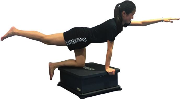
6                                                                                                                                                  Pain Research and Management

                                                               60.0
                                                                                                   ∗          ∗
              The difference of elative activation time (ms)    40.0

                                                               20.0

                                                                0.0
                                                                      Right ES        Left ES   Right MF   Left MF     Right TrA/IO Left TrA/IO   Right RA    Left RA

                                                              –20.0

                                                              –40.0

                                                              –60.0

                                                              –80.0

                                                        –100.0
                                                                      WBV group
                                                                      Control group
Figure 3: Between-group comparison of variation in relative activation time on shoulder flexion. Variation � post-test–pre-test, a negative
value indicated that the intervention shortened the relative activation time, and vice versa. WBV, whole-body vibration; ES, erector spinae;
MF, multifidus; TrA/IO, transversus abdominus/internal oblique; RA, rectus abdominis. The independent-samples t-test was used to
compare the variation. ∗ Significant at P < 0.05.

3.4. Comparison of Relative Activation Time between/within                                                           the two groups after the intervention. It was found that the
Groups on Shoulder Abduction. At baseline, when making                                                               change value in the relative activation time of the bilateral
the upper limbs abduction, the bilateral ES, bilateral MF,                                                           ES, right MF, and right RA in the WBV group was sig-
bilateral TrA/IO, and bilateral RA in two groups are acti-                                                           nificantly smaller than that in the control group (right ES:
vated after the prime mover muscles (delta middle muscle).                                                           t � −4.274, P < 0.001; left ES: t � −3.234, P � 0.003; right MF:
There was no significant difference in the relative activation                                                         t � −2.514, P � 0.016; right TrA/IO: t � −3.518, P � 0.001;
time of each muscle among two groups (P > 0.05) (Table 4).                                                           right RA: t � −3.717, P � 0.004). There was no significant
     After completing a single section of intervention, the                                                          difference in other muscles (Figure 4).
relative activation time of each muscle in participants of the
WBV group decreased. Except for the left TrA/IO and the
left RA, the relative activation time of other muscles was                                                           4. Discussion
significantly reduced (right ES: t � 3.847, P � 0.001; left ES:
t � 4.641, P < 0.001; right MF: t � 4.093, P � 0.001; left MF:                                                       The present study’s main objective was to evaluate the effects
t � 2.093, P � 0.009; right TrA/IO: t � 5.239, P < 0.001; right                                                      of a single-section WBV exercise on the activation time of
RA: t � 3.800, P � 0.001). In the control group, there was no                                                        core muscles in NSLBP patients. The results of this study
significant difference in the relative activation time of each                                                         demonstrate that (1) WBV exercise shorten the activation
muscle before and after the intervention (P > 0.05).                                                                 time of bilateral MF and bilateral TrA/IO on standing
     Using the independent-samples t-test to compare the post-                                                       shoulder flexion task, which means the deep core muscles
test data, it was found that after intervention, the relative ac-                                                    tend to be much easier to activated in maintain the sagittal
tivation time of the left ES, right MF, right TrA/IO and right RA                                                    balance after WBV exercise; (2) WBV exercise shorten the
in the WBV group was significantly less than that in the control                                                      activation time of bilateral ES, bilateral MF, right TrA/IO,
group (left ES: t � −3.283, P � 0.002; right MF: t � −2.552,                                                         and right RA on the standing shoulder abduction task, which
P � 0.015; right TrA/IO: t � −3.113, P � 0.004; right RA:                                                            means core muscles in lumbar and right abdomen tend to be
t � −3.984, P < 0.001). The relative activation time measured by                                                     much easier to activated in maintain the coronal balance
the remaining muscles after intervention in the WBV group                                                            after WBV exercise. In addition, with an eye to vibration in
was slightly reduced compared with the control group, how-                                                           relative activation time, MF’s relative activation time is
ever, with no significant difference (P > 0.05) (Table 4).                                                             significantly shortened by WBV exercise no matter in
                                                                                                                     shoulder flexion or abduction.
                                                                                                                         Previous literature has many different studies on muscle
3.5. Between-Group Comparison of Variation in Relative                                                               activation time. Based on the weight shift task, the current
Activation Time on Shoulder Abduction. Using the inde-                                                               studies stated that the onset of the sEMG activity of all trunk
pendent samples t-test to compare the variation                                                                      muscles occurred prior to that of the muscle in charge of
(Δ � posttest-pretest) in the relative activation time between                                                       limb movement in healthy individuals [5, 37]. This
Pain Research and Management 7
Table 4: Comparison of relative activation time between/within groups on shoulder abduction (x ± s, unit: ms).
WBV group (n � 20) Control group (n � 20) WBV – Control (95% CI) P‡ value
Right ES Pre-test 184.8 ± 99.8 147.3 ± 159.9 37.5 (−47.8 to 122.8) 0.379
Post-test 117.5 ± 115.4 178.0 ± 130.8 −60.4 (−139 to 18.5) 0.130
P‡ value 0.001∗ 0.052
Left ES Pre-test 162.5 ± 108.8 158.0 ± 104.2 4.5 (−63.7 to 72.7) 0.894
Post-test 57.7 ± 76.7 137.8 ± 77.5 −80.1 (−129.4 to −30.7) 0.002∗
P‡ value 0.000∗ 0.141
Right MF Pre-test 237.5 ± 97.4 257.6 ± 100.2 −20.1 (−83.4 to 43.1) 0.524
Post-test 144.1 ± 97.1 269.4 ± 196.9 −125.3 (−224.7 to −25.9) 0.015∗
P‡ value 0.001∗ 0.741
Left MF Pre-test 157.3 ± 90.3 163.0 ± 76.3 −5.7 (−59.2 to 47.8) 0.830
Post-test 105.4 ± 108.9 152.6 ± 81.2 −47.2 (−108.6 to 14.3) 0.129
P‡ value 0.009∗ 0.333
Right TrA/IO Pre-test 165.7 ± 100.4 186.3 ± 122.5 −20.6 (−92.6 to 51.1) 0.564
Post-test 69.4 ± 102.9 166.5 ± 94.4 −97.2 (−160.4 to −34.0) 0.004∗
P‡ value 0.000∗ 0.105
Let TrA/IO Pre-test 160.7 ± 143.4 195.1 ± 108.6 −34.5 (−115.9 to 47.0) 0.397
Post-test 124.6 ± 206.6 189.0 ± 100.4 −64.5 (−168.4 to 39.5) 0.217
P‡ value 0.403 0.508
Right RA Pre-test 231.3 ± 129.6 258.9 ± 123.1 −27.6 (−108.5 to 53.3) 0.494
Post-test 128.0 ± 99.3 265.3 ± 117.9 −137.3 (−207.1 to −67.5) 0.000∗
P‡ value 0.001∗ 0.584
Left RA Pre-test 108.8 ± 218.6 166.1 ± 181.9 −57.4 (−186.1 to 71.3) 0.373
Post-test 70.6 ± 127.7 160.1 ± 176.6 −89.5 (−188.1 to 9.2) 0.074
P‡ value 0.203 0.452
Relative activation time � muscle onset time-prime mover onset time (ms), a negative value indicated that the target muscle fired before the prime mover, and
vice versa. WBV, whole-body vibration; ES, erector spinae; MF, multifidus; TrA/IO, transversus abdominus/internal oblique; RA, rectus abdominis; CI,
confidence interval. Values are expressed as mean ± SD.† Analyzed by the independent-samples t-test;‡ analyzed by the paired-samples t-test;∗ significant at
P < 0.05.
60.0 ∗ ∗
40.0
∗
∗ ∗
The difference of relative activation time (ms)
20.0
0.0
Right ES Left ES Right MF Left MF Right TrA/IO Left TrA/IO Right RA Left RA
–20.0
–40.0
–60.0
–80.0
–100.0
–120.0
–140.0
WBV group
Control group
Figure 4: Between-group comparison of variation in relative activation time on shoulder abduction. Variation � post-test–pre-test, a
negative value indicated that the intervention shortened the relative activation time, and vice versa. WBV, whole-body vibration; ES, erector
spinae; MF, multifidus; TrA/IO, transversus abdominus/internal oblique; RA, rectus abdominis. The independent-samples t-test was used to
compare the variation. ∗ Significant at P < 0.05.8 Pain Research and Management
phenomenon contributes to the feedforward postural re- These biopsychosocial factors may affect the patients’
sponse. Furthermore, the anticipatory activation of trunk symptom after WBV intervention. In addition, a band-pass
muscles (e.g., TrA, ES and MF), known as APAs, is vital to filter was applied to minimize relevant artifacts in every
maintain lumbopelvic stability during predictable postural sEMG collection, but it unavoidably eliminated the actual
perturbations, just as those turned up during limb-oriented muscle activity signals. To make impartial contrasts, every
movements [4, 41]. APAs counteract the predictable in- muscle activity signal in our study performed the same
trinsic reactive forces induced by a focal movement through filtering process. Finally, every participant received only
preactivation of particular muscle groups [42]. Multiple single-section WBV exercise; the effect of the long-term
studies demonstrated low back pain patients have shown intervention should be performed in further study. Never-
anticipatory delays in the TrA/IO and MF during postural theless, this study offers a reasonable proposal for training
tasks [8, 43–45]. Hodges [46] claimed that delays in an- programs about WBV exercise, extending the knowledge
ticipatory muscle activation might be a central nervous about possible progressions to improve lumbar stability and
system adaptation to pain. Also, Hungerford suggested that muscle function, that is, WBV may shorten the activation
the delay in anticipatory muscle is associated with failure of time to improve APAs in NSLBP patients.
lumbopelvic stabilization [47]. These results are in line with
our study. We tested the relative activation muscle by the 5. Conclusions
weight-shifting task (shoulder flexion and abduction) for
NSLBP patients. Before intervention, irrespective of the In conclusion, single treatment of WBV exercise can ef-
WBV group or control group, the trunk muscles of ES, MF, fectively alleviate the delayed activation of core muscles in
TrA/IO, and RA showed a positive value of relative acti- NSLBP patients, but the long-term effects still need further
vation time, which means ES, MF, TrA/IO, and RA were study.
fired after deltoid. Anticipatory delays were observed.
Furthermore, previous studies focused on the activation Data Availability
time of deep fiber like MF and TrA; our study provided
available information about the trunk muscle containing ES The data used to support the findings of this study are in-
and RA to supply APA delays in NSLBP patients. cluded within the article.
As a noninvasive therapy method, WBV exercise acts
like a mild exercise on the body [48, 49]. In recent years, Conflicts of Interest
WBV exercise are performed for wild range of patients with
metabolic syndrome [50, 51] and musculoskeletal problems The authors declare that they have no conflicts of interest.
including low back pain [33], knee osteoarthritis [52, 53],
fibromyalgia [54], osteogenesis imperfecta [55], and so on. Acknowledgments
Although our research focused on the relative activation
time of trunk muscles for APAs in lumbar stability, the The authors gratefully acknowledge the valuable cooperation
intrinsic value of coactivation of core muscles for main- of Juan Zhang, Di Gong, Meng-Si Peng, Juan Wang, Jing-
taining lumbopelvic stabilization has been recognized in Zhao Yang, Chang-Cheng Chen, Bao Wu, and Ge Song
clinical knowledge. In our two previous studies, we inves- (Department of Sport Rehabilitation, Shanghai University of
tigated the effect of 12-week WBV exercise in young adults Sport). The authors also thank Yu-Lin Dong (Department of
with NSLBP; the results showed that WBV exercise im- Treatment, the Second Rehabilitation Hospital of Shanghai)
proved lumbar flexion and extension proprioception and for her Strong technical support. This study was supported
reduced pain [56]; then, the sEMG root mean square was by the Fok Ying-Tong Education Foundation of China
used to measure the core muscle activity influenced by WBV (161092); the scientific and technological research program
exercise in healthy young adults. The results shows that of the Shanghai Science and Technology Committee
plank, bridge with leg lift, and single plank can fully activate (19080503100); and the Shanghai Key Lab of Human Per-
MF, ES, IO, and RA [57]. Based on these studies, we designed formance (Shanghai University of Sport) (11DZ2261100).
this experiment to explore whether WBV exercise alleviates
anticipatory delays on the trunk muscle, leading an en- References
hancement clinical performance for NSLBP patients. Our
findings that WBV exercise shortens the activation time [1] B. W. Koes, M. W. Van Tulder, and S. Thomas, “Diagnosis and
differently in different shoulder movement may bolster this treatment of low back pain,” British Medical Journal, vol. 332,
point. no. 7555, pp. 1430–1434, 2006.
Our study has several limitations. First, this research [2] C. Maher, M. Underwood, and R. Buchbinder, “Non-specific
focuses on investigating sEMG onset activities of trunk low back pain,” The Lancet, vol. 389, no. 10070, pp. 736–747,
2017.
muscles after a single-section WBV exercise, and we
[3] R. D’Hooge, P. Hodges, and H. Tsao, “Altered trunk muscle
recruited a relatively small group of NSLBP patients. Hence, coordination during rapid trunk flexion in people in remis-
our findings might be cautious to popularize for the entire sion of recurrent low back pain. Journal of electromyography
population with NSLBP. Second, the patients were recruited and kinesiology,” Official Journal of the International Society
from different ways, so they have different educational of Electrophysiological Kinesiology, vol. 23, no. 1, pp. 173–181,
backgrounds, personalities, economic status, and so on. 2013.Pain Research and Management 9
[4] M. D. Bussey, D. Aldabe, J. Shemmell, and T. Jowett, “An- patients with nonspecific low back pain,” Medicine, vol. 96,
ticipatory postural control differs between low back pain and no. 36, p. e7991, 2017.
pelvic girdle pain patients in the absence of visual feedback,” [20] X.-Q. Wang, J.-J. Zheng, Z.-W. Yu et al., “A meta-analysis of
Human Movement Science, vol. 69, Article ID 102529, 2020. core stability exercise versus general exercise for chronic low
[5] P. W. Hodges and C. A. Richardson, “Delayed postural back pain,” PloS one, vol. 7, no. 12, Article ID e52082, 2012.
contraction of transversus abdominis in low back pain as- [21] Y. Dong, W. Wang, J. Zheng, S. Chen, J. Qiao, and X. Wang,
sociated with movement of the lower limb,” Journal of Spinal “Whole body vibration exercise for chronic musculoskeletal
Disorders, vol. 11, no. 1, pp. 46–56, 1998. pain: a systematic review and meta-analysis of randomized
[6] F. Deborah and P. W. Hodges, “Individualized exercise in- controlled trials,” Archives of Physical Medicine and Reha-
terventions for spinal pain,” Exercise and Sport Sciences Re- bilitation, vol. 100, no. 11, pp. 2167–2178, 2019.
views, vol. 45, no. 2, pp. 105–115, 2017. [22] P. Chung, C. Liu, H. Wang, Y. Liu, L. Chuang, and
[7] M. Sadeghi, S. Talebian, G. R. Olyaei, and B. Attarbashi T.-Y. Shiang, “Various performance-enhancing effects from
Moghadam, “Preparatory brain activity and anticipatory the same intensity of whole-body vibration training,” Journal
postural adjustments accompanied by externally cued of Sport and Health Science, vol. 6, no. 3, pp. 333–339, 2017.
weighted-rapid arm rise task in non-specific chronic low back [23] W.-W. Yang, L.-W. Chou, W.-H. Chen, T.-Y. Shiang, and
pain patients and healthy subjects,” Springer Plus, vol. 5, no. 1, C. Liu, “Dual-frequency whole body vibration enhances
p. 674, 2016. vertical jumping and change-of-direction ability in rugby
[8] S. L. Morris, B. Lay, and G. T. Allison, “Transversus abdominis players,” Journal of Sport and Health Science, vol. 6, no. 3,
is part of a global not local muscle synergy during arm pp. 346–351, 2017.
movement,” Human Movement Science, vol. 32, no. 5, [24] J. Luo, B. McNamara, and K. Moran, “The use of vibration
pp. 1176–1185, 2013. training to enhance muscle strength and power,” Sports
[9] H. Tsao, L. A. Danneels, and P. W. Hodges, “ISSLS prize Medicine, vol. 35, no. 1, pp. 23–41, 2005.
winner,” Spine, vol. 36, no. 21, pp. 1721–1727, 2011. [25] D. Bemben, C. Stark, and R. Taiar, “Relevance of whole-body
[10] P. Linek, P. Noormohammadpour, M. A. Mansournia, vibration exercises on muscle strength/power and bone of
T. Wolny, and D. Sikora, “Morphological changes of the elderly individuals. dose-response,” A Publication of Inter-
lateral abdominal muscles in adolescent soccer players with national Hormesis Society, vol. 16, no. 4, 2018.
low back pain: a prospective cohort study,” Journal of Sport [26] L. M. M. Santos, A. C. C. Oliveira, S. F Fonseca et al., “Whole-
and Health Science, vol. 9, no. 6, pp. 614–619, 2020. body vibration exercise in different postures on handgrip
[11] J. Wei, H.-B. Zhu, F. Wang, Y. Fan, and H.-J. Zhou, “Clinical strength in healthy women: a cross-over study,” Frontiers in
utility of flexion-extension ratio measured by surface elec- Physiology, vol. 11, Article ID 469499, 2020.
tromyography for patients with nonspecific chronic low-back [27] M. Moura-Fernandes, E. Moreira-Marconi, and A. Meirelles,
pain,” Journal of the Chinese Medical Association, vol. 82, “Acute effects of whole-body vibration exercise on pain level,
no. 1, pp. 35–39, 2019. functionality, and rating of exertion of elderly obese knee
[12] N. Kuriyama and H. Ito, “Electromyographic functional osteoarthritis individuals: a randomized study,” Applied Sci-
analysis of the lumbar spinal muscles with low back pain,” ences, vol. 10, 2021.
Journal of Nippon Medical School, vol. 72, no. 3, pp. 165–173, [28] C. Kurt and E. Pekünlü, “Acute effect of whole body vibration
2005. on isometric strength, squat jump, and flexibility in well-
[13] J. R. Steele, C. E. Coltman, and D. E. McGhee, “Effects of trained combat athletes,” Biology of Sport, vol. 32, no. 2,
obesity on breast size, thoracic spine structure and function, pp. 115–122, 2015.
upper torso musculoskeletal pain and physical activity in [29] D. J. Cochrane, “The potential neural mechanisms of acute
women,” Journal of Sport and Health Science, vol. 9, no. 2, indirect vibration,” Journal of Sports Science & Medicine,
pp. 140–148, 2020. vol. 10, no. 1, pp. 19–30, 2011.
[14] D. MacDonald, G. L. Moseley, and P. W. Hodges, “People [30] L. Burström, T. Nilsson, and J. Wahlström, “Whole-body
with recurrent low back pain respond differently to trunk vibration and the risk of low back pain and sciatica: a sys-
loading despite remission from symptoms,” Spine, vol. 99, tematic review and meta-analysis,” International Archives of
2010. Occupational and Environmental Health, vol. 88, no. 4,
[15] K. Kyle, R. J. Butler, and D. Tara, “Experimentally induced pp. 403–418, 2015.
pain alters the EMG activity of the lumbar multifidus in [31] J. H. Kim, L. S. Marin, and J. T. Dennerlein, “Evaluation of
asymptomatic subjects,” Manual Therapy, vol. 17, no. 3, commercially available seat suspensions to reduce whole body
pp. 236–240, 2012. vibration exposures in mining heavy equipment vehicle op-
[16] P. W. Hodges, G. Anna, and C. G. Simon, “Experimental erators,” Applied Ergonomics, vol. 71, pp. 78–86, 2018.
muscle pain changes feedforward postural responses of the [32] J. Rittweger, M. Mutschelknauss, and D. Felsenberg, “Acute
trunk muscles,” Experimental Brain Research, vol. 151, no. 2, changes in neuromuscular excitability after exhaustive whole
pp. 262–271, 2003. body vibration exercise as compared to exhaustion by
[17] P. Ferreira, M. Ferreira, and P. Hodges, “Changes in re- squatting exercise,” Clinical Physiology and Functional Im-
cruitment of the abdominal muscles in people with low back aging, vol. 23, no. 2, pp. 81–86, 2003.
pain,” Spine, vol. 12/01, no. 29, pp. 2560–2566, 2004. [33] Y.-L. Zheng, X.-F. Wang, B.-L. Chen et al., “Effect of 12-week
[18] M. Sadeghi, T. Saeed, and R. Gholam, “Preparatory brain whole-body vibration exercise on lumbopelvic proprioception
activity and anticipatory postural adjustments accompanied and pain control in young adults with nonspecific low back
by externally cued weighted-rapid arm rise task in non- pain,” Medical Science Monitor, vol. 25, pp. 443–452, 2019.
specific chronic low back pain patients and healthy subjects,” [34] J. A. Hwang, S. H. Bae, G. Do Kim, and K. Y. Kim, “The effects
SpringerPlus, vol. 5, no. 1, p. 674, 2016. of sensorimotor training on anticipatory postural adjustment
[19] H. Hu, Y. Zheng, X. Wang et al., “Correlations between of the trunk in chronic low back pain patients,” Journal of
lumbar neuromuscular function and pain, lumbar disability in Physical Therapy Science, vol. 25, no. 9, pp. 1189–1192, 2013.10 Pain Research and Management
[35] N. W. Mok, E. W. Yeung, J. C. Cho, S. C. Hui, K. C. Liu, and [50] D. Sá-Caputo, L. L. Paineiras-Domingos, A. Francisca-Santos
C. H. Pang, “Core muscle activity during suspension exer- et al., “Whole-body vibration improves the functional pa-
cises,” Journal of Science and Medicine in Sport, vol. 18, no. 2, rameters of individuals with metabolic syndrome: an ex-
pp. 189–194, 2015. ploratory study,” BMC Endocrine Disorders, vol. 19, no. 1, p. 6,
[36] P. Marshall and B. Murphy, “The validity and reliability of 2019.
surface EMG to assess the neuromuscular response of the [51] L. L. Paineiras-Domingos, D. Sá-Caputo, A. Francisca-Santos
abdominal muscles to rapid limb movement,” Journal of et al., “Can whole body vibration exercises promote im-
Electromyography and Kinesiology, vol. 13, no. 5, pp. 477–489, provement on quality of life and on chronic pain level of
2003. metabolic syndrome patients? A pseudorandomized cross-
[37] T. Suehiro, H. Ishida, K. Kobara, H. Osaka, and S. Watanabe, over study,” Journal of Applied Physiology, vol. 128, no. 4,
“Altered trunk muscle recruitment patterns during lifting in pp. 934–940, 2020.
individuals in remission from recurrent low back pain,” [52] X. Li, X. Wang, and B. Chen, “whole-body vibration exercise
Journal of Electromyography and Kinesiology, vol. 39, for knee osteoarthritis: a systematic review and meta-analy-
pp. 128–133, 2018. sis,” Evidence-Based Complementary and Alternative Medi-
[38] A. C. L. Sakamoto, L. F. Teixeira-Salmela, F. R. de Paula- cine: eCAM, vol. 2015, Article ID 758147, 12 pages, 2015.
Goulart, C. D. C. de Morais Faria, and C. Q. Guimarães, [53] E. Moreira-Marconi, Y. Teixeira-Silva, and A. G. de Meirelles,
“Effect of whole-body vibration on the functional responses of
“Muscular activation patterns during active prone hip ex-
the patients with knee osteoarthritis by the electromyographic
tension exercises,” Journal of Electromyography and Kinesi-
profile of thevastus lateralismuscles during the five-repetition
ology, vol. 19, no. 1, pp. 105–112, 2009.
chair stand test: a randomized crossover trial,” Applied Sci-
[39] T. Suehiro, M. Mizutani, H. Ishida, K. Kobara, H. Osaka, and
ences, vol. 12, p. 10, 2020.
S. Watanabe, “Individuals with chronic low back pain
[54] B. Julia, J. Angela, and V. Ina, “Whole body vibration exercise
demonstrate delayed onset of the back muscle activity during
training for fibromyalgia,” The Cochrane Database of Sys-
prone hip extension,” Journal of Electromyography and Ki- tematic Reviews, vol. 9, 2017.
nesiology, vol. 25, no. 4, pp. 675–680, 2015. [55] H. Wolfgang, S. Janis, and B. Nick, “The effect of whole body
[40] K. Chance-Larsen, C. Littlewood, and A. Garth, “Prone hip vibration training on bone and muscle function in children
extension with lower abdominal hollowing improves the with osteogenesis imperfecta,” The Journal of Clinical Endo-
relative timing of gluteus maximus activation in relation to crinology and Metabolism, vol. 102, no. 8, pp. 2734–2743,
biceps femoris,” Manual Therapy, vol. 15, no. 1, pp. 61–65, 2017.
2010. [56] W. Xue, W. Gu, and B. Chen, “Effects of whole-body vibration
[41] N. W. Mok, S. G. Brauer, and P. W. Hodges, “Postural re- exercise for non-specific chronic low back pain: an assessor-
covery following voluntary arm movement is impaired in blind, randomized controlled trial,” Clinical Rehabilitation,
people with chronic low back pain,” Gait & Posture, vol. 34, vol. 33, no. 9, pp. 1445–1457, 2019.
no. 1, pp. 97–102, 2011. [57] B. Chen, Y. Dong, and J. Guo, “Effects of whole-body vi-
[42] V. E. Belen’Kii, V. S. Gurfinkel’, and E. I. Pal’Tsev, “On the bration on lumbar-abdominal muscles activation in healthy
control elements 589 of voluntary movements,” Biofizika, young adults: a pilot study medical science monitor,” Inter-
vol. 12, no. 1, p. 135, 1967. national Medical Journal of Experimental and Clinical Re-
[43] M. Lorimer and W. H. Paul, “Are the changes in postural search, vol. 25, pp. 1945–1951, 2019.
control associated with low back pain caused by pain inter-
ference?” The Clinical Journal of Pain, vol. 21, no. 4,
pp. 323–329, 2005.
[44] M. Paul and M. Bernadette, “Delayed abdominal muscle
onsets and self-report measures of pain and disability in
chronic low back pain,” Official Journal of the International
Society of Electrophysiological Kinesiology, vol. 20, no. 5,
pp. 833–839, 2010.
[45] P. W. Hodges and C. A. Richardson, “Contraction of the
abdominal muscles associated with movement of the lower
limb,” Physical Therapy, vol. 77, no. 2, pp. 132–142, 1997.
[46] P. W. Hodges, “Moving differently in pain: a new theory to
explain the adaptation to pain,” Pain, vol. 152, pp. S90–S98,
2011.
[47] B. H. Hungerford, “Evidence of altered lumbopelvic muscle
recruitment in the presence of sacroiliac joint pain,” Spine,
vol. 28, no. 14, pp. 1593–1600, 2003.
[48] A. Godinez, B. Liston, R Ayzenberg, and B. William, “G-
loading and vibration effects on heart and respiration rates,”
Aviation, Space, and Environmental Medicine, vol. 85, no. 9,
pp. 949–953, 2014.
[49] E. Moreira-Marconi, V. D. Caiado, and Y. Teixeira-Silva,
“Whole-body vibration as antihypertensive non-pharmaco-
logical treatment in hypertensive individuals with knee os-
teoarthritis: randomized cross-over trial,” Sustainability,
vol. 21, p. 12, 2020.You can also read