Topical Delivery of Geranium/Calendula Essential Oil-Entrapped Ethanolic Lipid Vesicular Cream to Combat Skin Aging - Hindawi.com
←
→
Page content transcription
If your browser does not render page correctly, please read the page content below

Hindawi
BioMed Research International
Volume 2021, Article ID 4593759, 13 pages
https://doi.org/10.1155/2021/4593759

Research Article
Topical Delivery of Geranium/Calendula Essential Oil-Entrapped
Ethanolic Lipid Vesicular Cream to Combat Skin Aging

 Alka Lohani ,1 Anurag Verma ,1,2 G. Hema,3 and Kamla Pathak 4

 1
 School of Pharmaceutical Sciences, IFTM University, 244102, Moradabad, India
 2
 Teerthanker Mahaveer College of Pharmacy, Teerthanker Mahaveer University, 244102, Moradabad, India
 3
 Department of Biotechnology, Maharani’s Science College for Women, 560001, Bangalore, India
 4
 Pharmacy College Saifai, Uttar Pradesh University of Medical Sciences, 206130, Uttar Pradesh, India

 Correspondence should be addressed to Anurag Verma; anuragverma_iftm@yahoo.co.in

 Received 8 May 2021; Revised 9 August 2021; Accepted 16 August 2021; Published 13 September 2021

 Academic Editor: Dr. Abdul Ahad

 Copyright © 2021 Alka Lohani et al. This is an open access article distributed under the Creative Commons Attribution License,
 which permits unrestricted use, distribution, and reproduction in any medium, provided the original work is properly cited.

 The present study deals with the evaluation of the age-defying potential of topical cream formulations bearing Geranium
 essential oil/Calendula essential oil-entrapped ethanolic lipid vesicles (ELVs). Two types of cream formulations were
 prepared, viz., conventional and ELVs spiked o/w creams. Essential oil- (EO-) loaded ELVs were characterized by vesicle
 size, polydispersity index, encapsulation efficiency, and scanning electron microscopy. The cream formulations were
 evaluated for homogeneity, spreadability, viscosity, pH, in vitro antioxidant capacity, sun protection factor, and in vitro
 collagenase and elastase inhibition capacity. Confocal laser scanning microscopy (CLSM) was performed to ascertain skin
 permeation of conventional and vesicular cream. The results of in vitro antioxidant studies showed that GEO-/CEO-loaded
 vesicular creams have notable antioxidant capacity when compared to nonvesicular creams. GEO- or CEO-loaded vesicular
 creams exhibited the highest SPF value 10.26 and 18.54, respectively. Both the EO-based vesicular creams showed in vitro
 collagenase and elastase enzyme inhibition capacity. CLSM images clearly depicted that vesicular cream deep into the skin
 layers. From the research findings, the age-defying potential and photoprotective effects of GEO and CEO were confirmed.
 It can be concluded that ELVs are able to preserve the efficiency of EOs and have the potential to combat skin aging.

1. Introduction when they reach their target sites present in the deeper layers
 of the skin, but the stratum corneum is the biggest obstacle
Finding solutions against various signs of skin aging has in delivering the actives deep into the skin layers. Several
been a natural human desire for centuries. Skin aging is a antiaging strategies have been developed during the past
complex biological process, influenced by a combination of years to overcome this barrier. One of the possibilities for
intrinsic (genetics, cellular metabolism, hormone, and meta- increasing the penetration of active ingredients is the use
bolic processes) and extrinsic factors (chronic light expo- of vesicular delivery systems such as liposomes, niosomes,
sure, pollution, ionizing radiation, chemicals, and toxins) and ethosomes [2]. These vesicles can act either as a carrier
[1]. These triggers cause the skin to deteriorate over a time- system or as penetration enhancers. Lipid vesicles indicate
frame, affecting the wellbeing, wellness, and physical appear- their potential as carriers of cosmetics for plant extracts,
ance of a person. Because of the fact that skin health and phytochemicals, and other active ingredients which are
beauty are considered among the principal factors represent- poorly soluble, poorly absorbed, and unstable constituents.
ing overall well-being in humans, age-defying cosmetic Lipid vesicles have shown tremendous potential to improve
product market is observed to be one of the rising markets the effectiveness and efficiency of the delivery of cosmeceuti-
in today’s world. Age-defying cosmetics can act efficaciously cals and bioactive compounds [3]. Conventional liposomes,
2 BioMed Research International

on the other hand, appear to be limited to the upper layers of and geraniol were found to be the highly abundant chemical
the skin and act as a local reservoir for active ingredients constituents of the GEO. The eminently ample chemical
with very low permeation into deeper skin layers [4]. constituents present in CEO were trans-β-ocimene, dihy-
Therefore, many strategies have been proposed to over- drotagetone, cis-tagetone, neo-allo-ocimene, 1,8 cineole,
come the disadvantages of liposome vesicles. One of the and α-pinene. Our findings showed that GEO and CEO have
interesting approaches is the use of ethanolic lipid vesicle. the potential to reduce or prevent oxidative stress and can be
ELVs consist of phospholipid, ethanol, and water. The used in skincare regimens to slow down skin aging via its
presence of ethanol in these vesicles makes the vesicular antioxidant properties [9].
membrane highly flexible and malleable, and due to the
solvent effect of ethanol, fluidity of stratum corneum lipids 3.2. Ethics Declaration. The present investigation was con-
increases that leads to enhanced permeability of active ducted according to the ethical principles and was approved
ingredient. by the Institutional Animal Ethical Committee, School of
 One of the widely used plant extracts in cosmetics is Pharmaceutical Sciences, IFTM University Moradabad,
essentials oils (EOs). EO is the complex mixtures contain- India (Registration No. 837/ac/04/CPCSEA).
ing dozens of substances of varying chemical compositions 3.3. Formulation of Ethanolic Lipid Vesicles (ELVs)
at different concentrations. They are a very important part
of the perfume and cosmetic industry, but in the present 3.3.1. Preliminary Optimization Studies. Preliminary studies
scenario, their use is not limited to being used as fra- were done to optimize the methodology. Cold method
grances only. EOs confer several benefits including antifun- (described later) was used to prepare blank ELVs by varying
gal [5], antibacterial [6], and antiviral properties [7], and the concentration of Lipoid S-75 (LS75) in the range of
most of these oils also boast powerful antioxidant benefits, 0.5%w/vto 5.0%w/vand ethanol in the range of 10-40% v/v
which means they have the power to scavenge free radicals and analyzed by photomicrographs taken through optical
to protect the skin from damage [8]. However, their com- photomicrograph (HICON, Delhi, India) at 100x.
ponents are labile and volatile, and the sensory perception
can be changed as a consequence of oxidation, heating, 3.3.2. Preparation of Ethanolic Lipid Vesicles. ELVs were pre-
volatilization, or chemical interactions. These chemical pared by the cold method [5], composed of LS75 (2-3%
and physical effects, which can alter the quality of prod- w/v), ethanol (20-30% v/v), and propylene glycol (PG)
ucts, can be effectively minimized by encapsulating the (10% v/v). LS75 was dissolved along with the EO in etha-
essentials oils. nol. This mixture was heated to 30° C ± 1° C, and a fine
 In the present work, an attempt has been made to encap- stream of distilled water was added slowly, with constant
sulate Geranium/Calendula essential oil(s) in ethanolic lipid mixing at 1000 rpm with a mechanical stirrer in a closed
vesicles to prevent their evaporation and to increase their container. The preparation was left to cool at room tem-
availability and efficacy in cosmetic products to combat skin perature for 30 min, and then, it was sonicated at 4°C for
aging. two cycles of 2 min each with a minute rest between cycles
 using a sonicator. Various ELV formulations were pre-
2. Material pared by varying the concentration of LS75, ethanol, and
 oil (Table 1).
LipoidS-75 was obtained as a gift sample from Lipoid GmbH
 3.4. Characterization of Essential Oil-Loaded ELVs
(Ludwigshafen, Germany). 2,2-Diphenyl-1-picryl-hydrazyl
(DPPH) was obtained as a gift sample from HiMedia Labo- 3.4.1. Vesicle Size Measurement. The vesicle size and polydis-
ratories, Mumbai. Coconut oil, olive oil, naphthylethylene persity index (PDI) of vesicular colloidal suspension were
diamine dihydrochloride, sodium nitroprusside, and sulpha- analyzed by a dynamic light scattering technique with
nilamide were procured from Sigma-Aldrich Chemical Pvt. Malvern Zetasizer Nano-ZS, Malvern, U.K. with DTS
Limited, Bangalore. Surfactants (tween 60 and span 60), (Nano) software set at an angle of 173°. For vesicle size mea-
ascorbic acid, geraniol, α-pinene, HPLC grade acetonitrile, surement, the vesicular suspension was diluted with distilled
water, and methanol were obtained from Central Drug water (1 : 10) and put into the cuvettes of Malvern Zetasizer.
House (P) Ltd., New Delhi. All the chemicals used were of Then, the measurements were conducted at 25°C.
analytical grade. The reagents were prepared using double
distilled water. 3.4.2. Encapsulation Efficiency. Geraniol and α-pinene, one
 of the major components of GEO and CEO, respectively,
3. Methods were chosen as an index for the determination of encapsula-
 tion efficacy (EE). The encapsulation of geraniol and α-
3.1. Extraction of Essential Oils. In our previous work, we pinene was measured by HPLC analysis. The vesicular
have reported extraction of Geranium essential oil (GEO) suspension was transferred into a centrifuge tube and centri-
from Pelargonium graveolens leaves and Calendula essential fuged for one hour at 30,000 rpm at 4°C using a cooling
oil (CEO) from Calendula officinalis flowers using a Cleven- centrifuge (R-4C, Remi centrifuge, Vasai, India). After cen-
ger apparatus. The extracted EOs were subjected to GC-MS trifugation, the supernatant and sediment were recovered
(Gas Chromatography-Mass Spectrometry) analysis to get and their volume was measured. Then, the supernatant
information about their chemical composition. Citronellol was lysed using ACN : water (85 : 15) and filtered through a

BioMed Research International 3

 Table 1: Composition of ethanolic lipid vesicles. the preparation of nonvesicular cream, EO was added as
 such without encapsulation into the ELVs.
 LipoidS- Propylene
Formulation GEO CEO Ethanol
 75 glycol 3.6. Characterization of Cream Formulations
code (% w/v) (% w/v) (% v/v)
 (% w/v) (% v/v)
G1 2 4 — 20 10 3.6.1. Physical Characterization. All the prepared cream for-
G2 3 4 — 20 10 mulations were characterized for color, odor, phase separa-
 tion, and grittiness by visual observation. A small quantity
G3 4 4 — 20 10
 of cream formulation was pressed between the thumb and
G4 2 4 — 30 10 index finger. The consistency of the cream was noticed
G5 3 4 — 30 10 (whether homogeneous or not), if there were any coarse par-
G6 4 4 30 10 ticles that appeared on the fingers. Also, the homogeneity
C1 2 — 4 20 10 was also detected by rubbing a small quantity of cream on
C2 3 — 4 20 10 the skin back of the hand. The grittiness was also observed
C3 4 — 4 20 10 in the same manner.
C4 2 — 4 30 10
 3.6.2. Spreadability, pH, and Viscosity. The spreadability of
C5 3 — 4 30 10 cream formulations was calculated by an apparatus sug-
C6 4 — 4 30 10 gested by Multimer [12] which is modified accordingly and
 used for the spreadability study. For the measurement of
 pH, cream formulations were diluted with distilled water in
nylon filter disc (0.22 μm). The index constituent was the ratio of 1 : 10 (cream : water) and mixed properly and
assayed both in the sediment and in the supernatant using their pH was measured by using a digital pH meter [13].
HPLC to determine the EE [10]. The viscosity of prepared cream formulations was measured
 by a Brookfield viscometer using T-spindle S-93 at 20 rpm.
 Ct − Cs The temperature was maintained at 25° C ± 1° C. All the pro-
 %Encapsulation efficiency = × 100: ð1Þ
 Cs cedure was repeated three times, and observations are
 recorded as mean.
 Ct is the total amount of oil detected both in supernatant
and sediment; Cs is the amount of oil detected in superna- 3.6.3. Determination of Percent Essential Oil Content. The
tant. The EE was determined in triplicate. percentage content of oil present in the cream was deter-
 mined by taking 10 mg of the cream and diluting it to
3.4.3. Scanning Electron Microscopy (SEM). Optimized ELVs 10 ml with the suitable solvent (ACN : water; 85 : 15). The
were visualized using a scanning electron microscope (Hita- sample was mixed by using a vortex shaker for 40 min and
chi-H7500). A drop of 1% aqueous solution of phospho- examined by HPLC to determine the percentage of oil pres-
tungstic acid was added and left in contact with the sample ent in the cream by measuring the index constituent(s).
for 5 min. The surplus solution was removed, and the sample
was dried at room temperature, and then, the ELVs were 3.6.4. Stability Studies of Cream Formulations. The stability
viewed under SEM operating at an acceleration voltage of of the cream formulation(s) was assessed by storing the for-
80 kV. mulation at different storage conditions, namely, 8 ± 2°C,
 room temperature (25-28°C) and at 40 ± 2°C. The physical
3.5. Preparation of Cream Formulations
 attributes (color, look, and feel), organoleptic parameters
3.5.1. Base Cream. The cream formulation was prepared by (phase separation, and liquefaction), pH, viscosity, spread-
the phase inversion technique [11]. The cream composition ability, and oil content were also observed at various inter-
is given in Table 2. The composition and amount of emulsi- vals for 30 days [14, 15].
fying agents were calculated by the HLB method. First of all,
the oil constituents like cetyl alcohol, stearic acid, coconut 3.7. Determination of Antioxidant Capacity. The antioxidant
oil, olive oil, and span-60 were mixed in a magnetic stirrer capacity of GEO- and CEO-based vesicular and nonvesi-
at 100 rpm at 60°C. The aqueous phase contained aloe vera cular cream formulations was determined by the follow-
gel and tween 60 as an emulsifying agent. The aqueous phase ing methods:
was added to the oil phase at 60°C with continuous mixing. DPPH radical scavenging capacity: different dilutions of
When the mixture temperature reduced to 50°C, phase standard antioxidant (ascorbic acid) and cream formulation
inversion took place and the viscosity of the emulsion was were prepared (10-250 μg/ml) in methanol. DPPH solution
increased. in methanol (0.1 mM) was added to the equal volume of dif-
 ferent dilutions of the sample and standard antioxidant. All
3.5.2. Essential Oil-Loaded Vesicular/Nonvesicular Cream. the tubes were incubated (30°C) for 30 min in the dark.
The constituents and procedure for vesicular cream prepara- The absorbance of each solution was measured at 517 nm
tion were the same as those for the base cream, but while using a UV-visible spectrophotometer [9].
preparing vesicular cream, care was taken and the oil- Nitric oxide scavenging capacity: sodium nitroprusside
loaded vesicles were added at a temperature below 30°C. In solution (10 mM) in phosphate buffer (pH 7.4) was added
4 BioMed Research International

Table 2: Composition of various cream formulations, i.e., base cream; vesicular cream (GEO loaded: GC1, GC2, and GC3 and CEO loaded:
CC1, CC2, and CC3); nonvesicular cream GEO6 and CEO6 for GEO and CEO, respectively.

Ingredients (% w/w) Base cream GC1 GC2 GC3 GEO6 CC1 CC2 CC3 CEO6
Beeswax 8 8 8 8 8 8 8 8 8
Stearic acid 4 4 4 4 4 4 4 4 4
Cetyl alcohol 3 3 3 3 3 3 3 3 3
Olive oil 12 12 12 12 12 12 12 12 12
Coconut oil 16 16 16 16 16 16 16 16 16
Span60 1.4 1.4 1.4 1.4 1.4 1.4 1.4 1.4 1.4
Tween60 1.5 1.5 1.5 1.5 1.5 1.5 1.5 1.5 1.5
Aloe vera gel qs qs qs qs qs qs qs qs qs
GEO loaded in vesicles — 2 4 6 — — — — —
CEO loaded in vesicles — — — — — 2 4 6 —
Free GEO — — — — 6 — — — —
Free CEO — — — — — — — — 6

to the different dilutions (10-250 μg/ml) of sample and stan- photometric absorbance values at wavelength λ. The values
dard (ascorbic acid) in methanol. The tubes were incubated of EE ðλÞ × I are constants.
(25°C) for 2 hrs. After that, 0.5 ml Griess reagent was added
to the incubated tubes and absorbance was measured at 3.9. In Vitro Enzyme Inhibition Assay. After getting the
546 nm using a UV-visible spectrophotometer [9]. results from in vitro antioxidant assays and SPF determina-
 DPPH and nitric oxide radical scavenging capability was tion, optimized cream formulations (GC3 and CC3) were
calculated by using following equation: selected for the in vitro enzyme inhibition assay.
 3.9.1. In Vitro Collagenase Inhibition Assay. For this assay, 1,
 Ac − As
 %Inhibition = × 100, ð2Þ 10, 50, 100, 500, and 1000 μg/ml concentration of selected
 Ac cream samples in ethanol was prepared. 5 μl of each concen-
where Ac is the absorbance of control and As is absorbance tration of test sample was taken in a reaction mixture along
of cream sample/standard. with enzyme in total volume of 80 μl, and the reaction mix-
 A linear regression equation was obtained by plotting ture was incubated for 15 minutes. After incubation, 20 μl of
percent inhibition on they-axis and concentration (μg/ml) the substrate (FALGPA) was added to each reaction mixture
on the x-axis in a graph, and from this equation, the IC50 and readings were recorded at 345 nm/660 nm for 10 min at
value was calculated. 1 min interval. Epigallocatechin gallate (EGCG) was taken as
 Percent inhibition of each sample dilution was plotted by positive control.
taking on the y-axis and concentration (μg/ml) on the x-axis 3.9.2. In Vitro Elastase Inhibition Assay. In this assay, 1, 10,
in a graph to obtain a linear regression equation, and from 50, 100, 500, and 1000 μg/ml concentration of selected
this equation, the IC50 value (concentration of the sample cream samples in ethanol was prepared. 5 μl of each concen-
required to scavenge 50% free radical.) was calculated. The tration was taken in reaction mixture along with elastase
experiment was done in triplicate. enzyme in total volume of 90 μl, and the reaction mixture
3.8. Determination of Sun Protection Factor (SPF) of Cream was incubated for 15 minutes. After incubation, 10 μl of
Formulations. For practical, economical, and ethical reasons, the substrate (N-succinyl-Ala-Ala-Ala-p-nitroanilide) was
the in vitro SPF measurement techniques represent an added to each reaction mixture and the readings were
acceptable and speedy tool for shortening in vivo risks and recorded at 405 nm/660 nm for 10 min at 1 min interval. Epi-
experiments related to UV exposure of human subjects. In gallocatechin gallate (EGCG) was taken as positive control.
vitro SPF of cream formulations was determined as per the In both the enzyme inhibition assays, the percent inhibi-
COLIPA standards [16] which include the measurement of tion was calculated by using the following equation:
the percent transmittance of a sunscreen product across
the UV spectrum weighted by the erythemal weighting fac- AbC − AbS
 %Inhibition = × 100, ð4Þ
tors at different wavelengths [17]. AbC

 320 where AbC is absorbance of control and AbS is absorbance
 SPFspectrophotometric = CF × 〠 EEðλÞ × I ðλÞ × AbsðλÞ, ð3Þ of sample/standard.
 290
 3.10. Skin Irritation Study. Albino rats of either sex, weigh-
where CF is correction factor (10), EE(λ) is erythmogenic ing 150-180 g, were used for skin irritation study (n = 3 in
effect of radiation with wavelength λ, and Abs ðλÞ is spectro- each group). The animals were divided into two groups,

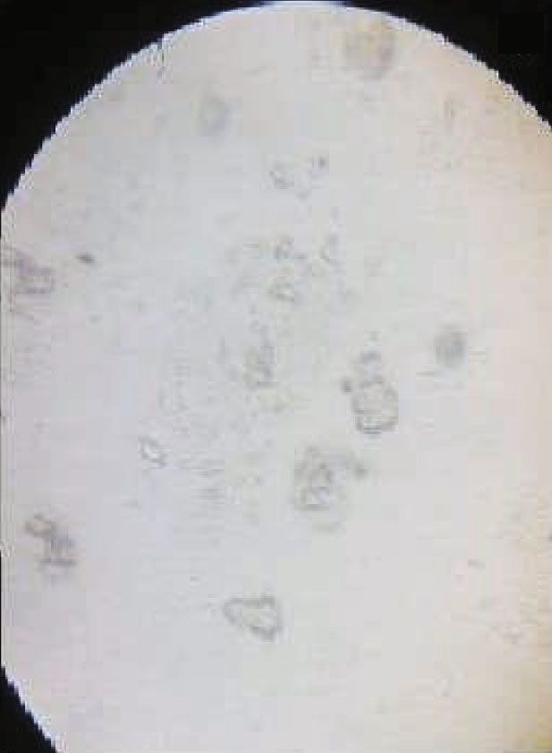
BioMed Research International 5

 (a) (b) (c)

Figure 1: Optical microscopic image of prepared vesicles at different concentrations of LS75: (a) below 2% w/v (vesicles could not form); (b)
2-4% w/v (spherical vesicles with well-defined boundaries); (c) above 4% w/v (larger vesicles with irregular shape).

namely, the controlled and test groups. Before three days of enclosed material. The optimum concentration of ethanol
starting the study, hair was shaved from the back of rats and enhances the membrane elasticity and fluidity of vesicles
a 5 cm2 area was marked. The cream formulations were that may contribute to high skin permeability.
applied to the marked site, and the site was observed for Preliminary screening studies were carried out to iden-
any reaction or sensitivity and slight/moderate or severe ery- tify the effect of variables that influence the physicochemical
thema till 3 days after application. To score the skin reac- properties of vesicles and to optimize the methodology. Pla-
tions for erythema, scar, and edema, the Draize skin cebo (blank) ELVs were prepared by using varied concentra-
irritation scoring system was selected [18]. The reactions, tion of LS75 (0.5%-4.0% w/v) and ethanol (10-40% v/v). The
defined as erythema and edema, were evaluated according results showed that below 2% w/v concentration of LS75, the
to the scoring system for skin reactions. The score of pri- vesicles could not form (Figure 1(a)). It was observed that
mary irritation (SPI) was calculated for each rat. Scores for between 2-4% w/v of LS75, the vesicles were uniformly dis-
erythema and edema at 24, 48, and 72, hours were summed tributed with spherical shape and well-defined boundaries
and divided by the number of the observations for the (Figure 1(b)) and above 4% w/v larger vesicles were formed
treated sites. The SPI for the control sites were calculated with irregular shape (Figure 1(c)). ELVs were formulated by
in the same fashion as the test. using LS75 in the concentration range of 2-4% w/v and eth-
 anol in the range of 30-40% v/v.
3.11. Confocal Laser Scanning Microscopy (CLSM) Study.
Skin permeation depth and mechanism of rhodamine red- 4.2. Characterization of Essential Oil-Loaded ELVs
loaded vesicles were examined by CLSM. Rhodamine-
loaded vesicular cream was formulated by adding the dye 4.2.1. Vesicle Size Measurement. Vesicle size is one of the
to the mixture of LS75 in ethanol and PG, and the prepared important parameters that affect the permeability across
dye-loaded vesicles were incorporated into the cream base. the skin. Developed EO-loaded ELVs were varied in the size
Dye-loaded vesicular and the nonvesicular cream formula- range of 192.0 nm-543.1 nm (Table 3). The vesicle size distri-
tion was applied to the dorsal rat skin for 8 h. The rats were bution diagram of optimized formulations (G6 and C6) is
sacrificed, and the skin was excised and washed. The skin shown in Figure 2. The results showed that vesicle size was
sections were prepared and examined with CLSM (Fluoview directly proportional to the concentration of LS75 and indi-
FV 1000, Olympus, Japan) [19]. rectly proportional to the concentration of ethanol [20], i.e.,
 as the concentration of LS75 was increased, vesicle size was
4. Results and Discussion also increased and upon increasing the concentration of eth-
 anol, the size of vesicles got reduced. It has been reported in
4.1. Formulation of ELVs. Phospholipid and ethanol are the the previous studies that a high concentration of ethanol
basic materials composing ELVs and play an important role leads to the interpenetration of the ethanol hydrocarbon
in vesicle characteristics such as size, entrapment efficacy, chain, which results in slimming down of vesicle membrane
and stability. The phospholipid is responsible for the formu- thickness and hence causes a reduction in vesicle size. Some
lation of a lipid bilayer that affects the stability of the vesicle researchers have suggested that this may be due to the fact
and also enhances the rigidity and prevents leakage of the that high concentration of ethanol modifies the vesicular
6 BioMed Research International

Table 3: Results of vesicle size, polydispersity index, and 4.3. Selection of Optimized ELVs. Based on the results
encapsulation efficiency of ethanolic lipid vesicles. obtained from the characterization of ELVs, G6 (GEO
 encapsulated) and C6 (CEO encapsulated) were selected
S. Formulation Vesicle Polydispersity Encapsulation for incorporation into the cream base.
No. code size (nm) index (PDI) efficiency (%)
1 G1 411.3 0.292 58:92 ± 0:33
 4.4. Characterization of Cream Formulations
2 G2 458.9 0.089 62:62 ± 0:64
3 G3 543.1 0.274 78:95 ± 0:69 4.4.1. Physical Characterization. All the cream formulations
 were white in color with a mild characteristic odor of the
4 G4 218.3 0.131 68:24 ± 0:87
 EO used. The prepared cream formulations were homoge-
5 G5 245.9 0.288 80:95 ± 0:49 neous with a complete absence of lumps and grittiness.
6 G6 199.6 0.063 89:00 ± 0:85
7 C1 339.5 0.208 60:24 ± 0:78 4.4.2. Spreadability, Viscosity, and pH. A cream should not
8 C2 399.1 0.292 69:52 ± 1:64 generate friction while applied on the skin and should
 spread easily. Spreading quality of cream helps in the uni-
9 C3 424.7 0.301 78:39 ± 0:61
 form application to the skin. The spreadability result of pre-
10 C4 185.0 0.172 72:81 ± 0:46 pared creams was found in the range of 17:68 ± 0:34
11 C5 220.1 0.045 84:60 ± 0:82 gm·cm/sec to 25:50 ± 0:45 gm·cm/sec. The results express
 90:95 ± 0:29
 the ability of the creams to spread on the application of a
12 C6 192.0 0.107
 small amount of shear. The spreadability characteristics are
 also influenced by viscosity. The viscosity of the cream formu-
 lations ranged between 2713:5 ± 1:02 and 6011:2 ± 1:20 cp
surface characteristics and alters the net charge, which could which indicate substantial consistency. Furthermore, the pH
lead to a decrease in mean vesicle size [21]. values of all developed formulations were in the range 6:8 ±
 0:052 gm·cm/sec to 7:1 ± 0:050 gm·cm/sec (Table 4). The pH
4.2.2. Polydispersity Index (PDI). PDI number is a descrip- values lie in the normal pH range of the skin and would not
tion of the dispersion of size populations within a given sam- produce irritation upon application to the skin.
ple. The stability of vesicular formulation depends upon the
homogenous populations of vesicles of a certain size. PDI
number ranges from 0.0 to 1.0. The best PDI value is 0.0 4.4.3. Determination of Percent Essential Oil Content. The
which indicates homogenous dispersion with respect to the EO content determination results indicated that the EO
vesicle size, and the PDI value 1.0 indicates an extremely was uniformly distributed throughout the vesicular cream
polydisperse sample with multiple vesicular size populations formulation. It was interesting to observe that in the case
[22]. In the case of lipid-based vesicular carriers, a PDI value of free EO-loaded creams, the EO content results were sig-
of 0.3 and below is considered to be agreeable and shows a nificantly reduced; this might be due to the loss of free EO
homogenous dispersion of lipid vesicles [23, 24]. The results during the cream formulation process (Table 4).
showed that all the vesicular formulations have a PDI value
less than 1.0 and ranged from 0.045 to 0.301, which indicates 4.5. Stability Studies of Cream Formulations. The motive
that vesicles are homogenously distributed (Table 3). behind stability testing of a cosmetic product is to confirm
 that the tested product meets the intended chemical and
4.2.3. Encapsulation Efficiency. EE of vesicular formulations physical quality standards, functionality, and aesthetics
is the part of total oil entrapped in the prepared vesicles, when stored under suitable storage conditions. The freshly
which determines the oil holding capacity and ultimately prepared creams were white in color, but a slight change in
the delivery potential to the particular site. The EE was color was observed for GC1 formulation after 30 days and
found to be in the range from 58:92 ± 0:33% to 90:95 ± for CC1 after 21 days when stored at 40°C; this may be
0:29% (Table 3). Results showed that both the amount of due to the separation of the oil phase at high temperature.
LS75 and the concentration of ethanol influence the encap- No sign of liquefaction was observed in the tested creams
sulation of oil in vesicles positively. It was noticed that with at different storage conditions for 30 days of observation.
the increase in the concentration of LS75 from 2% to 4% and There was no phase separation in any tested creams after
ethanol from 20% to 30% ,the entrapment of oil inside centrifugation and freeze and thaw test. It was observed that
the vesicles also increased. Based on the physicochemical the freezing lowered the viscosity of tested creams [25]. The
characterization, the optimized vesicular formulations G6 absence of any sign of liquefaction and phase separation
(GEO encapsulated) and C6 (CEO encapsulated) were provided strong evidence for the stability of the creams
selected for visualization. under investigation.
 There was no significant change in the pH value of tested
4.2.4. Scanning Electron Microscopy (SEM). SEM results of creams at various storage conditions. The oil content in
optimized ethanolic lipid vesicles loaded with GEO and creams decreased at higher temperature suggesting storage
CEO, i.e., G6 and C6, respectively, disclosed the dominance at room temperature or cool place. None of the stability
of spherical vesicular carriers as shown in Figure 3. parameters changed significantly at room temperature.
BioMed Research International 7

 100
 100

 10
 75
 75

 Cumulative intensity (%)
 Differential intensity (%)
 Cumulative intensity (%)
 5
 Differential intensity (%)

 50 50
 5

 25 25

 0 0 0 0
 100.0 1000.0 100.0 1000.0
 Diameter (nm) Diameter (nm)

 G6 C6

 Figure 2: Vesicle size distribution diagram of optimized ethanolic lipid vesicles (G6 and C6).

 G6 C6
 C6

 15 kV X10,000 1 m 0001 15 30 SEI 15 kV X10,000 1 m 0001 15 30 SEI

 Figure 3: SEM image of optimized ethanolic lipid vesicles: G6 (GEO encapsulated) and C6 (CEO encapsulated).

 Table 4: Characterization of GEO-/CEO-based vesicular and nonvesicular cream formulations.

Formulation code Spreadability (gm·cm/sec) pH Viscosity (cp) EO content (%)
Base cream 17:68 ± 0:34 6:8 ± 0:052 2713:5 ± 1:02 —
GC1 20:68 ± 0:56 7:0 ± 0:091 5413:5 ± 1:60 90:46 ± 1:98
GC2 22:82 ± 0:82 6:9 ± 0:056 5402:0 ± 1:58 93:78 ± 1:05
GC3 25:50 ± 0:45 6:9 ± 0:048 6011:2 ± 1:20 98:23 ± 1:60
CC1 18:06 ± 0:92 6:8 ± 0:060 5424:5 ± 2:60 95:62 ± 1:90
CC2 20:42 ± 0:42 7:1 ± 0:050 4302:5 ± 2:01 97:48 ± 2:01
CC3 22:49 ± 0:60 7:0 ± 0:078 4413:5 ± 1:98 98:66 ± 1:50
GEO6 20:60 ± 0:74 7:0 ± 0:028 2878:5 ± 1:02 74:78 ± 2:20
CEO6 18:68 ± 0:90 6:9 ± 0:040 4028:6 ± 1:00 71:89 ± 1:82

4.6. Determination of Antioxidant Capacity. Antioxidants is a free radical that reacts with oxygen under aerobic condi-
are capable to guard against skin damage and slow down tions and generates nitrite ions. The principle of this scav-
the skin aging process. The antioxidant capacities of enging technique relies on the measurement of the capacity
extracted GEO and CEO have been previously reported by of the antioxidant to trap nitric oxide, leading to a decreased
us [9], and their assessment of the antioxidant capacity after production of nitrite ions. The results of nitric oxide scav-
formulation as ELV-based cream was carried out. enging capacity are shown in Figure 4. The nitric oxide
 scavenging capacity of the standard (ascorbic acid) was
4.6.1. Nitric Oxide Scavenging Capacity. Sodium nitroprus- 89:19 ± 0:05% at 250 μg/ml. The nitric oxide scavenging
side decomposes in an aqueous physiological solution (at capacity of extracted EOs was previously reported by us as
pH 7.4) results in the generation of nitric oxide. Nitric oxide 85:15 ± 0:09% and 72:48 ± 0:12% for GEO and CEO,
8 BioMed Research International

 100

 80

 % Inhibition
 60

 40

 20

 0

 AA

 GEO

 CEO

 Base

 GC1

 GC2

 GC3

 GEO6

 CC1

 CC2

 CC3

 CEO6
Figure 4: Comparison of nitric oxide scavenging capacity of standard (AA); essential oils (GEO and CEO); base cream; GEO-loaded
vesicular creams (GC1, GC2, and GC3); CEO-loaded vesicular creams (CC1, CC2, and CC3); and nonvesicular cream GEO6 and CEO6
for GEO and CEO, respectively.

 100
 % Inhibition

 50

 0
 AA

 GEO

 CEO

 Base

 GC1

 GC2

 GC3

 GEO6

 CC1

 CC2

 CC3

 CEO6
Figure 5: Comparison of DPPH radical scavenging capacity of standard (AA); essential oils (GEO and CEO); base cream; GEO-loaded
vesicular creams (GC1, GC2, and GC3); CEO-loaded vesicular creams (CC1, CC2, and CC3); and nonvesicular cream GEO6 and CEO6
for GEO and CEO, respectively.

respectively [9]. In the case of GEO-/CEO-loaded vesicular remarkable in comparison to standard antioxidant and EO
and nonvesicular cream formulations (Figure 3), the maxi- alone.
mum inhibition was shown by GC3 (80:96 ± 0:20%) and The results of both the antioxidant assay showed that
CC3 (75:21 ± 0:31%) formulation for GEO and CEO, GEO- or CEO-loaded vesicular cream formulations have
respectively. notable antioxidant capacity when compared to EO alone.
 The results were eye-catching when the antioxidant capacity
4.6.2. DPPH Scavenging Capacity. In this method, the degree of vesicular cream formulations was compared with the
of DPPH radical discoloration is often used as an index of cream formulation containing the same concentration of
the antioxidant capacity of the tested samples. In our previ- unentrapped or free EO. It was interesting to observe that
ous work [9], we observed that GEO has a higher power to with both the EOs, the scavenging capacity was reduced
diminish the dark violet color of DPPH radical to yellow for nonvesicular cream formulation in comparison to the
diphenylpicrylhydrazine radical in comparison to CEO and same concentration of vesicular cream. This may be due to
that GEO was found to be rich in monoterpenoid and cit- the instability or loss of EO during the formulation of non-
ronellol and geraniol were the major component of the oil vesicular cream formulation.
and they also have been previously identified as a potential
antioxidant [26, 27]. The DPPH radical scavenging capac- 4.7. Determination of In Vitro Sun Protection Factor. In vitro
ity of extracted EOs was previously reported by us as SPF determination is a useful test for screening ingredients
85:51 ± 0:020% (for GEO) and 78:06 ± 0:04% (for CEO) during the development stage of a cosmetic product. The
at 250 μg/ml [9]. higher the SPF, the more the protection offered by sunscreen
 The highest DPPH radical scavenging capacity (99:08 ± against UV light. An EO should absorb major UV radiations
0:08%) was obtained by standard ascorbic acid in the (290-400 nm) to be adequately used in cosmetic formula-
concentration of 250 μg/ml. At the same concentration, the tions to prevent photoaging, sunburn, skin wrinkles, and
maximum percent inhibition was shown by GC3 (89:82 ± other skin damages. In our previous work, the SPF value of
0:11%) and CC3 (80:89 ± 1:03%) formulation, respectively GEO and CEO was found to be 6.45 and 8.36, respectively
(Figure 5). The DPPH radical scavenging capacities of EO- [9]. EO-based vesicular cream formulations showed remark-
loaded vesicular cream formulations were found to be ably high SPF in comparison to EO alone. The SPF value of
BioMed Research International 9

 20

 15

 SPF
 10

 5

 0

 GEO

 CEO

 Base

 GC1

 GC2

 GC3

 GEO6

 CC1

 CC2

 CC3

 CEO6
Figure 6: Comparison of in vitro SPF values of essential oils (GEO and CEO), base cream, GEO-loaded vesicular creams (GC1, GC2, and
GC3), CEO-loaded vesicular creams (CC1, CC2, and CC3), and free oil-loaded nonvesicular cream GEO6 and CEO6 for GEO and CEO,
respectively.

GEO-loaded vesicular cream was found to be 6.02, 8.35, and inhibitors prevent the degradation of FALGPA [31]. The
10.26 for GC1, GC2, and GC3, respectively (Figure 6). The EOs incorporated in the creams have potential for collage-
SPF of CEO-loaded vesicular cream was found to be 9.28, nase inhibition that was assessed by the assay. From the
12.68, and 18.54 for CC1, CC2, and CC3, respectively. The results of the collagenase inhibition assay, it was observed
SPF of free EO-loaded nonvesicular cream formulation was that both the formulations showed anticollagenase activity
7.82 (for GEO) and 9.02 (for CEO) which is very low in in a concentration-dependent manner (Figure 7). It was
comparison to the vesicular cream at the same concentration observed that the anticollagenase activity increases as the
of EO. The overall results showed that CEO-based cream concentration increases from 1 to 100 μg/ml. In the case
formulation had higher SPF in comparison to GEO-based of GC3, formulation maximum inhibition (28.4%) was
cream formulation. observed at 1000 μg/ml, whereas at the same concentra-
 tion, CC3 showed the maximum inhibition of 22.9%.
4.8. In Vitro Enzyme Inhibition Assay. One of the main rea-
sons behind various signs of skin aging is the damage and 4.8.2. In Vitro Elastase Inhibition Assay. The elastase inhibi-
loss of two key proteins: collagen and elastin [28]. These tion assay is based on the fact that the enzyme (elastase)
proteins are essential for maintaining youthful skin, which causes the breakdown of the elastin substrate (N-succinyl-
are found in the deeper layers of the skin. Collagen plays a Ala-Ala-Ala-p-nitroanilide) and yields fluorescent frag-
major role in skin strengthening, and elastin allows the body ments that are measured using a fluorescence microplate
tissues to return to their original shape either by contracting reader. This enzymatic hydrolysis is interrupted by the elas-
or stretching. These proteins are essential for skin and work tase inhibitors [32]. The elastase inhibition activity of the
together to create skin firmness and help to hold the skin developed creams was assessed to determine their antiaging
shape and strength [29, 30]. The main cause of degradation potential. The antielastase activities of both the opti-
of these proteins is matrix metalloproteinases (MMPs). mized cream formulations (GC3 and CC3) are shown in
MMP enzymes such as collagenase and elastase are respon- Figure 8. The antielastase activity was found to be in a
sible for the breakdown of collagen and elastin protein, concentration-dependent manner. In the case of GEO-
respectively. Degradation of the two essential skin proteins loaded vesicular cream formulation (GC3), it was surprising
by these enzymes accelerates skin wrinkling and sagging skin to see that the antielastase inhibition was higher (144.0%)
appearance and leads to skin aging. Thus, it is important to than ECGC (122.4%) at the concentration of 1000 μg/ml.
determine the enzyme inhibiting activity of the developed CEO-loaded vesicular cream formulation (CC3) also showed
formulations in order to assess the antiaging potential. This significant elastase inhibition activity. The highest antielas-
is the first-time research to investigate the antielastase, antic- tase inhibition was observed at 108.2% at the same concen-
ollagenase potential of GEO-/CEO-based formulation. The tration i.e., 1000 μg/ml.
optimized cream formulations (GC3 and CC3) from each The findings have verified that the GEO-/CEO-loaded
EO were used for further investigation of enzyme inhibition vesicular creams may contribute to the fight against skin
assay. aging by preventing collagen and elastin degradation under-
 neath the skin and can help to restore skin strength and elas-
4.8.1. In Vitro Collagenase Inhibition Assay. The assay is ticity and thereby slow down the skin wrinkling process.
based on an enzyme-substrate interaction, i.e., between col-
lagenase enzyme and the synthetic collagen substrate N-[3- 4.9. Skin Irritation Study. In this study, rats were divided
(2-furyl) acryloyl]-Leu-Gly-Pro-Ala (FALGPA). Proteolytic into 5 groups (n = 3 in each group). In group 1, base cream
degradation of this collagen substrate due to the collagenase was applied, in groups 2 and 3 optimized vesicular cream
enzyme results in a decrease in absorbance. Collagenase of each EO (GC3 and CC3), and in groups 4 and 5
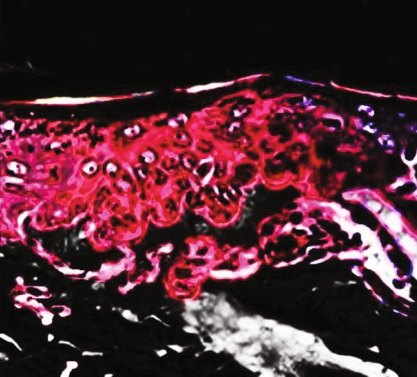
10 BioMed Research International

 % Collagenase inhibition
 45.0
 40.0
 35.0
 30.0
 25.0
 20.0
 15.0
 10.0
 5.0
 0.0
 1 5 10 100 500 1000 EGCG
 Concentration ( g/ml) (+ve control)
 (a)
 % Collagenase inhibition

 45.0
 40.0
 35.0
 30.0
 25.0
 20.0
 15.0
 10.0
 5.0
 0.0
 1 5 10 100 500 1000 EGCG
 Concentration ( g/ml) (+ve control)
 (b)

Figure 7: In vitro collagenase inhibition assay: (a) % collagenase inhibition shown by GEO-loaded optimized cream formulation GC3; (b) %
collagenase inhibition shown by CEO-loaded optimized cream formulation CC3.

 160.0
 % Elastase inhibition

 140.0
 120.0
 100.0
 80.0
 60.0
 40.0
 20.0
 0.0
 1 5 10 100 500 1000 EGCG
 Concentration ( g/ml) (+ve control)

 (a)
 140.0
 % Elastase inhibition

 120.0
 100.0
 80.0
 60.0
 40.0
 20.0
 0.0
 1 5 10 100 500 1000 EGCG
 Concentration ( g/ml) (+ve control)

 (b)

Figure 8: In vitro elastase inhibition assay: (a) % elastase inhibition shown by GEO-loaded optimized cream formulation GC3; (b) % elastase
inhibition shown by CEO-loaded optimized cream formulation CC3.

nonvesicular cream formulation of each EO (GEO6 and and 0.49 for CEO6 cream formulation) which indicates slight
CEO6). The results obtained from the primary skin irritation skin irritation in comparison to vesicle-based cream formula-
studies (Table 5) interpreted according to Draize test [17] tions of the same EO.
which says that test samples that produce PII scores of 2 or
less are considered negative, i.e., no skin irritation. Since, 4.10. Confocal Laser Scanning Microscopy (CLSM) Study.
the score between 0 and 2 suggests no to mild irritation, thus, Cream formulations loaded with and without lipid vesicles
low PII of vesicular cream formulation (0.27 for GC3 and were prepared, and penetration across the skin was mea-
0.16 for CC3 cream formulations) in comparison to base sured by CLSM study. Skin permeation of rhodamine
cream (0.16) observed in the study depicted nonirritancy of (marker) from rhodamine-loaded nonvesicular cream and
the cream formulation and could be considered safe for use, rhodamine-loaded vesicular cream formulation was visualized
while the primary skin irritation studies of free EO-loaded through a confocal laser scanning microscope. It was observed
nonvesicular cream showed high PII values (0.55 for GEO6 that permeation from rhodamine-loaded nonvesicular cream
BioMed Research International 11

Table 5: Skin irritation study scores for primary irritation and primary irritation index measured at 24, 48, and 72 hrs after applying the
cream formulations on the rat skin.

 (a)

 Group 1 (cream base) Group 2 (formulation GC3) Group 3 (formulation CC3)
Skin reaction Time (hrs)
 Rat-1 Rat-2 Rat-3 Rat-1 Rat-2 Rat-3 Rat-1 Rat-2 Rat-3
 24 1 1 1 1 1 1 1 0 0
Erythema 48 0 0 0 0 0 0 0 0 0
 72 0 0 0 0 0 0 0 0 0
 24 0 0 0 1 0 1 1 1 0
Edema 48 0 0 0 0 0 0 0 0 0
 72 0 0 0 0 0 0 0 0 0
Score of primary irritation 0.16 0.16 0.16 0.33 0.16 0.33 0.33 0.16 0.0
Primary irritation index 0.16 0.27 0.16

 (b)

 Group 4 (GEO6) Group 5 (CEO6)
Skin reaction Time (hrs)
 Rat-1 Rat-2 Rat-3 Rat-1 Rat-2 Rat-3
 24 1 1 1 1 1 1
Erythema 48 1 0 1 1 1 1
 72 0 0 0 0 0 0
 24 1 1 1 1 1 0
Edema 48 1 0 1 0 1 0
 72 0 0 0 0 0 0
Score of primary irritation 0.66 0.33 0.66 0.50 0.66 0.33
Primary irritation index 0.55 0.49

 (a) (b)

Figure 9: CLSM of (a) Rhodamine-loaded nonvesicular cream confined to the upper layer of the skin epidermis; (b) enhanced skin
permeation observed by rhodamine-loaded vesicular cream.

(Figure 9(a)) was confined only to the upper layer of the skin observed deep into the skin layers. This shows that prepared
epidermis, while in the case of rhodamine-loaded vesicular ELVs have the ability to carry antiaging EO deep into the skin
cream (Figure 9(b)), enhanced permeation of rhodamine was where the root cause of skin aging exists.
12 BioMed Research International

5. Conclusion [3] T. H. Chou, “Current application of lipid- and surfactant-
 based vesicles for cosmeceuticals: a review,” Current pharma-
Finding solutions against the signs of skin aging has been a ceutical biotechnology, vol. 16, no. 12, pp. 1035–1044, 2015.
natural human desire for centuries. Because of this, the mar- [4] E. Touitou, N. Dayan, L. Bergelson, B. Godin, and M. Eliaz,
ket is flooded with beauty care products claiming magical “Ethosomes – novel vesicular carriers for enhanced delivery:
results within a short period of application. It is to be noted characterization and skin penetration properties,” Journal of
that natural aging is genetically determined, but extrinsic Controlled Release, vol. 65, no. 3, pp. 403–418, 2000.
aging can be slowed down with the use of scientifically [5] K. Knobloch, A. Pauli, B. Iberl, H. Weigand, and N. Weis,
designed and evaluated cosmetic formulations. The need of “Antibacterial and antifungal properties of essential oil com-
the hour is to put serious efforts into the development of ponents,” Journal of Essential Oil Research, vol. 1, no. 3,
such products, which actually translate the stated claims in pp. 119–128, 1989.
the case of extrinsic aging. From the research findings, the [6] A. M. Janssen, J. J. Scheffer, and A. B. Svendsen, “Antimicro-
antioxidant potential, ability to inhibit collagenase and elas- bial activities of essential oils,” Pharmaceutisch Weekblad,
tase enzyme, and photoprotective effects of GEO/CEO vesic- vol. 9, no. 4, pp. 193–197, 1987.
ular cream were confirmed. Results clearly showed that [7] Y. M. Brand, V. C. Roa-Linares, L. A. Betancur-Galvis, D. C.
ELVs were able to preserve the efficiency of essential oils Durán-García, and E. Stashenko, “Antiviral activity of Colom-
and have the potential to deliver the actives deeper into the bian Labiatae and Verbenaceae family essential oils and mono-
skin. Essential oil-encapsulated vesicular creams were able terpenes on human herpes viruses,” Journal of Essential Oil
 Research, vol. 28, no. 2, pp. 130–137, 2016.
to provide antioxidant defence mechanism with high SPF
and are able to protect the skin in comparison to free essen- [8] M. T. Baratta, H. D. Dorman, S. G. Deans, A. C. Figueiredo,
 J. G. Barroso, and G. Ruberto, “Antimicrobial and antioxidant
tial oil-loaded nonvesicular creams. The cumulative effect of
 properties of some commercial essential oils,” Flavour and fra-
ELVs and cream composition and essential oil will collec- grance journal, vol. 13, no. 4, pp. 235–244, 1998.
tively produce a protective effect to combat skin aging.
 [9] A. Lohani, A. K. Mishra, and A. Verma, “Cosmeceutical poten-
 tial of geranium and calendula essential oil: determination of
Data Availability antioxidant activity and in vitro sun protection factor,” Journal
 of Cosmetic Dermatology, vol. 18, no. 2, pp. 550–557, 2019.
Data are available in Supplementary files. Supplementary [10] C. Sebaaly, A. Jraij, H. Fessi, C. Charcosset, and H. Greige-
data supporting the findings of this study includes the Gerges, “Preparation and characterization of clove essential
image of optimized cream formulations, detailed results oil-loaded liposomes,” Food Chemistry, vol. 178, pp. 52–62,
of antioxidant capacity, and SPF values of prepared cream 2015.
formulations. [11] C. D. Kaur and S. Saraf, “Topical vesicular formulations of
 Curcuma longa extract on recuperating the ultraviolet radia-
 tion–damaged skin,” Journal of Cosmetic Dermatology,
Conflicts of Interest vol. 10, no. 4, pp. 260–265, 2011.
The authors declare that they have no conflicts of interest. [12] M. Multimer, “Spreadability determination by an apparatus,”
 Journal of the American Pharmaceutical Association, vol. 45,
 pp. 212–214, 1956.
Acknowledgments [13] S. A. Mortazavi and S. Pishrochi, “Formulation and in-vitro
 evaluation of tretinoin microemulsion as a potential carrier
This work is a Ph.D. research work from IFTM University for dermal drug delivery,” Iranian journal of pharmaceutical
Moradabad, Uttar Pradesh, India. The authors are grateful research: IJPR, vol. 12, pp. 599–602, 2013.
to IFTM University, Moradabad, for providing necessary [14] S. Smaoui, H. B. Hlima, I. B. Chobba, and A. Kadri, “Develop-
facilities for this research work. ment and stability studies of sunscreen cream formulations
 containing three photo-protective filters,” Arabian Journal of
Supplementary Materials Chemistry, vol. 10, pp. S1216–S1222, 2017.
 [15] N. Akhtar, B. A. Khan, T. Mahmood, R. Parveen,
Supplementary data supporting the findings of this study M. Qayum, and M. Anwar, “Formulation and evaluation
includes the image of optimized cream formulations, of antisebum secretion effects of sea buckthorn w/o emul-
detailed results of antioxidant capacity, and SPF values of sion,” Journal of Pharmacy & Bioallied Sciences, vol. 2,
prepared cream formulations. (Supplementary Materials) no. 1, pp. 13–17, 2010.
 [16] COLIPA, CTFA-SA, and CTFA JCIA, COLIPA guidelines:
References International sun protection factor (SPF) test method, vol. 46,
 COLIPA, 2006.
 [1] B. A. Gilchrest, “Skin aging and photoaging: an overview,” [17] P. J. Matts, V. Alard, M. W. Brown et al., “The COLIPA
 Journal of the American Academy of Dermatology, vol. 21, in vitro UVA method: a standard and reproducible measure
 no. 3, pp. 610–613, 1989. of sunscreen UVA protection,” International Journal of Cos-
 [2] P. N. Gupta, P. Singh, V. Mishra, S. Jain, P. K. Dubey, and S. P. metic Science, vol. 32, no. 1, pp. 35–46, 2010.
 Vyas, “Topical immunization: mechanistic insight and novel [18] J. H. Draize, G. Woodward, and H. O. Calvery, “Method for
 delivery systems,” Indian Journal of Biotechnology, vol. 3, the study of irritation and toxicity of substances applied topi-
 pp. 9–21, 2004. cally to the skin and mucous membranes,” The Journal of
BioMed Research International 13

 Pharmacology and Experimental Therapeutics, vol. 82,
 pp. 377–390, 1944.
[19] R. Alvarez-Román, A. Naik, Y. N. Kalia, H. Fessi, and R. H.
 Guy, “Visualization of skin penetration using confocal laser
 scanning microscopy,” European Journal of Pharmaceutics
 and Biopharmaceutics, vol. 58, no. 2, pp. 301–316, 2004.
[20] S. Jain, N. Patel, P. Madan, and S. Lin, “Quality by design
 approach for formulation, evaluation and statistical optimiza-
 tion of diclofenac-loaded ethosomes via transdermal route,”
 Pharmaceutical Development and Technology, vol. 20, no. 4,
 pp. 473–489, 2015.
[21] I. M. Abdulbaqi, Y. Darwis, N. A. Khan, R. A. Assi, and A. A.
 Khan, “Ethosomal nanocarriers: the impact of constituents
 and formulation techniques on ethosomal properties, in vivo
 studies, and clinical trials,” International Journal of Nanomedi-
 cine, vol. 11, pp. 2279–2304, 2016.
[22] S. Singh, A. Lohani, A. K. Mishra, and A. Verma, “Formulation
 and evaluation of carrot seed oil-based cosmetic emulsions,”
 Journal of Cosmetic and Laser Therapy, vol. 21, no. 2, pp. 99–
 107, 2019.
[23] M. Chen, X. Liu, and A. Fahr, “Skin penetration and deposi-
 tion of carboxyfluorescein and temoporfin from different lipid
 vesicular systems: in vitro study with finite and infinite dosage
 application,” International Journal of Pharmaceutics, vol. 408,
 no. 1-2, pp. 223–234, 2011.
[24] T. C. Khom, H. K. Yadav, A. Raizaday, N. Manne, H. S.
 Kumar, and S. N. Kumar, “Development of mucoadhesive
 nanoparticulate system of ebastine for nasal drug delivery,”
 Tropical Journal of Pharmaceutical Research, vol. 13, no. 7,
 pp. 1013–1019, 2014.
[25] C. Li, Q. Huang, S. Ma, and C. Ji, “An experimental study on
 the viscosity of water-in-oil emulsions,” Journal of Dispersion
 Science and Technology, vol. 37, no. 3, pp. 305–316, 2016.
[26] D. Giustarini, R. Rossi, A. Milzani, and I. Dalle-Donne,
 “Nitrite and nitrate measurement by Griess reagent in human
 plasma: evaluation of interferences and standardization,”
 Methods in enzymology, vol. 440, pp. 361–380, 2008.
[27] Z. Al Mahmud, S. C. Bachar, C. M. Hasan, T. B. Emran,
 N. Qais, and M. M. Uddin, “Phytochemical investigations
 and antioxidant potential of roots of Leea macrophylla
 (Roxb.),” BMC research notes, vol. 10, no. 1, pp. 245–250, 2017.
[28] G. Jenkins, “Molecular mechanisms of skin ageing,” Mecha-
 nisms of Ageing and Development, vol. 123, no. 7, pp. 801–
 810, 2002.
[29] D. Harman, “The free radical theory of aging,” Antioxidants &
 Redox Signaling, vol. 2, pp. 557–561, 2003.
[30] B. Poljsak and R. Dahmane, “Free radicals and extrinsic skin
 aging,” Dermatology Research and Practice, vol. 1, 10 pages,
 2012.
[31] S. A. T. Tamsyn, H. Pauline, and P. N. Pauline, “Anti-collage-
 nase, anti-elastase and anti-oxidant activities of extracts from
 21 plants,” BMC complementary and alternative medicine,
 vol. 9, pp. 27–35, 2009.
[32] M. Chatatikun and A. Chiabchalard, “Thai plants with high
 antioxidant levels, free radical scavenging activity, anti-
 tyrosinase and anti-collagenase activity,” BMC complementary
 and alternative medicine, vol. 17, no. 1, pp. 487–492, 2017.
You can also read