U-Net with Hierarchical Bottleneck Attention for Landmark Detection in Fundus Images of the Degenerated Retina
←
→
Page content transcription
If your browser does not render page correctly, please read the page content below
U-Net with Hierarchical Bottleneck Attention
                                               for Landmark Detection in Fundus Images
                                                       of the Degenerated Retina

                                                Shuyun Tang1 , Ziming Qi1 , Jacob Granley1 , and Michael Beyeler1

                                                          University of California, Santa Barbara, CA 93106
                                                          {shuyun,zimingqi,jgranley,mbeyeler}@ucsb.edu
arXiv:2107.04721v1 [eess.IV] 9 Jul 2021

                                                Abstract. Fundus photography has routinely been used to document
                                                the presence and severity of retinal degenerative diseases such as age-
                                                related macular degeneration (AMD), glaucoma, and diabetic retinopa-
                                                thy (DR) in clinical practice, for which the fovea and optic disc (OD) are
                                                important retinal landmarks. However, the occurrence of lesions, drusen,
                                                and other retinal abnormalities during retinal degeneration severely com-
                                                plicates automatic landmark detection and segmentation. Here we pro-
                                                pose HBA-U-Net: a U-Net backbone enriched with hierarchical bottle-
                                                neck attention. The network consists of a novel bottleneck attention
                                                block that combines and refines self-attention, channel attention, and
                                                relative-position attention to highlight retinal abnormalities that may
                                                be important for fovea and OD segmentation in the degenerated retina.
                                                HBA-U-Net achieved state-of-the-art results on fovea detection across
                                                datasets and eye conditions (ADAM: Euclidean distance (ED) of 25.4
                                                pixels, REFUGE: 32.5 pixels, IDRiD: 32.1 pixels), on OD segmentation
                                                for AMD (ADAM: Dice coefficient (DC) of 0.947), and on OD detection
                                                for DR (IDRiD: ED of 20.5 pixels). We further validated the design of
                                                our network with an ablation study. Our results suggest that HBA-U-Net
                                                may be well suited for landmark detection in the presence of a variety of
                                                retinal degenerative diseases.

                                                Keywords: deep learning · landmark detection · segmentation · self-
                                                attention · fundus · fovea · optic disc · retinal degeneration · age-related
                                                macular degeneration · diabetic retinopathy · glaucoma

                                          1    Introduction
                                          Age-related macular degeneration (AMD), glaucoma, and diabetic retinopathy
                                          (DR) are three of the most common causes of blindness in the world [2]. Fundus
                                          photography has routinely been used to document the presence and severity of
                                          these retinal degenerative diseases in clinical practice. Among the landmarks of
                                          interest are the fovea, which is a small depression in the macula, and the optic
                                          disc (OD), which is where the optic nerve and blood vessels leave the retina.
                                          However, detecting retinal abnormalities associated with these diseases (e.g.,
                                          drusen in AMD, hemorrhage in DR) is a labor-intensive and time-consuming
                                          process, thus necessitating the need for automated fundus image analysis.
2      Tang et al.

     In recent years, numerous methods have been proposed for retinal struc-
ture detection. Jiang et al. [7] proposed an encoder-decoder network with deep
residual structure and recursive learning mechanism for robust OD localization,
followed by an end-to-end region-based convolutional neural network (R-CNN)
for joint optic disc and cup segmentation [8]. Similarly, numerous studies have
employed various convolutional neural network (CNN) models for fovea local-
ization (e.g., [1, 15]). Although fovea and OD are spatially correlated with each
other, only a few studies (e.g., [10, 22]) have focused on joint fovea and OD seg-
mentation. Furthermore, models trained on healthy eyes tend not to generalize
well to diseased eyes with retinal abnormalities. A notable exception is Kamble et
al. [9] who achieved state-of-the-art (SOTA) performance on landmark detection
for AMD and glaucoma using a modified U-Net++ with an EfficientNet encoder.
However, there is potential merit in combining convolutional backbone networks
with attentional mechanisms [19] to highlight retinal abnormalities that may be
important for landmark detection in the degenerated retina.
     To develop a segmentation model that is well suited for retinal degeneration,
we propose HBA-U-Net: a U-Net backbone enriched with hierarchical bottleneck
attention. The main contributions of this work are:
1. We propose a hierarchical bottleneck attention (HBA) block: a novel at-
   tention mechanism that combines and refines self-attention [19], channel
   attention [21], and relative-position attention [13] to highlight retinal ab-
   normalities important for landmark detection in the degenerated retina.
2. We integrate the HBA block into bottleneck skip connections across all layers
   of a U-Net backbone network to form HBA-U-Net, and test the network’s
   performance on three benchmark datasets for retinal degeneration: ADAM
   [4] for AMD, REFUGE [11] for glaucoma, and IDRiD [12] for DR.
3. We validate the design of HBA-U-Net with an ablation study.
4. We demonstrate SOTA performance on fovea detection across datasets and
   eye conditions, on OD segmentation for AMD, and on OD detection for DR.

2     Methods
2.1   Model Architecture
HBA-U-Net The proposed network architecture is illustrated in Fig. 1. First,
ImageNet pretrained ResNet-50 blocks were used as encoders to obtain feature
maps at different spatial resolutions. These feature maps, along with the original
image, were then fed into a modified U-Net structure [10, 14] with HBA blocks
added to skip connections. The outputs of the HBA blocks were up-sampled and
aggregated to produce the final fovea and OD segmentation mask.
    Our goal was to incorporate HBA blocks into the U-Net without drastically
increasing the computational complexity. Consistent with [16], we noticed that
adding a self-attention mechanism to the bottleneck layers (a shrinking path, the
attention module, and an expanding path) significantly boosted the network’s
performance. However, the original U-Net contains only a single bottleneck layer
Joint Fovea and OD Segmentation for Retinal Degeneration 3
Output
Input Sigmoid
(512,512,1)
(512x512x3)
R1
(256x256x64)
R2
(128x128x256)
(128,128,128)
R3
(64x64x512)
R4 (64x64x192)
(32x32x1024)
(32x32x256)
(16x16x1024)
Down-sampling Up-sampling Skip-connections
Encoder activation Convolutional HBA ResNet50
layer extraction Block Block Encoder
Fig. 1. HBA-U-Net architecture. A U-Net enriched with a novel attention block and re-
designed skip-connection paths jointly locates the fovea and segments the optic disc.
ResNet-50 was used as encoder. Note that the number of local bottlenecks and the
down/up-sampling projection rate depends on the image dimensions.
(between the last down-sampling block and the first up-sampling block). To
incorporate multiple HBA blocks into the network, we therefore re-designed the
U-Net by creating local bottleneck structures in each skip-connection pair (see
Fig. 1). After each down-convolution block, the features were down-sampled by
pooling and passed to the HBA block, followed by up-sampling to the original
size. In this way, the pairs of down/up-sampling convolution blocks could be
treated as local bottleneck structures operating at different spatial resolutions.
HBA block Recently, attention mechanisms have seen widespread adoption
in various tasks [19]. Inspired by [16, 21], our HBA block (Fig. 2) consisted
of channel, content, and relative-position attention modules, each described in
detail below. We denote the query, key, value, input feature map, relative height
logit, and relative weight logit as q, k, v, F, Rh , Rw , respectively.
In the proposed HBA block, content attention (blue box in Fig. 2) attended
to individual pixels in each spatial feature map. For each attention head, dense4 Tang et al.
F'
FC sigmoid softmax HBA Block
FS FR
shared
MLP qk T
qRh,w T
1x1 1x1
v k q
Ave Max WV WK WQ
Pooling Pooling Rh Rw
self relative
channel attention self content attention
position attention
F
Dense Layer Pooling Layer Relative Logits Multiply Add
Fig. 2. HBA block architecture, consisting of channel attention (green box outputs FC ),
content attention using multi-head self-attention (blue box outputs FS ), and relative
position attention (pink box outputs FR ).
layers (WQ , WK , WV ) were used to calculate the query (q = WQ (F )), key (k =
WK (F )), and value (v = WV (F )) for each pixel. The output of the content
attention was an attention score (FS ) between key (k) and query vectors (q):
FS = qk T . (1)
Inspired by [13, 16], we included relative-position attention (pink box in
Fig. 2) to encode the relative position of different retinal landmarks (e.g., to
relate the fovea to the OD location). Relative logits were used to store the x and
y offsets (Rh and Rw ) between each key and query. These were added and the
relative positional attention score FR was computed using the dot product:
FR = q(Rh + Rw )T . (2)
In a U-Net, spatial information is encoded to different channels through
down/up-sampling. We believe channel-wise attention is well suited to utilize
this information in the bottleneck layers, which usually have many channels. We
therefore used the channel attention module proposed in [21] (green box in Fig.
2). The input feature map F was passed in parallel to average pooling and max
pooling layers, compressing each channel to one value. These two feature maps
were forwarded through a single, shared multi-layer perceptron (MLP) with one
hidden layer and added to compute the final channel attention score (FC ):
FC = M LP (AvgP ool(F )) + M LP (M axP ool(F )). (3)Joint Fovea and OD Segmentation for Retinal Degeneration 5
In contrast to a conventional transformer, the value vector was scaled not
only by the content attention score (FS ), but also according to the relative-
position attention score (FR ) and the channel attention score (FC ). The output
of the HBA block (F0 ) is given as follows:
F0 = sof tmax(FS + FR )σ(FC )v, (4)
where the softmax was applied across attention heads and σ denotes the sigmoid
function.
2.2 Datasets
We evaluated our model on three prominent datasets for retinal degeneration:
ADAM [4] for AMD, REFUGE [11] for glaucoma, and IDRiD [12] for DR.
ADAM was released as part of a Grand Challenge at a satellite event of
the ISBI 2020 conference. The dataset contains 400 fundus images at either
2124 × 2056 or 1444 × 1444 resolution, 87 of which depict eyes at various stages
of AMD progression (typical signs include the presence of drusen, exudation, and
hemorrhage), and the rest are from healthy controls. ADAM includes ground-
truth OD segmentation masks and fovea image coordinates.
REFUGE was released as part of a Grand Challenge of the OMIA5 workshop
at MICCAI 2018. The dataset contains 1200 fundus images at either 2124×2056
or 1634 × 1634 resolution, 120 of which depict eyes with glaucoma, and the rest
are from healthy controls. REFUGE includes ground-truth OD segmentation
masks and fovea image coordinates.
IDRiD was released as part of a Grand Challenge at ISBI 2018. The dataset
contains 516 images at 4288 × 2848 resolution divided into 413 train images
and 103 test images, all of which contain pathological conditions such as DR
and diabetic macular edema. IDRiD includes ground-truth image coordinates
for the fovea and OD center, but not segmentation masks.
2.3 Implementation Details
Data Preprocessing and Augmentation First, we resized every image in
the dataset to 512 × 512 pixels. Second, we followed [9] to generate circular
segmentation masks from the ground-truth fovea coordinates and combined them
with the ground-truth OD segmentation masks. Third, we applied random image
rotations (uniformly sampled from [−0.2, 0.2] rad), and horizontal/vertical flips
to augment the original dataset on-the-fly. Fourth, we split the data 85-15 into
train and test sets and held out 20% of the training images for validation.
Training Procedure The model was trained using the adam optimizer, the
Dice loss [17], and early stopping, with a custom learning rate scheduler (start
rate 0.0025, decay rate 0.985 after 150 epochs), and batch size 8 for 500 epochs.
Initial weights were pre-trained on ImageNet. The model was implemented using
Keras 2.4.3 (Python 3.7) and run on an NVIDIA Tesla K80 (12GB of RAM)
provided by Google Colab Pro. The code is available at github.com/anonymous.6 Tang et al.
Evaluation Metrics We evaluated the performance of the model using Eu-
clidean distance (ED) [10], where only image coordinates were given, and Dice
coefficient (DC), where segmentation masks were given. Since none of the three
datasets came with fovea segmentation masks, we followed [10] to create a cir-
cular disc centered over the ground-truth fovea coordinates, which was then
used to train our network. After training, we recovered predicted coordinates by
extracting the centroid of the predicted segmentation mask using scikit-image.
3 Experiments and Results
3.1 Joint Fovea and OD Detection in the Degenerated Retina
Table 1 summarizes our results on three prominent datasets for retinal degener-
ation: ADAM for AMD, REFUGE for glaucoma, and IDRiD for DR.
HBA-U-Net achieved SOTA performance on fovea detection across all datasets
(ADAM: ED 25.4 px; REFUGE: ED 32.5 px; IDRiD: 32.1 px) and thus across
eye conditions, despite the fact that these datasets were previously used in Grand
Challenges that featured convolutional [9], attentional [23], and adversarial [20]
approaches, some of which had a considerably larger number of trainable pa-
rameters. Because all three datasets are relatively new, the number of published
results is still relatively small.
HBA-U-Net also achieved SOTA performance on OD segmentation for AMD
(DC of 0.947, on par with [9]) and on OD detection for DR (ED of 20.5). Our
OD segmentation was slightly worse than competing models, with the SOTA
belonging to [20], a patch-based morphology-aware segmentation network.
However, please note that the test data of these challenges is not made avail-
able to the public. To offer a fair comparison across models, we therefore re-
implemented a number of commonly used alternative network architectures and
Table 1. Landmark detection on ADAM, REFUGE, and IDRiD. Note that Challenge
test data is not publicly available. ED: Euclidean Distance. DC: Dice Coefficient.
Fovea Optic Disc
Model ED ED DC
ADAM
Aira matrix [9] (ISBI 2020 Challenge Winner) 26.2 - 0.947
HBA-U-Net (this paper) 25.4 - 0.947
Fu et al. [5] - - 0.936
REFUGE
Zhang et al. [23] - - 0.953
Kamble et al. [9] 35.2 - 0.957
Wang et al. [20] - - 0.960
HBA-U-Net (this paper) 32.5 - 0.947
DeepDR (IDRiD Subchallenge-3 Winner, on-site) 64.5 21.1 -
IDRiD
ZJU-BII-SGEX (IDRiD Subchallenge-3 Winner, online) 45.9 25.6 -
HBA-U-Net (this paper) 32.1 20.5 -Joint Fovea and OD Segmentation for Retinal Degeneration 7
Table 2. Landmark detection for different reimplemented models tested on ADAM,
REFUGE, and IDRiD. ED: Euclidean Distance, DC: Dice Coefficient, F: Fovea, OD:
Optic Disc.
ADAM REFUGE IDRiD
Model EDF DCOD EDF DCOD EDF EDOD
U-Net [14] 70.7 0.741 65.2 0.806 87.1 53.7
EfficientNet encoded U-Net++ [9] 26.9 0.867 37.6 0.935 50.4 28.1
HBA-U-Net (this paper) 25.4 0.947 32.5 0.947 32.1 20.5
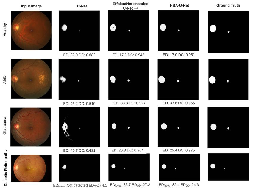
Fig. 3. Representative example predictions for a healthy eye (top row ), AMD (second
row ), glaucoma (third row ), and DR (bottom row ). Predictions are shown for a re-
implemented U-Net (second column), EfficientNet encoded U-Net++ with scSE blocks
(third column), and HBA-U-Net (fourth column), and compared against ground truth
(rightmost column). Error rates are given below each prediction panel.
compared their performance using our own train/test split. These alternative
networks included the classical U-Net [14] and an EfficientNet [18] encoded U-
Net++ with scSE blocks (similar to [9]). Results are given in Table 2 and example
predictions are shown in Fig. 3. HBA-U-Net outperformed the baseline models
on all three datasets.8 Tang et al.
Table 3. Ablation studies on each network component. Starting from a U-Net back-
bone [10, 14], we gradually added a ResNet-50 encoder [6], a standard self-attention
block [13] (‘Self-Att’), a single HBA block in the bottleneck (‘HBA-1’), and HBA blocks
across all levels of the hierarchy (‘HBA-all’).
U-Net ResNet Self-Att HBA-1 HBA-all Params Fovea ED OD DC
8.7M 70.7 0.741
20.8M 34.8 0.902
21.1M 29.8 0.925
21.3M 25.8 0.920
22.2M 25.4 0.947
3.2 Ablation Study
To measure the impact of the HBA block on different versions of our proposed
model architecture, we performed an ablation study on ADAM (see Table 3).
Starting with the original U-Net [10, 14] as a baseline, we were able to re-
duce fovea ED by a factor of two by adding a ResNet-50 encoder [6]. Adding
the original self-attention block [13] (without relative position and channel-wise
attention; labeled ‘Self-Att’ in Table 3) at the bottleneck part of the U-Net
improved fovea ED by ∼ 5%, but led to a ∼ 2% decrease in DC for OD segmen-
tation. Upgrading the self-attention block to our proposed HBA block at the
bottleneck part of the U-Net (labeled ‘HBA-1’) resulted in both the ED and DC
improving by ∼ 4%. Finally, creating local bottlenecks with HBA blocks at each
skip connection in the hierarchy (labeled ‘HBA-all’) led to SOTA performance.
4 Conclusions
We have proposed a re-designed U-Net architecture with hierarchical bottleneck
attention and demonstrated its utility for fundus analysis. The proposed net-
work achieved SOTA performance on fovea detection across datasets and eye
conditions, on OD segmentation for AMD, and on OD detection for DR.
Although self-attention, channel attention, and relative-position have been
deployed separately in other computer vision tasks, here we refined, simplified,
and combined their potential in segmenting retinal abnormalities. Furthermore,
our ablation study demonstrates the benefit of the local bottleneck structures
and HBA blocks for retinal landmark segmentation. Compared to content self-
attention alone, HBA does not add much overhead: relative position attention
does not have any learnable parameters and channel attention consists of a
shared MLP with one hidden layer. Compared to other pure attention networks
such as ViT [3], HBA blocks are more resourceful and better suited to work in
combination with convolutional modules commonly used in segmentation tasks.
Overall our results suggest that HBA-U-Net may be well suited for landmark
detection in the presence of a variety of retinal degenerative diseases.Joint Fovea and OD Segmentation for Retinal Degeneration 9
References
1. Alais, R., Dokládal, P., Erginay, A., Figliuzzi, B., Decencière, E.: Fast macula
detection and application to retinal image quality assessment. Biomedical Signal
Processing and Control 55, 101567 (Jan 2020)
2. Blindness, G.., Collaborators, V.I.: Causes of blindness and vision impairment in
2020 and trends over 30 years, and prevalence of avoidable blindness in relation
to VISION 2020: the Right to Sight: an analysis for the Global Burden of Disease
Study. The Lancet Global Health 9(2), e144–e160 (Feb 2021), publisher: Elsevier
3. Dosovitskiy, A., Beyer, L., Kolesnikov, A., Weissenborn, D., Zhai, X., Unterthiner,
T., Dehghani, M., Minderer, M., Heigold, G., Gelly, S., Uszkoreit, J., Houlsby, N.:
An image is worth 16x16 words: Transformers for image recognition at scale (2021)
4. Fu, H.: ADAM: Automatic Detection challenge on Age-related Macular degenera-
tion (Jan 2020), publisher: IEEE type: dataset
5. Fu, H., Cheng, J., Xu, Y., Zhang, C., Wong, D.W.K., Liu, J., Cao, X.: Disc-Aware
Ensemble Network for Glaucoma Screening From Fundus Image. IEEE Transac-
tions on Medical Imaging 37(11), 2493–2501 (Nov 2018), conference Name: IEEE
Transactions on Medical Imaging
6. He, K., Zhang, X., Ren, S., Sun, J.: Deep Residual Learning for Image Recognition.
In: 2016 IEEE Conference on Computer Vision and Pattern Recognition (CVPR).
pp. 770–778 (Jun 2016), iSSN: 1063-6919
7. Jiang, S., Chen, Z., Li, A., Wang, Y.: Robust Optic Disc Localization by Large
Scale Learning. In: Fu, H., Garvin, M.K., MacGillivray, T., Xu, Y., Zheng, Y.
(eds.) Ophthalmic Medical Image Analysis. pp. 95–103. Lecture Notes in Computer
Science, Springer International Publishing, Cham (2019)
8. Jiang, Y., Duan, L., Cheng, J., Gu, Z., Xia, H., Fu, H., Li, C., Liu, J.: JointRCNN:
A Region-Based Convolutional Neural Network for Optic Disc and Cup Segmen-
tation. IEEE Transactions on Biomedical Engineering 67(2), 335–343 (Feb 2020),
conference Name: IEEE Transactions on Biomedical Engineering
9. Kamble, R., Samanta, P., Singhal, N.: Optic Disc, Cup and Fovea Detection from
Retinal Images Using U-Net++ with EfficientNet Encoder. In: Fu, H., Garvin,
M.K., MacGillivray, T., Xu, Y., Zheng, Y. (eds.) Ophthalmic Medical Image Anal-
ysis. pp. 93–103. Lecture Notes in Computer Science, Springer International Pub-
lishing, Cham (2020)
10. Meyer, M.I., Galdran, A., Mendonça, A.M., Campilho, A.: A Pixel-Wise Dis-
tance Regression Approach for Joint Retinal Optical Disc and Fovea Detection.
In: Frangi, A.F., Schnabel, J.A., Davatzikos, C., Alberola-López, C., Fichtinger,
G. (eds.) Medical Image Computing and Computer Assisted Intervention – MIC-
CAI 2018. pp. 39–47. Lecture Notes in Computer Science, Springer International
Publishing, Cham (2018)
11. Orlando, J.I., Fu, H., Barbosa Breda, J., van Keer, K., Bathula, D.R., Diaz-Pinto,
A., Fang, R., Heng, P.A., Kim, J., Lee, J., Lee, J., Li, X., Liu, P., Lu, S., Murugesan,
B., Naranjo, V., Phaye, S.S.R., Shankaranarayana, S.M., Sikka, A., Son, J., van
den Hengel, A., Wang, S., Wu, J., Wu, Z., Xu, G., Xu, Y., Yin, P., Li, F., Zhang,
X., Xu, Y., Bogunović, H.: Refuge challenge: A unified framework for evaluating
automated methods for glaucoma assessment from fundus photographs. Medical
Image Analysis 59, 101570 (2020)
12. Porwal, P., Pachade, S., Kokare, M., Deshmukh, G., Son, J., Bae, W., Liu, L.,
Wang, J., Liu, X., Gao, L., Wu, T., Xiao, J., Wang, F., Yin, B., Wang, Y., Danala,
G., He, L., Choi, Y.H., Lee, Y.C., Jung, S.H., Li, Z., Sui, X., Wu, J., Li, X.,10 Tang et al.
Zhou, T., Toth, J., Baran, A., Kori, A., Chennamsetty, S.S., Safwan, M., Alex,
V., Lyu, X., Cheng, L., Chu, Q., Li, P., Ji, X., Zhang, S., Shen, Y., Dai, L., Saha,
O., Sathish, R., Melo, T., Araújo, T., Harangi, B., Sheng, B., Fang, R., Sheet,
D., Hajdu, A., Zheng, Y., Mendonça, A.M., Zhang, S., Campilho, A., Zheng, B.,
Shen, D., Giancardo, L., Quellec, G., Mériaudeau, F.: Idrid: Diabetic retinopathy
– segmentation and grading challenge. Medical Image Analysis 59, 101561 (2020)
13. Ramachandran, P., Parmar, N., Vaswani, A., Bello, I., Levskaya, A., Shlens, J.:
Stand-Alone Self-Attention in Vision Models. arXiv:1906.05909 [cs] (Jun 2019),
arXiv: 1906.05909
14. Ronneberger, O., Fischer, P., Brox, T.: U-Net: Convolutional Networks for Biomed-
ical Image Segmentation. In: Navab, N., Hornegger, J., Wells, W.M., Frangi, A.F.
(eds.) Medical Image Computing and Computer-Assisted Intervention – MICCAI
2015. pp. 234–241. Lecture Notes in Computer Science, Springer International
Publishing, Cham (2015)
15. Sedai, S., Tennakoon, R., Roy, P., Cao, K., Garnavi, R.: Multi-stage segmentation
of the fovea in retinal fundus images using fully Convolutional Neural Networks.
In: 2017 IEEE 14th International Symposium on Biomedical Imaging (ISBI 2017).
pp. 1083–1086 (Apr 2017), iSSN: 1945-8452
16. Srinivas, A., Lin, T.Y., Parmar, N., Shlens, J., Abbeel, P., Vaswani, A.: Bottle-
neck Transformers for Visual Recognition. arXiv:2101.11605 [cs] (Jan 2021), arXiv:
2101.11605
17. Sudre, C.H., Li, W., Vercauteren, T., Ourselin, S., Jorge Cardoso, M.: Gener-
alised Dice Overlap as a Deep Learning Loss Function for Highly Unbalanced
Segmentations. In: Cardoso, M.J., Arbel, T., Carneiro, G., Syeda-Mahmood, T.,
Tavares, J.M.R., Moradi, M., Bradley, A., Greenspan, H., Papa, J.P., Madabhushi,
A., Nascimento, J.C., Cardoso, J.S., Belagiannis, V., Lu, Z. (eds.) Deep Learning
in Medical Image Analysis and Multimodal Learning for Clinical Decision Support.
pp. 240–248. Lecture Notes in Computer Science, Springer International Publish-
ing, Cham (2017)
18. Tan, M., Le, Q.V.: EfficientNet: Rethinking Model Scaling for Convolutional Neural
Networks. arXiv:1905.11946 [cs, stat] (Sep 2020), arXiv: 1905.11946
19. Vaswani, A., Shazeer, N., Parmar, N., Uszkoreit, J., Jones, L., Gomez, A.N., Kaiser,
L., Polosukhin, I.: Attention is All you Need. Advances in Neural Information
Processing Systems 30 (2017)
20. Wang, S., Yu, L., Yang, X., Fu, C.W., Heng, P.A.: Patch-Based Output Space
Adversarial Learning for Joint Optic Disc and Cup Segmentation. IEEE Transac-
tions on Medical Imaging 38(11), 2485–2495 (Nov 2019), conference Name: IEEE
Transactions on Medical Imaging
21. Woo, S., Park, J., Lee, J., Kweon, I.S.: CBAM: convolutional block attention mod-
ule. CoRR (2018)
22. Yu, H., Barriga, S., Agurto, C., Echegaray, S., Pattichis, M., Zamora, G., Bauman,
W., Soliz, P.: Fast localization of optic disc and fovea in retinal images for eye dis-
ease screening. In: Medical Image Computing and Computer Assisted Intervention
– MICCAI 2011. vol. 7963, p. 796317 (Mar 2011)
23. Zhang, Z., Fu, H., Dai, H., Shen, J., Pang, Y., Shao, L.: ET-Net: A Generic Edge-
aTtention Guidance Network for Medical Image Segmentation. In: Shen, D., Liu,
T., Peters, T.M., Staib, L.H., Essert, C., Zhou, S., Yap, P.T., Khan, A. (eds.) Med-
ical Image Computing and Computer Assisted Intervention – MICCAI 2019. pp.
442–450. Lecture Notes in Computer Science, Springer International Publishing,
Cham (2019)You can also read