UNIVERSITÄTSKLINIKUM HAMBURG-EPPENDORF - ediss.sub.hamburg
←
→
Page content transcription
If your browser does not render page correctly, please read the page content below
UNIVERSITÄTSKLINIKUM HAMBURG-EPPENDORF

               Martini-Klinik Prostatakarzinomzentrum

                    Prof. Dr. med. Markus Graefen

Impact of Sarcopenia on Functional and Oncological Outcomes
                After Radical Prostatectomy

                            Dissertation

         zur Erlangung des Grades eines Doktors der Medizin
        an der Medizinischen Fakultät der Universität Hamburg.

                            vorgelegt von:

                           Markus Angerer
                            aus Leipzig

                           Hamburg 2021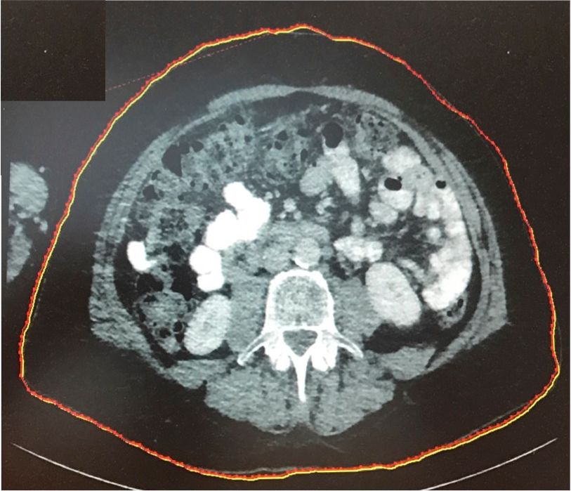
Angenommen von der
Medizinischen Fakultät der Universität Hamburg am: 18.08.2021

Prüfungsausschuss, der Vorsitzende:      Prof. Dr. Udo Schumacher

Prüfungsausschuss, zweiter Gutachter:    Prof. Dr. Georg Salomon

Prüfungsausschuss, dritte Gutachterin:   Prof. Dr. Katja Weisel

                                                                    2
Inhaltsverzeichnis

1. Manuskript und Letter of Acceptance             4-17

2. Darstellung der Publikation                     18-29

3. Literaturverzeichnis                            30-34

4. Zusammenfassung                                 35-36

5. Erklärung des Eigenanteils an der Publikation   37

6. Danksagung                                      38

7. Lebenslauf                                      39

8. Eidesstattliche Versicherung                    40

                                                           3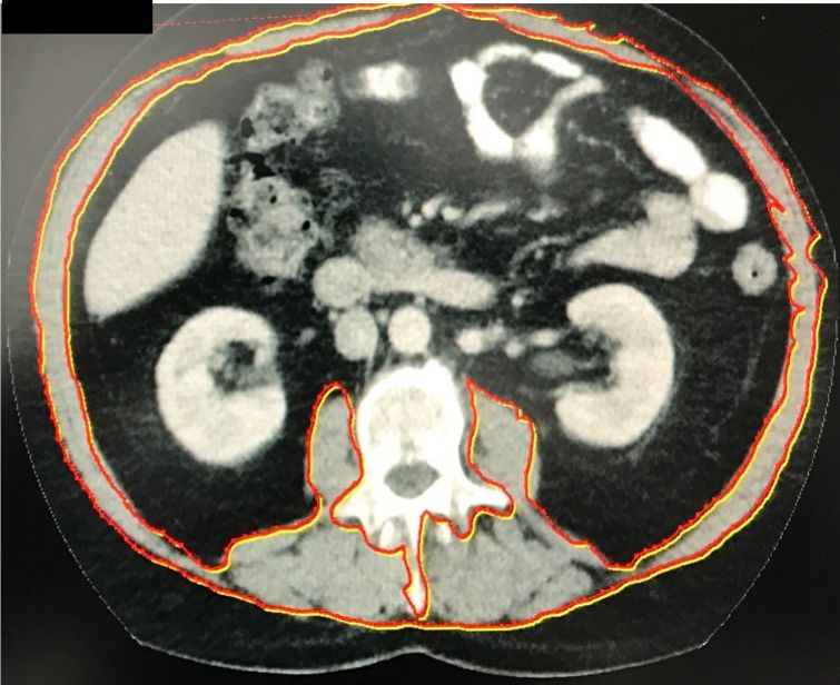
1.                      Manuskript und Letter of Acceptance

Letter of Acceptance:

Dear Dr Angerer,

Frontiers Surgery Editorial Office has sent you a message. Please click 'Reply' to send a
direct response

I am pleased to inform you that your manuscript Impact of Sarcopenia on Functional and
Oncological Outcomes After Radical Prostatectomy has been approved for production and
accepted for publication in Frontiers in Surgery, section Genitourinary Surgery.
A proof is being prepared for you to check before publication. A temporary version of the
abstract is currently online but will be replaced with the full version when your article is
published. Please do not communicate any changes until you receive your proof.

Manuscript title: Impact of Sarcopenia on Functional and Oncological Outcomes After
Radical Prostatectomy
Journal: Frontiers in Surgery, section Genitourinary Surgery
Article type: Original Research
Authors: Markus Angerer, Georg Salomon, Dirk Beyersdorff, Margit Fisch, Markus Graefen,
Clemens Mathias Rosenbaum
Manuscript ID: 620714
Edited by: Athanasios Papatsoris

Due to lockdown orders in various countries you may experience a delay in the production
and publication of your article but please be assured that we are working to provide them as
soon as possible and ask for your patience.

You can click here to access the final review reports and manuscript: http://
www.frontiersin.org/Review/EnterReviewForum.aspx?
activationno=1d1c29f9-01ca-4956-8e9e-a99c0188fc0f

As an author, it is important that you maintain your Frontiers research network (Loop) profile
up to date, as your publication will be linked to your profile allowing you and your publications
to be more discoverable. You can update profile pages (profile pictures, short bio, list of
publications) using this link: http://loop.frontiersin.org/people/

Kind regards,

Your Frontiers in Surgery team

Frontiers | Editorial Office - Collaborative Peer Review Team
www.frontiersin.org
12 Moorgate,
EC2R 6DA, London, UK
Office T 44 203 5144 082

                                                                                               4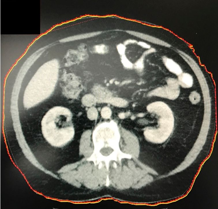
Impact of Sarcopenia on Functional and Oncological Outcomes
                              After Radical Prostatectomy

Markus Angerer 1,2*, Georg Salomon1, Dirk Beyersdorff 3, Margit Fisch2, Markus Graefen1 and
Clemens M. Rosenbaum4

1Martini-Klinik Prostate Cancer Center, University Hospital Hamburg-Eppendorf, Hamburg, Germany,
2 Department of Urology, University Hospital Hamburg-Eppendorf, Hamburg, Germany, 3 Department
of Radiology, Interventional Radiology and Nuclear Medicine, University Hospital Hamburg-Eppendorf,
Hamburg, Germany, 4 Department of Urology, Asklepios Hospital Barmbek, Hamburg, Germany

Introduction and Objectives: Knowledge about the significance of sarcopenia (muscle
loss) in prostate cancer (PCa) patients is limited. The aim of this study was to determine
the influence of skeletal muscle index (SMI) on early functional and pathological outcome
in patients undergoing radical prostatectomy (RP).

Materials and Methods: One hundred randomly chosen patients who received RP
between November 2016 and April 2017 at Martini-Klinik (Hamburg, Germany) were
retrospectively assessed. SMI (skeletal muscle mass cross-sectional area at L3/m2)
was measured by preoperative staging computed tomography scans at L3 level. Cox
regression analysis was applied to determine the impact of SMI on post-operative
outcome. Follow-up was 12months. Continence was defined as nomore than one safety
pad per day.

Results: Mean age of the cohort was 63.6 years. Mean SMI was 54.06 cm2/m2
(range, 40.65–74.58 cm2/m2). Of the patients, 41.4% had pT2, 28.7% had pT3a, and
29.9% had pT3b or pT4 PCa. SMI revealed to be without significant correlation on tumor
stage. Follow-up data of 55 patients were available for early functional outcome analysis.
SMI showed no significant influence on erectile function in multivariable Cox regression
analysis. In multivariable Cox regression analysis, SMI turned out to have no influence on
continence rates 6 weeks after surgery.

Conclusion: The present study shows that patients undergoing RP have a wide range
of SMI. Unlike in other urological malignancies, there was no significant impact of SMI on
early functional outcome and pathological outcome. A larger cohort is needed to confirm
these results.

Keywords: prostate cancer, sarcopenia, radical prostatectomy, oncological outcome, functional outcome

                                                                                                    5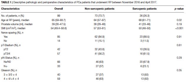
INTRODUCTION

Prostate cancer (PCa) is the most common cancer and the third most common cause
of cancer death among men in the western world (1). According to the German health
report in corporation with the Robert-Koch-Institute, 49,000 cases of PCa are
reported per annum; the incidence is 120 in all age classes in Germany (2).

       Radical prostatectomy (RP), brachytherapy (BT), and the advanced technique
of radiation using intensity-modulated radiation therapy (IMRT) are the three most
common treatment procedures for localized prostate cancer. All techniques show no
significant contrariness in overall survival (3, 4). RP embodies
one of the most often used treatment option in localized prostate cancer, mainly
implemented as either retropubic open RP or laparoscopic/robot-assisted RP (5).
       The most recognized risk factors for developing PCa are increasing age,
ethnic origin, and family history (6). The familiar predisposition suggests an inherited
genetic component to PCa (7, 8). Preoperative prostate-specific antigen (PSA),
pathological stage, Gleason score, and surgical margins status predicted BCR after
RP (9).
       “Sarcopenia is a progressive and generalized skeletal muscle disorder that is
associated with increased likelihood of adverse outcomes including falls, fractures,
physical disability, and mortality” as defined by the European Working Group on
Sarcopenia in Older People (EWGSOP) (10). Sarcopenia is increasingly recognized
as a risk factor for a worse performance especially in patients suffering from a
malignant tumor disease (11). Lately, the presence of sarcopenia has been identified
as a “prognostic marker of disease recurrence, cancer-specific mortality (CCM), and
all-cause mortality (ACM)” in patients
with not particularly urological malignancies but also, e.g., gynecological and
gastrointestinal cancer diseases (12–15).
       The definition of sarcopenia is based on the skeletal muscle index (SMI). The
muscle volume can reproducibly be measured by computed tomography (CT) or
magnetic resonance imaging
(MRI) (10).
       Among others, potential risk factors of perioperative complications are BMI
>30 and Charlson comorbidity index (CCI) 1 (16). Performance status and

                                                                                      6
comorbidity are generally subjective and difficult to define. The American Association
of Anesthesiologists (ASA) score, the Eastern Cooperative Oncology Group (ECOG)
performance status, and CCI are commonly calculated prognostic factors for
analyzing postoperative outcomes. Yet, they have been doubted to identify those
patients at highest risk of perioperative morbidity and mortality, despite the
successfully recognition of all status (14, 17). Sarcopenic patients have been
demonstrating a higher rate of perioperative complications (18–21).
This resulted to proclaim sarcopenia as an important acknowledging factor in
treatment planning, decision-making, and gaining information regarding patients peri-
and postoperative
outcome (17). In men diagnosed with prostate cancer, little is known about the role of
sarcopenia influencing the functional and oncological outcome. One study concluded
that sarcopenia does not predict the oncological outcome after RP (22). Another
study that investigated men undergoing radiotherapy for PCa identified a significant
impact of skeletal muscle reduction on non-cancer mortality (23).
We hypothesized that sarcopenia may be correlate with a higher complication
rate and worse oncological outcome in men undergoing RP. Consequently, we
examined the association between sarcopenia and perioperative as well as
oncological outcome in men undergoing RP (17).
MATERIALS AND METHODS
We retrospectively analyzed 100 patients who were treated with RP, either open
retropubic RP or laparoscopic, robot-assisted RP at a high-volume center (Martini-
Klinik Prostate Cancer Center, Hamburg-Eppendorf, Germany) between November
2016 and April 2017. RP was only performed consistently by eight highly trained
surgeons performing RRP and robot-assisted RP regularly.
We have identified the patients randomly within our database. Staging CT
scans were obtained by patients with intermediate and high-risk PCa defined by
D’Amico as a clinical T stage >cT2c, a Gleason score >8, or a PSA >20 ng/ml (24).
CT images were obtained from the patients preoperative CT scans of the
abdomen or pelvis. Included were only patients with sufficient quality of CT images.
Patients’ informed consent for data collection was obtained. The cross-sectional area
of all skeletal muscle at third lumbar vertebrae 3 (L3) has a high correlation to the
7body’s general muscle volume (18). Lumbal SMI is calculated by the cross-sectional
area of all skeletal muscle at L3 by height squared (m2) and reported as cm2/m2.
Clinical, blood sample results, and oncological data were collected from the hospitals’
documenting program, Soarian, and
Martini Data Registry.
A single axial image at the level of L3 was selected, and the cross-sectional
area of all skeletalmuscle at L3 wasmeasured after identifying the muscle-specific
attenuation thresholds (−29–150 HU). For the measurement, musculus rectus
abdominus; internal, external, and lateral musculus obliquus abdominis; musculus
psoas; musculus quadratus lumborum; and musculus erector spinae were included.
Axial CT images at L3 vertebra depicting patient without sarcopenia are shown in
Figures 1A,B as compared to patients with different BMIs and significantly different
SMI shown in Figures 1C,D. The radiologist program Centricty Viewer GE was used
for image analysis. Image analysis was performed by the same investigator who was
unaware about the patients’ cancer-specific data.
Clinical and pathological data were collected. Clinical data are include
information on age, clinical TNM classification (clinical tumor and lymph node stage),
preoperative PSA, continence by the number of pad usage per day, as well as
preoperative androgen deprivation therapy. Pathological data collected included
prostate biopsy Gleason score, pathological specimen Gleason score, pathological
TN classification (pathological tumor and lymph node stage), and surgical margin
status.
Taking into consideration the EWGSOP definition of sarcopenia, SMI was
based on sex- and BMI-specific cutoffs for menFIGURE 1 | (A,B) Axial CT-image at L3 vertebra depicting patient without sarcopenia. (C,D) Axial CT-image at L3
vertebra depicting patient with BMI and significantly different SMI (sarcopenic patient). The red marked area
represents the cross-sectional area of all skeletal muscle at L3 including the rectus abdominus; internal, external,
and lateral obliques; psoas; quadratus lumborum; and erector spinae muscles. The red marked line in the image
represents patients abdominal circumference.
Statistical Analysis
Clinical and pathological variables were compared between the sarcopenic and non-
sarcopenic patients. Age, BMI (in kg/m2), pathological tumor and lymph node stage,
pathological surgical margin status, PSA, and Gleason score are taken into account
for comparison of the two groups. Continuous features were summarized with
medians and interquartile ranges (IQRs). Categorical features were summarized with
frequency counts and percentages and compared using the chi-square test. The
primary interest was to evaluate the functional and oncological outcome.
Logistic regression analysis was used to estimate the oncological outcome
and biochemical recurrence (BCR). BCR was defined as PSA value >0.2 ng/ml after
RP. Urine continence was assessed by univariable and multivariable Cox
proportional hazards regression models and summarized with hazards ratios (HRs)
and 95% confidence intervals (95% CIs).
Furthermore, statistically significant prognosticators on univariable analysis
were also analyzed in multivariable models. A p < 0.05 was considered to be
statistically significant. For follow-up assessment, patients were evaluated for urinary
continence and erectile function (EF) after 6 weeks and 12 months after RP. Patient-
reported outcomes were registered by standardized Martini–Klinik questionnaires (5).
RESULTS
We included for the first analysis 99 patients from our database who fulfilled the
inclusion criteria. All of them were operated between November 2016 and April 2017.
One patient was excluded due to missing data.
9SMI measurements of all 99 patients were conducted based on SMI definition;
26 patients (26.3%) were classified as sarcopenic. Descriptive pathological and
perioperative characteristics are
shown in Table 1.
Overall, sarcopenic patients were older than non-sarcopenic patients (mean
age, 68.0 vs. 64 years, p = 0.02). There was no difference between sarcopenic and
non-sarcopenic patients in local and lymphonodal pathologic stage or Gleason score.
There was no significant difference between sarcopenic and nonsarcopenic patients
regarding nerve-sparring surgery (84.9 vs. 88.5%, p = 0.91).
In addition, there was no significant difference in urine continence at 1 year after
surgery between sarcopenic and nonsarcopenic patients in multivariable logistic
regression analysis [odd’s ratio (OR), 1.05; 95% confidence interval (CI), 0.96–1.16;
p = 0.26].
Results are shown in Table 2.
10In Cox regression analysis, the incidence of BCR did not differ significantly 1 year
after surgery between sarcopenic and nonsarcopenic patients [hazard ratio (HR),
0.97; 95% CI, 0.3–3.08; p = 0.953].
DISCUSSION
Sarcopenia represents “a response to both nutrient deprivation and systemic stress,
resulting in critical anatomic and functional deficits” (17). Sarcopenia is a major public
health issue. Using the definition with highest prevalence estimates, the number of
individuals with sarcopenia would rise from 19,740 million in
2016 to 32,338 million in 2045 only in Europe, corresponding to an increase from
20.2 to 22.3% (26).
In this current study, we examined the association between sarcopenia and
functional and oncological outcome after RP.Our hypothesis that sarcopenia
significantly effects functional and
oncologic outcome in men undergoing RP could not be proven.
We noted several findings of interest. First, we determined that in this cohort
of patients with RP, 26.3% of patients were classified as sarcopenic preoperatively.
The median age of sarcopenic patients was significantly older.
The correlation between BMI and outcome after RP has been investigated
often in past. An increase in BMI showed a significant increase risk of peri- and post-
operative complications; prolonged operative time, increased blood loss, increased
11open conversions, longer hospitalization, and higher positive surgical margin rate
(27). BMI has known associations with diabetes, coronary artery disease, and
hypertension (27). Obesity also has a significant impact on mortality in cancer
patients (24). Freedland et al. concluded that elevated BMI has been associated with
biochemical failure after radical prostatectomy, due to inferior surgery, which caused
a higher rate of positive surgical margin. Also in their cohort, obese men after RP
showed worse outcomes, suggesting that obesity may be associated with a
biologically more aggressive form of prostate cancer (28–30). Still, it remains
controversial regarding the effect on BCR (22).
As mentioned before, McDonald et al. assessed in their study the cross-
sectional area at the L4–5 level after radiotherapy for localized prostate cancer
retrospective of 653 men (23). They were concluding that sarcopenia significantly
increased risk of non-cancer mortality after radiotherapy. Analyzing their cohort,
the conclusion is due to the fact that cross-sectional area of all total skeletal muscle
was measured at L4–5 and relatively few patients. Furthermore, this study had
muscle L4–5 values below the sarcopenic threshold.
Mason et al. published in June 2018 the association between sarcopenia and
oncological outcome after RP in a cohort of totally 698 patients and 310 patients
identified as sarcopenic (22). They concluded that sarcopenia has no significant
association with either perioperative complications or oncological outcome after RP.
This study showed a representative number of patients classified sarcopenic
(55.6%). Furthermore, there were no significant differences in clinical T or N stage or
biopsy Gleason score.
Two different cohort of men with prostate cancer showed contradictory
associations of sarcopenia. This may be because of the different populations or
different cancer-specific criteria. Patients for RP selected by urologists favoring
patients younger in age with a longer life expectancy and reduced comorbidities. Our
data reveal that SMI has neither significant influence on pathological outcome nor on
BCR rates after RP.
Furthermore, SMI had no impact on post-operative urine continence in our
cohort. These results may suggest that sarcopenia is not a prognostic marker for
functional and oncological outcome after RP.
In our study, we acknowledge several limitations to this study. First, we cannot
rule out a bias due to random selection of included patients. Not all patients between
12the period of November 2016 and April 2017 who underwent RP have been selected
for analysis and follow-up. Exclusion was caused by missing CT scans, either not
readable, poor quality for analysis or missing import; or low-risk PCa patients
accordingly to D’Amico classification, which have not received a preoperative CT
staging. Another major limitation is that our cohort only figured 99 patients. Therefore,
additional subanalyses of risk classifications
are necessary. SMI was onlymeasured by preoperative scans. The change in SMI is
not considered. Ha et al. showed a significant change in sarcopenia and SMI 1 year
after radical cystectomy and might be an effective marker for oncological outcome
(31). Another limitation of this study is the time of follow-up after
RP, which limits the statement of sarcopenia effecting BCR. The results currently
show the 12 months questionnaire feedback. The effect of BCR cannot safely be
clarified; hence, the followup time must be prolonged. We are continuing to assess
followup data.
Nevertheless, little is known about the association of sarcopenia on functional
and oncological outcome after RP. Our study presents that sarcopenia is not
significantly associated with influencing the oncological outcome, urine continence, or
BCR
after RP.
CONCLUSION
Sarcopenia was not significantly associated with worse functional and oncological
outcome after RP. In addition, sarcopenia has no significant effect on BCR. Thus,
sarcopenia is not a prognostic marker for patients with prostate cancer after RP.
DATA AVAILABILITY STATEMENT
The raw data supporting the conclusions of this article will be made available by the
authors, without undue reservation.
13ETHICS STATEMENT
The studies involving human participants were reviewed and approved by
Ethikkommission Hamburg. The
patients/participants provided their written informed consent to participate in this
study.
AUTHOR CONTRIBUTIONS
MA was responsible for conceiving the presented idea, designed the study,
developed the theory, and performed the computations, literature research, data
collection, writing of the manuscript with the help of CR, CT image analysis, and
statistical calculations. GS contributed with literature research, investigated, and
supervised the findings of this work, helped with manuscript correction and revision.
DB contributed with the help of CT image analysis and providing the software, also
helped with manuscript correction and revision. MF contributed with the help of
manuscript correction and revision. MG contributed with literature, verified the
analytical methods, helped with manuscript correction and revision. CR contributed
with planning and supervision of the work, helped with statistical calculations,
manuscript correction and revision. All authors discussed the results and contributed
to the final manuscript.
14REFERENCES
1. Siegel, R.L., K.D. Miller, and A. Jemal, Cancer statistics, 2020. CA Cancer J
Clin, 2020. 70(1): p. 7-30.
2. Rohde, V., A. Katalinic, J. Wasem, and P. Aidelsburger,
Gesundheitsberichterstattung des Bundes Heft 36 Prostataerkrankungen.
Robert Koch Institut; Statistisches Bundesamt, 2007. 36.
3. Hayashi, N., Y. Yokomizo, Kimito Osaka, K. Makiyama, N. Nakaigawa, M. Yao,
et al., Ten year outcomes of treatment for localized prostate cancer in a single
institution; comparison of radical prostatectomy vs radiation therapy -
prospensity score matching analysis -. The Journal of Urology, 2016. 195.
4. Mottet, N., J. Bellmunt, E. Briers, M. Bolla, L. Bourke, P. Cornford, et al., EAU -
ESTRO - ESUR - SIOG Guidelines on Prostate cancer. Edn. presented at the
EAU Annual Congress Amsterdam 2020. EAU Guidelines Office, 2020.
978-94-92671-07-3.
5. Haese, A., S. Knipper, H. Isbarn, H. Heinzer, D. Tilki, G. Salomon, et al., A
comparative study of robot-assisted and open radical prostatectomy in 10 790
men treated by highly trained surgeons for both procedures. BJU Int, 2019.
123(6): p. 1031-1040.
6. Key, T., Risk factors for prostate cancer. Cancer Surv, 1995. 23: p. 63-77.
7. Lichtenstein, P., N.V. Holm, P.K. Verkasalo, A. Iliadou, J. Kaprio, and M.
Koskenvuo, Environmental and heritable factors in the causation of cancer –
analyses of cohorts of twins from Sweden, Denmark, and Finland. N Engl J
Med, 2000. 343(2): p. 78-85.
8. Oh, S.J., P. Mandel, F.K.H. Chun, P. Tennstedt, S. Peine, J.L. Hohenhorst, et al.,
AB0/Rhesus Blood Group Does Not Influence Clinicopathological Tumor
Characteristics or Oncological Outcome in Patients Undergoing Radical
Prostatectomy. Front Surg, 2017. 4: p. 75.
9. Ouzzane , A., P. Koenig, C. Ballereau, L. Zini, T. Ghoneim, F. Maladry, et al.,
Oncologic Outcomes After Radical Prostatectomy: French Validation of the
D'Amico Risk Group Classification. Prog Urol, 2010. 20(13): p. 1206-12.
10. Cruz-Jentoft, A.J., G. Bahat, J. Bauer, Y. Boirie, O. Bruyere, T. Cederholm, et
al., Sarcopenia: revised European consensus on definition and diagnosis. Age
Ageing, 2019. 48(1): p. 16-31.
11. Joglekar, S., P.N. Nau, and J.J. Mezhir, The impact of sarcopenia on survival
and complications in surgical oncology: a review of the current literature. J
Surg Oncol, 2015. 112: p. 503-509.
1512. Pinsky, P.F., B.S. Kramer, D. Reding, and S. Buys, Reported family history of
cancer in the prostate, lung, colorectal, and ovarian cancer screening trial. Am
J Epidemiol. 157(9): p. 792–9.
13. Del Fabbro, E., H. Parsons, C.L. Warneke, K. Pulivarthi, J.K. Litton, R. Dev, et
al., The relationship between body composition and response to neoadjuvant
chemotherapy in women with operable breast cancer. Oncologist, 2012.
17(10): p. 1240-5.
14. Peng, P., O. Hyder, A. Firoozmand, P. Kneuertz, R.D. Schulick, D. Huang, et al.,
Impact of sarcopenia on outcomes following resection of pancreatic
adenocarcinoma. J Gastrointest Surg, 2012. 16(8): p. 1478-86.
15. Hamaguchi, Y., T. Kaido, S. Okumura, Y. Fujimoto, K. Ogawa, A. Mori, et al.,
Impact of quality as well as quantity of skeletal muscle on outcomes after liver
transplantation. Liver Transpl, 2014. 20(11): p. 1413-9.
16. Liu, X.-j., L. Liu, K. Chang, D.-w. Ye, Y.-f. Zheng, and X.-d. Yao, Risk factors of
perioperative complications in patients undergoing radical retropubic
prostatectomy: A ten-year experience. Journal of Huazhong University of
Science and Technology, 2017.
17. Psutka, S.P., A. Carrasco, G.D. Schmit, M.R. Moynagh, S.A. Boorjian, I. Frank,
et al., Sarcopenia in Patients With Bladder Cancer Undergoing Radical
Cystectomy Impact on Cancer-Specific and All-Cause Mortality. Cancer, 2014.
120(18): p. 2910-8.
18. Shen, W., M. Punyanitya, Z. Wang, D. Gallagher, M.-P. St.-Onge, J. Albu, et al.,
Total body skeletal muscle and adipose tissue volumes: estimation from a
single abdominal cross-sectional image. J Appl Physiol, 2004. 97: p.
2333-2338.
19. Tan, B.H., L.A. Birdsell, L. Martin, V.E. Baracos, and K.C. Fearon, Sarcopenia in
an overweight or obese patient is an adverse prognostic factor in pancreatic
cancer. Clin Cancer Res, 2009. 15(22): p. 6973-9.
20. Wan, F., Y. Zhu, C. Gu, X. Yao, Y. Shen, B. Dai, et al., Lower skeletal muscle
index and early complications in patients undergoing radical. World Journal of
Surgical Oncology, 2014. 12(14).
21. Rutten, I.J., J. Ubachs, R.F. Kruitwagen, D. Van Dijk, R. Beets-Tan, L.
Massuger, et al., The influence of sarcopenia on survival and surgical
complications in ovarian cancer patients undergoing primary debulking
surgery. Eur J Surg Oncol, 2017. 43: p. 717-24.
22. Mason, R.J., S.A. Boorjian, B. Bhindi, L. Rangel, I. Frank, R.J. Karnes, et al.,
The Association Between Sarcopenia and Oncologic Outcomes After Radical
Prostatectomy. Clin Genitourin Cancer, 2018. 16(3): p. e629-e636.
1623. McDonald, A., T. Swain, D. Mayhew, R. Cardan, C. Baker, D. Harris, et al., CT
Measures of Bone Mineral Density and Muscle Mass Can Be Used to Predict
Noncancer Death in Men with Prostate Cancer. Radiology, 2017. 282(2).
24. Chang, A.J., K.A. Autio, M. Roach, 3rd, and H.I. Scher, High-risk prostate
cancer-classification and therapy. Nat Rev Clin Oncol, 2014. 11(6): p. 308-23.
25. Martin, L., L. Birdsell, N. MacDonald, T. Reiman, M.T. Clandinin, L.J. McCargar,
et al., Cancer Cachexia in the Age of Obesity: Skeletal Muscle Depletion Is a
Powerful Prognostic Factor, Independent of Body Mass Index. Journal of
Clinical Oncology, 2013. 31(12): p. 1539-1547.
26. Ethgen, O., C. Beaudart, F. Buckinx, O. Bruyere, and J.Y. Reginster, The Future
prevalence of sarcopenia in Europe: A claim for public health action. Calcif
tissue int, 2017(100): p. 229-234.
27. Herman, M.P., J.D. Raman, S. Dong, D. Samadi, and D.S. Scherr, Increasing
Body Mass Index Negatively ImpactsOutcomes Following Robotic Radical
Prostatectomy. Journal of the Society of Laparoendoscopic Surgeons, 2007.
11(438-442).
28. Calle, E.E., C. Rodriguez, K. Walker-Thurmond, and M. Thun, Overweight,
obesity, and mortality from cancer in a prospectively studied cohort of U.S.
adults. N Engl J Med, 2003. 348: p. 1625-1638.
29. Freedland, S.J., M.K. Terris, J.C.J. Presti, C.L. Amling, C.J. Kane, B. Trock, et
al., Obesity and biochemical outcome following radical prostatectomy for
organ confined disease with negative surgical margins. J Urol, 2004. 172: p.
520-524.
30. Ruszat, R., A. Bachmann, S. Wyler, T. Forster, M. Zimmermann, and T. Sulser,
Einfluss des Body-Mass-Index (BMI) auf die perioperativen Ergebnisse der
endoskopischen radikalen Prostatektomie. Aktuelle Urol, 2006. 37: p. 122.
31. McDonald, A., T. Swain, D. Mayhew, R. Cardan, C. Baker, D. Harris, et al., CT
Measures of Bone Mineral Density and Muscle Mass Can Be Used to Predict
NoncancerDeath in Men with Prostate Cancer. Radiology, 2017. 282(2).
32. Ha, Y.S., S.W. Kim, T.G. Kwon, S.K. Chung, and E.S. Yoo, Decrease in skeletal
muscle index 1 year after radical cystectomy as a prognostic indicator in
patients with urothelial bladder cancer. Int Braz J Urol, 2019. 45(4): p.
686-694.
172. Darstellung der Publikation
Das Prostatakarzinom (PCa) ist die häufigste Tumorerkrankung und die dritthäufigste
Todesursache bei Männern in der westlichen Welt. [1] Entsprechend dem deutschen
Gesundheitsbericht in Zusammenarbeit mit dem Robert-Koch-Institut werden jährlich
rund 49.000 Fälle von PCa gemeldet, die Inzidenz beträgt 120 in allen Altersklassen
in Deutschland. [2]
In einer Zeit geprägt von demographischem Wandel und einer älter werdenden
Gesellschaft rücken degenerative Alterungsprozesse und ihre Folgen mehr in den
Fokus des klinischen Interessens. Alterungsprozess umfasst multiple physiologische
Veränderungen; zu den bedeutendsten zählt der altersbedingte Muskelschwund.
1989 gab Rosenberg et al. dem Phänomen den Namen „Sarkopenie“. [3] Der Begriff
ist zusammengesetzt aus den griechischen Wörtern „Sarx“ für Fleisch und „Penia“ für
Armut und hat sich im klinischen Gebrauch etabliert.
"Sarkopenie ist eine fortschreitende und generalisierte Skelettmuskelerkrankung, die
mit einer erhöhten Wahrscheinlichkeit von unerwünschten Ereignissen wie Stürzen,
Frakturen, körperlicher Behinderung und Mortalität verbunden ist", wie von der
Europäischen Arbeitsgruppe für Sarkopenie bei älteren Menschen (EWGSOP)
definiert. [4] Die Skelettmuskulatur spielt eine zentrale Rolle in der Stabilisierung des
Halteapparats. Sarkopenie liefert Informationen zu bedeutsamen Parameter wie
Energiereserven und den Ernährungsstatus. Mit dem Rückgang der Muskulatur
kommt es zeitgleich zu einer Zunahme von Fettgewebe. Es kommt zum Phänomen
des stabilen Körpergewichts, aber zur unterschiedlichen Körperzusammensetzung
(Fett, Muskulatur, etc.). Hier spricht von einer sarkopenischen Übergewichtigkeit,
besser bekannt durch den englischen Ausdruck sarcopenic obesity. [3]
Sarkopenie wird zunehmend als Risikofaktor für eine schlechtere Leistung
anerkannt, insbesondere bei Patienten mit einer malignen Tumorerkrankung. [5] In
letzter Zeit wurde das Vorhandensein von Sarkopenie als "prognostischer Marker für
das Wiederauftreten von Krankheiten, die krebsspezifische Mortalität (CCM) und die
Gesamtmortalität (ACM)" bei Patienten nicht nicht nur urologischen malignen
Tumorerkrankungen, sondern auch bei z.B. gynäkologischen und gastrointestinalen
malingen Tumorerkrankungen. [6-9]
18Bei gastrointestinalen- und Lungentumoren waren 44%-57% der Patienten
betroffen. In einer publizierten Studie von von Martin et al. wurden 1.473 Patienten
mit malginen Gastrointestinal/-oder Lungentumor untersucht. Die durchschnittliche
Überlebensrate sarkopenischer Patienten betrug 13 Monaten vs. 20 Monate bei
Patienten mit normalen Muskelverhältnisse. [10]
Die Definition von Sarkopenie basiert auf dem Skelettmuskelindex (SMI). Die
Gesamtmuskelquerschnittsfläche auf Höhe von Lendenwirbelkörper 3 (L3) genormt
an die Körpergröße ergibt den SMI. Das Muskelvolumen kann reproduzierbar mittels
Computertomographie (CT) oder Magnetresonanztomographie (MRT) gemessen
werden. [4] CT-, und MRT-Bildgebung gilt hier als Goldstandard zur Messung von
Sarkopenie. Basierend auf einer Publikation von Shen et al. konnte eine hohe
Korrelation (r2 = 0,86) zwischen dem Gesamtmuskelvolumen des Körpers und der
totalen Muskelquerschnittsfläche auf Höhe von L3 nachgewiesen werden. [11]
Mögliche Risikofaktoren für perioperative Komplikationen sind unter anderem BMI>
30 und ein Charlson-Komorbiditäts-Index (CCI) ≥1. [12] Leistungsstatus und
Komorbidität sind im Allgemeinen subjektiv und schwer zu definieren. Der Score der
American Association of Anaesthesiologists (ASA), der Leistungsstatus der Eastern
Cooperative Oncology Group (ECOG) sowie CCI sind häufig berechnete
Prognosefaktoren für die Analyse postoperativer Ereignisse. Es wurde jedoch
bezweifelt, diejenigen Patienten zu identifizieren, bei denen das höchste Risiko für
eine perioperative Morbidität und Mortalität besteht, obwohl alle Scores erfolgreich
erkannt wurden und weltweit benutzt werden. [8,13] Sarkopenische Patienten zeigten
eine höhere Rate perioperativer Komplikationen. [14-17]
Dies führte dazu, dass Sarkopenie als wichtiger anerkennender Faktor bei der
Behandlungsplanung, Entscheidungsfindung und Informationsgewinnung über das
peri- und postoperative Ergebnis oder Ereignis von Patienten proklamiert wurde. [13]
Bei Männern mit diagnostiziertem PCa ist wenig über die Rolle bei vorliegen einer
Sarkopenie bekannt, in wieweit das funktionelle und onkologische Ergebnis
beeinflusst wird. Eine Studie kam zu dem Schlussfolgerung, dass Sarkopenie keinen
Einfluss auf das onkologische Ergebnis nach RP hat. [18] In einer weiteren Studie, in
der das Prostatakarzinom mittels Strahlentherapie therapiert wurde, konnte ein
19signifikanter Einfluss von Sarkopenie auf die nicht-krebsspezifische Sterblichkeit
(krebsspezifische Sterblichkeit; CCS= cancer specific survival) nachgewiesen
werden. [19]
Die Radikale Prostatektomie (RP), die Brachytherapie (BT) und die fortschrittliche
Strahlentechnik, intensitätsmodulierte Strahlentherapie (IMRT) sind die drei
häufigsten Behandlungsverfahren für das lokalisierte PCa. Alle Techniken zeigen
keine signifikanten Unterschiede im Gesamtüberleben. [20,21] Die RP ist eine der
am häufigsten verwendeten Behandlungsoptionen bei lokalisiertem PCa, die
hauptsächlich als retropubische offene RP oder als laparoskopische bzw.
roboterunterstützte (DaVinci) RP durchgeführt wird. [22] Die bekanntesten
Risikofaktoren für die Entwicklung eines PCa sind zunehmendes Alter, ethnische
Herkunft und die positive Familienanamnese hinsichtlich einer
Prosatatakarzinomerkrankungen. [23] Die Veranlagung im Familienstammbaum
deutet auf eine vererbte genetische Komponente für die Erkrankung eines PCa hin.
[24,25] Der präoperative Prostata-spezifische-Antigen (PSA) Wert, das
pathologisches Stadium, der Gleason-Score und der intraoperative Absetztungsrand
prognostizierten das biochemische Rezidiv (BCR) nach RP. [26]
Wir stellten die Hypothese auf, dass Sarkopenie mit einer höheren Komplikationsrate
und einem schlechteren onkologischen Ergebnis nach RP korreliert. Infolgedessen
untersuchten wir den Zusammenhang zwischen Sarkopenie und perioperativem
sowie onkologischem Ergebnis bei Männern nach RP. [13]
Wir haben retrospektiv 100 Patienten analysiert, die zwischen November 2016 und
April 2017 in einem high-volume Zentrum (Martini-Klinik Prostatakrebszentrum,
Hamburg-Eppendorf, Deutschland) radikal prostatektomiert wurden, entweder mit
offener retropubischer RP oder mit laparoskopischer bzw. robotergestützter RP. Die
RP wurde nur von acht hochqualifizierten Chirurgen durchgeführt, die regelmäßig
offene retropubische RP und roboterunterstützte RP durchführen.
Wir haben die Patienten zufällig aus unserer Datenbank identifiziert. Staging-CT
Untersuchungen wurden bei Patienten mit vorliegenden mittlerem Risiko und hohem
Risiko PCa durchgeführt, entsprechend der Definition nach D`Amico als klinisches T-
20Stadium ≥ cT2c, Gleason-Score ≥ 8 oder PSA> 20 ng / ml. [27] Die CT-Bilder zur
Auswertung wurden aus den präoperativen CT-Untersuchungen des Abdomens oder
des Beckens des Patienten gewonnen. Eingeschlossen wurden nur Patienten mit
ausreichender Qualität der CT-Bilder. Die Einwilligung der Patienten zur
Datenerfassung wurde eingeholt. Die Querschnittsfläche des gesamten
Skelettmuskels auf L3 korreliert stark mit dem allgemeinen Muskelvolumen des
Körpers. [14] Der lumbale SMI wird anhand der Querschnittsfläche des gesamten
Skelettmuskels auf Höhe L3 zum Quadrat (m2) berechnet und als cm2/m2
angegeben. Ein einzelnes axiales Bild auf Höhe von L3 wurde ausgewählt und die
Querschnittsfläche des gesamten Skelettmuskels auf Höhe von L3 wurde gemessen,
nachdem die muskelspezifischen Schwächungsschwellen (-29 bis 150 Hounsfield-
Einheiten) identifiziert worden. Für die Messung wurden folgende Muskel
eingeschlossen: Musculus rectus abdominus; innerer, äußerer und seitlicher
Musculus obliquus abdominis; Musculus psoas; musculus quadratus lumborum; und
Musculus erector spinae. Für die Bildanalyse/Berechnung des SMI wurde das
radiologische Programm Centricty Viewer GE verwendet. Die Berechnung des SMI
wurde nur durch den Promovenden durchgeführt, der die krebsspezifischen
Patientendaten vorher nicht kannte.
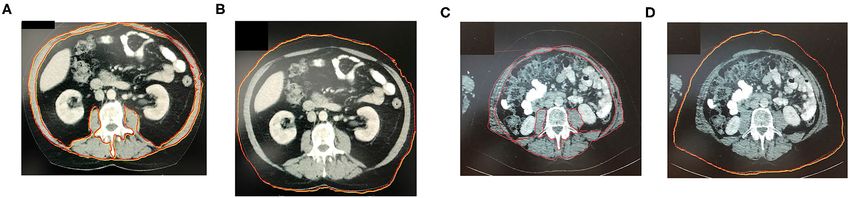
Axiale CT-Bilder auf Höhe von L3, von einem Patienten ohne Nachweis einer
Sarkopenie, sind in Abbildung 1a und 1b dargestellt. Im Vergleich in Abbildung 1c
und Abbildung 1d einen sarkopenischen Patient mit unterschiedlichem BMI und
signifikant unterschiedlichem SMI.
Abbildung 1a Abbildung 1b
Die rote Linie repräsentiert die Querschnittsfläche des Die rot Linie repräsentiert den Bauchumfang
gesamten Skelettmuskels auf Höhe von L3
21Abbildung 1c Abbildung 1d
Die rote Linie repräsentiert die Querschnittsfläche des Die rot Linie repräsentiert den Bauchumfang
gesamten Skelettmuskels auf Höhe von L3
Klinische, laborchemische, onkologische sowie pathologische Daten wurden aus
dem Krankenhausdokumentationsprogramm Soarian und aus dem Martini-Data-
Register gesammelt.
Zu den klinischen Daten gehören das Alter, klinische TNM-Klassifizierung
(klinisches Tumor- und Lymphknotenstadium), präoperativer prostataspezifischen
Antigen (PSA) Wert, bezüglich der Kontinenz die Anzahl an verwendeten
Sicherheitsvorlagen pro Tag sowie zur das Vorhandensein einer präoperativen
Androgenentzugstherapie. Zu den erhobenen pathologischen Daten gehörten der
Gleason-Score der Prostatastanzbiopsie, der Geason-Score des pathologischen
Präparats, die pathologische TN-Klassifikation (pathologisches Tumor- und
Lymphknotenstadium) und der Status des chirurgischen intraoperativen
Absetzungsrands. Unter Berücksichtigung der EWGSOP-Definition für Sarkopenie
gibt es geschlechtsspezifische und BMI-spezifische Grenzwerte zur Differenzierung
eines sarkopenen Patienten; für MännerKlinische und pathologische Variablen wurden zwischen sarkopenen und nicht sarkopenen Patienten verglichen. Alter, BMI (in kg / m2), pathologisches Tumor- und Lymphknotenstadium, positiver chirurgischer Absetzungsrand, PSA und Gleason- Score wurde für den Vergleich der beiden Gruppen berücksichtigt. Die kontinuierlichen Variablen wurden mit Medianen und IQRs angegeben. Die kategorialen Variablen wurden mit Häufigkeitszählungen und Prozentsätzen zusammengefasst und unter Verwendung des Chi-Quadrat-Tests verglichen. Das Hauptinteresse lag in der Auswertung und Bewertung des funktionellen und onkologischen Ergebnisses. Für die statistische Auswertung des onkologischen Ergebnis und des BCR, wurde eine logistische Regressionsanalyse durchgeführt. Das BCR wurde als ein PSA-Wert> 0,2 ng / ml nach RP definiert. Die Urinkontinenz wurde durch univariable und multivariable Cox-Regressionsmodelle statistisch ausgewertet und mit Hazards Ratios (HRs) und 95% -Konfidenzintervallen (95% -KI) angegeben. Darüber hinaus wurden auch statistisch signifikante Ergebnisse in der univariablen Analyse, weitergehend in multivariablen Modellen analysiert. Ein p-Wert
pathologischen Stadium oder im Ergebnis des pathologischen Gleason-Score des
intraoperativen Präparats der Prostata. Es gab keinen signifikanten Unterschied
zwischen sarkopenen und nicht sarkopenen Patienten hinsichtlich einer Nerv-
erhaltenden (nerve-sparing) Operation (84,9% gegenüber 88,5%, p-Wert 0,91).

  Tabelle 1                    Deskriptive pathologische und perioperative
                                         Patientencharakteristika

      Parameter           Gesamt        nicht-sarkopene   sarkopene      p-Wert
                                           Patienten      Patienten
Patienten, n (%)             99             73 (73.7)      26 (26.3)

Alter zum Zeitpunkt     65 (59-68.7)       64 (57-67)     68 ( 61-71)        0.02
der RP(y), median
median
Prostatavolumen         39 (30-47.5)       38 (28-48)     41 (33-46)         0.19
(ml), median
SMI (cm2/m2),                54            57 (53-61)     50 (46-50)     4+4              59                 46 (63)    13 (50)

Darüber hinaus zeigte sich 1 Jahr nach RP kein signifikanter Unterschied in der
Urinkontinenz zwischen sarkopenen und nicht sarkopenen Patienten in einer
multivariablen logistischen Regressionsanalyse (Odd's Radtio [OR] 1,05; 95%
-Konfidenzintervall [CI], 0,96-1,16; P 0,26).
       Die Ergebnisse sind in Tabelle 2 gezeigt.

                                                                                    24
Tabelle 2 Urinkontinenz 1 Jahr nach radikaler Prostatektomie
Parameter Odd´s Ratio 95% CI p-Wert
Alter zum Zeitpunkt der RP 1.05 0.95-1.17 0.31
Prosatavolumen 1.02 0.98-1.06 0.4
Nerverhalt
ja Reference
nein 0.38 0.03-3.35 0.4
pT-Stadium
pT2 Reference
pT3a 0.53 0.1-2.46 0.43
pT3b/pT4a 1.99 0.35-10.92 0.42
SMI 1.05 0.96-1.16 0.26
In der Cox-Regressionsanalyse unterschied sich die BCR-Inzidenz 1 Jahr nach der
Operation zwischen sarkopenischen und nicht-sarkopenischen Patienten nicht
signifikant (Hazard Ratio [HR] 0,97; 95% -Konfidenzintervall [CI] 0,3-3,08; P 0,953).
Zusammenfassend ist Sarkopenie „eine Reaktion sowohl auf einen Nährstoffmangel
als auch auf systemischen Stress, der zu kritischen anatomischen und funktionellen
Defiziten führt“. [13] Sarkopenie ist ein wichtiges und ernstzunehmendes Problem
der allgemeinen Gesundheit. Unter Verwendung der aktuellen Definition der
EWGSOP, würde die Zahl der Personen mit Sarkopenie von 19.740 Millionen im Jahr
2016 auf 32.338 Millionen im Jahr 2045 nur in Europa steigen, was einem Anstieg
von 20,2% auf 22,3% entspricht. [29]
25Bezüglich eines Zusammenhang zwischen Sarkopenie und PCa ist wenig bekannt.
Die Krebsspezifische Mortalität von Prostatakarzinom war die letzten 10 Jahre stets
weniger als 10%. [30] Möglicherweise erklärt dies die begrenzte Studienlage zu dem
Thema dazu. Verglichen mit den anderen urologischen Tumoren wurde Sarkopenie
kaum als prädiktiver Faktor untersucht.
Cushen et al. analysierte die Gesamtkörperzusammensetzung von
Prostatakarzinom Patienten. [31] Martin et al. konnte in 47% seiner Kohorte
Patienten mit Sarkopenie festgestellen, von denen 26.7% sarcopenisch-adipös
waren. Weder Sarkopenie noch Sarcopenic-obesity waren mit Gesamtüberleben
assoziiert, noch gab es Unterschiede zwischen sarkopensichen und nicht-
sarkopensichen Paitenten. [10]
Bei anderen urologischen malignen Tumorerkrankungen in Bezug auf
Sarkopenie ist die Datenlage wesentlich umfangreicher. Zwischen den Jahren 2013
und 2017 gab es 7 Studien, die den Zusammenhang zwischen Sarkopenie und dem
nicht-metastasierten, muskelinvasiven Urothelkarzinom der Blase untersuchten. In
den Studien mit der größten Fallzahl schwankte die Prävalenz von Sarkopenie
zwischen 47.8 -68.8%. Patienten mit Sarkopenie waren signifikant älter. In allen
Studien war Sarkopenie mit einem signifikant schlechteren CSS und
Gesamtüberlebensrate (OS=overall survival) assoziert. [32-34] Psutka et al.
beschrieb eine verminderte 5 Jahres Überlebensrate (49% vs 72% P=.003) und
reduzierte OS bei Patienten mit Sarkopenie im Vergleich zu nicht-sarkopenen
Patienten (39% vs. 70% P=.003).
Der Zusammenhang bezüglich Sarkopenie und des Nierenzellkarzinom
wurden ebenfalls zwischen 2010 und 2017 in 8 Studien thematisiert. Einige Studien
beschrieben ferner eine Korrelationen zwischen Sarkopenie und Anzahl der
Metastasen, perioperativer Blutverluste und schlechterem MSKCC-Score, während
dieselben Variablen sich in anderen Studien nicht signifikant von Nicht-sarkopenen
Patienten unterschieden hat. [35-37]
In einer multivariablen Analyse war Sarkopenie ein unabhängiger Prädiktor für das
Gesamtüberleben (HR 2.58, p = 0.015) [13], das progressionsfreies Überleben (HR:
2.54, p = 0.0163) [35], sowie dem und erhöhtem CSS (HR 1.70, p=0.047) [36]
In einer Metaanalyse aus 6 Studien mit insgesamt 559 Patienten konnte ein
Zusammenhang zwischen erniedrigtem SMI und erhöhter Gesamtmortalität beim
metastasierten Nierenzellkarzinom (HR= 1.48, 95% CI: 1.08–2.03) nachgewiesen
26werden. [39] Vrieling et al beschreibt in seiner Metaanalyse, dass es beim
metastasierten Nierenzellkarzinom mit niedrigem SMI häufiger zu Dosislimitierungen
aufgrund einer Chemotoxizität kommt. [39]
In dieser aktuellen Studie untersuchten wir den Zusammenhang zwischen
Sarkopenie und dem funktionellem sowie onkologischem Ergebnis nach RP. Unsere
Hypothese, dass Sarkopenie das funktionelle und onkologische Ergebnis bei
Männern nach RP signifikant beeinflusst, konnte nicht nachgewiesen werden. Wir
haben mehrere interessante Ergebnisse festgestellt. Zunächst stellten wir fest, dass
in unserer Kohorte 26,3% der Patienten präoperativ als sarkopen eingestuft wurden.
Das Durchschnittsalter der sarkopenen Patienten war signifikant älter. Die Korrelation
zwischen BMI und Ergebnis nach RP wurde in der Vergangenheit häufig untersucht.
Ein Anstieg des BMI zeigte ein signifikant erhöhtes Risiko für peri- und postoperative
Komplikationen; verlängerte Operationszeit, erhöhten Blutverlust, erhöhte Rate an
Konversionen auf eine offene RP, längerer Krankenhausaufenthalt und höhere
positive Absetzungsränder. [40] Ein erhöhter BMI (Adipositas) hat bekannte
Zusammenhänge mit anderen Erkrankungen wie z.B. Diabetes mellitus Typ II,
Erkrankungen der Herzkranzgefäße und Bluthochdruck. [40] Fettleibigkeit hat auch
einen signifikanten Einfluss auf die Mortalität bei Tumorpatienten. [24] Freedland et
al. schlussfolgernden, dass ein erhöhter BMI nach einer radikalen Prostatektomie mit
einem BCR verbunden war, entweder aufgrund einer minderwertigen Operation, die
eine höhere Rate positiver Absetzungsränder verursachte. Auch in dieser Kohorte
zeigten fettleibige Männer nach RP schlechtere Ergebnisse, was darauf hindeutet,
dass Fettleibigkeit mit einer biologisch aggressiveren Form von PCa verbunden sein
kann. [41-43] Die Auswirkungen auf das BCR sind nach wie vor umstritten. [18]
Wie bereits erwähnt, untersuchten McDonald et al. in ihrer Studie die
Querschnittsfläche auf L4-5-Niveau retrospektive von 653 Männern nach
Strahlentherapie bei lokalisierten PCa. [44] Sie schlussfolgernden, dass Sarkopenie
das Risiko einer Nichtkrebssterblichkeit nach Strahlentherapie signifikant erhöhte.
Die Schlussfolgerung beruht auf der Tatsache, dass die Querschnittsfläche des
gesamten Skelettmuskels auf Höhe von LWK 4-5 und relativ geringen Population
gemessen wurde. Darüber hinaus zeigte diese Studie SMI-Werte auf Höhe von LWK
L4-5 unterhalb der sarkopenen Schwelle.
27Mason et al. veröffentlichten im Juni 2018 den Zusammenhang zwischen
Sarkopenie und onkologischem Ergebnis nach RP in einer Kohorte von insgesamt
698 Patienten. 310 Patienten wurden als sarkopen identifiziert. [18] Sie
schlussfolgernden, dass Sarkopenie weder mit perioperativen Komplikationen noch
mit dem onkologischen Ergebnis nach RP einen signifikanten Zusammenhang
aufweist. Diese Studie zeigte eine repräsentative Anzahl von Patienten, die als
sarkopen klassifiziert wurden (55,6%). Darüber hinaus gab es keine signifikanten
Unterschiede im klinischen T- oder N-Stadium oder im Prostatastanzbiopsie
Gleason-Score.
Zwei verschiedene Kohorten von Männern mit PCa zeigten eine
widersprüchliche Assoziationen der Sarkopenie. Dies kann die Ursache für die
unterschiedliche Populationen oder unterschiedlicher krebsspezifischer
Kohortendaten sein. In der Auswahl der Patienten für eine geplante RP, wurden
Patienten bevorzugt, die jünger waren, eine längere Lebenserwartung hatten und
weniger Komorbiditäten aufwiesen.
Unsere Daten zeigen, dass SMI weder einen signifikanten Einfluss auf das
pathologische Ergebnis noch auf die BCR-Rate nach RP hat. Darüber hinaus hatte
der SMI in unserer Kohorte keinen Einfluss auf die postoperative Urinkontinenz.
Diese Ergebnisse könnten darauf hindeuten, dass Sarkopenie kein prognostischer
Marker für das funktionelle und onkologische Ergebnis nach RP ist.
In unserer Studie konnten wir einige Limitationen identfizieren. Erstens, kann es zu
eine eine Verzerrung der Daten aufgrund der zufälligen Auswahl der
eingeschlossenen Patienten nicht ausschlossen werden. Nicht alle Patienten, die
zwischen November 2016 und April 2017 radikal prostatektomiert wurden sind für die
Kohorte und Nachsorge unserer Studie ausgewählt wurden. Der
Patientenausschluss basierte aufgrund fehlender CT-Untersuchungen, die entweder
nicht lesbar waren, eine schlechte Auswertungsqualität aufwiesen oder einen
fehlenden Import aufwiesen. Zudem PCa-Patienten mit geringem Risiko gemäß
D´Amico-Klassifikation, brauchen leitlinengerecht präoperativ kein CT-Staging. Eine
weitere wichtige Limitation besteht darin, dass unsere Kohorte nur 99 Patienten
umfasst. Daher sind zusätzlich Metaanalysen der einzelnen Risikoklassifizierungen
erforderlich. Der SMI wurde nur durch präoperative CT-Staging-Untersuchungen
gemessen. Eine Veränderung des SMI zwischen Zeitpunkt des CT-Stagings und der
28Operation wurde nicht berücksichtigt. Ha et al. zeigte ein Jahr nach radikaler
Zystektomie eine signifikante Veränderung des SMI und somit ein Zunahme an
sarkopener Patienten und könnte ein wirksamer Marker für das onkologische
Ergebnis sein. [45] Postoperative Auswertungen des SMI im Zusammenhang bei
Prostatakarzinompatienten könnte für die Aussagekraft des onkologischen Ergebnis
bei sarkopenen Patienten hilfreich sein. Eine weitere Einschränkung dieser Studie ist
die Dauer des Follow-up. Die Aussagekraft ob Sarkopenie ein Auswirkung auf das
BCR hat, wird hierdurch limitiert. Unsere Ergebnisse wiederspiegeln den Zeitpunkt
des 12-Monats-Fragebogens postoperativ. Die Auswirkung von Sarkopenie auf das
BCR kann nicht sicher geklärt werden, daher muss das Follow-up weitergeführt
werden. Über den Zusammenhang von Sarkopenie und dem funktionellen und
onkologischen Ergebnis nach RP ist jedoch wenig bekannt. Unsere Studie zeigt,
dass Sarkopenie nicht signifikant mit der Veränderung des onkologischen
Ergebnisses, der Urinkontinenz oder des BCR nach RP assoziiert ist.
Somit schlussfolgern wir, dass Sarkopenie nach RP nicht signifikant mit einem
schlechteren funktionellen und onkologischen Ergebnis assoziiert ist. Darüber hinaus
hat Sarkopenie keinen signifikanten Einfluss auf das BCR. Somit ist Sarkopenie kein
prognostischer Marker für Patienten mit PCa nach RP, im Gegensatz Sarkopenie
sich sowohl beim Urothelkarzinom der Blase und beim Nierenzellkarzinom
erfolgreich als neuer Prognosefaktor bewähren konnte.
In Addition zu seiner prognostischen Funktion kann die Identifikation von
sarkopenen Patienten auch die Möglichkeiten bieten, die Gesundheit der Patienten
zu verbessern, da ein Skelettmuskel-Verlust nachweislich auch modifizierbar und
unter Umständen reversibel ist. [46] Resistenztraining kombiniert mit leichten Aerobic
Übungen konnte erfolgreich Muskelmasse und Kraft bei Älteren verbessern, sowie
eine erhöhte Lebensqualität, Fitness und höheren Leistungsstatus erzielen. [47]
Ernährungsveränderungen wie Proteinergänzungen, Leucin, Vitamin D und
Antioxidantien konnten vorläufige Erfolge erzielen, um die Progression der
Sarkopenie aufzuhalten und die Muskelproteinsynthese zu stimulieren. [48]
Diese allgemeinen und nicht-invasiven Möglichkeiten könnte jedem Patienten
mit Sarkopenie angeboten werden.
293. Literaturverzeichnis
1. Siegel, R.L., K.D. Miller, and A. Jemal, Cancer statistics, 2020. CA Cancer J
Clin, 2020. 70(1): p. 7-30.
2. Rohde, V., A. Katalinic, J. Wasem, and P. Aidelsburger,
Gesundheitsberichterstattung des Bundes Heft 36 Prostataerkrankungen.
Robert Koch Institut; Statistisches Bundesamt, 2007. 36.
3. Rosenberg, I.H., Sarcopenia: Origins and Clinical Relevance. J Nutr, 1197. 127:
p. 990-991.
4. Cruz-Jentoft, A.J., G. Bahat, J. Bauer, Y. Boirie, O. Bruyere, T. Cederholm, et
al., Sarcopenia: revised European consensus on definition and diagnosis. Age
Ageing, 2019. 48(1): p. 16-31.
5. Joglekar, S., P.N. Nau, and J.J. Mezhir, The impact of sarcopenia on survival
and complications in surgical oncology: a review of the current literature. J
Surg Oncol, 2015. 112: p. 503-509.
6. Pinsky, P.F., B.S. Kramer, D. Reding, and S. Buys, Reported family history of
cancer in the prostate, lung, colorectal, and ovarian cancer screening trial. Am
J Epidemiol. 157(9): p. 792–9.
7. Del Fabbro, E., H. Parsons, C.L. Warneke, K. Pulivarthi, J.K. Litton, R. Dev, et
al., The relationship between body composition and response to neoadjuvant
chemotherapy in women with operable breast cancer. Oncologist, 2012.
17(10): p. 1240-5.
8. Peng, P., O. Hyder, A. Firoozmand, P. Kneuertz, R.D. Schulick, D. Huang, et al.,
Impact of sarcopenia on outcomes following resection of pancreatic
adenocarcinoma. J Gastrointest Surg, 2012. 16(8): p. 1478-86.
9. Hamaguchi, Y., T. Kaido, S. Okumura, Y. Fujimoto, K. Ogawa, A. Mori, et al.,
Impact of quality as well as quantity of skeletal muscle on outcomes after liver
transplantation. Liver Transpl, 2014. 20(11): p. 1413-9.
10. Martin, L., L. Birdsell, N. MacDonald, T. Reiman, M.T. Clandinin, L.J. McCargar,
et al., Cancer Cachexia in the Age of Obesity: Skeletal Muscle Depletion Is a
Powerful Prognostic Factor, Independent of Body Mass Index. J Clin Oncol,
2013. 31(12): p. 1539-1547.
11. Shen, W., M. Punyanitya, Z. Wang, D. Gallagher, M.P. St.-Onge, J. Albu, et al.,
Total body skeletal muscle and adipose tissue volumes: estimation from a
single abdominal cross-sectional image. J Appl Physiol, 2004. 97: p.
2333-2338.
12. Liu, X.-j., L. Liu, K. Chang, D.-w. Ye, Y.-f. Zheng, and X.-d. Yao, Risk factors of
perioperative complications in patients undergoing radical retropubic
30prostatectomy: A ten-year experience. Journal of Huazhong University of
Science and Technology, 2017.
13. Psutka, S.P., A. Carrasco, G.D. Schmit, M.R. Moynagh, S.A. Boorjian, I. Frank,
et al., Sarcopenia in Patients With Bladder Cancer Undergoing Radical
Cystectomy Impact on Cancer-Specific and All-Cause Mortality. Cancer, 2014.
120(18): p. 2910-8.
14. Shen, W., M. Punyanitya, Z. Wang, D. Gallagher, M.-P. St.-Onge, J. Albu, et al.,
Total body skeletal muscle and adipose tissue volumes: estimation from a
single abdominal cross-sectional image. J Appl Physiol, 2004. 97: p.
2333-2338.
15. Tan, B.H., L.A. Birdsell, L. Martin, V.E. Baracos, and K.C. Fearon, Sarcopenia in
an overweight or obese patient is an adverse prognostic factor in pancreatic
cancer. Clin Cancer Res, 2009. 15(22): p. 6973-9.
16. Wan, F., Y. Zhu, C. Gu, X. Yao, Y. Shen, B. Dai, et al., Lower skeletal muscle
index and early complications in patients undergoing radical. World Journal of
Surgical Oncology, 2014. 12(14).
17. Rutten, I.J., J. Ubachs, R.F. Kruitwagen, D. Van Dijk, R. Beets-Tan, L.
Massuger, et al., The influence of sarcopenia on survival and surgical
complications in ovarian cancer patients undergoing primary debulking
surgery. Eur J Surg Oncol, 2017. 43: p. 717-24.
18. Mason, R.J., S.A. Boorjian, B. Bhindi, L. Rangel, I. Frank, R.J. Karnes, et al.,
The Association Between Sarcopenia and Oncologic Outcomes After Radical
Prostatectomy. Clin Genitourin Cancer, 2018. 16(3): p. e629-e636.
19. McDonald, A., T. Swain, D. Mayhew, R. Cardan, C. Baker, D. Harris, et al., CT
Measures of Bone Mineral Density and Muscle Mass Can Be Used to Predict
Noncancer Death in Men with Prostate Cancer. Radiology, 2017. 282(2).
20. Hayashi, N., Y. Yokomizo, Kimito Osaka, K. Makiyama, N. Nakaigawa, M. Yao,
et al., Ten year outcomes of treatment for localized prostate cancer in a single
institution; comparison of radical prostatectomy vs radiation therapy -
prospensity score matching analysis -. The Journal of Urology, 2016. 195.
21. Mottet, N., J. Bellmunt, E. Briers, M. Bolla, L. Bourke, P. Cornford, et al., EAU -
ESTRO - ESUR - SIOG Guidelines on Prostate cancer. Edn. presented at the
EAU Annual Congress Amsterdam 2020. EAU Guidelines Office, 2020.
978-94-92671-07-3.
22. Haese, A., S. Knipper, H. Isbarn, H. Heinzer, D. Tilki, G. Salomon, et al., A
comparative study of robot-assisted and open radical prostatectomy in 10 790
men treated by highly trained surgeons for both procedures. BJU Int, 2019.
123(6): p. 1031-1040.
23. Key, T., Risk factors for prostate cancer. Cancer Surv, 1995. 23: p. 63-77.
31You can also read