MicroRNA-3613-3p functions as a tumor suppressor and represents a novel therapeutic target in breast cancer
←
→
Page content transcription
If your browser does not render page correctly, please read the page content below
Chen et al. Breast Cancer Research    (2021) 23:12
https://doi.org/10.1186/s13058-021-01389-9

 RESEARCH ARTICLE                                                                                                                                    Open Access

MicroRNA-3613-3p functions as a tumor
suppressor and represents a novel
therapeutic target in breast cancer
Chong Chen1,2†, Yundi Pan1,2†, Lipeng Bai1,2,3, Huilin Chen1,2, Zhaojun Duan1,2, Qin Si1,2, Ruizhe Zhu1,2,
Tsung-Hsien Chuang4 and Yunping Luo1,2*

  Abstract
  Background: MicroRNAs have been reported to participate in tumorigenesis, treatment resistance, and tumor
  metastasis. Novel microRNAs need to be identified and investigated to guide the clinical prognosis or therapy for
  breast cancer.
  Method: The copy number variations (CNVs) of MIR3613 from Cancer Genome Atlas (TCGA) or Cancer Cell Line
  Encyclopedia (CCLE) were analyzed, and its correlation with breast cancer subtypes or prognosis was investigated.
  The expression level of miR-3613-3p in tumor tissues or serum of breast cancer patients was detected using in situ
  hybridization and qPCR. Gain-of-function studies were performed to determine the regulatory role of miR-3613-3p
  on proliferation, apoptosis, and tumor sphere formation of human breast cancer cells MDA-MB-231 or MCF-7. The
  effects of miR-3613-3p on tumor growth or metastasis in an immunocompromised mouse model of MDA-MB-231-
  luciferase were explored by intratumor injection of miR-3613-3p analogue. The target genes, interactive lncRNAs,
  and related signaling pathways of miR-3613-3p were identified by bioinformatic prediction and 3′-UTR assays.
  Results: We found that MIR3613 was frequently deleted in breast cancer genome and its deletion was correlated
  with the molecular typing, and an unfavorable prognosis in estrogen receptor-positive patients. MiR-3613-3p level
  was also dramatically lower in tumor tissues or serum of breast cancer patients. Gain-of-function studies revealed
  that miR-3613-3p could suppress proliferation and sphere formation and promote apoptosis in vitro and impeded
  tumor growth and metastasis in vivo. Additionally, miR-3613-3p might regulate cell cycle by targeting SMS, PAFA
  H1B2, or PDK3 to restrain tumor progression.
  Conclusion: Our findings indicate a suppressive role of miR-3613-3p in breast cancer progression, which may
  provide an innovative marker or treatment for breast cancer patients.
  Keywords: miR-3613-3p, Tumor suppressor, Tumor biomarker, Cancer cell proliferation, Cancer stem cell, Cell cycle

* Correspondence: ypluo@ibms.pumc.edu.cn
†
 Chong Chen and Yundi Pan contributed equally to this work.
1
 Department of Immunology, Institute of Basic Medical Sciences, Chinese
Academy of Medical Sciences; School of Basic Medicine, Peking Union
Medical College, Beijing 100005, China
2
 Collaborative Innovation Center for Biotherapy, Institute of Basic Medical
Sciences, Chinese Academy of Medical Sciences; School of Basic Medicine,
Peking Union Medical College, Beijing 100005, China
Full list of author information is available at the end of the article

                                         © The Author(s). 2021 Open Access This article is licensed under a Creative Commons Attribution 4.0 International License,
                                         which permits use, sharing, adaptation, distribution and reproduction in any medium or format, as long as you give
                                         appropriate credit to the original author(s) and the source, provide a link to the Creative Commons licence, and indicate if
                                         changes were made. The images or other third party material in this article are included in the article's Creative Commons
                                         licence, unless indicated otherwise in a credit line to the material. If material is not included in the article's Creative Commons
                                         licence and your intended use is not permitted by statutory regulation or exceeds the permitted use, you will need to obtain
                                         permission directly from the copyright holder. To view a copy of this licence, visit http://creativecommons.org/licenses/by/4.0/.
                                         The Creative Commons Public Domain Dedication waiver (http://creativecommons.org/publicdomain/zero/1.0/) applies to the
                                         data made available in this article, unless otherwise stated in a credit line to the data.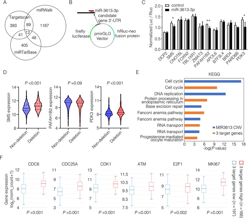
Chen et al. Breast Cancer Research   (2021) 23:12                                                             Page 2 of 13

Introduction                                                   deletion was associated with the breast cancer subtypes
Breast cancer is the most common cancer and the second         and the survival of estrogen receptor-positive (ER+)
cause of cancer-related death in women [1]. Improved           breast cancer patients. Moreover, miR-3613-3p level was
treatments with combination of surgery, radiotherapy, and      also dramatically decreased in tumor tissues or serum of
chemotherapy have increased the overall survival rate for      breast cancer patients. We also proved the tumor sup-
breast cancer patients, but some patients still undergo        pressive roles of mir-3613-3p in human breast cancer
tumor relapse, metastasis, or therapy resistance. Dysregu-     cell lines in vitro and in vivo. Furthermore, miR-3613-3p
lated proliferation is a typical feature of cancer cells and   targets (coding or non-coding RNAs) were predicted by
an essential target of cancer therapy [2]. Cancer stem cells   bioinformatic analysis and verified by biological experi-
(CSCs) possess a potent capacity for self-renewal and dif-     ments. These results elucidated the novel anti-tumor ef-
ferentiation and, therefore, are responsible for the unre-     fect of mir-3613-3p in breast cancer.
stricted growth of tumors [3]. Treatments specifically
targeting CSCs may be a more effective strategy to elimin-     Methods
ate the source of tumor growth, ultimately leading to con-     Cell lines and human serum samples
siderable clinical benefit.                                    The human breast cancer cell lines MDA-MB-231 and
   MicroRNAs (miRNAs) are endogenous, small non-               MCF-7, and human embryonic kidney cell line
coding RNAs that repress target genes expression by            HEK293T, were obtained from American Type Culture
pairing to the 3′-untranslated regions (3′-UTR) of             Collection (ATCC) (October 2011) and cultured accord-
mRNAs [4]. A certain miRNA often has many targets              ing to guidelines. The serum of breast cancer patients
and can participate in several pathways. miRNAs play a         and healthy controls was obtained from Jiangxi Cancer
crucial role in various cancer and have potential applica-     Hospital and stored at − 20 °C.
tions in cancer diagnosis, prognosis, and therapy [5].
The strategy of miRNA-based therapies is either to re-         TCGA and CCLE database analysis
store tumor suppressive miRNAs or to block oncogenic           Gene-level copy number of miR-3613 was estimated by
miRNAs. Tumor suppressive miRNAs can be replen-                using the GISTIC2 method, and clinicopathological data
ished by chemically modified miRNA mimics and onco-            for TCGA breast cancer cohort were retrieved from
genic miRNAs can be reduced by complementary                   UCSC Xena (data downloaded on August 17, 2018) [19].
oligonucleotides, delivered by some novel systems like         Gene-level copy number data for cancer cell lines of
nanoparticles [6]. Some microRNA-based therapies have          Broad Institute Cancer Cell Line Encyclopedia (CCLE)
progressed into clinical trials [7]. Specially, different      were retrieved from UCSC Xena (data downloaded on
types or subtypes of cancer seem to have different             July 30, 2018) and were classified with a low-level
miRNA expression profiles [8]. It is important to identify     threshold 0.3 and a high-level threshold 1 as described
specific miRNAs associated with the tumor subtype              before [20, 21]. The significance of copy number vari-
when developing miRNA-targeted therapeutics.                   ation was analyzed by GISTIC2.0, available through the
   Copy number variations (CNVs) are genomic amplifi-          Broad Institute TCGA copy number portal, filtered for
cations or deletions, which are quite common in human          Q-values less than 0.25 [12].
genome and only a few of germline (or inherited) CNVs
are associated with diseases [9]. Cancer harbors many de       In situ hybridization
novo CNVs, called somatic CNVs. It has been demon-             The tissue microarray used for analysis of miR-3613-3p
strated that amplification of oncogenic genes and dele-        expression in breast cancer was purchased from Shang-
tion of tumor suppressor genes are relevant with               hai Outdo Biotech. miRCURY™ LNA™ Detection probe,
tumorigenesis [10, 11]. Some somatic CNVs are com-             5′-DIG and 3′-DIG labeled (Art.No. YD00611450-BCG,
mon in cancer tissues and those frequently deleted are         Exiqon, Denmark), was used for in situ hybridization at
likely to contain tumor suppressor genes [12].                 a final concentration of 500 nM. The pictures of immu-
   Hsa-mir-3613-3p was first identified through ultra-         nohistochemistry were captured by microscope (Leica,
high throughput sequencing at moderate abundance in            Germany).
human cervical cells in 2010 [13]. A few studies have re-
ported the change of mir-3613-3p associated with sev-          RNA isolation and quantitative real-time PCR
eral types of cancer other than breast cancer, but the         Total RNAs were harvested from cultured cells or ani-
results are inconsistent and lack functional evidence          mal tumor tissues using TRIzol (Invitrogen, USA) and a
[14–18]. In this study, we found that MIR3613 locus was        RNeasy Mini Kit (Qiagen, Germany) according to the
frequently deleted in breast cancer tissues depending on       manufacturer’s instructions. Serum miRNAs were har-
whole exome sequencing data from The Cancer Genome             vested from 200 μL serum of each sample by using miR-
Atlas (TCGA). It is important to note that MIR3613             Neasy Serum/Plasma Kit (Catalog no. 217184, Qiagen,
Chen et al. Breast Cancer Research   (2021) 23:12                                                             Page 3 of 13

Germany) and miRNeasy Serum/Plasma Spike-In Con-             constructs with miRNA mimic were cotransfected into
trol (Catalog no. 219610, Qiagen, Germany) was added         HEK293T or MCF-7 cells. Twenty-four hours after
as a normalization control.                                  transfection, cells were analyzed for luciferase activity
  Total RNA underwent reverse transcription using First      using the Dual-Glo® Luciferase Assay System (Promega,
Strand cDNA Synthesis Kit (Thermo Scientific technolo-       USA). Normalized firefly luciferase activity (firefly lucif-
gies, USA) and each individual microRNAs’ sequence is        erase activity/Renilla luciferase activity, normalized to
shown in Supplementary Table 1. For microRNAs detec-         the pmirGLO Vector no-insert control) was compared.
tion, stem–loop RT-PCR [22] was carried out by using         For each transfection, luciferase activity was averaged
an SYBR Green PCR master mix (TransGen Biotechnol-           from five replicates.
ogy, Beijing) and primer sequences are shown in Supple-
mentary Table 2. All samples were normalized to              Bioinformatic analysis
internal controls and fold changes were calculated. Re-      For functional annotation, the database DAVID Bio-
verse transcription and real-time PCR of mir-3613-3p         informatics Tool was used to search for the most
were also performed by using TaqMan probe (miR-              enriched Kyoto Encyclopedia of Genes and Genomes
3613-3p probe Art.No. 4427975, TaqMan), TaqMan               (KEGG) pathways in selected gene lists. For target pre-
MicroRNA Reverse Transcription Kit (Art.No. 4366596,         diction, TargetScan, miRWalk, and miRTarBase were
TaqMan), and TaqMan Universal PCR Master Mix II              used to identify the mRNAs as miR-3613-3p targets
(Art.No. 4440043, TaqMan) normalized to U6 small nu-         [25–27]. DIANA-LncBase was used to identify the
clear RNA (U6 probe Art.No. 4427975, TaqMan).                LncRNAs interacting with miR-3613-3p [28]. Gene
                                                             Expression Profiling Interactive Analysis (GEPIA) was
MicroRNA transfection                                        used to analyze the expression of candidate lncRNAs in
miRNA mimic (Ribobio, China) were transfected using          breast cancer and normal tissues [29]. The prognostic
Lipofectamine 2000 (Invitrogen, USA) with a final            value of each lncRNAs in breast cancer was analyzed by
miRNA concentration of 50 nM. Forty-eight hours after        using Kaplan–Meier Plotter [30].
transfection, total RNA was isolated as before, and trans-
fection efficiency was confirmed by miRNA qPCR.              Animal study
                                                             Female NOD/SCID mice, 6 to 8 weeks of age, were pur-
Cell proliferation                                           chased from Vital River Laboratories. Approximately 1 ×
Forty-eight hours after transfection, MCF7 cells were        106 of luciferase-transfected MDA-MB-231 cells, mixed
plated in 96-well dishes at 6000 cells/well and MDA-MB-      with Matrigel (1:1), were transplanted into 4th mam-
231 cells at 4000 cells/well. Cell growth rates were moni-   mary gland fat pad of NOD/SCID mice. Mice were
tored using a Cell Counting Kit-8 (CCK-8) (Bimake,           randomly divided into experimental groups with 5 mice
China) according to the manufacturer’s instructions.         in each group. After 12 days, 1 nmol of microRNA ago-
                                                             mir (Ribobio, China) in 20 μl PBS was infused into the
Tumor sphere assay                                           mammary gland once every 3 days for a total of 12 days
Single-cell suspensions were seeded in 6-well or 24-well     (four times). Primary and metastatic tumors in mice
ultra-low attachment plates (Corning, USA) in sphere-        were detected by PET scan (MicroPET Focus 120,
culturing medium (Stemcell Technologies, Canada) for         Siemens, Germany). Lung tissues of each mice were
7 days. Tumor sphere formation was monitored using an        fixed in formalin and embedded in paraffin for histologic
inverted Leica microscope fitted with a camera as de-        analysis. There were four mice in each group in subse-
scribed previously [23] and CSCs frequency was ana-          quent experiments.
lyzed by the extreme limiting dilution analysis (ELDA)
online software [24].                                        Statistical analysis
                                                             All quantified data represents an average of triplicate
Apoptosis assay                                              samples or as indicated. All experiments were repeated
Apoptosis was gauged 24 h post-transfection using FITC       at least three times with similar results each time. Data
Annexin V Apoptosis Detection Kit II (BD, USA) ac-           are represented as mean ± S.E.M or as indicated. Com-
cording to the manufacturer’s instructions.                  parisons between groups were analyzed with tow-tailed
                                                             Student’s t test or Mann–Whitney test dependent on
Luciferase assay                                             whether data conform to distribution normality. Chi-
To generate the luciferase reporter vectors, the 3′-un-      square test was used for correlation analyses between
translated regions (UTR) of genes of interest were syn-      MIR3613 copy number and PAM50 subtypes and mul-
thesized and cloned into the pmirGLO Dual-Luciferase         tiple comparison analysis was adjusted by Bonferroni
miRNA Target Expression Vector. Above distinct               correction. Pearson test was used for correlation analyses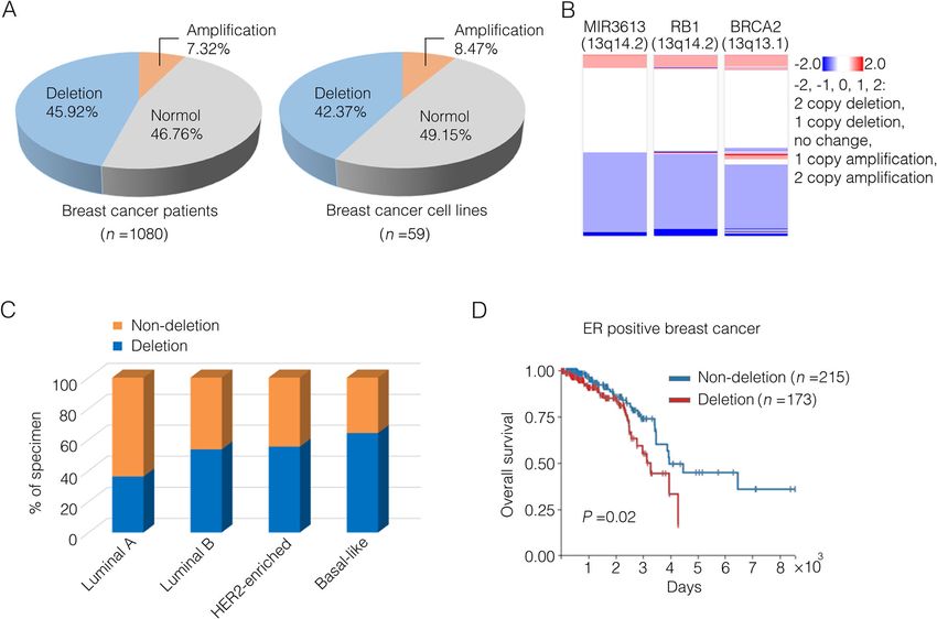
Chen et al. Breast Cancer Research (2021) 23:12 Page 4 of 13
between primary tumor bioluminescence intensity and Table 1 miR-3613 is significantly deleted in 13 independent
miR-3613-3p expression, which estimates a correlation cancer types
value r and a significance P value (0 < r < 1, positive cor- Cancer type Q-value Overall
relation; 0 > r > − 1, negative correlation). Overall survival frequency (%)
of patients from the TCGA cohort was evaluated by the Prostate adenocarcinoma 2.41×10-47 44.51
Kaplan–Meier method. Patients were divided into two Bladder urothelial carcinoma 1.78×10⁻²⁹ 37.01
groups based on the MIR3613 CNVs or the three target Liver hepatocellular carcinoma 8.42×10 -23
48.11
genes (SMS, PAFAH1B2, and PDK3) expression and the Glial cancers 1.34×10⁻¹⁷ 36.06
statistic differences of differentially expressed genes be- -16
Breast invasive adenocarcinoma 1.29×10 46.11
tween the two groups were evaluated by the log-rank
tests. Statistical analyses were performed using SPSS ver- Glioblastoma multiforme 3.7×10⁻¹² 42.98
sion 23. P < 0.05 was considered as the criterion for stat-
-11
Cervical squamous cell carcinoma 3.5×10 38.98
istical significance (*P < 0.05; **P < 0.01; ***P < 0.001). Head and neck squamous 2.55×10⁻⁷ 40.42
cell carcinoma
Results Brain lower grade glioma 2.23×10⁻⁵ 28.27
MiR3613 locus was frequently deleted in breast cancer Ovarian serous 2.63×10 -5
62.52
and associated with breast cancer subtypes and clinical cystadenocarcinoma
prognosis Lung squamous cell carcinoma 4.49×10⁻³ 68.06
The copy number variations (CNVs) of MIR3613, which Lung adenocarcinoma 1.84×10 -2
53.88
encoded miR-3613-3p, were analyzed across several can- Diffuse large B cell lymphoma 1.3×10⁻¹ 14.58
cer types in The Cancer Genome Atlas (TCGA) dataset
Significance (Q-value): Low Q-values (< 0.25) suggest that deletions at this
using TCGA Copy Number Portal [12]. The locus of locus are enriched by selective pressures and this locus has a possible role in
MIR3613 was significantly deleted across 13 diverse can- cancer initiation, growth, or survival. Overall frequency: Overall frequency
measures the fraction of cancers which exhibit any deletion at this locus
cer types including breast invasive adenocarcinoma
(Table 1). The proportion of MIR3613 deletions in
TCGA breast cancer cohort was then studied using the prognosis in the ER positive patients (Fig. 1d). Taken to-
UCSC Xena database [19]. Nearly 46% of TCGA breast gether, these results suggest that MIR3613 genomic loss
cancer tumor samples were subjected to either heterozy- is related to poor prognosis in breast cancer patients.
gous or homozygous deletions at MIR3613 locus and ap-
proximately 42% of breast cancer cell lines from Cancer MiR-3613-3p is downregulated in tumor tissues or serum
Cell Line Encyclopedia (CCLE) showed copy number de- of breast cancer patients
letions of MIR3613 (Fig. 1a). Interestingly, MIR3613 To further evaluate its clinical relevance, miR-3613-3p
locus (13q14.2) located near tumor suppressor genes expression in breast cancer tissues from 30 individual
RB1 (13q14.2) and BRCA2 (13q13.1) on chromosome 13 breast cancer patients was detected by using in situ
and copy number of this gene segment in breast cancer hybridization and their clinical information was shown
was frequently altered (Fig. 1b, Supplementary Fig. 1A) in Supplementary Table 3. These patients were divided
[12]. These results suggest that MIR3613 deletion is of into a miR-3613-3p low expression group and a miR-
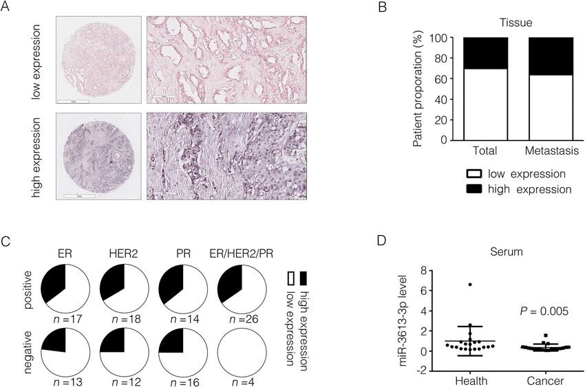
common occurrence in breast cancer patients. 3613-3p high expression group and the representative
The genomic study in 2012 gave a chance to examine images were shown in Fig. 2a (in situ hybridization) and
the relationship between miR-3613 copy numbers and Supplementary Fig. 2 (H&E). Low expression of miR-
PAM50 subtypes [31]. The PAM50 gene signature classi- 3613-3p was shown in 21 out of all 30 breast cancer pa-
fies breast cancer into 4 intrinsic subtypes according to tients (accounting for 70%) and in 9 out of 14 patients
50 genes expression [32]. A total of 499 cancer samples having lymph node metastasis (accounting for 64%)
from TCGA dataset having PAM50 subtype identities which demonstrated a conspicuously lower expression of
were divided into the non-deletion (CNV ≥ 0, n = 254) miR-3613-3p in cancer tissues (Fig. 2b). In accord with
group and the deletion (CNV < 0, n = 232) group accord- the results from TCGA database (Fig. 1c), patients of
ing to whether there was a deletion of MIR3613. The lu- miR-3613-3p low-expression from breast cancer tissue
minal A subtype specifically contained more non- array accounted for a larger proportion in patients with
deletion samples and the basal-like subtype contained negative clinical marker (ER, HER2 or PR) accordingly
more deletion samples (Fig. 1c). Although no significant compared to patients with positive clinical marker
correlation was observed between MIR3613 copy num- (Fig. 2c). Besides that, we also collected blood serum
ber and survival in the whole cohort or ER negative sub- samples from other 20 breast cancer patients, and 20
set of breast cancer patients (Supplementary Fig. 1B), healthy women and their clinical information are shown
deletion of MIR3613 was associated with an unfavorable in Supplementary Table 4. It was noteworthy that the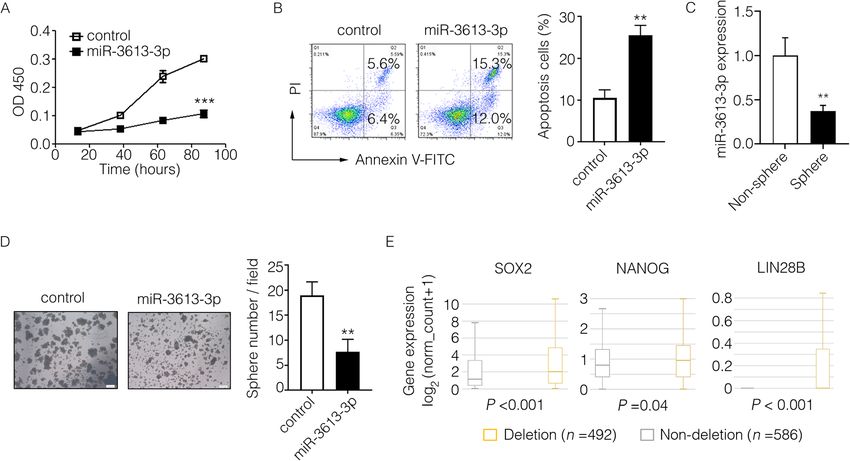
Chen et al. Breast Cancer Research       (2021) 23:12                                                                                       Page 5 of 13

 Fig. 1 The relationship between copy number of miR-3613 and breast cancer subtypes and survival rate. a Left: Proportion description of diverse
 groups in breast cancer patients distinguished by their genomic copy number values (CNVs) of MIR3613 from TCGA database. Homozygous
 deletion or single copy deletion (CNV < 0) represented as deletion group, diploid normal copy (CNV = 0) represented as normal group, low-level
 copy amplification or high-level copy amplification (CNV > 0) represented as amplification group. Right: Proportion description of diverse groups
 in breast cancer cell lines distinguished by the MIR3613 CNV from Cancer Cell Line Encyclopedia (CCLE). b The CNVs of MIR3613, RB1 and BRCA2
 in breast cancer patients from TCGA database. c MIR3613 CNV profiles in breast cancer subtypes from TCGA database. Different breast cancer
 subtypes contained diverse non-deletion (high-CNV) or deletion (low-CNV) patients’ distribution indicated by the Pearson chi-squared test (P <
 0.001). Luminal A, n = 224; Luminal B, n = 127; HER2-enriched, n = 56; Basal-like, n = 92. d Kaplan–Meier survival curves of ER-positive breast cancer
 patients from TCGA database were depicted by MIR3613 CNVs (P = 0.02). The non-deletion group contained samples with high-CNV of MIR3613
 (CNV ≥ 0, n = 215), while the deletion group contained samples with low-CNV of MIR3613 (CNV < 0, n = 173)

expression of miR-3613-3p in serum were dramatically                          neither proliferation rate of MDA-MB-231 cells nor the
decreased in cancer samples detected by RT-PCR                                apoptotic proportion of MCF7 cells was changed after
(Fig. 2d). Taken together, these findings suggest that                        miR-3613-3p overexpression (Supplementary Fig. 3A, B).
miR-3613-3p is distinctly downregulated in clinical                           Tumor spheres forming ability was a golden standard to
samples of breast cancer patients.                                            evaluate the self-renewal capacity of cancer stem cells
                                                                              (CSCs). After culturing and separating of tumor spheres,
MiR-3613-3p suppressed malignant phenotypes of breast                         we found that stemness associated genes (SOX2, OCT4,
cancer cells in vitro                                                         NANOG, and LIN28B) exhibited a high level of expres-
To address whether miR-3613-3p might play a key role                          sion whereas stemness-suppressive microRNAs (let7
in malignant characters in breast cancer cells, we first                      family and miR-146a) exhibited a low level of expression
measured the proliferation or apoptosis or human breast                       in spheres in accordance with previous studies (Supple-
cancer cell lines MCF7 and MDA-MB-231 cells trans-                            mentary Fig. 4A, B). Interestingly, we observed that miR-
fected with miR-3613-3p or control mimics, respectively.                      3613-3p expression was dramatically decreased in tumor
After overexpression of miR-3613-3p, the proliferation                        spheres (Fig. 3c) and it could obviously suppress the
rate of MCF7 cells was significantly suppressed (Fig. 3a),                    tumor spheres forming ability of MDA-MB-231 cells as
whereas the proportion of apoptotic cells was markedly                        well (Fig. 3d, Supplementary Fig. 5A, B), indicating the
increased in MDA-MB-231 cells (Fig. 3b). However,                             inhibitory effect of miR-3613-3p in cancer stemness
Chen et al. Breast Cancer Research (2021) 23:12 Page 6 of 13
Fig. 2 miR-3613-3p expression in clinical breast cancer patients. a miR-3613-3p was detected by in situ hybridization and representative images of low or high
expression from breast cancer tissue array were shown. b The proportion of patients with lymph node metastasis or not was calculated depending on miR-
3613-3p expression. c Fractions of two group patients divided based on the miR-3613-3p expression were analyzed in different subtypes of breast cancer from
breast cancer tissue array. d miR-3613-3p in serum of breast cancer patients was detected by RT-PCR (P = 0.005)
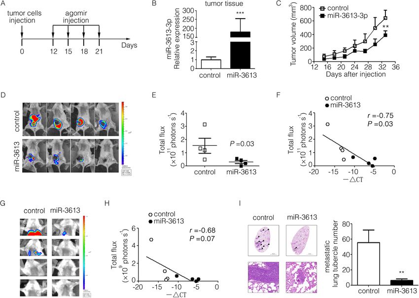
maintenance. We then analyzed the transcripts of differ- explore the function of microRNA in vivo previously [33].
entially expressed genes in two groups of breast cancer On the 12th day after tumor inoculation, miR-3613-3p ago-
patients divided according to MIR3613 CNVs from mir or negative control agomir were intratumorally injected
TCGA dataset (group of MIR3613 CNV ≥ 0, MIR3613 respectively and tumor growth or lung metastasis was moni-
non-deletion, n = 586; group of MIR3613 CNV < 0, tored (Fig. 4a). Results showed that the expression of miR-
MIR3613 deletion, n = 492). Interestingly, the expression 3613-3p in tumor was increased as expected (Fig. 4b) and
of stemness genes (SOX2, NANOG, and LIN28B) were volumes of tumor in situ were dramatically reduced by ad-
all significantly higher in the MIR3613 deletion group ministrating of miR-3613-3p agomir (Fig. 4c). In fact, pri-
compared to the non-deletion group (Fig. 3e). These re- mary tumor growth in groups of mice given by miR-3613-3p
sults suggest that miR-3613-3p can suppress the malig- agomir was clearly delayed as indicated by living imaging
nant phenotypes and cancer stemness of breast cancer and the signal intensity in primary tumor cells of mice was
cells and may serve as a tumor suppressor. negatively correlated with miR-3613-3p expression (r = −
0.75, P = 0.03) (Fig. 4d–f). Furthermore, the degree of pul-
MiR-3613-3p suppressed tumor growth and metastasis in monary metastasis in the miR-3613-3p group significantly re-
an immunocompromised mouse model of human breast duced and the signal intensity in pulmonary tumor cells of
cancer cell MDA-MB-231 mice was slightly negatively correlated with miR-3613-3p ex-
To investigate the effect of miR-3613-3p on tumor progres- pression (r = − 0.68, P = 0.07) (Fig. 4g, h). H&E staining of
sion in vivo, luciferase-labeled MDA-MB-231 cells were lung specimens indicated obviously that the number of meta-
injected into the 4th mammary gland fat pad of NOD/SCID static foci number in lung were much fewer in groups of
mice. Agomir, a chemically modified analogue of microRNA mice subjected to miR-3613-3p agomir treatment (Fig. 4i).
with enhanced stability and activity, was successfully used to Taken together, these results reveal a remarkable role ofChen et al. Breast Cancer Research (2021) 23:12 Page 7 of 13
Fig. 3 miR-3613-3p suppressed malignant phenotype of human breast cancer cells. a Proliferation of MCF7 cells transfected by miR-3613-3p
mimic or control mimic was analyzed by CCK-8 assay (***, P < 0.001). b Apoptosis of MDA-MB-231 cells transfected by miR-3613-3p mimic or
control mimic was analyzed by flow cytometry. The representative apoptosis images were visualized on the left and the proportion of apoptosis
cells were statistically analyzed on the right (**P < 0.05). c The expression of miR-3613-3p in non-spheres or spheres of MDA-MB-231 cells was
calculated by RT-PCR (**P < 0.05). d Tumor spheres of MDA-MB-231 cells transfected by miR-3613-3p mimic or control mimic was cultured with
serum-free medium for 7 days. The representative tumor sphere images were visualized by microscope on the left (scale bar, 200 mm) and the
sphere number per field were statistically analyzed on the right (**P < 0.05). e Transcripts of differentially expressed genes were analyzed in two
groups of breast cancer patients divided according to MIR3613 CNVs from TCGA dataset (group of MIR3613 CNV ≥ 0, MIR3613 non-deletion, n =
586; group of MIR3613 CNV< 0, MIR3613 deletion, n = 492). The expression of SOX2, NANOG, and LIN28B in above two groups were shown
according to the transcriptome sequencing data
miR-3613-3p in tumor suppression in vivo and likely provide vectors, gene fragments, which are approximately 400 to
a useful target for future therapeutic interventions in breast 800 base pairs, including the predicted combining loci in
cancer. the 3′-UTR, were synthesized and cloned into the pmir-
GLO Dual-Luciferase miRNA Target Expression Vector
Identification of miR-3613-3p target genes and related (Fig. 5b). Decreased luciferase activity was found in con-
signaling pathway structs for three genes (SMS, PAFAH1B2, and PDK3) in
To identify the mechanism(s) whereby miR-3613-3p HEK293T or MCF-7 cells (Fig. 5c) and their binding sites
exerted its biological effect on breast cancer cells, we first with miR-3613-3p are shown in Supplementary Fig. 11A.
ran a target prediction in silico using TargetScan, miR- More importantly, the highly amplified transcripts of SMS
Walk and miRTarBase. From 7395 target genes predicted and PDK3 were observed in breast cancer samples with
by TargetScan, we selected 535 genes with the highest ab- miR-3613 deletion compared to the non-deletion group
solute value of context score which indicated as the prob- from TCGA breast cancer cohort (Fig. 5d). We then ana-
ability of targeting. Functional pathway analysis revealed lyzed the signaling pathways potentially regulated by these
that the shared target genes predicted by TargetScan and 3 target genes or miR-3613-3p. On the one hand, differen-
miRWalk (n = 101) were mainly enriched in cancer- tially expressed genes and enriched signaling pathways
related signaling pathways (Supplementary Fig. 6A, B). were analyzed in two groups of breast cancer patients di-
Furthermore, 12 of miR-3613-3p target genes were identi- vided according to MIR3613 CNVs from TCGA dataset
fied as the overlaps among those predicted by TargetScan, (group of MIR3613 CNV ≥ 0, n = 586; group of MIR3613
miRWalk, and miRTarBase (Fig. 5a). Then, we con- CNV < 0, n = 492). On the other hand, differentially
structed the dual-luciferase reporter system of 3′-untrans- expressed genes and enriched signaling pathways were an-
lated region (UTR) from these 12 target genes to identify alyzed in two groups of breast cancer patients divided ac-
miR-3613-3p targets. To generate the luciferase reporter cording to SMS, PAFAH1B2, and PDK3 expression fromChen et al. Breast Cancer Research (2021) 23:12 Page 8 of 13 Fig. 4 miR-3613-3p suppressed tumor growth and metastasis of MDA-MB-231 cells in NOD/SCID mice. a The strategy of animal experiment was illustrated. After tumor volume reached about 50 mm3, miR-3613-3p or control agomir was intratumor injected respectively every 2 days for four times. b The expression of miR-3613-3p in tumor tissues from miR-3613-3p or control group were detected by RT-PCR after mice were killed on day 60 (***P < 0.001). c Tumor growth curve of mice injected with miR-3613-3p or control agomir (Shown are mean tumor volumes ± SEM; n = 4 per group; **P < 0.05). d Bioluminescence images of the primary tumors from miR-3613-3p or control mice were captured by Live animal imager on day 59. e Bioluminescence intensity of primary tumors were statistically analyzed (P = 0.03). f Correlation between primary tumor bioluminescence intensity and miR-3613-3p expression was calculated by Pearson test (r = − 0.75, P = 0.03). g Bioluminescence images of the pulmonary metastases were captured by Live animal imager on day 59. h Correlation between primary tumor bioluminescence intensity and miR-3613-3p expression was calculated by Pearson test (r = − 0.68, P = 0.07). i Representative images of the pulmonary metastases in mice by H&E staining (arrow marked typical metastases; Scale bar, 100 mm). j Metastatic lung tubercle numbers were calculated from H&E staining images (**P < 0.05) TCGA dataset (group of 3 target genes high expression, patients, regardless of ER status, high expression of n = 166; group of 3 target genes low expression, n = 197). PAFAH1B2 (P = 0.04), but not SMS (P = 0.35) or PDK3 It is worth noting that among the top 10 of KEGG signal- (P = 0.23), was associated with an unfavorable prognosis ing pathways, cell cycle was the most relevant pathway in (Supplementary Fig. 9A-C). In ER-positive patients, high both two-independent analysis (Fig. 5e, Supplementary expression of PAFAH1B2 (P = 0.02) or PDK3 (P = 0.04) Fig. 7). Further analysis demonstrated that the expression indicated a poor prognosis and patients with high ex- of cell cycle-related genes (CDC6, CDC25A, CDK1, ATM, pression of SMS (P = 0.09) showed a tendency towards a E2F1, and MKI67) were much higher in breast cancer pa- worse overall survival (Supplementary Fig. 9A-C). How- tients of 3 target genes high expression group or MIR3613 ever, in ER-negative patients, no significant correlation deletion group compared with their counterpart, respect- was observed between the expression of these three ively (Fig. 5f, Supplementary Fig. 8). genes and survival rates (Supplementary Fig. 9A-C). Fur- The overall survival rates of breast cancer patients thermore, in the ER-positive subsets, but not in the based on the expression of these three genes (SMS, whole subsets or ER-negative subsets, patients with sim- PAFAH1B2 and PDK3) were analyzed by UCSC Xena ultaneously high expression of all these three genes had (data retrieved on November 24, 2020) [19]. In all the markedly poorer prognosis compared to patients with
Chen et al. Breast Cancer Research       (2021) 23:12                                                                                      Page 9 of 13

 Fig. 5 Identification of miR-3613-3p target genes and related signaling pathway. a Venn diagram of predicted target genes of miR-3613-3p from
 Targetscan, miRWalk and miRTarBase online websites. b Schematic diagram of 3′-UTR dual-luciferase reporter system. c Predicted 12 target genes of
 miR-3613-3p were verified by 3′-UTR dual-luciferase reporter assays in HEK293T or MCF-7 cells (*P < 0.05). d The expression of SMS, PAFAH1B2, or
 SNHG16 was shown in two separate group samples (MIR3613 deletion group or MIR3613 non-deletion group) of breast cancer tissues from TCGA
 database. e Transcripts of differentially expressed genes were analyzed in two groups of breast cancer patients divided according to MIR3613 CNVs
 from TCGA dataset (group of MIR3613 CNV ≥ 0, n = 586; group of MIR3613 CNV < 0, n = 492), and enriched top five signaling pathways (blue) were
 shown. Transcripts of differentially expressed genes were analyzed in two groups of breast cancer patients divided according to SMS, PAFAH1B2, and
 PDK3 expression from TCGA dataset (group of 3 target genes high expression, n = 166; group of 3 target genes low expression, n = 197), and enriched
 top five signaling pathways (orange) were shown. f Transcripts of differentially expressed genes were analyzed in two groups of breast cancer patients
 divided according to SMS, PAFAH1B2, and PDK3 expression from TCGA dataset (group of 3 target genes high expression, n = 166; group of 3 target
 genes low expression, n = 197). The expression of CDC6, CDC25A, CDK1, ATM, E2F1, and MKI67 in above two groups were shown according to the
 transcriptome sequencing data

low expression of all of them (P = 0.03, Supplementary                        perhaps regulate cell cycling pathway by targeting SMS,
Fig. 9D). These results strongly suggested that higher                        PAFAH1B2, or PDK3 to restrain tumor progression.
level of SMS, PAFAH1B2, or PDK3 could predict worse
prognosis in ER-positive breast cancer.                                       Bioinformatic analysis of lncRNAs interacting with miR-
  However, the specific biological functions of SMS,                          3613-3p
PAFAH1B2, or PDK3 need to be further investigated in                          Long non-coding RNAs (lncRNAs) have been reported
breast cancer. These results suggest that miR-3613-3p                         to play important roles in carcinogenesis and acting as a
Chen et al. Breast Cancer Research   (2021) 23:12                                                              Page 10 of 13

sponge for several miRNAs in previous studies [34]. To         lower frequency of MIR3613 deletion was most re-
investigate the specific lncRNAs interacting with miR-         stricted in PAM50 subtypes with a comparatively favor-
3613-3p, the online software DIANA-LncBase (https://           able prognosis (luminal A subtype) and ER-positive
omictools.com/diana-lncbase-tool) was used, which of-          breast cancer patients with MIR3613 deletion possessed
fered miRNA–lncRNA interactions supported by either            a worse prognosis compared with the non-deletion
computational prediction or experimental verification.         group. Previously, our group identified serum miR-1915-
Forty-one lncRNAs were both experimentally supported           3p and miR-455-3p as biomarkers for breast cancer pa-
and in silico predicted targets, in which 6 were predicted     tients with lymph node metastasis [42]. This study fur-
targets specifically in breast tissue (Supplementary           ther revealed a lower expression of miR-3613-3p in the
Fig. 10A). Then, we explored the expression levels of          blood serum of breast cancer patients compared with
these 41 lncRNAs in breast cancer and their association        healthy controls and a decreased level was also shown in
with overall survival using the GEPIA database and the         breast cancer tissues of patients with or without metas-
Kaplan–Meier Plotter database. Out of the 30 available         tasis. These results strongly imply that miR-3613-3p
expression results, 7 lncRNAs had lower expression             serves as a molecular indicator or tumor suppressor for
levels in breast cancer tissues than in normal breast tis-     breast cancer initiation and progression.
sue (Supplementary Fig. 10B). Out of the 13 available             Uncontrolled proliferation is an essential characteristic
survival curve results, low expression of NEAT1 or high        of cancer and interruption to proliferative pathways is a
expression of SNHG16 was associated with significantly         promising strategy to fight cancer [3]. Overexpression of
shorter survival in 626 breast cancer patients (Supple-        miR-3613-3p significantly impairs the proliferation and
mentary Fig. 10C). The binding sites of NEAT1 and              promotes the apoptosis of breast cancer cells in vitro. In
SNHG16 with miR-3613-3p are shown in Supplemen-                line with our data, Zhang et al. recently discovered miR-
tary Fig. 11B, respectively. It is noteworthy that NEAT1       3613-3p could inhibit hepatoma cell proliferation [43].
was decreased and SNHG16 was increased in breast can-          However, our data revealed that overexpression of miR-
cer samples with MIR3613 deletion compared to the              3613-3p had almost no influence on the proliferation of
non-deletion group from TCGA breast cancer cohort              MDA-MB-231 cells and the apoptosis of MCF-7 cells.
(Supplementary Fig. 10D). These data potentially indi-         MCF7 (ER positive) and MDA-MB-231 (ER negative)
cate that NEAT1 or SNHG16 may interact with miR-               cells had different molecular pathways impacting their
3613-3p and perform their diverse biological functions         distinct malignant behaviors [44]. Since miR-3613-3p
in breast cancer.                                              could target multiple genes, it was probable that the
                                                               major targets or relative molecular pathways of miR-
Discussion                                                     3613-3p in ER-positive and ER-negative breast cancers
MicroRNAs, as an important class of non-coding small           were not the same. In addition, a BRCA2 mutation was
RNAs, almost affect all the cellular processes and have        frequently observed to associate with worse prognosis in
been found to be dysregulated in a multitude of diseases.      ER-positive breast cancer and the basic mechanism was
The study of Calin et al. reveals that miRNA loci are fre-     still unclear [45]. Our results suggested that MIR3613
quently located in fragile sites associated with cancers       locus (13q14.2) located near BRCA2 (13q13.1) in breast
[35]. The role of genes at fragile genomic locations is        cancer and they perhaps had some functional similarity
controversial, but emerging evidence support that a lot        and interactivity.
of these genes contribute to tumorigenesis [36, 37].              In our previous studies, we demonstrated that target-
Non-coding genes may have critical functions, as well as       ing CSCs or CSC-associated microRNA (miR-34a) could
protein-coding genes. Mir-101 is often deleted in non-         markedly improve the efficacy of cancer therapy [23, 46].
small cell lung cancer and displays anti-tumorigenic           In this study, the expression of miR-3613-3p was de-
properties [38]. Mir-383-3p is at a frequently missing         creased in tumor spheres and it could dramatically in-
genomic locus in prostate cancer and impedes cancer            hibit the self-renewal of CSCs (tumor sphere forming
initiation and metastasis [39]. In the present study, we       ability), which suggested a novel suppressive function of
first found a widespread loss of miR-3613-3p DNA frag-         miR-3613-3p in CSCs. It should be noticed that some
ment (13q14.2) in breast cancer patients genome, which         microRNAs regulating cancer stemness were supposed
located near the famous tumor suppressor genes RB1             as excellent therapeutic targets in previous studies [47,
(13q14.2) and BRCA2 (13q13.1). It is coincident that           48]. Importantly, miR-3613-3p repressed tumor growth
miR-15/16 located to 13q14 were also deleted in patients       and metastasis in an immunocompromised mouse
with chronic lymphocytic leukemia [40]. PAM50 signa-           model of human breast cancer cell MDA-MB-231.
ture assay has displayed additive values in breast cancer      Molecular and histologic analysis of tumor tissues has
prognosis and treatment decisions combined with stand-         provided microscopic evidence that the expression of
ard clinical and pathological factors [41]. Interestingly, a   miR-3613-3p was inversely correlated with tumor
Chen et al. Breast Cancer Research (2021) 23:12 Page 11 of 13
progression. Although cell migratory capacity did not cancer and our findings provided a novel direction in
change much in vitro, the pulmonary metastasis was the field of cancer-related lncRNAs for future research.
dramatically suppressed by miR-3613-3p agomir in mice,
implying the complexity of metastatic process in vivo Conclusions
[49]. These phenotype findings are consistent with previ- In this study, we discovered the frequent genomic dele-
ous genomic studies suggesting that loss-function of tion and low expression of miR-3613-3p in breast cancer
miR-3613-3p is an important mechanism in breast can- for the first time. In vitro and in vivo experimental valid-
cer progression and thus provide a novel therapeutic ation proved the important suppressive role of miR-
target. 3613-3p in breast cancer progression. Bioinformatic ana-
Functional annotation revealed that predicted target lysis revealed miR-3613-3p might regulate cell cycle sig-
genes of miR-3613-3p were mostly enriched in cancer naling pathway by targeting SMS, PAFAH1B2, or PDK3
related pathways. Bioinformatic prediction and 3′-UTR to restrain tumor progression. Our findings imply the
assay identified SMS, PAFAH1B2, and PDK3 could be important role of miR-3613-3p as an innovative progno-
regulated by miR-3613-3p. Spermine synthase, encoded sis marker or treatment target for breast cancer patients.
by SMS, catalyzed polyamine metabolism, whose dysreg-
ulation was associated with carcinogenesis [50]. High
Supplementary Information
SMS mRNA expression was related to poor survival and The online version contains supplementary material available at https://doi.
increased risk of metastasis in triple-negative breast can- org/10.1186/s13058-021-01389-9.
cer [51]. PAFAH1B2 encodes a subunit of platelet-
activating factor acetyl hydrolase and was identified to Additional file 1: Supplementary Figure 1. The genomic location of
MIR3613 and its CNV in breast cancer patients prognosis. A) The genomic
participate in metabolic cancer pathogenicity [52]. location of MIR3613, RB1 and BRCA2 (red lines): bands according to
Blockade of PAFAH1B2 and PAFAH1B3 causes a Ensembl, locations according to GeneLoc. B) Kaplan–Meier survival curves
heightened level of tumor-suppressing lipids and dam- of breast cancer patients from TCGA database were depicted by their
genomic copy number value (CNV) of miR-3613 (P = 0.18). The non-
ages cancer pathogenicity in breast cancer and several deletion group contained samples with high-CNV miR-3613 (CNV ≥ 0,
other types of cancer. Pyruvate dehydrogenase kinase 3 n = 265), while the deletion group contained samples with low-CNV miR-
(PDK3) could alter glucose metabolism in cancer by in- 3613 (CNV < 0, n = 242) (left). Kaplan–Meier survival curves of ER–negative
breast cancer patients from TCGA database were depicted by their miR-
activating pyruvate dehydrogenase kinase and overex- 3613 CNV (P = 0.52). (non-deletion group, n = 46; deletion group, n = 67)
pression of PDK is related to tumor invasion, metastasis, (right).
and drug resistance [53]. It is interesting to note that cell Additional file 2: Supplementary Figure 2. Histological analysis of
cycle, important for cell proliferation, was the most breast cancer tissues with low or high miR-3613-3p expression by using
hematoxylin-eosin staining.
enriched signaling pathway regulated by these three tar-
Additional file 3: Supplementary Figure 3. Proliferation of MDA-MB-
get genes or miR-3613-3p, which mightily suggested 231 cells and apoptosis of MCF-7 cells. A) Proliferation of MDA-MB-231
miR-3613-3p targeted SMS, PAFAH1B2, or PDK3 to cells transfected with miR-3613-3p or control mimic was analyzed by
suppress oncogenic pathway of cell cycle for tumor con- CCK-8 assay. B) Apoptosis of MCF-7 cells transfected with miR-3613-3p or
control mimic was analyzed by flow cytometry.
trol. Recently, miR-3613-3p was reported to regulate
Additional file 4: Supplementary Figure 4. The expression of
genes of EGFR signaling pathway in the epithelial- stemness associated genes and miRNAs in tumor spheres of MDA-MB-
mesenchymal transition of lung adenocarcinoma [54]. It 231 cells. A) The expression of stemness genes (SOX2, OCT4, NANOG and
is possible that miR-3613-3p affect cancer pathogenicity LIN28B) were analyzed by RT-PCT in non-spheres or spheres of MDA-MB-
231 cells. B) The expression of stemness associated miRNAs (let7 family
in different ways in diverse tumor types and further in- and miR-146a) were analyzed by RT-PCT in non-spheres or spheres of
vestigation need to be complete investigation. Further- MDA-MB-231 cells.
more, seven lncRNAs, downregulated in breast cancer Additional file 5: Supplementary Figure 5. Frequency of cancer stem
tissues and potentially interacting with miR-3613-3p, cells in MDA-MB-231 cells. A) Plot result of extreme limiting dilution ana-
lysis for MDA-MB-231 sphere formation. B) Frequency of CSCs was esti-
were identified by bioinformatic analysis. The expression mated based on MDA-MB-231 sphere formation assay.
of NEAT1 was markedly decreased in MIR3613 deletion Additional file 6: Supplementary Figure 6. Identification of miR-
group and its high expression tended to be associated 3613-3p target genes and related KEGG pathways. A) Venn diagram of
with prolonged survival in breast cancer patients. Previ- predicted target genes miR-3613-3p from Targetscan and miRWalk online
websites. B) Predicted 101 target genes of miR-3613-3p were enriched in
ous study revealed that NEAT1 emerged as an important KEGG pathways by DAVID online software.
regulator in cancer development and was downregulated Additional file 7: Supplementary Figure 7. Bioinformatic analysis of
in invasive breast cancer [55], which was consistent with signaling pathways regulated by miR-3613-3p or its 3 target genes. A)
our results. SNHG16, upregulated in miR-3613 deletion Transcripts of differentially expressed genes were analyzed in two groups
of breast cancer patients divided according to MIR3613 CNV from TCGA
group, may act as a sponge for miR-3613-3p and its high dataset (group of MIR3613 CNV ≥ 0, n = 586; group of MIR3613 CNV< 0,
expression was associated with a worse prognosis in n = 492). Top 10 KEGG signaling pathways were shown according to
breast cancer patients. The biological functions of above differentially expressed genes. B) Transcripts of differentially
expressed genes were analyzed in two groups of breast cancer patients
NEAT1 and SNHG16 are rarely investigated in breastChen et al. Breast Cancer Research (2021) 23:12 Page 12 of 13
divided according to SMS, PAFAH1B2 and PDK3 expression from TCGA Inter-Governmental S&T Cooperation Project from Ministry of Science and
dataset (group of 3 target genes expression high, n = 166; group of 3 tar- Technology of China: grant no. 2018YFE0114300.
get genes expression low, n = 197). Top 10 KEGG signaling pathways
were shown according to above differentially expressed genes. Availability of data and materials
The TCGA and CCLE dataset are available in the UCSC Xena (https://
Additional file 8: Supplementary Figure 8. The expression of xenabrowser.net).
proliferation related genes in breast cancer patients. Transcripts of Bioinformatic analysis is based upon data generated by the DAVID
differentially expressed genes were analyzed in two groups of breast Bioinformatics Tool (https://david.ncifcrf.gov), TargetScan (http://www.
cancer patients divided according to MIR3613 CNV from TCGA dataset targetscan.org/vert_72), miRWalk (http://mirwalk.umm.uni-heidelberg.de),
(group of MIR3613 CNV ≥ 0, MIR3613 non-deletion, n = 586; group of miRTarBase (http://mirtarbase.cuhk.edu.cn/php/index.php), DIANA-LncBase
MIR3613 CNV < 0, MIR3613 deletion, n = 492). The expression of CDC6, (http://carolina.imis.athena-innovation.gr/diana_tools/web/index.php?r=
CDC25A, CDK1, ATM, E2F1 and MKI67 in above two groups were shown lncbasev2%2Findex), GEPIA (http://gepia.cancer-pku.cn), and Kaplan-Meier
according to the transcriptome sequencing data. Plotter (http://kmplot.com/analysis). The data generated during the current
Additional file 9: Supplementary Figure 9. Survival analysis of breast study are available from the corresponding author on reasonable request.
cancer patients based on the expression of SMS, PAFAH1B2 or PDK3. A)
Kaplan-Meier survival curves of total, ER-negative or ER-positive breast Ethics approval and consent to participate
cancer patients from TCGA database were depicted by SMS expression. This study has been approved by INSTITUTIONAL REVIEW BOARD of Institute
B) Kaplan-Meier survival curves of total, ER-negative or ER-positive breast of Basic Medical Sciences, Chinese Academy of Medical Sciences.
cancer patients from TCGA database were depicted by PAFAH1B2 expres-
sion. C) Kaplan-Meier survival curves of total, ER-negative or ER-positive Consent for publication
breast cancer patients from TCGA database were depicted by PDK3 ex- Not applicable.
pression. D) Kaplan-Meier survival curves of total, ER-negative or ER-
positive breast cancer patients from TCGA database were depicted by
Competing interests
SMS, PAFAH1B2 and PDK3 expression.
The authors declare that they have no conflict of interest.
Additional file 10: Supplementary Figure 10. Bioinformatic analysis
of lncRNAs interacting with miR-3613-3p. A) Venn diagram of predicted Author details
targets from Targetscan and miRWalk online websites. B) The differential 1
Department of Immunology, Institute of Basic Medical Sciences, Chinese
expression of 7 lncRNAs in breast cancer tissues (T, n = 1085) or normal Academy of Medical Sciences; School of Basic Medicine, Peking Union
tissues (N, n = 291) were analyzed by using the GEPIA database. C) Medical College, Beijing 100005, China. 2Collaborative Innovation Center for
Kaplan–Meier survival curves of breast cancer patients were depicted by Biotherapy, Institute of Basic Medical Sciences, Chinese Academy of Medical
the expression of NEAT1 (HR = 0.72 (0.52–0.98)) or SNHG16 (HR = 1.76 Sciences; School of Basic Medicine, Peking Union Medical College, Beijing
(1.28–2.42)), respectively. D) The expression of NEAT1 or SNHG16 in two 100005, China. 3Department of Clinical Laboratory, Jiangxi Cancer Hospital,
separate group samples of breast cancer tissues from TCGA database di- Nanchang 330029, China. 4Immunology Research Center, National Health
vided by whether miR-3613 locus was deleted. Research Institutes, Zhunan, Miaoli, Taiwan.
Additional file 11: Supplementary Figure 11. The description of
binding sites of miR-3613-3p with its targets. A) The description of bind- Received: 10 September 2020 Accepted: 11 January 2021
ing sites of miR-3613-3p with the 3′-UTR of SMS, PAFAH1B2 or PDK3. B)
The description of binding sites of miR-3613-3p with the lncRNAs NEAT1
or SNHG16. References
Additional file 12: Supplementary Table 1. Sequences of microRNAs. 1. Siegel RL, Miller KD, Jemal A. Cancer statistics, 2019. CA Cancer J Clin. 2019;
Supplementary Table 2. Primer sequences of mRNAs or microRNAs for 69:7–34.
RT-PCR. Supplementary Table 3. The correlation of miR-3613-3p ex- 2. Hanahan D, Weinberg RA. Hallmarks of cancer: the next generation. Cell.
pression clinicopathological characteristics of patients from breast cancer 2011;144:646–74.
tissue array. Supplementary Table 4. Clinicopathological characteristics 3. Batlle E, Clevers H. Cancer stem cells revisited. Nat Med. 2017;23:1124–34.
of patients from serologic detection. 4. Bartel DP. MicroRNAs: target recognition and regulatory functions. Cell.
2009;136:215–33.
5. Hayes J, Peruzzi PP, Lawler S. MicroRNAs in cancer: biomarkers, functions
Abbreviations and therapy. Trends Mol Med. 2014;20:460–9.
CNVs: Copy number variations; TCGA: The Cancer Genome Atlas; CCLE: Broad 6. Labatut AE, Mattheolabakis G. Non-viral based miR delivery and recent
Institute Cancer Cell Line Encyclopedia; qPCR: Quantitative polymerase chain developments. Eur J Pharm Biopharm. 2018;128:82–90.
reaction; lncRNA: Long non-coding RNA; UTR: Untranslated region; 7. Rupaimoole R, Slack FJ. MicroRNA therapeutics: towards a new era for the
CSCs: Cancer Stem Cells; ATCC: American Type Culture Collection; management of cancer and other diseases. Nat Rev Drug Discov. 2017;16:
GEPIA: Gene Expression Profiling Interactive Analysis; KEGG: Kyoto 203–22.
Encyclopedia of Genes and Genomes; H&E: Hematoxylin and eosin 8. Goh JN, Loo SY, Datta A, Siveen KS, Yap WN, Cai W, et al. microRNAs in
breast cancer: regulatory roles governing the hallmarks of cancer. Biol Rev
Camb Philos Soc. 2016;91:409–28.
Acknowledgements
9. Kuiper RP, Ligtenberg MJ, Hoogerbrugge N, Geurts van Kessel A. Germline
We thank Lipeng Bai (Department of Clinical Laboratory, Jiangxi Cancer
copy number variation and cancer risk. Curr Opin Genet Dev. 2010;20:282–9.
Hospital, Nanchang, China) for the serum collection of breast cancer patients
10. Bowtell DD. The genesis and evolution of high-grade serous ovarian cancer.
or healthy persons.
Nat Rev Cancer. 2010;10:803–8.
11. Gu J, Ajani JA, Hawk ET, Ye Y, Lee JH, Bhutani MS, et al. Genome-wide
Authors’ contributions catalogue of chromosomal aberrations in barrett’s esophagus and
CC, YP, and HC carried out the bioinformatic analysis. CC and YP carried out esophageal adenocarcinoma: a high-density single nucleotide
the cellular and animal experiments. YP and RZ performed the statistical polymorphism array analysis. Cancer Prev Res (Phila). 2010;3:1176–86.
analysis. CC and YP drafted the manuscript. LB, ZD, and QS provided material 12. Beroukhim R, Mermel CH, Porter D, Wei G, Raychaudhuri S, Donovan J, et al.
support. YL, CC, and THC conceived of the study and participated in its The landscape of somatic copy-number alteration across human cancers.
design. All authors read and approved the final manuscript. Nature. 2010;463:899–905.
13. Witten D, Tibshirani R, Gu SG, Fire A, Lui W-O. Ultra-high throughput
Funding sequencing-based small RNA discovery and discrete statistical biomarker
This work was supported by grants from the National Natural Science analysis in a collection of cervical tumours and matched controls. BMC Biol.
Foundation of China (NSFC): grant no. 81972795 and 81672914; the Bilateral 2010;8:58.Chen et al. Breast Cancer Research (2021) 23:12 Page 13 of 13
14. Bibi F, Naseer MI, Alvi SA, Yasir M, Jiman-Fatani AA, Sawan A, et al. 38. Thu KL, Chari R, Lockwood WW, Lam S, Lam WL. miR-101 DNA copy loss is
microRNA analysis of gastric cancer patients from Saudi Arabian population. a prominent subtype specific event in lung cancer. J Thorac Oncol. 2011;6:
BMC Genomics. 2016;17:751. 1594–8.
15. Pu Q, Huang Y, Lu Y, Peng Y, Zhang J, Feng G, et al. Tissue-specific and 39. Bucay N, Sekhon K, Yang T, Majid S, Shahryari V, Hsieh C, et al. MicroRNA-
plasma microRNA profiles could be promising biomarkers of histological 383 located in frequently deleted chromosomal locus 8p22 regulates CD44
classification and TNM stage in non-small cell lung cancer. Thorac Cancer. in prostate cancer. Oncogene. 2017;36:2667–79.
2016;7:348–54. 40. Calin GA, Dumitru CD, Shimizu M, Bichi R, Zupo S, Noch E, et al. Frequent
16. Yan W, Yang W, Liu Z, Wu G. Characterization of microRNA expression in deletions and down-regulation of micro-RNA genes miR15 and miR16 at
primary human colon adenocarcinoma cells (SW480) and their lymph node 13q14 in chronic lymphocytic leukemia. Proc Natl Acad Sci U S A. 2002;99:
metastatic derivatives (SW620). Onco Targets Ther. 2018;11:4701–9. 15524–9.
17. Chong GO, Jeon HS, Han HS, Son JW, Lee YH, Hong DG, et al. Differential 41. Holm J, Eriksson L, Ploner A, Eriksson M, Rantalainen M, Li J, et al.
microRNA expression profiles in primary and recurrent epithelial ovarian Assessment of breast cancer risk factors reveals subtype heterogeneity.
cancer. Anticancer Res. 2015;35:2611–7. Cancer Res. 2017;77:3708–17.
18. Wang ZX, Deng TX, Ma Z. Identification of a 4-miRNA signature as a 42. Guo J, Liu C, Wang W, Liu Y, He H, Chen C, et al. Identification of serum
potential prognostic biomarker for pancreatic adenocarcinoma. J Cell miR-1915-3p and miR-455-3p as biomarkers for breast cancer. PLoSONE.
Biochem. 2019;120:16416–26. 2018;13:e0200716.
19. Goldman M, Craft B, Swatloski T, Cline M, Morozova O, Diekhans M, et al. 43. Zhang D, Liu E, Kang J, Yang X, Liu H. MiR-3613-3p affects cell proliferation
The UCSC Cancer Genomics Browser: update 2015. Nucleic Acids Res. 2015; and cell cycle in hepatocellular carcinoma. Oncotarget. 2017;8:93014–28.
43:812–7. 44. Bombonati A, Sgroi DC. The molecular pathology of breast cancer
20. Liu Y, Zhang J, Li L, Yin G, Zhang J, Zheng S, et al. Genomic heterogeneity progression. J Pathol. 2011;223(2):307–17.
of multiple synchronous lung cancer. Nat Commun. 2016;7:13200. 45. Metcalfe K, Lynch HT, Foulkes WD, et al. Estrogen receptor status and
21. Liu Y, Chen C, Xu Z, Scuoppo C, Rillahan CD, Gao J, et al. Deletions linked to survival in women with BRCA2-associated breast cancer. Br J Cancer. 2019;
TP53 loss drive cancer through p53-independent mechanisms. Nature. 2016; 120(4):398–403.
531:471–5. 46. Chen C, Bai L, Cao F, Wang S, He H, Song M, et al. Targeting LIN28B
22. Chen C, Ridzon DA, Broomer AJ, Zhou Z, Lee DH, Nguyen JT, et al. Real- reprograms tumor glucose metabolism and acidic microenvironment to
time quantification of microRNAs by stem-loop RT-PCR. Nucleic Acids Res. suppress cancer stemness and metastasis. Oncogene. 2019;38:4527–39.
2005;33:e179. 47. Yu F, Yao H, Zhu P, Zhang X, Pan Q, Gong C, et al. let-7 regulates self
23. Chen C, Cao F, Bai L, Liu Y, Xie J, Wang W, et al. IKKbeta enforces a LIN28B/ renewal and tumorigenicity of breast cancer cells. Cell. 2007;131:1109–23.
TCF7L2 positive feedback loop that promotes cancer cell stemness and 48. Hwang WL, Jiang JK, Yang SH, Huang TS, Lan HY, Teng HW, et al.
metastasis. Cancer Res. 2015;75:1725–35. MicroRNA-146a directs the symmetric division of Snail-dominant colorectal
24. Hu Y, Smyth GK. ELDA: extreme limiting dilution analysis for comparing cancer stem cells. Nat Cell Biol. 2014;16:268–80.
depleted and enriched populations in stem cell and other assays. J 49. McAllister SS, Weinberg RA. The tumour-induced systemic environment as a
Immunol Methods. 2009;347:70–8. critical regulator of cancer progression and metastasis. Nat Cell Biol. 2014;16:
25. Agarwal V, Bell GW, Nam J-W, Bartel DP. Predicting effective microRNA 717–27.
target sites in mammalian mRNAs. eLife. 2015;4:e05005. 50. Casero RA Jr, Murray Stewart T, Pegg AE. Polyamine metabolism and cancer:
26. Dweep H, Gretz N. miRWalk2.0: a comprehensive atlas of microRNA-target treatments, challenges and opportunities. Nat Rev Cancer. 2018;18:681–95.
interactions. Nat Methods. 2015;12:697. 51. Fahrmann JF, Vykoukal J, Fleury A, Tripathi S, Dennison JB, Murage E, et al.
27. Chou CH, Shrestha S, Yang CD, Chang NW, Lin YL, Liao KW, et al. Association between plasma diacetylspermine and tumor spermine
miRTarBase update 2018: a resource for experimentally validated microRNA- synthase with outcome in triple-negative breast cancer. J Natl Cancer Inst.
target interactions. Nucleic Acids Res. 2018;46:296–302. 2020;112:607–16.
28. Paraskevopoulou MD, Vlachos IS, Karagkouni D, Georgakilas G, Kanellos I, 52. Kohnz RA, Mulvihill MM, Chang JW, Hsu KL, Sorrentino A, Cravatt BF, et al.
Vergoulis T, et al. DIANA-LncBase v2: indexing microRNA targets on non- Activity-based protein profiling of oncogene-driven changes in metabolism
coding transcripts. Nucleic Acids Res. 2016;44:231–8. reveals broad dysregulation of PAFAH1B2 and 1B3 in cancer. ACS Chem
Biol. 2015;10:1624–30.
29. Tang Z, Li C, Kang B, Gao G, Li C, Zhang Z. GEPIA: a web server for cancer
53. Sradhanjali S, Reddy MM. Inhibition of pyruvate dehydrogenase kinase as a
and normal gene expression profiling and interactive analyses. Nucleic
therapeutic strategy against cancer. Curr Top Med Chem. 2018;18:444–53.
Acids Res. 2017;45:98–102.
54. Song J, Wang W, Wang Y, Qin Y, Wang Y, Zhou J, et al. Epithelial-
30. Györffy B, Lanczky A, Eklund AC, Denkert C, Budczies J, Li Q, et al. An online
mesenchymal transition markers screened in a cell-based model and
survival analysis tool to rapidly assess the effect of 22,277 genes on breast
validated in lung adenocarcinoma. BMC Cancer. 2019;19:680.
cancer prognosis using microarray data of 1,809 patients. Breast Cancer Res
55. Li S, Li J, Chen C, Zhang R, Wang K. Pan-cancer analysis of long non-coding
Treat. 2010;123:725–31.
RNA NEAT1 in various cancers. Genes Dis. 2018;5:27–35.
31. Koboldt DC, Fulton RS, McLellan MD, Schmidt H, Kalicki-Veizer J, McMichael
JF, et al. Comprehensive molecular portraits of human breast tumours.
Nature. 2012;490:61–70. Publisher’s Note
32. Parker JS, Mullins M, Cheang MC, Leung S, Voduc D, Vickery T, et al. Springer Nature remains neutral with regard to jurisdictional claims in
Supervised risk predictor of breast cancer based on intrinsic subtypes. J Clin published maps and institutional affiliations.
Oncol. 2009;27:1160–7.
33. Yang J, Zhang Z, Chen C, Liu Y, Si Q, Chuang TH, et al. MicroRNA-19a-3p
inhibits breast cancer progression and metastasis by inducing macrophage
polarization through downregulated expression of Fra-1 proto-oncogene.
Oncogene. 2014;33:3014–23.
34. Bartonicek N, Maag JL, Dinger ME. Long noncoding RNAs in cancer:
mechanisms of action and technological advancements. Mol Cancer. 2016;
15:43.
35. Calin GA, Sevignani C, Dumitru CD, Hyslop T, Noch E, Yendamuri S, et al.
Human microRNA genes are frequently located at fragile sites and genomic
regions involved in cancers. Proc Natl Acad Sci U S A. 2004;101:2999–3004.
36. Glover TW, Wilson TE, Arlt MF. Fragile sites in cancer: more than meets the
eye. Nat Rev Cancer. 2017;17:489–501.
37. Solimini NL, Xu Q, Mermel CH, Liang AC, Schlabach MR, Luo J, et al.
Recurrent hemizygous deletions in cancers may optimize proliferative
potential. Science. 2012;337:104–9.You can also read