Viral communities in the parasite Varroa destructor and in colonies of their honey bee host (Apis mellifera) in New Zealand - Nature
←
→
Page content transcription
If your browser does not render page correctly, please read the page content below
www.nature.com/scientificreports
OPEN Viral communities in the parasite
Varroa destructor and in colonies
of their honey bee host (Apis
mellifera) in New Zealand
Philip J. Lester1,5*, Antoine Felden1,5, James W. Baty1, Mariana Bulgarella1, John Haywood2,
Ashley N. Mortensen3, Emily J. Remnant4 & Zoe E. Smeele1
The parasitic mite Varroa destructor is a leading cause of mortality for Western honey bee (Apis
mellifera) colonies around the globe. We sought to confirm the presence and likely introduction of
only one V. destructor haplotype in New Zealand, and describe the viral community within both
V. destructor mites and the bees that they parasitise. A 1232 bp fragment from mitochondrial
gene regions suggests the likely introduction of only one V. destructor haplotype to New Zealand.
Seventeen viruses were found in bees. The most prevalent and abundant was the Deformed wing virus
A (DWV-A) strain, which explained 95.0% of the variation in the viral community of bees. Black queen
cell virus, Sacbrood virus, and Varroa destructor virus 2 (VDV-2) played secondary roles. DWV-B and
the Israeli acute paralysis virus appeared absent from New Zealand. Ten viruses were observed in V.
destructor, with > 99.9% of viral reads from DWV-A and VDV-2. Substantially more variation in viral
loads was observed in bees compared to mites. Where high levels of VDV-2 occurred in mites, reduced
DWV-A occurred in both the mites and the bees co-occurring within the same hive. Where there were
high loads of DWV-A in mites, there were typically high viral loads in bees.
The parasitic mite Varroa destructor is currently considered to be the most serious threat to Western honey bee
(Apis mellifera) colonies1–3 (Fig. 1a,b). Varroa destructor evolved on the Eastern honey bee, Apis cerana, but
was introduced to Western honey bees during the first half of the last century and has since dispersed to nearly
every country in the world4. The mite primarily feeds on the fat body of bees5, thereby suppressing the immune
response of parasitised bees or bee colonies1,6,7. Critical to the impact of the mite are the viruses that it vectors or
exacerbates. Viral infections only became a widespread and serious health issue for honey bees after V. destructor
infestation8,9. Varroa destructor are known to host a diverse viral c ommunity10–13. Of these viruses the Deformed
wing virus (DWV) is considered to be the key pathogen associated with honey bee over-wintering mortality14.
Direct DWV inoculation mediated by V. destructor can change the viral community within the bee h ost15. There
is a ‘swarm’ of DWV variants16 and many strains of DWV that can occur in bees without V. destructor, but after
the introduction of the parasite one or two strains predominate17,18. One emerging DWV strain is known as
DWV-B (previously referred to as Varroa destructor virus 1 or VDV-1). DWV-B appears to be more virulent
than the originally described variant DWV-A19. The DWV-B genotype seems to be replacing the DWV-A strain
in several c ountries20–22 and has been linked to overwintering honey bee losses23.
Varroa destructor was first identified in New Zealand in 2000, although it may have been present for 3–5 years
prior to this t ime24. A previous analysis of a 929 bp fragment of the cytochrome c oxidase I (MT-CO1) gene
region of six individual V. destructor from 2005 indicated a single introduction, consisting of one haplotype of
the ‘Korean’ s train25. It seems likely that the mite was introduced via the illegal importation of a honey bee queen
or queens accompanied by attending workers24. DWV was probably first introduced to New Zealand with these
bees and V. destructor26. Mites are now present in at least 65–70% of New Zealand hives and apiaries sampled
in autumn26. Mondet et al.27 previously examined viruses in both V. destructor and honey bees in New Zealand.
1
Centre for Biodiversity and Restoration Ecology, School of Biological Sciences, Victoria University of Wellington,
PO Box 600, Wellington 6012, New Zealand. 2School of Mathematics and Statistics, Victoria University of
Wellington, PO Box 600, Wellington 6012, New Zealand. 3The New Zealand Institute for Plant and Food Research
Limited, Private Bag 3230, Waikato Mail Centre, Hamilton 3240, New Zealand. 4Behaviour, Ecology and Evolution
Laboratory, School of Life and Environmental Sciences, University of Sydney, Science Road, Sydney, NSW 2006,
Australia. 5These authors contributed equally: Philip J. Lester and Antoine Felden. *email: Phil.Lester@vuw.ac.nz
Scientific Reports | (2022) 12:8809 | https://doi.org/10.1038/s41598-022-12888-w 1
Vol.:(0123456789)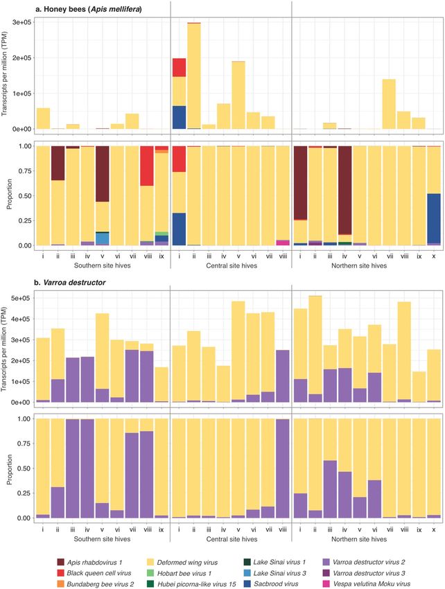
www.nature.com/scientificreports/
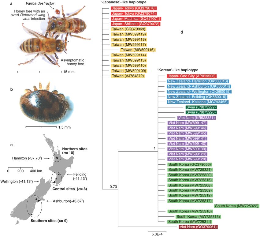
Figure 1. (a) A Western honey bee (Apis mellifera) showing an overt Deformed wing virus (DWV) infection,
with V. destructor mites, and another bee asymptomatic for this virus. (b) Varroa destructor mites were sampled
alive and typically moved quickly when disturbed. This mite was sampled from the Ashburton location. It
had slow movement and unusually dark staining, which might indicate it was old, although some pathogens
can also alter V. destructor physiology and b ehaviour37. (c) Sample sites in New Zealand showing the location
of hives from which both adult honey bees and V. destructor mites were sampled. The sites are grouped into
three regions for the statistical analysis. The arrows with latitude south coordinates show the location of the
five V. destructor samples used in the phylogenetic tree. An additional sample of nine mites showed no genetic
variation. (d) A Bayesian phylogeny showing the grouping of V. destructor mitochondrial haplotypes for 862 bp
of the cytochrome c oxidase I (MT-CO1) gene. The samples from New Zealand (coloured blue) clustered within
the Korean-like haplotype. The number between brackets is the GenBank accession number. Support for clades
correspond to posterior probabilities. Photographs by Phil Lester.
They specifically examined the infection prevalence and viral titres of seven viruses. These viruses, ordered from
most to least prevalent in honey bees (when in the presence of mites) were the Black queen cell virus (BQCV),
DWV, Sacbrood virus (SBV), Kashmir bee virus (KBV), and the Chronic bee paralysis virus (CBPV). The Acute bee
paralysis virus (ABPV) and the Israeli acute bee paralysis virus (IAPV) were not observed in New Zealand mites
or bees27. Other studies have since confirmed the absence of IAPV from New Z ealand28 and a high prevalence
of DWV and BQCV in b ees . In V. destructor, Mondet et al. found the prevalence of viruses was DWV, BQCV,
26 27
KBV, SBV, and CBPV, from most to least prevalent, respectively. Non-random patterns of viral communities
were observed in mite and bee samples, with the DWV titres in bees and V. destructor correlated. Their work
also suggested that viral communities changed either with increasing time since V. destructor arrival, or per-
haps in a latitudinal cline. Higher DWV titres were observed in northern apiaries where V. destructor had been
established the l ongest27. DWV and many other ‘honey bee’ viruses are now seen in a wide variety of arthropods
in New Z ealand29–33.
RNA-Seq technology has been used to enhance viral discovery and quantification for both honey bees and
V. destructor9,22. For example, Levin et al.34 used RNA-Seq to examine viruses in honey bees and mites in Israel.
Their analysis found eight viral species in bees. In V. destructor from the same hives, 22 viral species were found
Scientific Reports | (2022) 12:8809 | https://doi.org/10.1038/s41598-022-12888-w 2
Vol:.(1234567890)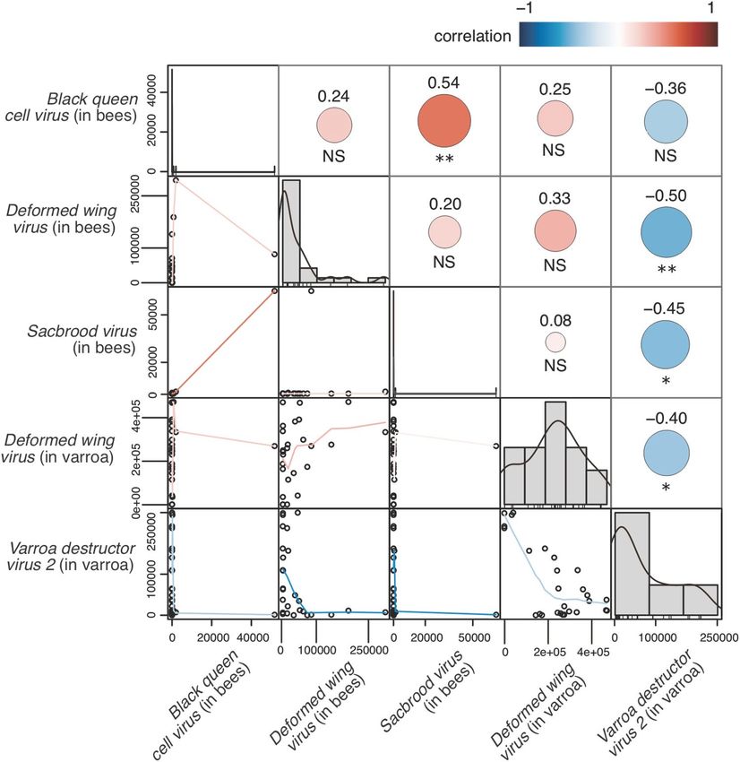
www.nature.com/scientificreports/
including two that had not been previously described. DWV-A and DWV-B (previously known as VDV-1)
dominated both communities as assessed by read abundance. The viral species newly discovered using RNA-Seq
were only found in the mites but not in bees they p arasitised34. Work on Middle East and African honey bees has
also shown a high viral diversity in V. destructor relative to their bee hosts. Other RNA-Seq studies from Asia
have similarly identified new viruses prevalent in V. destructor, sometimes demonstrating a V. destructor virus to
be present and replicating in associated honey b ees10,11. DNA viruses have similarly been recently discovered in
12,22,35
mites , some of which they may passively acquire from feeding on bees36. At least 59 putative viral species
have previously been identified from V. destructor (Supplementary Table S1). Two key conclusions can be drawn
from these studies. Firstly, several newly discovered viruses have recently been described from V. destructor and
bees, although their distribution, prevalence and effects on hosts are largely unknown. The viral communities
in V. destructor may influence the life history of the bees they parasitise6 as well as that of the mite themselves37.
Secondly, the abundance of several of these viral species in bees and mites may be correlated10,27, while other
viruses appear unique to each animal.
Our goals in this study were to: (i) confirm the presence and likely introduction of only one V. destructor
haplotype in New Zealand; (ii) assess and describe the over-wintering viral community within both V. destructor
mites and the bees that they parasitise, in order to facilitate our understanding of how the viral community in
mites might influence that of bees; and (iii) examine the viral landscape within New Zealand mites and honey
bees for regional variation.
Materials and methods
Sample collection. We collected both V. destructor and bees from 27 colonies within New Zealand dur-
ing autumn to winter (May–August) 2021. It is over the autumn–winter period that honey bee colony losses
have been linked with increased prevalence of viral infections, particularly with D WV23. We opportunistically
sampled hives from a mixture of hobbyist, commercial and research hives throughout New Zealand, which
were broadly categorised into regions or sites from the upper North Island, lower North Island and South Island
(Fig. 1c, Supplementary Table S2). Samples were taken directly from hives by the authors, or bees and mites were
sent by beekeepers to Victoria University of Wellington. Varroa destructor is listed as an ‘Unwanted Organism’ in
New Zealand under the Biosecurity Act 1993, so permission to send and handle this pest was obtained from the
Ministry for Primary Industries under sections 52 and 53 of the Biosecurity Act. A diversity of mite management
approaches had been used on the hives in the previous seasons ranging from no mite treatments, chemical pes-
ticides including amitraz and flumethrin, and organic approaches with oxalic acid. The relative density of mites
may have fluctuated considerably leading up to the point of collection and beekeepers who sent samples used a
variety of approaches in their collection. Hence, we did not record the mite control treatments used or the rela-
tive density of mites, instead focussing solely on collecting at least 10 mites and 10 bees from each hive. Pooled
samples of 5–10 individuals have been used in previous pathogen surveys from honey bee hives38–40. From two
samples, only 5 and 8 mites were able to be recovered. Live mite and bee samples were anesthetised using CO2
and placed directly into a − 80 ºC freezer or were snap-frozen in liquid nitrogen.
Mitochondrial DNA haplogroup assignment. We first sought to confirm the presence and likely intro-
duction of only one V. destructor haplotype in New Zealand. DNA was extracted from 13 individual mites, from
different hives in three locations in New Zealand to determine their mitochondrial DNA haplogroup (Fig. 1d,
Supplementary Table S3). DNA was extracted using GENEzol DNA reagent plant (Geneaid, Taiwan). Three
partial mitochondrial genes, cytochrome c oxidase I (MT-CO1), cytochrome c oxidase III (MT-CO3) and ATP
synthase membrane subunit 6 (MT-ATP6) were amplified using the primers developed by Navajas et al.41. We
performed PCR in 25 μL reactions, consisting of 2 μL mite DNA, 12.5 μL MyTaq Red Mix (Meridian Bioscience,
USA), 1 μM forward primer, 1 μM reverse primer, and nuclease-free water. Cycling conditions followed Navajas
et al.41 but with 40 cycles. A non-template control was included for each target. PCR products were visualised on
2% agarose gels and were then treated with Exo-CIP Rapid PCR Cleanup Kit (New England BioLabs, USA) prior
to sequencing. Samples were sequenced on an ABI3730 DNA Analyzer at Massey Genome Service (Palmerston
North, New Zealand). Sequence base-calls were checked by eye using Geneious42. We aligned sequences from
different mites with the Geneious alignment algorithm, using global alignment with free ends and gaps and a
cost matrix equal to 93% similarity.
To compare V. destructor mites found in New Zealand with those in other countries, we downloaded V.
destructor sequences from GenBank that overlapped with the gene portions that we sequenced. Sequences were
aligned as explained above. Clade probabilities were obtained from the posterior distribution using the MrBayes
v.3.2.6 plug-in43 for Geneious. Bayesian analyses were replicated twice, each with four Markov chains of 1 million
generations. Trees were sampled every 2,500 generations, of which the first 150,000 generations were discarded
as burn-in. Sequences have been archived in GenBank (accession numbers in Supplementary Table S3).
Viral communities in V. destructor and bees using RNA‑Seq. For each bee sample, 10 adult bees
from the same hive were pooled into a 7 mL reinforced tube with three 3.2 mm stainless steel beads (Next
Advance Inc., USA). The samples were homogenised for 3 cycles of 30 s each at 7500 rpm in a Precellys Evolu-
tion homogeniser (Bertin Instruments, France) with the dry ice on the top compartment to keep the samples
cold. We added ~ 0.5 g of the bee homogenate to a centrifuge tube containing 600 µL of cold TRIzol reagent
(Life Technologies, USA). The remainder of the extraction procedure followed the Direct-Zol RNA MiniPrep
kit protocol (Zymo Research, USA). The concentration and purity of RNA samples were determined using a
NanoPhotometer NP80 (Implen, Germany), and samples stored immediately at − 80 °C. For each pooled mite
sample, RNA was extracted as described above using the Direct-Zol RNA MicroPrep kit (Zymo Research, USA).
Scientific Reports | (2022) 12:8809 | https://doi.org/10.1038/s41598-022-12888-w 3
Vol.:(0123456789)www.nature.com/scientificreports/
Bee and mite samples were shipped dried in Gentegra RNA tubes (Gentegra, USA) via Custom Science
(Auckland, New Zealand) for 150 base pair (bp) paired-end sequencing with NovaSeq 6000 (Illumina, USA).
Raw reads were processed using Trimmomatic 0.39 to remove bases with a mean Phred score < 20 over a sliding
window of 4 bp as well as reads shorter than 25 b p44. Adapters were trimmed off using the adapter sequences
which were provided by the sequencing company, i.e. universal Illumina adapters P7 and P5. Clean read qual-
ity was checked using FASTQC 0.11.7 (Babraham Institute, UK). We aligned bee and V. destructor clean reads
using HISAT2 2.1.045 onto the honey b ee46 and V. destructor47 genomes, respectively. Files containing unmapped
reads generated by HISAT2 were used as input for de novo transcriptome assembly with Trinity 2.13.248. We
ran separate assemblies for bee and V. destructor. In order to identify viral transcripts, we used DIAMOND49
to align the resulting assembled transcripts on the viral protein NCBI database50 downloaded on 06/10/2021.
Significant alignments with an e-value < 1 × 10⁻⁵ were then re-aligned to the non-redundant protein database
using DIAMOND in order to remove false-positives and include viral transcripts only deposited in the NCBI
non-redundant protein database50 downloaded on 04/11/2021. The outputs from DIAMOND were processed
using a custom R script that selected for the hits with the best bit score, e-value and percent identity, in that order.
Final DIAMOND hits were verified using manual BLASTn51. We obtained viral transcript abundance expressed
as transcripts per million (TPM) using Salmon implemented within Trinity (align_and_estimate_abundance.pl
script)52. We further normalised TPM values to the number of host reads taken from the HISAT2 step statistics
(i.e. reads that aligned to the V. destructor or honey bee reference genomes). We only retained viruses known
to infect arthropods.
All analysis were performed within the R statistical e nvironment53. We used two separate principal compo-
nents analyses (PCA) to represent the unscaled variation in the viral loads from bees and from V. destructor,
as in our previous work32. From both PCAs, we retained only the viruses associated with the first two principal
components, which accounted for almost all (> 99.9%) of the total variance in each case. We then calculated pair-
wise Spearman correlations for the viruses retained from each PCA, examining viral loads between and within
bees and V. destructor. In order to investigate the associations between DWV in bees and VDV-2 plus DWV in
V. destructor, we also investigated pairwise linear regressions. We tested for differences in viral communities
between regions in bees and V. destructor, using PERMANOVA from the adonis function in the vegan package
with both the Bray–Curtis (abundance) and Jaccard (presence/absence) methods54.
PCR and replication assays for selected RNA viruses. We further interrogated our samples for the
presence of DWV-A, DWV-B (previously described as VDV-1), BQCV, KBV, IAPV, VDV-2, VDV-3 and VDV-
5, using reverse-transcription (RT)-PCR. Sample RNA was combined resulting in one bee RNA pool and one
V. destructor RNA pool for each region, which were then used to generate cDNA using Quanta qScript cDNA
SuperMix (Quantabio, USA) following the manufacturer’s instructions. Equal amounts of cDNA from each
region for V. destructor and bee samples were then pooled to create one V. destructor cDNA master mix and one
bee cDNA master mix that was used for PCR assays. The samples were pooled specifically because we wanted
only to provide evidence on whether viral replication was occurring in mites or in bees (rather than determining,
for example, how frequently replication was occurring in each species). PCR was then carried out using primer
sets described in Supplementary Table S4. PCR reactions consisted of 7.5 uL MyTaq Red Mix, 1 µL of 10 µM
forward primer, 1 µL of 10 µM reverse primer, 1 µL cDNA, made to 15 µL with nuclease-free water. PCR cycling
conditions were: 95 °C for 5 min; 35 cycles of 95 °C for 15 s, 58 °C for 30 s, 72 °C for 60 s, with a final extension
at 72 °C for 5 min and a hold step at 4 °C. PCR products were separated using 2% agarose gel electrophoresis and
visualized using SYBR Safe DNA gel stain (Invitrogen/ThermoFisher Scientific, USA). To prepare PCR products
for Sanger sequencing, samples were digested with ExoSAP-IT PCR Product Cleanup Reagent (Applied Biosys-
tems/ThermoFisher Scientific, USA). Samples were then sent to Massey Genome Service (Palmerston North,
New Zealand) for sequencing. Geneious was used to analyze sequences42.
For positive-sense single-stranded RNA viruses, the presence of the negative-sense strands is indicative of
viral replication. Strand-specific RT-PCR assays were used to detect the negative-sense viral strands for DWV-A,
DWV-B, BQCV, KBV, IAPV, VDV-2, VDV-3 and VDV-5. Tagged-forward primers (Supplementary Table S4)
were used to generate cDNA from the negative-strand using the Super-Script IV First-Strand Synthesis System
(Invitrogen/ThermoFisher Scientific, USA). PCR was then conducted as above.
DNA virus confirmation assays. We also examined our samples for the Apis mellifera filamentous virus
(AmFV), which is a large double stranded DNA virus from honey bees. DNA was extracted from pools of 10
mites, and separately 10 bees, taken from hives within three regions: the upper North Island, lower North Island
and South Island (n = 3, Fig. 1c, Supplementary Table S5). Briefly, each 10-mite or 10-bee sample was homog-
enized by bead-beating in microcentrifuge tubes containing 1 mL of GENEzol plant DNA reagent (Geneaid
Biotech, Taiwan) and 5 µL of β-mercaptoethanol (Sigma Aldrich, USA). Samples were homogenised for 3 cycles
of 15 s each at 6,000 rpm, followed by 1 cycle of 10 s at 9,900 rpm. We used a 24:1 chloroform–isoamyl alcohol
mixture (BioUltra from Merck, USA) to isolate the nucleic acids, followed by isopropanol precipitation (BioRea-
gent from Merck, USA), and a 70% ethanol purification step (VWR Chemicals, UK). Mite DNA was eluted in 25
µL and bee DNA in 100 µL of nuclease-free water.
We screened these pooled mite and bee samples respectively for AmFV via PCR. We used the primer sets
designed by Cornman et al.35 to amplify two portions of the pathogen genome35: a ribonucleotide reductase
small subunit gene and a thymidylate synthase gene. We amplified each locus in a 15 μL PCR containing 1 μL
DNA, 1 μM forward primer, 1 μM reverse primer, water and 7.5 μL MyTaq Red Mix. Cycling conditions were:
1 min at 95 °C; 35 cycles of 15 s at 95 °C, 15 s at 54 °C, and 10 s at 72 °C; followed by a final extension of 5 min
at 72 °C. We included a non-template control in each reaction. PCR products were visualised on a 2% agarose
Scientific Reports | (2022) 12:8809 | https://doi.org/10.1038/s41598-022-12888-w 4
Vol:.(1234567890)www.nature.com/scientificreports/
Honey bees Varroa destructor
Virus Average S.E Range Infected (%) Average S.E Range Infected (%)
Deformed wing virus
40,737 13,183 (76– 294,978) 100 238,459 27,545 (1265– 472,301) 100
(DWV-A)
Sacbrood virus (SBV) 3030 2937 (0.05– 64,693) 81 31 15 (0.07– 265) 74
Black queen cell virus
2188 2066 (0.04– 51,721) 93 12 7 (0.02– 75) 48
(BQCV)
Lake Sinai virus 3 (LSV-3) 175 – – 4 – – – –
Apis rhabdovirus 1
150 54 (0.02– 941) 93 44 22 (0.42– 592) 100
(ARV-1)
Kashmir bee virus (KBV) 92 75 (0.03– 1333) 67 37 21 (0.30– 174) 33
Lake Sinai virus 1 (LSV-1) 34 23 (0.02– 185) 30 – – – –
Drosophila subobscura
14 – – 4 – – – –
Nora virus
Chronic bee paralysis virus
13 13 (0.04– 40) 11 – – – –
(CBPV)
Varroa destructor virus 2
11 2 (0.06– 30) 96 81,853 17,255 (1,295– 251,109) 100
(VDV-2)
Bundaberg bee virus 2 9 2 (7– 10) 7 3 2 (0.46– 6) 11
Varroa destructor virus 5
6 2 (4– 10) 15 124 29 (1– 551) 100
(VDV-5)
Hobart bee virus 1 5 4 (0.23– 12) 11 – – – –
Vespa velutina Moku virus 4 3 (1– 16) 19 – – – –
Aphid lethal paralysis virus
4 3 (1– 16) 19 – – – –
(ALPV)
Hubei picorna-like virus
4 3 (0.17– 28) 37 – – – –
15
Apis rhabdovirus 2
1 0.46 (0.04– 4) 33 34 8 (1– 174) 100
(ARV-2)
Hubei picorna-like virus
– – – – 9 7 (0.09– 37) 19
22
Table 1. Viruses observed in honey bees and mites from the RNA-Seq analysis. The counts are expressed as
the average transcript abundance, expressed in transcripts per million (TPM). The viruses are ordered by the
most to least common virus observed in honey bees. ‘S.E.’ is the standard error; ‘Infected’ is the percentage
of the 27 hives from which the mite or bee samples had reads for each virus. ‘−’ indicates no virus reads were
found in a sample. For the average, standard error, and range calculations we excluded the 0 values.
gel. We cleaned up the samples using the Monarch DNA Gel Extraction Kit (New England BioLabs, USA). The
identities of the products were confirmed by Sanger sequencing (GenBank accession numbers in Table S5).
Sequence base-calls were checked by eye using Geneious42. We used the BLASTn algorithm to search against the
nucleotide database in NCBI GenBank. Gene identifications were assigned to genes on the database based on
sequence identity > 97%. We aligned the sequences with the Geneious alignment algorithm.
Results
Mitochondrial DNA haplogroup assignment. Our first goal was to confirm the presence and likely
introduction of only one V. destructor haplotype in New Zealand. We sequenced a total of 1232 bp from three
mitochondrial gene regions of V. destructor taken from 13 spatially separated hives (Fig. 1c, Supplementary
Table S3). There was no variation between any of these sequences (Fig. 1d). Further, our samples showed no
variation from one 811 bp sequence from two New Zealand V. destructor samples that were deposited on Gen-
Bank sampled in 2016 from Kaikohe and Gisborne55. This single New Zealand haplotype is most similar to the
Korean-like haplotype of V. destructor56. Because of the absence of any genetic variation within the New Zealand
haplotypes, it seems likely that there has been only one successful V. destructor introduction and establishment
here. We note, however, that it would be impossible to prove this conclusion without widespread and extensive
sampling throughout the country. Mitochondrial DNA sequences of similar length from other countries show
variation indicative of multiple haplotype introductions (Fig. 1d).
Viral communities in bees and V. destructor using RNA‑Seq. We found strong evidence for 17
viruses in our honey bee samples (Table 1; Fig. 2a; Supplementary Table S6). Each individual hive contained
multiple RNA viral species infections within the inhabiting bees. The median number of RNA viruses observed
from a bee sample within a hive was 7 (range: 4–11 across all 27 hives). DWV-A was found in all 27 bee samples
with viral loads representing between 7.1 and > 99.9% of the total viral load in our samples. We found SBV in 22
samples (< 0.01–49.7% of total viral loads), BQCV in 25 samples (< 0.01–39.9% of total viral loads), and VDV-2
in 26 samples accounting for < 0.01–3.9% of total viral loads. Apis rhabdovirus 1 was present in 25 samples, typi-
cally in small numbers of transcripts but represented more than a third of total viral loads in four samples (over-
Scientific Reports | (2022) 12:8809 | https://doi.org/10.1038/s41598-022-12888-w 5
Vol.:(0123456789)www.nature.com/scientificreports/
Figure 2. Viruses in honey bees and V. destructor mites. (a) DWV-A was the most abundant and prevalent in
honey bees, occurring in all bee samples from all 27 hives that we sampled, although with substantial variation
between samples. A total of 17 virus species were seen in these bee samples. (b) In comparison to the bees,
the viral loads in mites showed much less variation. The viral community of V. destructor was dominated by
DWV-A and VDV-2. Eight other viruses were observed in relatively low abundance from these mites.
Scientific Reports | (2022) 12:8809 | https://doi.org/10.1038/s41598-022-12888-w 6
Vol:.(1234567890)www.nature.com/scientificreports/
all < 0.01–88.6% of the total viral loads). Apis rhabdovirus 2 was found in nine samples but at low levels (up to
0.02% of total viral loads). We observed Vespa velutina Moku virus in five samples (< 0.01–4.6% total viral loads),
VDV-5 in four samples (< 0.01–2.9% of total viral loads) and Hobart bee virus in three samples (< 0.01–3.7%
total viral loads). Lake Sinai virus 1 was found in eight samples (< 0.01–1.5% of total viral loads), and Lake Sinai
virus 3 was only found in one sample, but in this sample accounted for 10.5% of the total viral load. Other less
abundant viruses were only found at < 1% of total viral loads in samples where they were present. For example,
KBV was found in 18 samples but only accounted for ≤ 0.7% of viral loads. Aphid lethal paralysis virus was only
found in five North and Central region samples (< 0.01–0.4% of total viral loads). CBPV was found in three sam-
ples (0.01–0.3% of total viral loads). There was evidence for additional viruses present in our sequences, though
we excluded these for the purpose of our analyses due to their low sequence identity or high e-value scores. In
our PERMANOVA we found no significant differences in bee viral communities between regions, using either
abundance data (F = 1.899, R2 = 0.137, p = 0.160) or presence/absence data (F = 0.696, R2 = 0.124, p = 0.154).
We found 10 viruses in V. destructor, including nine shared with honey bees. Each individual hive contained
multiple RNA viral species infections within the inhabiting mites. The median number of RNA viruses observed
from a V. destructor sample within a hive was 7 (range: 5–10 across all 27 hives). DWV-A and VDV-2 were
found in all samples, accounting for 0.6–99.2% and 0.8–99.3% of total viral loads, respectively (Table 1; Fig. 2b;
Supplementary Table S7). These two viruses accounted for > 99.9% of the observed average viral loads in these
mites (Table 1). All the other viruses were observed at low viral loads, with each accounting for < 0.04% of the
average number of viral loads observed. ARV-1, ARV-2 and VDV-5 were found in all samples, but only accounted
for ≤ 0.3% of any sample total viral loads. SBV, BQCV and KBV were found in 20, 13 and nine samples, respec-
tively. Hubei picorna-like virus 22 was present in five samples (≤ 0.008% of total viral loads) and Bundaberg bee
virus 2 in three samples (≤ 0.002% of total viral loads). We found no significant differences in V. destructor viral
communities between regions, using either abundance data (F = 1.257, R2 = 0.095, p = 0.254) or presence/absence
data (F = 0.988, R2 = 0.076, p = 0.434).
We found no evidence in the RNA-Seq data for the viruses DWV-B, IAPV, and VDV-3. Only the DWV-A
strain was detected.
Principal component analyses and association between viruses. In bees, DWV-A was strongly
associated with the first principal component ( PC1bees, Supplementary Table S8), which explained 95.0% of the
total variance in the dataset. The second principal component ( PC2bees) was associated predominantly with SBV
and BQCV. Together the first two principal components explained more than 99.99% of the total variance. In the
V. destructor analysis, DWV and VDV-2 were effectively the only viruses associated with the first two principal
components, which together explained 99.9% of the total variance (Supplementary Table S9).
Our pairwise comparisons focused on the viruses identified as key contributors in the PCAs, since we were
interested only in associations between the highly abundant viruses. We therefore selected DWV-A, SBV and
BQCV for further scrutiny in bees, plus DWV-A and VDV-2 in V. destructor. Specifically, in the analysis of
individual hive replicates, where there were high levels of VDV-2 in mites, reduced DWV-A occurred in both the
mites (Spearman’s correlation: rs = − 0.404, p = 0.037; linear regression: β = − 1.145, r2 = 0.515, p < 0.001; Fig. 3, Sup-
plementary Fig. S1) and the bees co-occurring within the same hive (rs = − 0.498, p = 0.009; β = − 0.301, r2 = 0.155,
p = 0.042; Fig. 3, Supplementary Fig. S1). Where there were high loads of DWV-A in mites, there were typically
high loads in bees although the relationship was not statistically significant at a conventional 5% level (rs = 0.334,
p = 0.088; β = 0.163, r2 = 0.117, p = 0.081; Fig. 3, Supplementary Fig. S1). Strong correlations were observed for
other pairs of viruses, including SBV and BQCV (Fig. 3), although given the extraordinarily low abundances of
these viruses the associations are not likely to be biologically relevant.
PCR and replication assays for selected RNA viruses. The presence of DWV-A, KBV and BQCV
in the pooled bee and V. destructor samples was confirmed by RT-PCR, while VDV-2 and VDV-5 were only
detected using RT-PCR in the pooled V. destructor sample (Table 2). The negative-sense, replicative strands of
DWV-A and KBV were detected in both the pooled bee and V. destructor samples. We detected the negative-
sense strand of VDV-2 and VDV-5 only in the pooled V. destructor samples. The negative-strand of BQCV was
not detected in any of the samples using two different sets of tagged primers (Supplementary Table S4). IAPV
and VDV-3 were not detected.
We used two sets of primers to examine for the presence of DWV-B/VDV-1. Firstly, specific primers (labelled
DWV-B_L_F and DWV-B_L_R; Bradford et al.57) successfully amplified a 357 bp fragment in bee and V. destruc-
tor samples, however, a BLAST analysis of the sequenced PCR products revealed these to best match DWV-A.
Testing these DWV-B/VDV-1 primers on the DWV-A reference genome (AY292384.1) in Geneious software
showed that the DWV-B/VDV-1 forward primer could bind to DWV-A. In a second analysis, different DWV-B/
VDV-1 primers (labelled DWV-B_F and DWV-B_R57), which targeted a shorter 116 bp region within the 357 bp
region, were also tested against the DWV-A reference genome (AY292384.1) in Geneious, yet did not bind to
any region. Likewise, PCR screening of V. destructor and bee samples using this second set of DWV-B/VDV-1
primers (DWV-B_F and DWV-B_R) failed to amplify any fragments in the samples.
DNA virus confirmation assays. AmFV was found in the three pooled regional V. destructor samples
and the three pooled regional bee samples (Supplementary Table S5). The two genes amplified in this analysis
matched AmFV sequences in GenBank with > 97% identity. This DNA virus therefore appears to be present
throughout New Zealand.
Scientific Reports | (2022) 12:8809 | https://doi.org/10.1038/s41598-022-12888-w 7
Vol.:(0123456789)www.nature.com/scientificreports/
Figure 3. Pairwise correlations between virus loads in honey bees and V. destructor. The V. destructor and
bee samples were paired, taken from the same hive. The size of the circles is representative of the Spearman
correlation coefficient, which is shown above each circle with statistical significance indicated below. Only
viruses that were identified in the PCA as important in explaining the variation in the communities were used in
the analysis. * p < 0.05; ** p < 0.01. The counts are in transcripts per million.
RT-PCR detection Negative-strand detection
Virus or virus strain Honey bee V. destructor Honey bee V. destructor
DWV-A + + + +
DWV-B (VDV-1) − − − −
BQCV + + − −
KBV + + + +
IAPV − − − −
VDV-2 − + − +
VDV-3 − − − −
VDV-5 − + − +
Table 2. RT-PCR and replication assay results for viruses in honey bees and V. destructor. Pooled V. destructor
and honey bee samples were screened for eight viruses (DWV-A, DWV-B (VDV-1), BQCV, KBV, IAPV,
VDV-2, VDV-3 and VDV-5). Positive test results are indicated with ( +) while (−) indicates that no band was
observed. A positive result for the negative-strand detection assay is indicative that the virus is parasitising the
host cells. Primer pairs used in this analysis are shown in Supplementary Table S4.
Scientific Reports | (2022) 12:8809 | https://doi.org/10.1038/s41598-022-12888-w 8
Vol:.(1234567890)www.nature.com/scientificreports/
Discussion
Our results indicate that it is likely that there has been only one introduction of V. destructor into New Zealand.
An analysis of the mitochondrial gene cytochrome c oxidase I (MT-CO1) from six mites sampled from Auck-
land soon after the mites discovery in the year 2 00025 indicated the presence of the Korean-like haplotype of V.
destructor56. Unfortunately, these samples are not available for sequence comparison. One sequence from one
mite was available on GenBank from a collection in 2016 in Kaikohe, in the upper North Island of New Zealand,
that consisted of a 811 bp fragment of MT-CO155. In the same study an additional mite was sampled from Gis-
borne (approximately 520 km from Kaikohe), which shared the exact same haplotype sequence. These samples
also shared identical MT-CO1 sequences to our samples. While we cannot rule out the possibility of multiple
introductions from a genetically similar source, only one invasion event for New Zealand seems likely given the
higher genetic variation in V. destructor populations observed in other countries where multiple introductions
have occurred (Fig. 1). Elsewhere, substantial genetic diversity in V. destructor populations has been found even
within individual h ives58. A high level of genetic diversity in countries such as Argentina has been hypothesised
to allow spatial genetic differentiation of V. destructor populations due to different mite genotypes adapting to
different climatic c onditions59. A single introduction event to New Zealand will probably have limited the ability
of V. destructor to adapt or evolve to environmental conditions, or potentially to evolve pesticide resistance. A
single introduction event will also have limited the viral diversity and community introduced with this parasite.
DWV was likely to have been absent from New Zealand prior to the introduction of V. destructor in 200027.
All colonies that we sampled were positive for DWV-A in both V. destructor and bees, which was much higher
than the 50% and 90% prevalence in a previous PCR-based study in New Zealand in 2 01427. It is now typically
the predominant virus present in both bees and mites. Mites were found to carry DWV-A loads that were on
average 5.85-fold higher than in bees (as measured in transcripts per million reads). The RNA virome of several
of our mites and bees samples had > 99% of the observed virus reads as DWV-A for both mites and bees, which
is consistent with other s tudies10,34,60. Five of the 27 mite samples, however, had low numbers of DWV-A reads
representing < 15% of the observed virome. In another study from Germany, many but not all mites in a popula-
tion appear to carry DWV i nfections61. How and why some populations had such low DWV-A reads is unknown
and worthy of more study. Our assays and other studies62,63 have found that DWV-A appears to replicate in both
mites and bees, in contrast to more recent work where authors have concluded that this virus seems unable to
replicate in mites61,64. We note the debate around using negative strand assays for DWV replication and that other
approaches may be more definitive, as PCR approaches could give false-positive results after mites acquire nega-
tive strands from bees via their f eeding61. We also observed no evidence of DWV-B (originally named VDV-1)
from either the RNA-Seq or PCR analyses. DWV-B has become more prevalent and more virulent to honey bees
in several countries1,20,21. The lack of this DWV-B is good news for beekeepers and may be due to the apparent
single V. destructor introduction from a period prior to DWV-B becoming globally widespread, in addition to
New Zealand’s complete ban on honey bee or honey bee product imports. Importing live bees continues to be a
key contributing factor in emerging bee disease and colony loss elsewhere65,66.
The viral community in our bees from New Zealand was associated with that in V. destructor, which was
previously observed in New Zealand bee and V. destructor communities27, but unlike in other studies such as
Roberts et al.67 who analysed viruses in bees and V. jacobsoni in Papua New Guinea. The viruses observed in
the bees examined by Roberts et al.67 formed a near completely different and distinct community compared to
the mites that were parasitising the same hives. Bees in this region appear to tolerate mite infestations, which
was attributed to a lack of DWV in mites and b ees67. Similarly, honey bees on the remote island of Fernando
de Noronha (Brazil) also tolerate V. destructor infestations, possibly because of extremely low DWV levels68.
DWV is known to have an immunosuppressive effect that serves to enhance the reproduction and fitness of the
parasitic mite6. Perhaps due to these immunosuppressive effects when DWV is prevalent, viral communities in
bees and V. destructor become substantially more similar. The introduction of V. destructor and its associated
DWV strains can alter the viral landscape of b ees17 and even their p redators18. These studies suggest that DWV
plays a central role in not only influencing bee health but also the entire viral communities in V. destructor, bee
hosts, and other insects.
Our analysis showed that after DWV-A, BQCV and SBV contributed most to the variance observed in the
viral communities in honey bees. All but two of our 27 honey bee samples were positive for BQCV, and 22 were
positive for SBV. Both of these viruses were found in the V. destructor samples, but at substantially lower loads.
These observed infection rates were consistent with previous work in New Z ealand27. There is no evidence that
V. destructor can vector either SBV or B 1
QCV , but there is evidence that SBV can modify mite behaviour after
it becomes infected as a result of mite feeding37. Of note for our samples was the near absence of CBPV, which
was present at extremely low levels in only three of the 27 honey bee samples and absent from mites. CBPV was
present in 20–40% of samples from one previous NZ s tudy27, but a more recent analysis showed apiaries have
infection rates < 20%26. This virus has been identified as a major emerging threat to honey bees elsewhere65.
VDV-2 and VDV-5 were found in both bees and V. destructor in our RNA-Seq analysis. The read counts
were on average 7,441-fold and 21-fold higher in V. destructor than in bees, respectively. Other researchers have
observed similar results to ours, with both present but without evidence of replication in bees11,69. Their pres-
ence in bees seems to be related to feeding by the mite3 wherein they acquire a small number of viral particles.
Such a small number of particles probably explains why we observed both VDV-2 and VDV-5 in bees using the
RNA-Seq analysis, but not in PCR approaches. Our assembled contigs for VDV-2 and VDV-5 showed 85–92%
nucleotide identity compared to published genome sequences. This suggests that the sequences are clearly vari-
ants of VDV-2 and VDV-5. There was, however, considerable strain diversity, particularly in the VDV-2 strains.
This level of diversity was surprising given the probable single introduction of V. destructor to New Zealand and
is currently under further investigation.
Scientific Reports | (2022) 12:8809 | https://doi.org/10.1038/s41598-022-12888-w 9
Vol.:(0123456789)www.nature.com/scientificreports/
Seventeen RNA viruses were observed in honey bees and 10 in V. destructor. These viruses represent a subset
of the 87 and 59 viruses that have previously been described to infect honey b ees9 and V. destructor (Supple-
mentary Table S1), respectively. Several viruses, including IAPV, were not found in our analyses and have been
concluded as absent from New Zealand in previous studies that have used PCR-based a ssays26–28. Many of the
viruses that we observed in honey bees were prevalent in only a few samples or were in low abundance. KBV was
present in 66% of bee samples and 33% of mites, typically in extremely low relative abundance. These KBV preva-
lence rates are similar to other work on New Zealand honey bees26,27. Mondet et al.27 observed that KBV peaked
in abundance 2 years after V. destructor invasion but appeared to then disappear from colonies entirely and be
replaced by DWV. Our results with extremely low titre of KBV are consistent with this pattern. In contrast, our
work on KBV in invasive wasps from New Zealand demonstrated infection in every single individual from every
nest we examined, incurring fitness costs for the w asps70. We also found three Lake Sinai virus (LSV) strains, but
only low relative abundance in honey bees. Other researchers have described LSV from V. destructor71. In addi-
tion to these RNA viruses, the large double stranded DNA virus of honey bees, Apis mellifera filamentous virus
(AmFV), was observed in mites and bees. This virus may be ubiquitous in honey bees72. It has been described
as weakly pathogenic to honey bees, but it can alter bee behaviour and physiology when in high a bundance73.
AmFV has previously been observed in other bee species73 and V. destructor mites36.
Our RNA-Seq approach tentatively indicated a diversity of other viruses or viral strains. We excluded these
species from the analysis presented here due to low confidence in their similarity to known viruses. More work
is needed to define and describe many of these viral species that were typically rare and in extremely low rela-
tive abundance. We strongly suspect there were more than just 10 virus species present in V. destructor and in
the honey bees, especially as at least 59 putative viruses have previously been described from this invasive mite
(Supplementary Table S1). Our RNA-Seq data can be explored further with the potential to describe new viral
species, strains, and host associations. It is also possible that additional sampling within New Zealand would
identify additional viruses, or even that additional sampling might identify further haplotypes of V. destructor.
Our results also suggest a relationship between interspecific viral interactions within mites, which influences
those interactions in bees. Specifically, where we observed high levels of VDV-2 in mites, reduced DWV-A
occurred in both the mites and the bees co-occurring within the same hive. Where there were high loads of
DWV-A in mites, there were typically high loads in bees. Perhaps as both VDV-2 and DWV-A belong to the
Iflaviridae viral family, they may compete for similar resources within host cells. There is evidence of competi-
tion between viruses in honey bees from other studies74, with this virus-virus competition potentially mediat-
ing colony c ollapse75. Other work has found that the DWV load in mites is dynamic and can rapidly increase
or decrease depending on the DWV load of the honey bees upon which they are feeding64. Overt infections of
DWV-A might only occur after the viral titre has exceeded a certain threshold62, with VDV-2 restraining DWV-A
infections. We do note, however, that such a relationship between viruses such as DWV and VDV-2 was not
observed in V. destructor examined in Herrero et al.69. Experimental approaches are needed to better understand
how the viral community in V. destructor and honey bees interact.
We found no evidence of different viral communities occurring in mites or bees in the different regions of
New Zealand. Mondet et al.27 suggested that a ‘dynamic and turbulent pathological landscape’ forms that settles
into a more stable and predictable pattern 2- 3 years after V. destructor invasion. Our results are consistent with
their hypothesis. We did, however, still observe high levels of variation within regions. Understanding the mecha-
nisms for this variation may help beekeepers cope with what appears to be a worsening V. destructor problem.
Data availability
Clean reads from which we assembled and quantified viral transcripts can be accessed from the NCBI SRA
repository at http://www.ncbi.nlm.nih.gov/bioproject/820512 under BioProject PRJNA820512.
Received: 10 December 2021; Accepted: 11 May 2022
References
1. Traynor, K. S. et al. Varroa destructor: A complex parasite, crippling honey bees worldwide. Trends Parasitol. 36, 592–606. https://
doi.org/10.1016/j.pt.2020.04.004 (2020).
2. Rosenkranz, P., Aumeier, P. & Ziegelmann, B. Biology and control of Varroa destructor. J. Invertebr. Pathol. 103, S96–S119. https://
doi.org/10.1016/j.jip.2009.07.016 (2010).
3. Noel, A., Le Conte, Y. & Mondet, F. Varroa destructor: how does it harm Apis mellifera honey bees and what can be done about it?.
Emerg. Top. Life Sci. 4, 45–57. https://doi.org/10.1042/ETLS20190125 (2020).
4. Boncristiani, H. et al. World honey bee health: the global distribution of western honey bee (Apis mellifera L.) pests and pathogens.
Bee World 98, 2–6 (2020).
5. Ramsey, S. D. et al. Varroa destructor feeds primarily on honey bee fat body tissue and not hemolymph. Proc. Natl. Acad. Sci. U.S.A.
116, 1792–1801. https://doi.org/10.1073/pnas.1818371116 (2019).
6. Di Prisco, G. et al. A mutualistic symbiosis between a parasitic mite and a pathogenic virus undermines honey bee immunity and
health. Proc. Natl. Acad. Sci. USA 113, 3203–3208. https://doi.org/10.1073/pnas.1523515113 (2016).
7. Mondet, F. et al. Antennae hold a key to Varroa-sensitive hygiene behaviour in honey bees. Sci. Rep. 5, 10454. https://doi.org/10.
1038/srep10454 (2015).
8. McMenamin, A. J. & Genersch, E. Honey bee colony losses and associated viruses. Curr. Opin. Insect Sci. 8, 121–129. https://doi.
org/10.1016/j.cois.2015.01.015 (2015).
9. Beaurepaire, A. et al. Diversity and global distribution of viruses of the western honey bee Apis mellifera. Insects 11, 239. https://
doi.org/10.3390/insects11040239 (2020).
10. Levin, S. et al. New viruses from the ectoparasite mite Varroa destructor infesting Apis mellifera and Apis cerana. Viruses 11, 94.
https://doi.org/10.3390/v11020094 (2019).
11. Chen, G. et al. A new strain of virus discovered in China specific to the parasitic mite Varroa destructor poses a potential threat
to honey bees. Viruses 13, 679. https://doi.org/10.3390/v13040679 (2021).
Scientific Reports | (2022) 12:8809 | https://doi.org/10.1038/s41598-022-12888-w 10
Vol:.(1234567890)www.nature.com/scientificreports/
12. Kraberger, S. et al. Genome sequences of two single-stranded DNA viruses identified in Varroa destructor. Genome Announc. 6,
e00107-00118. https://doi.org/10.1128/genomeA.00107-18 (2018).
13. Haddad, N., Horth, L., Al-Shagour, B., Adjlane, N. & Loucif-Ayad, W. Next-generation sequence data demonstrate several patho-
genic bee viruses in Middle East and African honey bee subspecies (Apis mellifera syriaca, Apis mellifera intermissa) as well as their
cohabiting pathogenic mites (Varroa destructor). Virus Genes 54, 694–705. https://doi.org/10.1007/s11262-018-1593-9 (2018).
14. Wilfert, L. et al. Deformed wing virus is a recent global epidemic in honeybees driven by Varroa mites. Science 351, 594–597.
https://doi.org/10.1126/science.aac9976 (2016).
15. Remnant, E. J., Mather, N., Gillard, T. L., Yagound, B. & Beekman, M. Direct transmission by injection affects competition among
RNA viruses in honeybees. Proc. Royal Soc. B 286, 20182452. https://doi.org/10.1098/rspb.2018.2452 (2019).
16. Martin, S. J. & Brettell, L. E. Deformed wing virus in honeybees and other insects. Ann. Rev. Virol. 6, 49–69. https://doi.org/10.
1146/annurev-virology-092818-015700 (2019).
17. Martin, S. J. et al. Global honey bee viral landscape altered by a parasitic mite. Science 336, 1304–1306. https://doi.org/10.1126/
science.1220941 (2012).
18. Loope, K. J., Baty, J. W., Lester, P. J. & Wilson Rankin, E. E. Pathogen shifts in a honeybee predator following the arrival of the
Varroa mite. Proc. Royal Soc. B 286, 20182499. https://doi.org/10.1098/rspb.2018.2499 (2019).
19. McMahon, D. P. et al. Elevated virulence of an emerging viral genotype as a driver of honeybee loss. Proc. Royal Soc. B https://doi.
org/10.1098/rspb.2016.0811 (2016).
20. Grindrod, I., Kevill, J. L., Villalobos, E. M., Schroeder, D. C. & Ten Martin, S. J. years of Deformed wing virus (DWV) in Hawaiian
honey bees (Apis mellifera), the dominant DWV-A variant is potentially being replaced by variants with a DWV-B coding sequence.
Viruses 13, 969. https://doi.org/10.3390/v13060969 (2021).
21. Kevill, J. L., Stainton, K. C., Schroeder, D. C. & Martin, S. J. Deformed wing virus variant shift from 2010 to 2016 in managed and
feral UK honey bee colonies. Arch. Virol. 166, 2693–2702. https://doi.org/10.1007/s00705-021-05162-3 (2021).
22. Grozinger, C. M. & Flenniken, M. L. Bee viruses: Ecology, pathogenicity, and impacts. Annu. Rev. Entomol. 64, 205–226. https://
doi.org/10.1146/annurev-ento-011118-111942 (2019).
23. Natsopoulou, M. E. et al. The virulent, emerging genotype B of Deformed wing virus is closely linked to overwinter honeybee
worker loss. Sci. Rep. 7, 5242. https://doi.org/10.1038/s41598-017-05596-3 (2017).
24. Iwasaki, J. M., Barratt, B. I., Lord, J. M., Mercer, A. R. & Dickinson, K. J. The New Zealand experience of varroa invasion highlights
research opportunities for Australia. Ambio 44, 694–704. https://doi.org/10.1007/s13280-015-0679-z (2015).
25. Solignac, M. et al. The invasive Korea and Japan types of Varroa destructor, ectoparasitic mites of the Western honeybee (Apis
mellifera), are two partly isolated clones. Proc. Royal Soc. B 272, 411–419. https://doi.org/10.1098/rspb.2004.2853 (2005).
26. Hall, R. J. et al. Apicultural practice and disease prevalence in Apis mellifera, New Zealand: A longitudinal study. J. Apic. Res. 60,
644–658. https://doi.org/10.1080/00218839.2021.1936422 (2021).
27. Mondet, F., de Miranda, J. R., Kretzschmar, A., Le Conte, Y. & Mercer, A. R. On the front line: quantitative virus dynamics in
honeybee (Apis mellifera L) colonies along a new expansion front of the parasite Varroa destructor. PLoS Pathog. 10, e1004323
(2014).
28. McFadden, A. M. J. et al. Israeli acute paralysis virus not detected in Apis mellifera in New Zealand in a national survey. J. Apic.
Res. 53, 520–527. https://doi.org/10.3896/ibra.1.53.5.03 (2015).
29. Dobelmann, J., Felden, A. & Lester, P. J. Genetic strain diversity of multi-host RNA viruses that infect a wide range of pollinators
and associates is shaped by geographic origins. Viruses 12, 358. https://doi.org/10.3390/v12030358 (2020).
30. Gruber, M. A. M. et al. Single-stranded RNA viruses infecting the invasive argentine ant Linepithema humile. Sci. Rep. 7, 3304.
https://doi.org/10.1038/s41598-017-03508-z (2017).
31. Brenton-Rule, E. C. et al. The origins of global invasions of the German wasp (Vespula germanica) and its infection with four honey
bee viruses. Biol. Invasions 20, 3445–3460. https://doi.org/10.1007/s10530-018-1786-0 (2018).
32. Lester, P. J., Buick, K. H., Baty, J. W., Felden, A. & Haywood, J. Different bacterial and viral pathogens trigger distinct immune
responses in a globally invasive ant. Sci. Rep. 9, 5780. https://doi.org/10.1038/s41598-019-41843-5 (2019).
33. Lester, P. J. et al. No evidence of enemy release in pathogen and microbial communities of common wasps (Vespula vulgaris) in
their native and introduced range. PLoS ONE 10, e0121358. https://doi.org/10.1371/journal.pone.0121358 (2015).
34. Levin, S., Sela, N. & Chejanovsky, N. Two novel viruses associated with the Apis mellifera pathogenic mite Varroa destructor. Sci.
Rep. 6, 37710. https://doi.org/10.1038/srep37710 (2016).
35. Cornman, S. R. et al. Genomic survey of the ectoparasitic mite Varroa destructor, a major pest of the honey bee Apis mellifera.
BMC Genom. 11, 602. https://doi.org/10.1186/1471-2164-11-602 (2010).
36. Gauthier, L. et al. The Apis mellifera filamentous virus genome. Viruses 7, 3798–3815. https://doi.org/10.3390/v7072798 (2015).
37. Giuffre, C., Lubkin, S. R. & Tarpy, D. R. Does viral load alter behavior of the bee parasite Varroa destructor?. PLoS ONE 14,
e0217975. https://doi.org/10.1371/journal.pone.0217975 (2019).
38. De Smet, L. et al. BeeDoctor, a versatile MLPA-based diagnostic tool for screening bee viruses. PLoS ONE 7, e47953. https://doi.
org/10.1371/journal.pone.0047953 (2012).
39. Runckel, C. et al. Temporal analysis of the honey bee microbiome reveals four novel viruses and seasonal prevalence of known
viruses, Nosema, and Crithidia. PLoS ONE 6, e20656. https://doi.org/10.1371/journal.pone.0020656 (2011).
40. Remnant, E. J. et al. A diverse range of novel RNA viruses in geographically distinct honey bee populations. J. Virol. 91, e00158.
https://doi.org/10.1128/JVI.00158-17 (2017).
41. Navajas, M. et al. New Asian types of Varroa destructor: a potential new threat for world apiculture. Apidologie 41, 181–193. https://
doi.org/10.1051/apido/2009068 (2010).
42. Kearse, M. et al. Geneious basic: an integrated and extendable desktop software platform for the organization and analysis of
sequence data. Bioinformatics 28, 1647–1649. https://doi.org/10.1093/bioinformatics/bts199 (2012).
43. Huelsenbeck, J. P. & Ronquist, F. MRBAYES: Bayesian inference of phylogenetic trees. Bioinformatics 17, 754–755. https://doi.org/
10.1093/bioinformatics/17.8.754 (2001).
44. Bolger, A. M., Lohse, M. & Usadel, B. Trimmomatic: A flexible trimmer for Illumina sequence data. Bioinformatics 30, 2114–2120.
https://doi.org/10.1093/bioinformatics/btu170 (2014).
45. Kim, D., Langmead, B. & Salzberg, S. L. HISAT: A fast spliced aligner with low memory requirements. Nat. Methods 12, 357–360.
https://doi.org/10.1038/nmeth.3317 (2015).
46. Wallberg, A. et al. A hybrid de novo genome assembly of the honeybee, Apis mellifera, with chromosome-length scaffolds. BMC
Genom. 20, 275. https://doi.org/10.1186/s12864-019-5642-0 (2019).
47. Techer, M. A. et al. Divergent evolutionary trajectories following speciation in two ectoparasitic honey bee mites. Commun. Biol.
2, 357. https://doi.org/10.1038/s42003-019-0606-0 (2019).
48. Haas, B. J. et al. De novo transcript sequence reconstruction from RNA-seq using the Trinity platform for reference generation
and analysis. Nat. Protoc. 8, 1494–1512. https://doi.org/10.1038/nprot.2013.084 (2013).
49. Buchfink, B., Xie, C. & Huson, D. H. Fast and sensitive protein alignment using Diamond. Nat. Methods 12, 59–60. https://doi.
org/10.1038/nmeth.3176 (2015).
50. National Center for Biotechnology Information (NCBI). Bethesda (MD), National Library of Medicine (US), National Center for
Biotechnology Information; [1988]–[cited 2017 Apr 06]. Available from: https://www.ncbi.nlm.nih.gov/.
Scientific Reports | (2022) 12:8809 | https://doi.org/10.1038/s41598-022-12888-w 11
Vol.:(0123456789)www.nature.com/scientificreports/
51. Camacho, C. et al. BLAST+: Architecture and applications. BMC Bioinform. 10, 421. https://doi.org/10.1186/1471-2105-10-421
(2009).
52. Patro, R., Duggal, G., Love, M. I., Irizarry, R. A. & Kingsford, C. Salmon provides fast and bias-aware quantification of transcript
expression. Nat. Methods 14, 417–419. https://doi.org/10.1038/nmeth.4197 (2017).
53. R: A language and environment for statistical computing v. 4.0.2 (R Foundation for Statistical Computing, Vienna, Austria, 2020).
54. Oksanen, J. et al. vegan: community ecology package. (R package version 2.4–0. https://CRAN.R-project.org/package=vegan.,
2016).
55. Li, D. et al. Molecular detection of small hive beetle Aethina tumida Murray (Coleoptera: Nitidulidae): DNA barcoding and
development of a real-time PCR assay. Sci. Rep. 8, 9623. https://doi.org/10.1038/s41598-018-27603-x (2018).
56. Anderson, D. L. & Trueman, J. W. H. Varroa jacobsoni (Acari: Varroidae) is more than one species. Exp. Appl. Acarol. 24, 165–189.
https://doi.org/10.1023/a:1006456720416 (2000).
57. Bradford, E. L., Christie, C. R., Campbell, E. M. & Bowman, A. S. A real-time PCR method for quantification of the total and major
variant strains of the Deformed wing virus. PLoS ONE 12, e0190017. https://doi.org/10.1371/journal.pone.0190017 (2017).
58. Dynes, T. L. et al. Fine scale population genetic structure of Varroa destructor, an ectoparasitic mite of the honey bee (Apis mel-
lifera). Apidologie 48, 93–101. https://doi.org/10.1007/s13592-016-0453-7 (2017).
59. Maggi, M. et al. Genetic structure of Varroa destructor populations infesting Apis mellifera colonies in Argentina. Exp. Appl. Acarol.
56, 309–318. https://doi.org/10.1007/s10493-012-9526-0 (2012).
60. Hasegawa, N., Techer, M. & Mikheyev, A. S. A toolkit for studying Varroa genomics and transcriptomics: preservation, extraction,
and sequencing library preparation. BMC Genom. 22, 54. https://doi.org/10.1186/s12864-020-07363-7 (2021).
61. Gisder, S. & Genersch, E. Direct evidence for infection of Varroa destructor mites with the bee-pathogenic Deformed wing virus
variant B, but not variant A, via fluorescence in situ hybridization analysis. J. Virol. 95, e01786. https://doi.org/10.1128/JVI.01786-
20 (2021).
62. Gisder, S., Aumeier, P. & Genersch, E. Deformed wing virus: replication and viral load in mites (Varroa destructor). J. Gen. Virol.
90, 463–467. https://doi.org/10.1099/vir.0.005579-0 (2009).
63. Yue, C. & Genersch, E. RT-PCR analysis of Deformed wing virus in honeybees (Apis mellifera) and mites (Varroa destructor). J.
Gen. Virol. 86, 3419–3424. https://doi.org/10.1099/vir.0.81401-0 (2005).
64. Posada-Florez, F. et al. Deformed wing virus type A, a major honey bee pathogen, is vectored by the mite Varroa destructor in a
non-propagative manner. Sci. Rep. 9, 12445. https://doi.org/10.1038/s41598-019-47447-3 (2019).
65. Budge, G. E. et al. Chronic bee paralysis as a serious emerging threat to honey bees. Nat. Commun. 11, 2164. https://doi.org/10.
1038/s41467-020-15919-0 (2020).
66. Graystock, P. et al. The Trojan hives: pollinator pathogens, imported and distributed in bumblebee colonies. J. Appl. Ecol. 50,
1207–1215. https://doi.org/10.1111/1365-2664.12134 (2013).
67. Roberts, J. M. K., Simbiken, N., Dale, C., Armstrong, J. & Anderson, D. L. Tolerance of honey bees to Varroa mite in the absence
of deformed wing virus. Viruses https://doi.org/10.3390/v12050575 (2020).
68. Brettell, L. E. & Martin, S. J. Oldest Varroa tolerant honey bee population provides insight into the origins of the global decline of
honey bees. Sci. Rep. 7, 45953. https://doi.org/10.1038/srep45953 (2017).
69. Herrero, S. et al. Identification of new viral variants specific to the honey bee mite Varroa destructor. Exp. Appl. Acarol. 79, 157–168.
https://doi.org/10.1007/s10493-019-00425-w (2019).
70. Dobelmann, J. et al. Fitness in invasive social wasps: the role of variation in viral load, immune response and paternity in predicting
nest size and reproductive output. Oikos 126, 1208–1218. https://doi.org/10.1111/oik.04117 (2017).
71. Shojaei, A., Nourian, A., Khanjani, M. & Mahmoodi, P. The first molecular characterization of Lake Sinai virus in honey bees (Apis
mellifera) and Varroa destructor mites in Iran. J. Apic. Res. https://doi.org/10.1080/00218839.2021.1921467 (2021).
72. Hartmann, U., Forsgren, E., Charriere, J. D., Neumann, P. & Gauthier, L. Dynamics of Apis mellifera filamentous Virus (AmFV)
infections in honey bees and relationships with other parasites. Viruses 7, 2654–2667. https://doi.org/10.3390/v7052654 (2015).
73. Nanetti, A., Bortolotti, L. & Cilia, G. Pathogens spillover from honey bees to other arthropods. Pathogens 10, 1044. https://doi.
org/10.3390/pathogens10081044 (2021).
74. Norton, A. M., Remnant, E. J., Buchmann, G. & Beekman, M. Accumulation and competition amongst Deformed wing virus
genotypes in naive Australian honeybees provides insight into the increasing global prevalence of genotype B. Front. Microbiol.
11, 620. https://doi.org/10.3389/fmicb.2020.00620 (2020).
75. Mordecai, G. J. et al. Superinfection exclusion and the long-term survival of honey bees in Varroa-infested colonies. ISME J. 10,
1182–1191. https://doi.org/10.1038/ismej.2015.186 (2016).
Acknowledgements
We thank New Zealand beekeepers for submitting or enabling the collection of bees and varroa mites. Two
anonymous referees provided valuable feedback and improved a draft of this manuscript. The bioinformatic
analyses were performed on Rāpoi, the high-performance computing cluster of Victoria University of Wel-
lington. This work was supported by Victoria University of Wellington, and the ApiNZ Peter Molan Award for
Excellence in Apiculture Science to P.J.L.
Author contributions
P.J.L. conceptualized the study and created the design; P.J.L., A.F., J.W.B., M.B., J.H., A.N.M. and Z.E.S. performed
data acquisition and analysis; all authors contributed to interpreting the data; P.J.L., A.F., J.W.B., M.B., E.J.R. &
Z.E.S. wrote the initial manuscript text and prepared figures; all authors edited and contributed to the overall
final version of this manuscript.
Competing interests
The authors declare no competing interests.
Additional information
Supplementary Information The online version contains supplementary material available at https://doi.org/
10.1038/s41598-022-12888-w.
Correspondence and requests for materials should be addressed to P.J.L.
Reprints and permissions information is available at www.nature.com/reprints.
Publisher’s note Springer Nature remains neutral with regard to jurisdictional claims in published maps and
institutional affiliations.
Scientific Reports | (2022) 12:8809 | https://doi.org/10.1038/s41598-022-12888-w 12
Vol:.(1234567890)www.nature.com/scientificreports/
Open Access This article is licensed under a Creative Commons Attribution 4.0 International
License, which permits use, sharing, adaptation, distribution and reproduction in any medium or
format, as long as you give appropriate credit to the original author(s) and the source, provide a link to the
Creative Commons licence, and indicate if changes were made. The images or other third party material in this
article are included in the article’s Creative Commons licence, unless indicated otherwise in a credit line to the
material. If material is not included in the article’s Creative Commons licence and your intended use is not
permitted by statutory regulation or exceeds the permitted use, you will need to obtain permission directly from
the copyright holder. To view a copy of this licence, visit http://creativecommons.org/licenses/by/4.0/.
© The Author(s) 2022
Scientific Reports | (2022) 12:8809 | https://doi.org/10.1038/s41598-022-12888-w 13
Vol.:(0123456789)You can also read