Ambulatory electrocardiography, heart rate variability, and pharmacologic stress testing in cats with subclinical hypertrophic cardiomyopathy - Nature
←
→
Page content transcription
If your browser does not render page correctly, please read the page content below
www.nature.com/scientificreports
OPEN Ambulatory electrocardiography,
heart rate variability,
and pharmacologic stress
testing in cats with subclinical
hypertrophic cardiomyopathy
Ashley L. Walker1, Yu Ueda2, Amanda E. Crofton1, Samantha P. Harris3 & Joshua A. Stern1*
The utility of ambulatory electrocardiography (AECG) to evaluate cats with subclinical hypertrophic
cardiomyopathy (HCM) for arrhythmias and heart rate variability (HRV) is not well defined but may
provide information regarding risk stratification. This prospective study used AECG to evaluate
ectopy and HRV in subclinical HCM cats compared to healthy controls and is the first to implement
a pharmacologic cardiac stress test. Twenty-three purpose-bred, Maine coon cross cats (16 HCM, 7
control) underwent 48-h of continuous AECG. Terbutaline (0.2–0.3 mg/kg) was administered orally at
24 and 36 h. Heart rate, ectopy frequency and complexity and HRV parameters, including standard
deviation of normal R-R intervals (SDNN), were compared pre-terbutaline and post-terbutaline and
across phenotype, genotype and sex. Genotype for an HCM-causative mutation was significantly
associated with the frequency of supraventricular (P = 0.033) and ventricular (P = 0.026) ectopy
across all cats. Seven HCM cats and zero healthy cats had a sinus arrhythmia. Mean heart rate was
significantly higher post-terbutaline (p < 0.0001). HCM cats had significantly greater HRV compared
to controls (SDNN: p = 0.0006). Male cats had significantly higher HRV (SDNN: p = 0.0001) and lower
mean heart rates (p = 0.0001). HRV decreased post-terbutaline (SDNN: p = 0.0008) and changes in HRV
observed between sexes were attenuated by terbutaline.
Hypertrophic cardiomyopathy (HCM) is the most common cardiac disease in cats, affecting approximately 15%
of the general cat population1–3. HCM is characterized as thickening of the left ventricle myocardium in the
absence of other hemodynamic or metabolic etiologies4. Feline HCM can be caused by an autosomal dominant
missense mutation in the myosin binding protein C gene (MYBPC3) in the Maine Coon and Ragdoll breeds5–7.
In people, mutations in more than eleven sarcomeric genes have been associated with HCM8. Feline HCM rep-
resents a highly comparable animal model for human HCM with similar pathophysiology, echocardiographic
changes, and histologic findings. Potential outcomes of feline HCM are also similar to those in people, with some
cats experiencing sudden cardiac death (SCD) as the first presenting c omplaint9–11. While cats that die suddenly
are thought to have developed malignant arrhythmias, the antemortem clinical factors that increase the risk for
SCD in subclinically affected cats remain incompletely understood.
In humans with HCM the development of non-sustained ventricular tachycardia is positively associated with
the likelihood of experiencing SCD12–14. Ambulatory electrocardiogram (AECG) monitoring is commonly used
in both human and veterinary medicine as a superior diagnostic tool for the identification of cardiac arrhythmias
compared to standard in-hospital ECG15,16. A study of 178 human patients with HCM using AECG found a high
prevalence of ventricular arrhythmias including ventricular premature complexes and non-sustained ventricular
tachycardia17. Another human study using AECG and cardiovascular magnetic resonance imaging found that
patients with greater myocardial fibrosis also had an increased frequency of ventricular t achyarrhythmias18.
These patients had mild or asymptomatic HCM, suggesting that the development of myocardial fibrosis does
not necessarily translate into clinical symptoms, however it does indicate a greater risk of SCD. While cats
1
Department of Medicine & Epidemiology, School of Veterinary Medicine, University of California-Davis, 2108
Tupper Hall, Davis, CA 95616‑8732, USA. 2Department of Clinical Sciences, College of Veterinary Medicine, North
Carolina State University, Raleigh, NC 27607, USA. 3Department of Cellular and Molecular Medicine, University of
Arizona, Tucson, AZ 85724, USA. *email: jstern@ucdavis.edu
Scientific Reports | (2022) 12:1963 | https://doi.org/10.1038/s41598-022-05999-x 1
Vol.:(0123456789)
www.nature.com/scientificreports/
experiencing SCD have been identified to have a higher prevalence of interstitial fibrosis, subendocardial fibrosis
and intramural arteriolosclerosis at necropsy, there have been no studies showing a correlation between fibrosis,
ventricular arrhythmias and risk of SCD in cats19. Identifying cats with subclinical HCM that are arrhythmic,
and particularly those with ventricular tachyarrhythmias on AECG, may be useful in guiding treatment, risk
stratification, and preventing SCD.
Previous studies comparing the arrhythmia frequency and severity of cats with asymptomatic HCM to healthy
cats using 24-h AECG monitoring have found differing results: one study found that cats with asymptomatic
HCM had more frequent and complex ventricular and supraventricular arrhythmias, while the other failed to
find a significant difference between groups, potentially due to differences in environmental stressors between
the cats s tudied20,21. Another more recent study evaluated cats with compensated and decompensated HCM
compared to healthy controls and found both HCM groups to have more ventricular arrhythmias but did not
find a difference between HCM groups or a correlation to prognosis22. Ventricular arrhythmias in humans with
HCM during exercise-induced cardiac stress are positively associated with a greater risk for SCD, possibly even
more than the presence of ventricular arrhythmias when not undergoing stress testing23. Therefore, it is possible
that cats with asymptomatic HCM that are at higher risk for SCD are more susceptible to the development of
arrhythmias when faced with stressors and experiencing increased sympathetic drive. Ambulatory 24 or 48 h
ECG monitoring and exercise stress testing are commonly performed in people with HCM to identify higher
risk populations but have thus far not been established for use in cats with HCM in the clinical setting24.
In addition to the identification and quantification of arrhythmias, AECG monitoring can be used to evalu-
ate heart rate variability (HRV). HRV is highly influenced by the autonomic nervous system, and therefore can
be used as a measurement of sympathovagal balance and as a potential marker of cardiovascular d isease25–27. In
humans, a reduction in HRV has been shown to be a predictor of death in patients with progressive heart failure
and a risk factor for development of left ventricular dilation in patients post-myocardial infarction28,29. Several
studies evaluating HRV in people with HCM have found reductions in HRV parameters, with some differences
depending on age or stage of disease30–32. For example, HRV was found to be more significantly reduced in
patients that were symptomatic and in those pediatric patients that later experienced SCD. Spectral parameters of
HRV have previously been evaluated in healthy cats using AECG to compare heart rate and HRV in the hospital
versus in home setting, finding significantly higher heart rates in the hospital and changes to HRV indicating
increased sympathetic tone33. HRV has not previously been assessed in cats with subclinical HCM and might be
a useful tool for risk stratification for clinical outcome.
The aims of this study were firstly to use AECG monitoring to characterize the frequency and severity of
arrhythmias as well as the HRV in a colony of Maine Coon-cross cats with subclinical HCM compared to
healthy controls. Secondly, to create and implement a feline pharmacological cardiac stress test using terbutaline
administration and assess its effects on heart rate and HRV as a means of validation in this population of cats.
Materials and methods
Animals. This study was approved by the Institutional Animal Care and Use Committee (IACUC) at the
University of California, Davis. All methods were carried out in accordance with university guidelines, the
approved IACUC committee protocol, and the ARRIVE guidelines and regulations. The animals used in the
study were selected from a colony of Maine Coon-cross cats that were bred and raised at the UC Davis Feline
HCM Research Laboratory, School of Veterinary Medicine. All cats were housed individually in an indoor enclo-
sure throughout the study period. Prior to and following the study period cats were group housed. Cats were
observed a minimum of twice daily and received water ad libitum and twice daily feedings. Diet was uniform
across the colony without any supplements or medications beyond those in the study protocol. Cats underwent
annual cardiovascular screenings including a physical examination, echocardiogram with simultaneous ECG,
NTproBNP measurements and cardiac troponin-I measurements.
All cats were genotyped for the A31P mutation in the MYBPC3 gene, previously identified as causative for
HCM in the Maine Coon b reed6. Cats were chosen for the study if they had an echocardiogram performed within
the previous ten months, their genotype for the A31P mutation was known, and their temperament would allow
administration of oral medications. Genotypes were converted into mutant allele count for statistical analyses,
with wild type = 0, heterozygous positive = 1, and homozygous positive = 2. The age, sex (intact male or female),
weight, and most recent NTproBNP measurement for each included cat was recorded. No cats showing any
clinical signs or evidence of being in congestive heart failure were included in the study.
Echocardiography. Echocardiography was performed by a board-certified veterinary cardiologist (JS) with
cats restrained in right and left lateral recumbency sequentially. Cats were sedated with intramuscular butor-
phanol (0.2 mg/kg) and acepromazine (0.05 mg/kg) at the time of echocardiography. Standard 2-dimensional,
M-mode, and Doppler examinations were performed and recorded with analysis conducted using standard
offline software. The maximal left ventricular wall thickness (septum or free wall) was recorded for each cat
in both right parasternal long-axis 2D views and using 2D of M-mode, excluding insertion sites of moderator
bands. Cats were classified as having an HCM phenotype if they met the following criteria prior to inclusion
in the study: (1) left ventricular wall thickness of the interventricular septum, left ventricular free wall or both
were ≥ 6 mm when measured from 2D or M-mode images or (2) left ventricular wall thickness of the interven-
tricular septum, left ventricular free wall or both were ≥ 5.5 mm when measured from 2D or M-mode images
and the cat had an NTproBNP measurement of > 99 pmol/L34. If cats did not meet either of these criteria for an
HCM diagnosis, they were classified as healthy controls. The maximal left ventricular wall measurement (sep-
tum or free wall, whichever was greater) via M-mode from each cat’s most recent echocardiogram was recorded.
Scientific Reports | (2022) 12:1963 | https://doi.org/10.1038/s41598-022-05999-x 2
Vol:.(1234567890)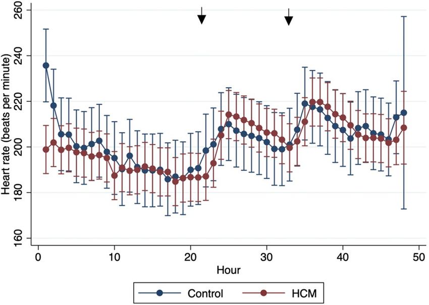
www.nature.com/scientificreports/
Left ventricular outflow tract obstruction was noted to be present when turbulence was observed via color Dop-
pler within the left ventricular outflow tract and the spectral Doppler velocity was > 1.9 m/s.
Sedation and Holter placement. All cats were sedated with 0.25 mg/kg midazolam and 2.0 mg/kg of
alfaxalone intramuscularly approximately 10 min prior to Holter device placement. The thorax of each cat was
shaved, cleaned with 70% isopropyl alcohol, and dried with gauze. A commercially available, AECG device
(H3 + Digital Holter Recorder, Mortara Instrument, Inc.) with three-channel recording was attached to each cat
with three electrodes placed on the right thorax and two on the left thorax in a position to optimize ECG signal.
Each monitor was initialized for 48 h of continuous recording. Monitors and leads were secured to each subject
using skin-safe elastic adhesive tape and bandaging tape. Following placement, each subject was monitored until
alert and able to stand at which time they were returned to the colony and placed in a separate enclosure from
other individuals for the entire 48 h of AECG recording. After 48 h, cats were sedated with the same initial pro-
tocol for device removal, monitored for a minimum of 30 min and until determined to be fully recovered and
then returned to their group housing enclosure.
Terbutaline administration. After approximately 24 h of continuous AECG recording each cat was
administered a dose of oral terbutaline sulfate. As an orally administered, beta agonist that is used clinically
in cats, terbutaline sulfate was chosen as the best option. Based on the authors’ clinical experience, terbutaline
would result in cardiovascular stimulatory effects with minimal invasiveness when compared to intravenous
pharmacologic stress testing options used in humans such as dobutamine. The dose for each cat was between
0.2 mg/kg and 0.3 mg/kg, with cats weighing less than 4.0 kg receiving 0.625 mg and cats weighing more than
4.0 kg receiving 1.25 mg. After 36 h of continuous AECG each cat received a second, matched dose of oral terb-
utaline sulfate.
Holter data acquisition and processing. All data from the AECG monitors was uploaded to the soft-
ware analysis system, Vision 5 software (Mortara Instrument, Inc.) following device removal from each subject.
The uploaded data was randomized via a standard random number generation system to allow for blinded
analysis of the uploaded recordings. The authors that performed analysis of the uploaded recordings were
therefore blinded to animal identification, previous echocardiographic findings, and genotype status. Analysis
and interpretation of the uploaded AECG recording was made prospectively, in a similar manner to previous
publication35. The software analysis system automatically annotates normal and abnormal complexes, however
incorrect labeling of beat type and QRS timing occurs frequently in feline recordings. Therefore, all recordings
were visually inspected on a beat-by-beat basis in their entirety and all mis-labeled beats were corrected in order
to accurately determine the frequency and complexity of ectopy. QRS complexes labeled as normal but with
incorrect timing, for example over the T or P wave, were manually corrected to allow for accurate HRV analysis.
Portions of the recordings with motion-related artifact that was significant enough to preclude accurate labeling
and interpretation was labeled as artifact, discarded and not quantified for analysis.
Supraventricular and ventricular arrhythmias were classified based on a complexity scale: 0 = no arrhythmias
present, 1 = only single premature complexes, 2 = couplets, 3 = triplets, or 4 ≥ 4 consecutive ectopic beats. Ventricu-
lar arrhythmias were classified based on the instantaneous heart rate (HR) as premature (HR ≥ 160), accelerated
idioventricular (HR = 100–159), or escape (HR ≤ 99 bpm) complexes. A sinus arrhythmia was noted to be present
when the rhythm alternated between slower and faster heart rates in a cyclical pattern and all complexes in this
pattern were sinus in origin. HRV was analyzed using standard time-domain techniques in accordance with
published recommendations36,37. All of the time-domain measures of HRV were calculated for each one-hour
period and averaged over the full disclosure using the Vision 5 ECG analysis software. The normal-to-normal
(NN) intervals were calculated from one R wave to the next R wave for all sinus beats by the software. Based on
the NN intervals, the mean, minimal, and maximal HR were recorded. The standard deviation of all NN intervals
(SDNN), the standard deviation of the average NN intervals over 5 min (SDANN), the square root of the mean
squared differences of successive NN interval (RMSSD), and the number of interval differences of successive NN
intervals > 50 ms divided by the total number of NN intervals (pNN50) were obtained for each disclosure. The
NN intervals were also converted to the triangular index value, defined as the integral of the density distribution
of the NN intervals divided by the maximal density distribution.
Statistical analysis. Data was tested for normality using the Shapiro–Wilk test. Normally distributed data
are reported as mean and standard deviation; non-parametric data are reported as median and interquartile
range. Parametric and non-parametric baseline population data were compared using an unpaired t-test and
Mann–Whitney U test, respectively. Comparisons of ectopy frequency before and after terbutaline were made
using a paired t-test for parametric data and the Wilcoxon matched pairs signed rank test for non-parametric
data. Categorical variables were compared using a Fisher’s exact test. Direct comparisons of HRV for the HCM
status, LVOTO, sex, and A31P genotypes over the observation period were performed using the Mann–Whitney
U test or Kruskal Wallis test. Analysis of repeated measurements before and after terbutaline administration was
performed using a linear mixed model to assess the effect of HCM status, LVOTO, sex, and A31P genotype as
fixed effects. Simple and multiple linear regression via the least-squares model were used to assess for predictors
of the total number of supraventricular and ventricular ectopic beats. Statistical significance was considered to
be a p-value < 0.05.
Scientific Reports | (2022) 12:1963 | https://doi.org/10.1038/s41598-022-05999-x 3
Vol.:(0123456789)www.nature.com/scientificreports/
Population characteristic All cats (n = 23) Control (n = 7) HCM (n = 16) p-value
Age (years) 3.64 ± 1.32 4.13 ± 1.51 3.43 ± 1.22 0.25
Sex (% male) 14/23 (60.9%) 3/7 (42.9%) 11/16 (68.8%) 0.36
Body weight (kg) 5.07 (3.10–5.68) 4.15 (3.05–5.20) 5.25 (3.53–6.57) 0.19
MYBPC3 A31P allele count 1 (0–2) 0 (0–1) 1.5 (1–2)
0 or wild type (%) 8/23 (34.8%) 5/7 (71.4%) 3/16 (18.8%)
0.0131
1 or heterozygous (%) 7/23 (30.4%) 2/7 (28.6%) 5/16 (31.3%)
2 or homozygous (%) 8/23 (34.8%) 0/8 (0.0%) 8/16 (50.0%)
NTproBNP level (pmol/L) 52.0 (28.0–111.0) 26.0 (24.0–34.0) 63.0 (46.8–150.0) 0.0004
Maximal LV wall thickness (mm) 5.77 ± 0.98 4.86 ± 0.69 6.17 ± 0.82 0.0013
LVOTO present (%) 7/23 (30.4%) 0/7 (0.0%) 7/16 (43.8%) 0.057
Table 1. Study population characteristics including signalment, genotype for the A31P MYBPC3 mutation,
NTproBNP measurement, maximal left ventricular wall thickness, and presence of left ventricular outflow
tract obstruction on echocardiogram. Data are displayed as mean ± SD or median (IQR). Comparison is made
between groups with p-values displayed.
Holter parameter Overall Resting Stressed p-value (resting vs. stressed)
Heart rate 201.7 ± 18.6 195.0 ± 22.56 209.2 ± 16.1 < 0.0001
SV ectopic beats 2 (0–7) 1 (0–5) 0 (0–2) 0.181
SV ectopy complexity 1 (0–1) 1 (0–1) 0 (0–1) 0.0781
No ectopy n=6 n=7 n = 15 –
Level 1 n = 13 n = 13 n=7 –
Level 2 n=3 n=3 n=0 –
Level 3 n=0 n=0 n=0 –
Level 4 n=1 n=0 n=1 –
V ectopic beats 8 (2–38) 5 (2–19) 3 (0–13) 0.0587
V ectopy complexity 1 (1–4) 1 (1–1) 1 (0–2) 0.7351
No ectopy n=1 n=4 n=7 –
Level 1 n = 14 n = 14 n = 10 –
Level 2 n=1 n=3 n=2 –
Level 3 n=0 n=0 n=0 –
Level 4 n=6 n=2 n=4 –
Table 2. Holter recording data for the 23 cats for both the entire recording and the portion of recordings
before and after terbutaline administration. Data are displayed as mean ± SD if normally distributed or median
(IQR) if non-parametric.
Results
Characteristics of cats. A total of 23 cats were enrolled in the study and underwent 48 h of continuous
AECG monitoring. Sixteen cats met the established criteria for a diagnosis of HCM based on echocardiogram
and NTproBNP measurements, while seven cats were classified as healthy controls. A balanced distribution
of genotypes for the A31P MYBPC3 mutation was achieved: 8/23 cats had 0 mutant alleles (wild type), 7/23
cats had one copy of the mutation (heterozygous), and 8/23 cats had two mutated alleles (homozygous). Base-
line characteristics for the study population are displayed in Table 1. Of these, mutant allele count (p = 0.013),
NTproBNP measurement (p < 0.0005), and maximal left ventricular wall thickness (p = 0.001) were significantly
different between cats in the HCM phenotype group and healthy controls. All cats had a 48-h AECG monitor
placed, however due to artifact, the length of recording analyzed varied. The median time of ECG recording ana-
lyzed was 39.85 h (IQR = 37.23–43.58). Seven cats were administered a dose of 0.625 mg oral terbutaline and 16
cats were given an oral dose of 1.25 mg at each dosing time per the study protocol, with all cats receiving a dose
between 0.2–0.3 mg/kg. The first dose was administered 21–23 h into the analyzed recording and the second
dose was administered 33–35 h into the recording time.
Heart rate, rhythm, and heart rate variability. The median number of analyzed QRS complexes for
each cat was 490,858 (IQR = 447,345–526,074). The mean heart rate over the entirety of ECG recordings was
201.7 ± 18.6 bpm (Table 2). When comparing the mean heart rates of cats in the resting state (prior to terbutaline
administration) and stressed state (post-terbutaline administration), there was a significant difference (195.0 ±
22.56 bpm versus 209.2 ± 16.1 bpm, p < 0.0001) with higher mean heart rates in the stressed state. Analysis of the
linear mixed model also reported significant differences with higher mean heart rate after terbutaline adminis-
Scientific Reports | (2022) 12:1963 | https://doi.org/10.1038/s41598-022-05999-x 4
Vol:.(1234567890)www.nature.com/scientificreports/
Figure 1. Mean heart rate (HR) plotted by hour for the entire AECG recordings separated by phenotype
(subclinical HCM vs. control group). Arrows indicate when terbutaline was administered.
Figure 2. Example of ventricular tachycardia (complexity level 4) obtained via AECG in one cat from the HCM
group.
tration (p < 0.0001). There was no significant difference in the mean heart rate for cats in the HCM group versus
the control group (p = 0.42). Figure 1 shows mean heart rate over the entirety of the recordings, demonstrating
similar rates between the two phenotypic groups and the rise in heart rate for both groups in the stressed state.
The median number of supraventricular ectopic beats over the entire recording period was 2 (IQR = 0–7) with
a median complexity level of 1 (IQR = 0–1). Seventeen of the 23 cats (73.9%) had at least one supraventricular
premature complex over the recording period, the majority of which were single isolated complexes. The median
number of ventricular ectopic beats over the recording period was 8 beats (IQR = 2–38) with a median complex-
ity level of 1 (IQR = 1–4). Twenty-one of the 23 cats (91.3%) had at least one ventricular premature complex on
their ECG recording. Most cats (14/23) only experienced single ventricular premature complexes, however there
were six cats (26%) that had ventricular tachycardia (four or more complexes in a row) (Fig. 2). Two of these cats
were in the control group and four were in the HCM group. The frequency and complexity of ventricular and
supraventricular ectopic beats between the two groups are shown in Table 3.
Simple linear regression was performed for both the total number of supraventricular and ventricular ectopic
beats, assessing phenotype, sex, age, mutant allele count, NTproBNP, maximal left ventricular wall thickness,
and the presence or absence of left ventricular outflow tract obstruction as possible predictors. Of these vari-
ables, none were significantly correlated with the total number of supraventricular ectopic beats. Only mutant
allele count (genotype) was significantly correlated with the number of ventricular ectopic beats (p = 0.049;
R2 = 0.17). Using multiple linear regression to evaluate the same variables as predictors of ectopy frequency,
mutant allele count was significantly correlated with the total number of both ventricular (p = 0.026; R 2 = 0.66)
2
and supraventricular (p = 0.033; R
= 0.66) ectopic beats (Table 4). When comparing the number and complexity
of both supraventricular and ventricular ectopic beats before and after terbutaline administration there were
no significant differences. Seven cats (30.4%) were noted to have a sinus arrhythmia at some point during the
AECG recording. Of these, all seven cats were within the HCM phenotype group with zero cats in the control
group having a sinus arrhythmia present on their recording (p = 0.06).
Scientific Reports | (2022) 12:1963 | https://doi.org/10.1038/s41598-022-05999-x 5
Vol.:(0123456789)www.nature.com/scientificreports/
Holter parameter HCM (n = 16) Control (n = 7)
Heart rate 200.4 ± 19.8 204.7 ± 16.8
SV ectopic beats 1.5 (0–200) 2 (0–23)
SV ectopy complexity 1 (0–4) 1 (0–2)
No ectopy n=5 n=1
Level 1 n=9 n=4
Level 2 n=1 n=2
Level 3 n=0 n=0
Level 4 n=1 n=0
V ectopic beats 10 (1–202) 6 (0–151)
V ectopy complexity 1 (1–4) 1 (0–4)
No ectopy n=0 n=1
Level 1 n = 11 n=4
Level 2 n=1 n=0
Level 3 n=0 n=0
Level 4 n=4 n=2
Table 3. Ectopy frequency and complexity for the 23 cats for the entire recordings based on group. Data are
displayed as median (minimum–maximum) and the number of cats with each complexity level.
Ventricular ectopy Supraventricular ectopy
Variable p-value 95% CI p-value 95% CI
Phenotype 0.640 − 57.5–90.7 0.162 − 20.7–113.1
Sex 0.540 − 42.3–77.6 0.462 − 35.0–73.2
Age (years) 0.211 − 7.45–31.0 0.634 − 13.4–21.3
Mutant allele count (0–2) 0.026 − 89.1–− 6.60 0.033 − 78.4–− 3.90
NTproBNP (pmol/L) 0.110 − 0.15–1.30 0.223 − 0.26–1.04
Maximal LV wall thickness (mm) 0.130 − 8.14–57.3 0.516 − 20.3–38.7
Presence of LVOTO 0.103 − 157.0–15.9 0.133 − 136.1–19.8
Table 4. Multiple linear regression evaluating for predictors of total ventricular and supraventricular ectopic
beats over 48-h Holter recordings.
As SDNN is generally considered the Gold Standard measurement of HRV for clinical purposes and cardiac
risk stratification36, only results for SDNN are presented here. Results for the other time-domain measurements
of HRV can found in the Supplementary Material. Cats within the HCM group showed greater HRV compared to
cats within the control group, with a higher median SDNN value (22 [17–27] ms vs. 21 [14–26] ms; p = 0.0006).
Similarly, there was a significant difference between cats based on mutant allele count, with those cats that had
zero mutant alleles for the A31P mutation having a lower median (IQR) SDNN measurement (0 alleles = 20
[14–27] ms, 1 allele = 23 [19–27] ms, 2 alleles=22 [16–28] ms; p = 0.0001). There was no significant difference
in SDNN between cats with or without left ventricular outflow tract obstruction (22 [16–26] ms and 22 [16–27]
ms; p = 0.29). In the linear mixed model, significant differences in SDNN were also found between cats in the
HCM versus control group (p = 0.002) and mutant allele counts (p = 0.0001).
Male cats were found to have significantly lower minimum (164 [151–179] vs 172 [155–197] bpm; p = 0.0001),
mean (197 [184–212] vs 212 [194–230] bpm; p = 0.0001), and maximum (245 [230–261] vs 259 [242–273] bpm;
p = 0.0001) heart rates compared to female cats across the entire AECG recordings (Fig. 3A). While there was a
higher percentage of male cats within the HCM group (69%) versus the control group (43%), the difference was
not statistically significant (p = 0.36). There was also a significant difference in HRV among sexes, with male cats
having a higher median SDNN compared to female cats (22 [18–28] ms vs. 20 [14–25] ms; p = 0.0001). Figure 3B
shows the median SDNN for the entire AECG recordings for males and females and demonstrates the higher
HRV of males. Figure 3 also demonstrates the effects of terbutaline administration. Namely, in the stressed state
(post-terbutaline) mean heart rate for both sexes increased, HRV measured by SDNN decreased and the dif-
ference seen between sexes for both mean heart rate and HRV is diminished. Heart rate variability measured
by SDNN was significantly lower in the stressed state compared to the resting state (p = 0.0008). Linear mixed
model analysis also revealed a significant difference of SDNN between male and female over time before and
after terbutaline administration (p = 0.0001) (Fig. 3B).
Scientific Reports | (2022) 12:1963 | https://doi.org/10.1038/s41598-022-05999-x 6
Vol:.(1234567890)www.nature.com/scientificreports/
Figure 3. Heart rate and heart rate variability shown between male and female cats. (A) Mean heart rate
(HR) plotted by hour for the entire AECG recordings separated by sex. (B) Standard deviation of NN intervals
(SDNN) plotted by hour for the entire AECG recordings separated by sex. Arrows indicate when terbutaline was
administered.
Discussion
Our study is the first to design, implement, and validate a pharmacological cardiac stress test in cats with sub-
clinical HCM. Herein, we show that administration of oral terbutaline is feasible and resulted in expected cardiac
changes indicative of increased sympathetic drive, presumably what would be seen with physical activity. Namely,
mean heart rate significantly increased and HRV significantly decreased. Exercise testing is performed in people
with asymptomatic HCM to better assess hemodynamic stability and prognosticate. Furthermore, evaluation
of exercise-induced arrhythmias has been shown to provide important information regarding the likelihood of
future SCD and survival times23. In feline veterinary medicine, an exercise test is not a feasible diagnostic, as our
patient population would not be compliant. Therefore, our study aimed to develop a pharmacologic analogue to
an exercise test with the goal of inducing increased sympathetic tone.
Despite effectively placing the studied cats in a state of cardiac stress, no effect on supraventricular or ventricu-
lar arrhythmia frequency or complexity was seen. This may be because the overall frequency and complexity of
arrythmias in the studied population was relatively low or because the cats studied were comprised of relatively
mildly affected cats (ACVIM Stage B1 HCM) and healthy controls. The low frequency and complexity of arrhyth-
mias in the studied population is further highlighted when comparing these results to a recent study assessing
cats with other non-HCM cardiomyopathies. This study assessed cats with more advanced disease, many in
congestive heart failure, and found the median number of ventricular ectopic beats to be 2031 over 24 h38. Fur-
thermore, it is possible that no change was seen because the studied population was made up of cats not prone
to develop arrhythmias in a stressed state. A larger, longitudinal study that includes cats with both subclinical
and later stage HCM would be helpful in determining if our pharmacologic stress test is able to reveal cats that
are more prone to arrhythmogenesis and at a greater risk of SCD. Additionally, 24 h of AECG monitoring may
not have been sufficiently long enough to evaluate arrhythmogenesis in our study population, especially given
day-to-day variability in arrhythmia frequency, and future studies should consider longer monitoring intervals.
Two prior studies have compared the arrhythmia frequency and severity of cats with asymptomatic HCM
to healthy cats using 24-h AECG monitoring20,21. While one of these studies found that cats with asymptomatic
HCM had more frequent and complex ventricular and supraventricular arrhythmias, the other failed to find a
significant difference between g roups20,21. We hypothesized that a possible explanation for these contradictory
Scientific Reports | (2022) 12:1963 | https://doi.org/10.1038/s41598-022-05999-x 7
Vol.:(0123456789)www.nature.com/scientificreports/
results was that the first study noted poor AECG device tolerance and therefore, that cats with asymptomatic
HCM may be more susceptible to the development of arrhythmias when in a stressed state. However, our results
do not support this hypothesis, as subclinical HCM cats did not become significantly more arrhythmic following
administration of terbutaline. More recently, 24-h AECG monitoring was used to compare arrhythmia frequency
and complexity between cats with decompensated HCM, compensated HCM, and healthy control cats22. This
study found that cats with both decompensated and compensated HCM had significantly more ventricular ectopy
compared to healthy cats but did not find a significant difference between the two HCM groups or a correlation
between ectopy and prognosis over the one-year follow-up period. However, of the four cats in this study that
died suddenly, three of them had complex arrhythmias: two with atrial fibrillation and ventricular ectopy and
one with ventricular tachycardia. As our study was not longitudinal in design, we cannot infer a relationship
between arrhythmia development and the likelihood of SCD. However, this study is the first to show a correla-
tion between the frequency of ventricular ectopy on AECG monitoring in cats and MYBPC3 mutant allele count
positivity. While statistically significant, this finding should be further evaluated as the low overall frequency of
ventricular ectopy in our results and the known day-to-day variability of arrhythmia frequency on 24-h AECG in
other species could have resulted in type I error. Further prospective, longitudinal studies are needed to evaluate
the relationship between genotype, phenotype, arrhythmogenesis and clinical outcome.
To our knowledge, HRV has not been previously studied in cats with HCM. One study previously assessed
HRV in healthy cats comparing spectral HRV parameters in the hospital versus in home settings33. Interestingly,
our results found that cats with an HCM phenotype had greater HRV and lower mean heart rates compared to
healthy controls. Additionally, all of the cats with a sinus arrhythmia present on AECG had an HCM phenotype,
which would suggest a relatively higher level of vagal tone. These findings contrast with what was expected given
previous studies of HRV in humans with cardiac disease. For example, SDNN was found to be reduced in patients
with left ventricular hypertrophy secondary to aortic valve stenosis, HCM or hypertension and the extent of
hypertrophy was a positive predictor for the degree of depression of H RV39. However, our findings may be result-
ant of the mild degree of disease in the studied population. Additional studies in people have shown that reduced
HRV is a predictor of death for those patients hospitalized for congestive heart failure and of left ventricular
dilation in patients suffering myocardial infarction. These patient populations represent more severe categories
of disease with clinical signs and hemodynamic compromise. Therefore, our results are likely representative of a
compensated population of cats with mild subclinical HCM compared to healthy controls. We hypothesize that
with disease progression to a clinical state, HRV would decrease as vagal influence is overridden by an increase
in sympathetic tone. Again, prospective studies that encompass both subclinical and decompensated cats with
HCM are needed to assess this. If this were the case, then repeated monitoring for decreases in HRV could
provide indication of worsening disease even before a clinical state. Furthermore, mean SDNN measurements
for all cats in this study were lower than expected for people, whom should have a SDNN greater than 50 ms in
health36. We propose that this is due to higher mean heart rates in cats. A previous study assessing heart rates
and HRV in rhesus macaques found mean SDNN measurements of 27.3 ms for the monkeys with left ventricular
hypertrophy and 35.0 ms for the healthy control group35. As the mean heart rates of the monkeys were between
136 and 147 bpm, which more closely reflects a cat’s heart rate compared to people, it is plausible that healthy cats
have an SDNN below 50 ms. Further studies with larger control groups of healthy cats are needed to assess this.
Male cats had both significantly lower heart rates (minimum, mean, and maximum) and significantly higher
HRV compared to female cats. These findings are logical, as individuals with higher mean heart rates are likely to
have lower HRV as a results of smaller inter-beat intervals. A previous study assessing heart rates in healthy cats
using AECG yielded similar results with females having significantly higher minimum and mean heart rates40.
These findings are also in alignment with the literature on HRV in people. A large meta-analysis assessing the
differences in HRV based on sex in healthy humans found that females had a significantly smaller beat to beat
interval, and thus higher heart rate, and lower HRV measured by SDNN compared to males, similar to our find-
ings in this feline population41. Furthermore, their findings indicated that although females had higher mean
heart rates, they showed greater vagal activity when power spectral density analysis of HRV was performed. It is
possible that this higher underlying parasympathetic tone is cardioprotective, despite higher heart rates and lower
HRV, as there is a male predisposition in both human and feline HCM. Proposed explanations for this difference
in cardiovascular autonomic balance between sexes include differing estrogen levels, differences in oxytocin
concentrations, and differences in neural control, specifically via the amygdala and hippocampus. Similar to these
human findings, our study highlights sex differences in heart rate, HRV, and cardiovascular autonomic balance
in cats. Future studies assessing HRV in cats should ensure to account for these differences between males and
females and is a further reminder on the importance of sex balance in study groups.
There were several limitations of this study, largely due to the population of cats included. Firstly, this study
included a relatively small number of cats which may result in Type II error. This is particularly possible for the
control group, which contained the smallest number of animals. Cats were classified into the control group if
they lacked left ventricular hypertrophy on echocardiography and had an NTproBNP < 99 pmol/L. Two cats in
this group were heterozygous for the A31P MYBPC3 mutation, and thus it is possible that these cats develop
HCM later in life or had histopathologic disease already present that was not appreciable on echocardiography or
resulted in an elevated NTproBNP. Additionally, all cats were selected from a closed colony of research animals
and therefore results may not be representative of a more general feline population. As such, influences of envi-
ronment or lifestyle may be different when comparing these animals to typical pet cats with or without HCM.
Cats were also sedated for placement of the AECG devices and this may have influenced study results, however
the sedation protocol used is relatively short-acting and unlikely to have significantly impacted results beyond
our42. Furthermore, cats were not acclimated to the AECG devices and had never experienced the
the first h
placement of one prior to this study. The mean heart rates, both in the resting and stressed state, are higher in our
study than those found in previous studies evaluating both client-owned, healthy cats in the home environment
Scientific Reports | (2022) 12:1963 | https://doi.org/10.1038/s41598-022-05999-x 8
Vol:.(1234567890)www.nature.com/scientificreports/
and cats with HCM in the home environment20,33,40. Despite attempts to minimize handling, use sedation, and
place cats back into an enclosure in the same building for the AECG recording, this may reflect a component of
environmental stress from the colony environment, placement of the AECG device, and/or administration of
oral terbutaline which could confound our results. Finally, there were limitations pertaining to the equipment
and software used to obtain and analyze the AECG recordings. In particular, the software used did not provide
options to perform spectral analysis of HRV and therefore this limits our ability to draw conclusions about
sympathetic and vagal tone in the study population or compare our results to the previously published literature
on spectral HRV in healthy cats.
In the present study, we show that administration of oral terbutaline is an effective and feasible cardiac
stress test in cats with subclinical HCM. Following dosage with oral terbutaline, heart rates increased and HRV
decreased indicating a predictable pattern of increased sympathetic tone. A low number of single ventricular
premature complexes was relatively common in the studied population of cats, however few cats had more fre-
quent or complex arrhythmias. Arrhythmias also did not differ before or after terbutaline. Cats with an HCM
phenotype had lower heart rates and higher HRV compared to control cats. Furthermore, we found significant
differences in heart rate and HRV between male and female cats, with male cats having lower heart rates and
higher HRV. Finally, cats positive for the A31P MYBPC3 mutation were more likely to have ventricular ectopy.
This finding may indicate that client-owned cats known to be heterozygous or homozygous for the A31P mutation
should be more proactively screened for arrhythmias compared to animals that are wild type. Further studies
that include more severe manifestations of HCM and a larger control group are necessary to determine if such
clinical recommendations are warranted.
Received: 24 July 2021; Accepted: 21 January 2022
References
1. Abbott, J. A. Feline hypertrophic cardiomyopathy: An update. Vet. Clin. N. Am. Small Anim. Pract. 40, 685–700 (2010).
2. Luis Fuentes, V. & Wilkie, L. J. Asymptomatic hypertrophic cardiomyopathy: Diagnosis and therapy. Vet. Clin. North Am. Small
Anim. Pract. 47, 1041–1054 (2017).
3. Payne, J. R., Brodbelt, D. C. & Luis Fuentes, V. Cardiomyopathy prevalence in 780 apparently healthy cats in rehoming centres (the
CatScan study). J. Vet. Cardiol. 17(Suppl 1), S244–S257 (2015).
4. Luis Fuentes, V. et al. ACVIM consensus statement guidelines for the classification, diagnosis, and management of cardiomyopathies
in cats. J. Vet. Intern. Med. 34, 1062–1077 (2020).
5. Meurs, K. M. et al. A cardiac myosin binding protein C mutation in the Maine Coon cat with familial hypertrophic cardiomyopathy.
Hum. Mol. Genet. 14, 3587–3593 (2005).
6. Meurs, K. M., Norgard, M. M., Ederer, M. M., Hendrix, K. P. & Kittleson, M. D. A substitution mutation in the myosin binding
protein C gene in ragdoll hypertrophic cardiomyopathy. Genomics 90, 261–264 (2007).
7. Kittleson, M. D., Meurs, K. M. & Harris, S. P. The genetic basis of hypertrophic cardiomyopathy in cats and humans. J. Vet. Cardiol.
17, S53–S73 (2015).
8. Maron, B. J., Maron, M. S. & Semsarian, C. Genetics of hypertrophic cardiomyopathy after 20 years: Clinical perspectives. J. Am.
Coll. Cardiol. 60, 705–715 (2012).
9. Payne, J. R., Borgeat, K., Brodbelt, D. C., Connolly, D. J. & Luis Fuentes, V. Risk factors associated with sudden death vs conges-
tive heart failure or arterial thromboembolism in cats with hypertrophic cardiomyopathy. J. Vet. Cardiol. 17(Suppl 1), S318–S328
(2015).
10. Spalla, I. et al. Survival in cats with primary and secondary cardiomyopathies. J. Feline Med. Surg. 18, 501–509 (2016).
11. Fox, P. R. et al. International collaborative study to assess cardiovascular risk and evaluate long-term health in cats with preclinical
hypertrophic cardiomyopathy and apparently healthy cats: The REVEAL Study. J. Vet. Intern. Med. 32, 930–943 (2018).
12. Maron, B. J. et al. Epidemiology of hypertrophic cardiomyopathy-related death. Circulation 102, 858–864 (2000).
13. Monserrat, L. et al. Non-sustained ventricular tachycardia in hypertrophic cardiomyopathy: An independent marker of sudden
death risk in young patients. J. Am. Coll. Cardiol. 42, 873–879 (2003).
14. Chatterjee, S. et al. Meta-analysis of left ventricular hypertrophy and sustained arrhythmias. Am. J. Cardiol. 114, 1049–1052 (2014).
15. Møller, M. Standard ECG versus 24-hour Holter monitoring in the detection of ventricular arrhythmias. Clin. Cardiol. 4, 322–324
(1981).
16. Petrie, J. P. Practical application of holter monitoring in dogs and cats. Clin. Tech. Small Anim. Pract. 20, 173–181 (2005).
17. Adabag, A. S., Casey, S. A., Kuskowski, M. A., Zenovich, A. G. & Maron, B. J. Spectrum and prognostic significance of arrhythmias
on ambulatory Holter electrocardiogram in hypertrophic cardiomyopathy. J. Am. Coll. Cardiol. 45, 697–704 (2005).
18. Adabag, A. S. et al. Occurrence and frequency of arrhythmias in hypertrophic cardiomyopathy in relation to delayed enhancement
on cardiovascular magnetic resonance. J. Am. Coll. Cardiol. 51, 1369–1374 (2008).
19. Wilkie, L. J., Smith, K. & Luis Fuentes, V. Cardiac pathology findings in 252 cats presented for necropsy; A comparison of cats with
unexpected death versus other deaths. J. Vet. Cardiol. 17, S329–S340 (2015).
20. Jackson, B. L., Lehmkuhl, L. B. & Adin, D. B. Heart rate and arrhythmia frequency of normal cats compared to cats with asymp-
tomatic hypertrophic cardiomyopathy. J. Vet. Cardiol. 16, 215–225 (2014).
21. Hanås, S., Tidholm, A. & Holst, B. S. Ambulatory electrocardiogram recordings in cats with primary asymptomatic hypertrophic
cardiomyopathy. J. Feline Med. Surg. 19, 158–164 (2017).
22. Bartoszuk, U. et al. Holter monitoring demonstrates that ventricular arrhythmias are common in cats with decompensated and
compensated hypertrophic cardiomyopathy. Vet. J. 243, 21–25 (2019).
23. Gimeno, J. R. et al. Exercise-induced ventricular arrhythmias and risk of sudden cardiac death in patients with hypertrophic
cardiomyopathy. Eur. Heart J. 30, 2599–2605 (2009).
24. Makavos, G. et al. Hypertrophic cardiomyopathy: An updated review on diagnosis, prognosis, and treatment. Heart Fail. Rev.
https://doi.org/10.1007/s10741-019-09775-4 (2019).
25. Bootsma, M. et al. Heart rate and heart rate variability as indexes of sympathovagal balance. Am. J. Physiol. Hear. Circ. Physiol.
266, 565 (1994).
26. Pagani, M. et al. Power spectral analysis of heart rate and arterial pressure variabilities as a marker of sympatho-vagal interaction
in man and conscious dog. Circ. Res. 59, 178–193 (1986).
27. Malik, M. & Camm, A. J. Heart rate variability. Clin. Cardiol. 13, 570–576 (1990).
Scientific Reports | (2022) 12:1963 | https://doi.org/10.1038/s41598-022-05999-x 9
Vol.:(0123456789)www.nature.com/scientificreports/
28. Nolan, J. et al. Prospective study of heart rate variability and mortality in chronic heart failure: Results of the United Kingdom
heart failure evaluation and assessment of risk trial (UK-Heart). Circulation 98, 1510–1516 (1998).
29. Dambrink, J. H. E. et al. Association between reduced heart rate variability and left ventricular dilatation in patients with a first
anterior myocardial infarction. Br. Heart J. 72, 514–520 (1994).
30. Bonaduce, D. et al. Heart rate variability in patients with hypertrophic cardiomyopathy: Association with clinical and echocar-
diographic features. Am. Heart J. 134, 165–172 (1997).
31. Yanagi, S. et al. Heart rate variability and ambulatory blood pressure monitoring in young patients with hypertrophic cardiomyo-
pathy. Circ. J. 68, 757–762 (2004).
32. Butera, G., Bonnet, D., Kachaner, J., Sidi, D. & Villain, E. Heart rate variability in children with hypertrophic cardiomyopathy.
Heart 89, 205–206 (2003).
33. Abbott, J. A. Heart rate and heart rate variability of healthy cats in home and hospital environments. J. Feline Med. Surg. 7, 195–202
(2005).
34. Fox, P. R. et al. Multicenter evaluation of plasma N-terminal probrain natriuretic peptide (NT-pro BNP) as a biochemical screening
test for asymptomatic (occult) cardiomyopathy in cats. J. Vet. Intern. Med. 25, 1010–1016 (2011).
35. Ueda, Y. et al. Heart rate and heart rate variability of rhesus macaques (Macaca mulatta) affected by left ventricular hypertrophy.
Front. Vet. Sci. 6, 1–11 (2019).
36. Shaffer, F. & Ginsberg, J. P. An overview of heart rate variability metrics and norms. Front. Public Heal. 5, 1–17 (2017).
37. Task Force of the European Society of Cardiology and the North American Society of Pacing and Electrophysiology. Heart rate
variability: Standards of measurement, physiological interpretation and clinical use. Circulation 93, 1043–1065 (1996).
38. Ferasin, L., Ferasin, H. & Borgeat, K. Twenty-four-hour ambulatory (Holter) electrocardiographic findings in 13 cats with non-
hypertrophic cardiomyopathy. Vet. J. 264, 105537 (2020).
39. Alter, P., Grimm, W., Vollrath, A., Czerny, F. & Maisch, B. Heart rate variability in patients with cardiac hypertrophy: Relation to
left ventricular mass and etiology. Am. Heart J. 151, 829–836 (2006).
40. Ware, W. A. Normal cats. J. Vet. 1, 175–180 (1999).
41. Koenig, J. & Thayer, J. F. Sex differences in healthy human heart rate variability: A meta-analysis. Neurosci. Biobehav. Rev. 64,
288–310 (2016).
42. Rodrigo-Mocholí, D. et al. Pharmacokinetics and effects of alfaxalone after intravenous and intramuscular administration to cats.
N. Z. Vet. J. 66, 172–177 (2018).
Acknowledgements
We acknowledge that staff of the UC Davis Feline HCM Research Laboratory that care for the cat colony and
assisted in this research project. This study was supported in part by an NIH T35 grant and the STAR Program
at the UC Davis School of Veterinary Medicine.
Author contributions
A.W. and J.S. were responsible for study design. Research and data analysis were conducted by A.W., Y.U., A.C.,
S.H., and J.S. Statistical analyses were performed by A.W. and Y.U. Manuscript preparation was completed by
A.W., Y.U. and J.S.
Competing interests
The authors declare no competing interests.
Additional information
Supplementary Information The online version contains supplementary material available at https://doi.org/
10.1038/s41598-022-05999-x.
Correspondence and requests for materials should be addressed to J.A.S.
Reprints and permissions information is available at www.nature.com/reprints.
Publisher’s note Springer Nature remains neutral with regard to jurisdictional claims in published maps and
institutional affiliations.
Open Access This article is licensed under a Creative Commons Attribution 4.0 International
License, which permits use, sharing, adaptation, distribution and reproduction in any medium or
format, as long as you give appropriate credit to the original author(s) and the source, provide a link to the
Creative Commons licence, and indicate if changes were made. The images or other third party material in this
article are included in the article’s Creative Commons licence, unless indicated otherwise in a credit line to the
material. If material is not included in the article’s Creative Commons licence and your intended use is not
permitted by statutory regulation or exceeds the permitted use, you will need to obtain permission directly from
the copyright holder. To view a copy of this licence, visit http://creativecommons.org/licenses/by/4.0/.
© The Author(s) 2022
Scientific Reports | (2022) 12:1963 | https://doi.org/10.1038/s41598-022-05999-x 10
Vol:.(1234567890)You can also read