Analysis of Polycerate Mutants Reveals the Evolutionary Co-option of HOXD1 for Horn Patterning in Bovidae - Oxford Academic Journals
←
→
Page content transcription
If your browser does not render page correctly, please read the page content below
Analysis of Polycerate Mutants Reveals the Evolutionary
          Co-option of HOXD1 for Horn Patterning in Bovidae
          Aurelie Allais-Bonnet,†,1,2,3 Aurelie Hintermann,†,4 Marie-Christine Deloche,1,2,3 Rapha€el Cornette,5
          Philippe Bardou,6,7 Marina Naval-Sanchez,8 Alain Pinton,6 Ashleigh Haruda,9 Cecile Grohs,10
          Jozsef Zakany,4 Daniele Bigi,11 Ivica Medugorac,12 Olivier Putelat,13,14 Ockert Greyvenstein,15
          Tracy Hadfield,16 Slim Ben Jemaa,17 Gjoko Bunevski,18 Fiona Menzi,19 Nathalie Hirter,19 Julia M. Paris,19
          John Hedges,20 Isabelle Palhiere,6 Rachel Rupp,6 Johannes A. Lenstra,21 Louisa Gidney,22 Josephine Lesur,23
          Renate Schafberg,9 Michael Stache,9 Marie-Dominique Wandhammer,24 Rose-Marie Arbogast,25
          Claude Guintard,26,27 Amandine Blin,28 Abdelhak Boukadiri,10 Julie Rivière,10,29 Diane Esquerre,30
          Cecile Donnadieu,30 Coralie Danchin-Burge,31 Coralie M. Reich,32 David G. Riley,15
          Este van Marle-Koster,33 Noelle Cockett,16 Benjamin J. Hayes,34 Cord Drögemüller,19 James Kijas,8

                                                                                                                                                       Downloaded from https://academic.oup.com/mbe/article/38/6/2260/6126410 by guest on 26 November 2021
          Eric Pailhoux,2,3 Gwenola Tosser-Klopp ,6 Denis Duboule,*,4,35,36 and Aurelien Capitan *,1,10
          1
           ALLICE, Paris, France
          2
           Universite Paris-Saclay, UVSQ, INRAE, BREED, Jouy-en-Josas, France
          3
           Ecole Nationale Veterinaire d’Alfort, BREED, Maisons-Alfort, France
          4
           Department of Genetics and Evolution, University of Geneva, Geneva, Switzerland
          5
           Institut de Systematique, Evolution, Biodiversite (ISYEB), Museum National d’Histoire Naturelle, CNRS, Sorbonne Universite, EPHE,
          Universite des Antilles, Paris, France
          6
           GenPhySE, Universite de Toulouse, INRAE, ENVT, Castanet-Tolosan, France
          7
           INRAE, Sigenae, Castanet-Tolosan, France
          8
           CSIRO Agriculture & Food, St. Lucia, QLD, Australia
          9
           Central Natural Science Collections, Martin Luther University Halle-Wittenberg, Halle (Saale), Germany
          10
            Universite Paris-Saclay, INRAE, AgroParisTech, GABI, Jouy-en-Josas, France
          11
            Dipartimento di Scienza e Tecnologie Agro-Alimentari, Alma Mater Studiorum University of Bologna, Bologna, Italy
          12
            Population Genomics Group, Department of Veterinary Sciences, Ludwig-Maximilians-University Munich, Munich, Germany
          13
            Archeologie Alsace, Selestat, France
          14
            UMR 7044, ARCHIMEDE, MISHA, Strasbourg, France
          15
            Department of Animal Science, Texas A&M University, College Station, TX, USA
          16
            Department of Animal, Dairy, and Veterinary Sciences, Utah State University, Logan, UT, USA
Article

          17
            Laboratoire des Productions Animales et Fourragères, Institut National de la Recherche Agronomique de Tunisie, Universite de
          Carthage, Ariana, Tunisia
          18
            Livestock Department, Faculty of Agricultural Sciences and Food Institute of Animal Biotechnology, University Ss. Cyril and
          Methodius, Skopje, North Macedonia
          19
            Institute of Genetics, Vetsuisse Faculty, University of Bern, Bern, Switzerland
          20
            Manx Loaghtan Sheep Breeders’ Group, Bassingbourn, Cambridgeshire, United Kingdom
          21
            Faculty of Veterinary Medicine, Utrecht University, Utrecht, The Netherlands
          22
            Rent a Peasant, Tow Law, Bishop Auckland, Durham County, United Kingdom
          23
            Unite Archeozoologie, Archeobotanique, Societes Pratiques et Environnements (AASPE), CNRS, Museum National d’Histoire
          Naturelle, Paris, France
          24
            Musee Zoologique de Strasbourg, Strasbourg, France
          25
            CNRS UMR 7044, ARCHIMEDE, MISHA, Strasbourg, France
          26
            Unite d’Anatomie Comparee, Ecole Nationale Veterinaire de l’Agroalimentaire et de l’Alimentation, Nantes Atlantique—ONIRIS,
          Nantes, France
          27
            Groupe d’Etudes Remodelage Osseux et bioMateriaux (GEROM), Universite d’Angers, Unite INSERM 922, LHEA/IRIS-IBS, CHU
          d’Angers, Angers, France
          28
            Museum National d’Histoire Naturelle, CNRS, UMS 2700 2AD, Paris, France
          29
            INRAE, Micalis Institute, AgroParisTech, Universite Paris-Saclay, Jouy-en-Josas, France

          ß The Author(s) 2021. Published by Oxford University Press on behalf of the Society for Molecular Biology and Evolution.
          This is an Open Access article distributed under the terms of the Creative Commons Attribution Non-Commercial License
          (http://creativecommons.org/licenses/by-nc/4.0/), which permits non-commercial re-use, distribution, and reproduction in any
          medium, provided the original work is properly cited. For commercial re-use, please contact journals.permissions@oup.com       Open Access
          2260                       Mol. Biol. Evol. 38(6):2260–2272 doi:10.1093/molbev/msab021 Advance Access publication February 2, 2021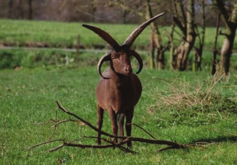
Evolutionary Co-option of HOXD1 for Horn Patterning in Bovidae . doi:10.1093/molbev/msab021 MBE
30
INRAE, US, 1426, GeT-PlaGe, Genotoul, Castanet-Tolosan, France
31
Institut de l’Elevage, Paris, France
32
Agriculture Victoria, AgriBio, Centre for AgriBioscience, Bundoora, VIC, Australia
33
Department of Science, University of Pretoria, Hatfield, South Africa
34
Queensland Alliance for Agriculture and Food Innovation (QAAFI), Centre for Animal Science, University of Queensland, St. Lucia,
QLD, Australia
35
Swiss Cancer Research Institute, EPFL, Lausanne, Switzerland
36
Collège de France, Paris, France
†
Shared first authors.
*Shared last authors and corresponding authors: E-mails: aurelien.capitan@inrae.fr; denis.duboule@epfl.ch.
Associate editor: Patricia Wittkopp
Downloaded from https://academic.oup.com/mbe/article/38/6/2260/6126410 by guest on 26 November 2021
Abstract
In the course of evolution, pecorans (i.e., higher ruminants) developed a remarkable diversity of osseous cranial appen-
dages, collectively referred to as “headgear,” which likely share the same origin and genetic basis. However, the nature
and function of the genetic determinants underlying their number and position remain elusive. Jacob and other rare
populations of sheep and goats are characterized by polyceraty, the presence of more than two horns. Here, we char-
acterize distinct POLYCERATE alleles in each species, both associated with defective HOXD1 function. We show that
haploinsufficiency at this locus results in the splitting of horn bud primordia, likely following the abnormal extension of
an initial morphogenetic field. These results highlight the key role played by this gene in headgear patterning and
illustrate the evolutionary co-option of a gene involved in the early development of bilateria to properly fix the position
and number of these distinctive organs of Bovidae.
Key words: Hox genes, co-option, regulatory mutation, goat and sheep genomics.
Introduction mapped in seven distinct populations to the same region of
chromosome 2 (Chr2), it has not yet been identified
In pecorans, successive environmental and behavioral adap-
(Greyvenstein et al. 2016; He et al. 2016; Kijas et al. 2016;
tations favored the emergence and sometimes the secondary
Ren et al. 2016). In contrast, polycerate goats are observed
loss of a variety of headgear, as exemplified by bovid horns,
only sporadically in the Alps. They are not present in archae-
cervid antlers, giraffid ossicones, or antilocaprid pronghorns
ological remains and have not been subject to any genetic
(Davis et al. 2011; Wang et al. 2019). As different as they are,
studies thus far. The oldest record of this condition in goat
these iconic organs share both a common cellular origin and a
dates back from 1786, when a four-horned billy-goat was
minimal structural organization: they derive from neural crest
transferred from the city of Bulle in Switzerland to the model
stem cells and consist of paired structures, located on the
farm of French Queen Marie-Antoinette in Versailles
frontal bones and composed of a bony core covered by in-
(Heitzmann 2006).
tegument (Davis et al. 2011; Wang et al. 2019) (fig. 1 and
In this study, we set up to determine the genetic bases of
supplementary fig. 1, Supplementary Material online).
these conditions in sheep and goats. We show that polyceraty
Although the development and evolution of headgear is a
in Bovidae is due either to a 4-bp deletion affecting the splic-
long-standing question, the underlying molecular and cellular
ing of the HOXD1 gene in sheep, or to the deletion of a large
mechanisms have been difficult to study, mostly because the
regulatory region controlling the same gene in goats. These
patterning and differentiation of headgear progenitor cells
results thus illustrate the evolutionary co-option of this gene
occur early during embryogenesis (Lincoln 1973; Allais-
normally involved in early development to help determine
Bonnet et al. 2013) and involve hundreds of genes (Wang
the position and number of horns. They also show that com-
et al. 2019).
parable phenotypes observed in distinct species and selected
In this context, natural mutations affecting headgear num-
and maintained for a long time are caused by the mis-
ber, shape, or position, such as the polycerate (multihorned)
regulation of the same gene.
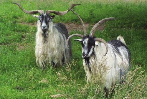
phenotype occurring in small ruminants (fig. 1a and b, OMIA
entries 000806-9940 and 000806-9925; https://omia.org/
home/, last accessed November 4, 2020), offer a valuable al- Results and Discussion
ternative (Capitan et al. 2012). Polyceraty was already ob- Characterization of POLYCERATE Mutations in Sheep
served ca 6000 BCE, in the oldest ovine remains from and Goats
Çatalhöyük, Turkey (Epstein 1971; Putelat 2005) and this To identify the genetic determinants of polyceraty, we rean-
dominant trait currently segregates in several sheep breeds alyzed the Illumina OvineHD Beadchip genotyping data (600
around the world. Even though the corresponding locus was k SNPs) of 111 case and 87 control sheeps generated by two
2261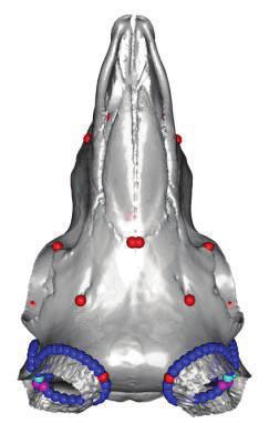
Allais-Bonnet et al. . doi:10.1093/molbev/msab021                                                                                                        MBE

                    (a)                                                                             (b)

                                                                                                                                                                       Downloaded from https://academic.oup.com/mbe/article/38/6/2260/6126410 by guest on 26 November 2021
                   (c)                             Chr2 minus strand                                (d)
                                  132 832 260 bp                       132 832 250 bp
  Heterozygous polycerate sheep

                                                                                                                              503-kb deleon

                                                                           4
                                                                           4
                                                                           4                                    115,5 Mb                 116,0 Mb            Chr2
                                                                                                                           137-kb duplicaon
                                                                           4
                                                                           4

                                  A A G A A A A G T A A G T A C T T
                                                          Splice        Deleon
                                                          donor                         intron 1
                                    HOXD1 exon 1                                                   48,0 Mb                        48,5 Mb                   Chr5

                                                                                                    e
                                                                                                     Mutant
                                                                                                      Chr2
                                                                                                                                     Chr5                Wt
                                                                                                                                                        Chr2
                                                           +1 +2 +3 +4 +5 +6 +7

FIG. 1. Polyceraty in sheep and goats and candidate genetic variants. (a) Polycerate Manx Loaghtan ram. (b) Wild-type and polycerate male goats
from a local German population. These individuals represent the most common phenotype. Polycerate animals with asymetric horns and partial
fusion of lateral horns are also regularly observed. (c) A 4-bp deletion causing polyceraty in sheep. Integrative Genome Viewer (IGV) screenshot
with the localization of the variant with respect to HOXD1. Below is a graphical representation of nucleotide conservation at the exon 1-intron
junction across 103 sarcopterigian and tetrapod species. (d) Plot of read coverage in a heterozygous polycerate goat animal carrying a deletion of
503-kb downstream the HOXD gene cluster on Chr2 and a duplication of 137 kb on Chr5. (e) FISH-mapping in a heterozygous polycerate goat with
BAC clones corresponding to the region deleted in Chr2 (labeled in red) and to the segment of Chr5 inserted at the deletion site (labeled in green).
Magnification: 1,000. Sheep and goat icons were made by “Monkik” from www.thenounproject.com (last accessed November 4, 2020).

previous studies (Greyvenstein et al. 2016; Kijas et al. 2016)                                          three nucleotides (þ4, þ5, þ6) of the consensus splice donor
(supplementary tables 1 and 2, Supplementary Material on-                                               site (Zhang 1998). Genotyping of this variant in 236 animals
line). Assuming autosomal dominant inheritance and genetic                                              from eight populations containing polycerate specimens
homogeneity in the three breeds investigated, we fine-                                                  showed a perfect genotype to phenotype association (sup-
mapped the ovine POLYCERATE locus between positions                                                     plementary tables 3 and 4, Supplementary Material online).
132,717,593 and 133,151,166 bp on Chr2 (Oar_v4.0 assembly;                                              Moreover, cross-species alignments revealed that the þ4 and
supplementary fig. 2, Supplementary Material online). By                                                þ5 nucleotides are conserved among 103 sarcopterigian and
comparing whole-genome sequences of 11 polycerate speci-                                                tetrapod species, indicating the occurrence of a genuine and
mens and 1,179 controls representing the world-wide sheep                                               consensual splice donor site and hence suggesting a detri-
diversity, we identified a single candidate variant in this inter-                                      mental effect of the micro-deletion in the splicing of HOXD1
val: a four-nucleotide deletion located at position þ4 to                                               precursor RNAs (fig. 1c and supplementary table 5,
þ7 bp after exon 1 of the HOXD1 gene                                                                    Supplementary Material online).
(g.132,832,249_132,832,252del; fig. 1c), that is, encompassing
2262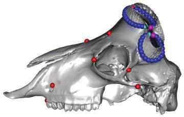
Evolutionary Co-option of HOXD1 for Horn Patterning in Bovidae . doi:10.1093/molbev/msab021                                  MBE
    We next mapped the caprine POLYCERATE mutation to a             (a)
542-kb large region orthologous to that of the ovine locus            posterior, late                                anterior, early
(Chr2:115,143,037–115,685,115 bp on ARS1 assembly;
Bickhart et al. 2017; supplementary fig. 2, Supplementary
                                                                     13 12 11       10 9     8            4          3          Hoxd1
Material online), by using a panel of 35 polycerate and 51
two-horned goats obtained from eight European populations
and genotyped with the Illumina GoatSNP50 BeadChip                                                                               50 kb
(Tosser-Klopp et al. 2014) (supplementary table 6,
Supplementary Material online). Within this interval, we iden-              HoxD       Mtx2
tified 36 private heterozygous variants in one heterozygous
polycerate goat versus 1,160 control individuals (supplemen-                         HOXD1Del(4bp)
tary table 7, Supplementary Material online). Genotyping of                                         HOXDDel(503kb)
five case–control pairs from distinct breeds reduced the list of

                                                                                                                                             Downloaded from https://academic.oup.com/mbe/article/38/6/2260/6126410 by guest on 26 November 2021
candidates to 15 short variants, affecting genomic regions not
conserved among 103 eutherian mammals, as well as a rare                             Hoxd1Lac
type of structural variation located 57 kb downstream of the                           BACHoxD
HOXD1 30 -UTR (supplementary tables 7 and 8,                                                  BACMtx2
                                                                                              HoxDDel(151kb)lac
Supplementary Material online). The latter involved the
                                                                    (b)
translocation of 137 kb from Chr5 to Chr2 by means of a
circular intermediate (Durkin et al. 2012) and the deletion
of       503 kb        from        the       insertion      site
(g.115,652,290_116,155,699delins137 kb; fig. 1d and e), as con-
firmed by PCR amplification and Sanger sequencing of the
regions containing the breakpoints (supplementary fig. 3,
Supplementary Material online). Consequently, the mutant
chromosome lacked the MTX2 gene and carried an exogenic
copy of both RASSF3 and the first ten exons of GNS.                              Hoxd1Lac                         TgBACHoxD
Genotyping of this variant in 77 case and 355 control goats
originating from 24 distinct populations revealed a 100 per-
cent association between polyceraty and heterozygosity for
the large insertion–deletion (indel, supplementary table 9 and
supplementary fig. 4, Supplementary Material online).
Homozygous mutants were not detected in our panel,
whereas at least 14 polycerate animals were born from poly-
cerate pairs of parents (binomial P ¼ 3.4  103; supplemen-
tary note 1, Supplementary Material online). Because the                        TgBACMtx2                      HoxDDel(151kb)lac
knockdown of Mtx2 in zebrafish is embryonic lethal at gas-
trulation (Wilkins et al. 2008) and newborn mice homozygous        FIG. 2. Regulation of Hoxd1 expression pattern in crest cell-derived
for a deletion including Mtx2 were never scored (binomial          head structures in mouse. (a) On top is the structure of the mouse
                                                                   HoxD gene cluster with arrows showing the timing and localization of
P ¼ 5.7  106; supplementary note 1, Supplementary
                                                                   gene expression along the body axis during development. The posi-
Material online), we concluded that homozygosity at the            tion of Hoxd1 is highlighted in red. Below is a 1-Mb view of the locus,
goat POLYCERATE locus is an early lethal condition.                with Hoxd1 in red as well as the relative positions of the POLYCERATE
                                                                   variants in sheep (black arrowhead) and goat (black line). Below are
Remote Hoxd1 Regulation in Transgenic Mice                         depicted the various murine alleles, with the lacZ insertion in Hoxd1
These mapping studies identified the HOXD gene cluster as          (blue arrowhead), the two BAC clones (thick blue lines) and the
being involved in the polycerate phenotype in both sheep           engineered deletion (black line). (b) Heads of E12.5–E13.5 mouse
and goats. This cluster contains nine homeobox genes encod-        fetuses after X-gal staining. The dashed circle highlights the absence
ing transcription factors involved in the organization of the      of Hoxd1 expression in the crown (corresponding to the localization
body plan during embryogenesis (Krumlauf 1994). Both their         of hornbuds in Bovidae), whereas the surrounding dermal cells are
                                                                   positive. The conservation of Hoxd1 expression in the back of the neck
timing of activation and their domains of expression are de-
                                                                   (black arrows) contrasts with the presence/absence of expression in
termined by their respective positions along the gene cluster
                                                                   the facial muscle precursors (white arrows) and in the eyelids (arrow-
(Kmita and Duboule 2003). Accordingly, the mouse Hoxd1             head). The comparison between the four strains indicate that Hoxd1
gene is expressed very early on and in the most rostral part of    expression in all these cranial derivatives is controlled by regulatory
the embryo (fig. 2a). In rodents, Hoxd1 is expressed in crest      elements located in a region orthologous to the proximal portion of
cell-derived structures (Frohman and Martin 1992), which           the segment deleted in polycerate goats. Sheep, goat and mouse icons
made this gene a particularly interesting candidate for poly-      were made by “Monkik” from www.thenounproject.com (last
ceraty. In addition, a DNA sequence conserved only among           accessed November 4, 2020).
pecoran species carrying headgear was identified 15 kb
                                                                                                                                    2263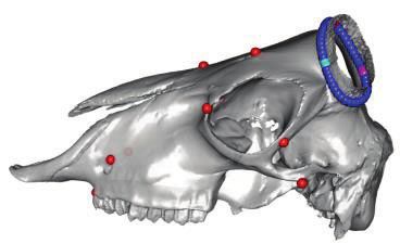
Allais-Bonnet et al. . doi:10.1093/molbev/msab021                                                                     MBE
downstream of HOXD1 (“HCE” in Wang et al. [2019]). This            expression was significantly lower in horn buds than in sur-
sequence, however, is not included in the large indel observed     rounding tissues (fig. 3a and b), reminiscent of the weak—if
in polycerate goats.                                               any—expression of Hoxd1 observed in a comparable region in
    We assessed whether the deletion present in goat may           the mouse. In heterozygous mutant goat fetuses, however,
impact the expression of HOXD1 in cranial crest cells by           HOXD1 RNA levels were equally low in all three samples
looking at a series of modified mouse strains either carrying      (fig. 3a), re-enforcing the idea that the caprine
transgenes or where a targeted deletion was induced at the         POLYCERATE variant negatively affects the expression of
orthologous locus (see Materials and Methods). First, the          HOXD1.
wide presence of cells expressing Hoxd1 both in the face               In sheep, when primers targeting the second exon of the
and in the cranial derma, the latter being of crest cell origin,   gene were used (fig. 3b, upper histogram and methods), het-
was detected in fetuses with a targeted integration of lacZ        erozygous mutants for the 4-bp deletion overlapping the
sequences into the Hoxd1 gene (fig. 2b, Hoxd1Lac). Expression      splice donor site and control samples displayed similar pro-
was however not scored in the crown region (fig. 2b, dashed        files of RNA expression despite some variation due to slight

                                                                                                                                     Downloaded from https://academic.oup.com/mbe/article/38/6/2260/6126410 by guest on 26 November 2021
circle), an area we presumably defined as corresponding to         differences in sampling. However, RT–qPCR with intronic
that of horn bud differentiation in Bovidae (Dove 1935;            primers revealed significant intron retention in all mutant
Capitan et al. 2011). Instead, Hoxd1 was expressed in various      tissues but horn buds, where expression was likely too low
amounts in other regions of the head including the eyelids         (fig. 3b lower histogram). Intron retention is predicted to
(fig. 2b, white arrow and arrowhead), an observation consis-       result in a nonfunctional protein, truncated two residues after
tent with the abnormal upper eyelids and eyebrows detected         the last amino acid encoded by exon 1 and thus lacking the
in a minority of polycerate sheep and goats (Gascoigne et al.      homeodomain, the DNA binding moiety (fig. 3c and supple-
2017; supplementary figs. 5–7, Supplementary Material on-          mentary fig. 9, Supplementary Material online). Therefore,
line), even though such alterations were not observed in mice      both POLYCERATE variants appear to reduce the amount
lacking Hoxd1.                                                     of functional HOXD1 RNAs in the horn bud region. We hy-
    We next tried to localize the underlying regulatory ele-       pothesize that this reduction leads to the extension of the
ments by using transgenic BACs with lacZ sequences intro-          cellular field permissive for horn bud development following
duced within Hoxd1. A BAC covering the HoxD cluster itself         the loss of the HOXD1 boundary. This extension may suffi-
did not show any expression in the head, suggesting that           ciently elongate the bud region to allow its separation into
regulatory sequences are not located in the gene cluster           two distinct organs.
(fig. 2b, TgBACHoxD). In contrast, a transgenic BAC extending
in the region upstream of Hoxd1 and including Mtx2 gave a          Morphometric Analyses and Topology of the Horn
subset of the staining observed with Hoxd1Lac (fig. 2b,            Field
TgBACMtx2), indicating that some regulatory sequences were         To substantiate this hypothesis, we analyzed variations in
located upstream Hoxd1, in a region including and surround-        horn topology in 61 ovine and 19 caprine skulls from various
ing Mtx2. The latter result was controlled by using an engi-       populations using 3D geometric morphometrics (supplemen-
neered 151-kb deletion of a largely overlapping region,            tary table 10, Supplementary Material online). We performed
including a lacZ reporter gene, which expectedly abrogated         a principal component analysis (PCA) using 16 anatomical
Hoxd1 expression in cranial cellular populations (fig. 2b,         landmarks (anatomically homologous) and 100 sliding semi-
HoxDDel(151 kb)lac). The weaker expression of Hoxd1Lac in the      landmarks (geometrically homologous [Bookstein 1992] after
dermal component also seemed to disappear in the latter            scaling and eliminating the effects of translation and rotation
deletion. As a positive control for the lacZ reporter system,      thanks to a generalized procrustes analysis [GPA]; Rohlf and
expression of Hoxd1 in neural derivatives driven by sequences      Slice 1990). This protocol, using sliding semilandmarks, makes
within the HoxD cluster was scored, as expected (fig. 2b, black    it possible to quantify, visualize, and compare anatomical
arrows). These analyses in mice demonstrated that regulatory       regions devoid of anatomical landmarks (Gunz and
sequences driving Hoxd1 expression in the head are located in      Mitteroecker 2013; see Materials and Methods). We then
a region largely comprised within the deletion determined in       plotted the first principal components (PCs) to visualize the
goats as causative of polyceraty, further suggesting that the      specimen distribution in the morphospace (fig. 4a and sup-
latter deletion abrogates HOXD1 expression in goat fetuses.        plementary figs. 10–12 and supplementary table 11,
                                                                   Supplementary Material online). The first two axes repre-
Expression of HOXD1 in Pecoran Fetuses                             sented 35.8% and 23.3% of the total shape variability and
To investigate whether the absence of mouse Hoxd1 expres-          distinguished the phenotypes and species categories, respec-
sion in the crown region of the head was also observed in          tively. Along the first axis, we individualized three subgroups
pecoran embryos, we isolated heterozygous polycerate and           of polycerate specimens in sheep, based on the distances
wild-type fetuses both at 70 dpc (days post-coı̈tum) in goat       between lateral horns (fig. 4a, dlh). Of note, the group dis-
and at 76 dpc in sheep, two stages where eyelids are fully         playing the largest dlh (i.e., that with the highest negative
grown and horn buds can be distinguished (supplementary            values along the x axis) had no equivalent in goat, possibly
fig. 8, Supplementary Material online). After microdissection      due to early lethal homozygosity (see above).
and reverse transcription-quantitative PCR (RT–qPCR), we               We looked at the association between genotypes and horn
noticed that in wild-type fetuses of both species, HOXD1           implantation within polycerate animals by measuring the
2264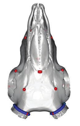
Evolutionary Co-option of HOXD1 for Horn Patterning in Bovidae . doi:10.1093/molbev/msab021 MBE
(a) fs fs distances both between the lateral horns on the left side of
hb hb the skull (dlhl), and between the upper horns (duh) in 29
rams (supplementary table 12, Supplementary Material on-
Goat 70 dpc
el el
line). We found a significant difference in the proportions of
homozygous and heterozygous specimens in animals with
+/+ +/- dlhlduh versus dlhl>duh (fig. 4b) and no heterozygous
animal was found to have dlhl>duh. We computed the the-
*
* *
oretical skull shape at the maximum and minimum of PC1
1
1.0 ** axis (fig. 4c) and the corresponding vectors of deformation
(fig. 4d). The results obtained were consistent with a splitting
HOXD1 0.5 of horn buds in polycerate animals. This splitting always oc-
0,5
exon 2 curred along the major axis of the ellipse formed by the wild-
type horn bud, with an extension of the hemi-horn buds in an
Downloaded from https://academic.oup.com/mbe/article/38/6/2260/6126410 by guest on 26 November 2021
0.0
0
area where HOXD1 expression was detected in wild-type
hb fs el hb fs el
(b) specimens (fig. 4d and above). In homozygous animals, the
fs h2 fs new cellular field was likely larger than in heterozygous, lead-
hb
Sheep 76 dpc
h1 ing to a clearer separation of hemi-horns, whereas heterozy-
bs el bs el gous specimens often displayed partially fused organs, a
situation markedly different from the production of addi-
tional horns, as observed in subspecies of Tetracerus quad-
+/+ +/- ricornis (Groves 2003) (supplementary fig. 13, Supplementary
Material online).
** * * *
# ** *
1,00
1.0 Conclusions
HOXD1 # From these results, we conclude that pecorans have an intrin-
0.5
0,50 @
exon 2 sic capacity to induce hornbuds within a presumptive head
@ territory. This capacity appears to be associated with the non-
0.0
0,00 expression of the HOXD1 transcription factor, which is present
bs hb fs el bs h1 h2 fs el in surrounding cells and may delimit this field, a function
* somewhat distinct from the ancestral role of Hox genes during
1,00
1.0 * development (Krumlauf 1994). Two independent haploinsuf-
* ficient conditions, in sheep and goat, both involving reduced
HOXD1
0.5
0,50 expression of HOXD1 presumably lead to the extension of this
intron1
territory, a condition fully achieved in the complete absence of
0.0
0,00
the wild-type HOXD1 allele in homozygous polycerate sheep.
bs bs hb h1 h2 fs fs el el Although a weak extension of this morphogenetic field may
correspond to the growth of twin horns, fused at their bases, a
(c) 4-bp deleon Amplicons full extension would induce the complete splitting of the horn
bud, thus generating a pair of lateral horns. We hypothesize
Ovine gene Exon 1 Exon 2 that the initial expression of HOXD1 in anterior crest cells
Intron 1
made this evolutionary co-option possible and thus helped
Wildtype protein HD to determine the position and number of horns, which be-
came the distinctive trait of Bovidae.
Putave Premature
mutant protein truncaon Materials and Methods
Exon juncon
Ethics Statement
FIG. 3. RT–qPCR gene expression analyses in sheep and goat fetuses. All experiments reported in this work comply with the ethical
(a and b) Schemes of the tissues sampled at stage 70 dpc in goat (a) guidelines of both the French National Research Institute for
and 76 dpc in sheep (b) in four control (þ/þ) and four heterozygous Agriculture, Food and Environment (INRAE) and the
(þ/) polycerate fetuses within each species. bs: skin from the back University of Geneva, Switzerland. Blood samples were
of the head; hb: skin from the hornbud; h1: skin from the lower horn
bud; and h2: skin from the upper horn bud in polycerate specimens; fs:
frontal skin; el: eyelids. RT–qPCR gene expression analyses in these means are not equal). For the sake of clarity, the symbols # and @ were
tissues are shown below (means and standard errors of the means also used to show significant differences (P < 0.05) between distant
from triplicate experiments). Gene expression was normalized using bars. (c) Schematic representation of the ovine HOXD1 gene and
GAPDH, H2AFZ, and HPRT1 as reference genes. *P < 0.05, **P < 0.01 corresponding wild-type and putative mutant proteins. The local-
(Welch two-sample t-test with the alternative hypothesis that the izations of the amplicons studied in (b) are indicated with double
arrows. HD: Homeodomain.
2265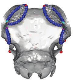
Allais-Bonnet et al. . doi:10.1093/molbev/msab021                                                                                                                            MBE

    (a)                                                                                                         (b)                                         Genotypes
                       duh                                                                                                                        +/-                  -/-
                                  dlh
                                                                                                                                                                 ***

                                                                                                                Number of rams of each genotype
                                                                   #                                                                                    1
                           @                                 0.1
                                                                         £
                                                         #                                       £
                                            @
                                                                                                  PC1
               -0.3            -0.2         -0.1                         0.1               0.2
                                                                                                                                                        17
                       &                                                                         35.8%

                                                                                                                                                                                   Downloaded from https://academic.oup.com/mbe/article/38/6/2260/6126410 by guest on 26 November 2021
                                                                         $                                                                                                  11

                                                         -0.1

                                                     €
                   &
                                                                                                                                                  dlhl 1≤ duh dlhl 2> duh

                                                         -0.2

                                                              PC2                      $
                                        €
                                                             23.3%

                                                                                                                (d)
    (c)                                                                                                                                                     fs

                                                                                                                                                                   Left
                                                                                                                                                                   horn

                                                                                                                   Orbit

                                                                                                                                                                       bs

FIG. 4. Results of 3D geometric morphometric analyses of 61 ovine and 19 caprine skulls. (a) Distribution of the specimens along the first two axes
of the PCA. The proportion of variance explained by the main principal components is indicated on each axis. Green dots: polycerate sheep with a
distance between lateral horns (dlh) larger than the distance between upper horns (duh); light blue: polycerate sheep with a dlhduh; blue:
polycerate sheep with at least two lateral horns partially fused at their basis; purple: wild-type sheep; black: polycerate goats; and red: wild-type
goats. Representative specimens illustrate each cluster and symbols are used to indicate their respective locations in the PCA analysis (see
supplementary fig. 10, Supplementary Material online, for intraspecies analyses and further information). (b) Number of heterozygous (þ/) and
homozygous (/) polycerate rams among groups of live animals with different dlhl (dlh on the left side) and duh relative sizes (see supple-
mentary table 12, Supplementary Material online, for further information); ***P value ¼ 3.5  107 (Fisher’s exact test). (c) Theoretical shapes
associated with the maximum (upper three) and minimum values (lower three) of PC1 axis for a sheep skull. Red dots correspond to anatomical
landmarks, whereas the other dots correspond to sliding semilandmarks; light blue and purple dots highlight the sites of division of lateral horns.
(d) Shape differences for the sliding semilandmarks located at the basis of the left horn. Light blue and purple dots correspond to the maximum
and minimum values of PC1 axis, respectively. Dashed squares indicate the estimated position of dissected tissues in figure 3 (bs: skin from the back
of the head; fs: frontal skin) in which HOXD1 expression was observed in fetuses.

2266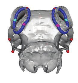
Evolutionary Co-option of HOXD1 for Horn Patterning in Bovidae . doi:10.1093/molbev/msab021                           MBE
collected on sheep and goats during routine blood sampling        (Schep et al. 2016). The BACs were selected based on their
(for annual prophylaxis, paternity testing, or genomic selec-     localization on the physical map of the mouse genome
tion purpose) by trained veterinarians and following standard     (Gregory et al. 2002) and obtained from the RPCI-23 and -
procedures and relevant national guidelines. Sample collec-       24 Mouse (C57BL/6J) BAC Libraries from the Children’s
tion of small ruminants in Switzerland was approved by the        Hospital Oakland Research Institute (https://bacpacresour-
Cantonal Committee for Animal Experiments (Canton of              ces.org/libraries.php, last accessed November 4, 2020). The
Bern; permit 75/16). Ovine and caprine fetuses were pro-          modified BACs were purified, linearized, and microinjected
duced in an INRAE experimental farm (Bressonvilliers,             into mouse fertilized oocytes to obtain each of these strains
France) and collected in the INRAE experimental slaughter-        in a mixed Bl6XCBA hybrid background, by standard proce-
house of Jouy-en-Josas (France). Experiments were performed       dures. Gene expression analyses were performed on hetero-
in strict accordance with the European directive 2010/63/UE       zygous specimens. A precise map of the orthologous Hoxd
and were approved by the local Institutional Animal Care and      region in mouse and goat was obtained by aligning on murine
Use Committee of AgroParisTech/INRAE (COMETHEA, per-              GRCM38/mm10 genome assembly the BAC end sequences

                                                                                                                                     Downloaded from https://academic.oup.com/mbe/article/38/6/2260/6126410 by guest on 26 November 2021
mit number 19/032). All experiments involving mice were           and goat genome sequences of 10-kb segments encompass-
performed in agreement with the Swiss law on animal pro-          ing the breakpoints of the large indel. Alignments were car-
tection (LPA), under license No GE 81/14 (to D.D.). All the       ried out using the BLAT tool from the UCSC Genome
samples and data analyzed in the present study were               Browser        (http://genome.ucsc.edu/cgi-bin/hgBlat,   last
obtained with the permission of breeders, breeding organiza-      accessed November 4, 2020).
tions, and research group providers.
                                                                  Animals Subject to Postmortem Clinical Examination
Animals                                                           The eyelids and eyes fundus were examined in a 3-week-old
Live Sheep and Goats                                              polycerate male Provençale kid who died from a natural cause
Animals from a wide diversity of breeds around the world          and a matched control, as well as in an 8-year-old polycerate
were involved in at least one of the analyses performed in this   Jacob ewe and her wild-type half-sister after slaughter.
study. Briefly, they fall into four categories: 1) Individuals
genotyped with Illumina OvineHD or GoatSNP50 (Tosser-             Ovine and Caprine Fetuses
Klopp et al. 2014) BeadChip for mapping the POLYCERATE            Fetuses were generated by mating heterozygous polycerate
locus in both species (supplementary tables 1 and 6,              males of the caprine Provençale and ovine Jacob breeds with
Supplementary Material online). 2) A set of whole-genome          wild-type cull females after oestrus synchronization. Oestrus
sequences used for identifying and filtering candidate muta-      cycles were synchronized using intravaginal sponge impreg-
tions (supplementary table 13, Supplementary Material on-         nated with progestagen for 15 days followed by PMSG
line). 3) Individuals genotyped by PCR and Sanger sequencing      (Pregnant Mare Serum Gonadotropin) injection 48 h after
for candidate mutations (supplementary tables 3 and 7,            sponge removal. Pregnant females were anesthetized by elec-
Supplementary Material online). 4) Polycerate sheep animals       tronarcosis and euthanized by immediate exsanguination on
genotyped for verifying putative differences between hetero-      day 70 or 76 post-coı̈tum in the INRAE slaughterhouse of
zygous and homozygous individuals in terms of distances           Jouy-en-Josas (France). Directly after, the fetuses were recov-
between the lateral horns and between the upper horns (sup-       ered from their genital tracts and exsanguinated. “Skin” sam-
plementary table 12, Supplementary Material online).              ples comprising the epidermis, dermis, and hypodermis were
                                                                  collected at different locations on the left side of the head of
Mouse Models                                                      the 70 dpc goat and 76 dpc sheep fetuses (see fig. 3) for
Five different transgenic mouse stocks were used (see supple-     expression studies. Of note, the skin of the back of the
mentary table 14, Supplementary Material online). The             head was sampled slightly more caudally in polycerate ani-
HoxD(Del365) allele was produced by CRISPR-Cas9 technology.       mals due to the specific localization of the posterior pair of
sgRNA were designed manually, ordered as DNA oligos at            horns. The same skin samples were collected on the right side
Eurogentec, and cloned into px330. sgRNAs were synthetized        of the head with the underlying bone for histological analyses.
with HiScribe T7 high yield RNA synthesis kit (New England        Four case fetuses and four sex-matched controls were se-
Biolabs), incubated together with Cas9 mRNA and electro-          lected in each species for expression studies. Finally, for ver-
porated into fertilized mouse zygotes (see also supplementary     ification, liver samples were also collected for DNA extraction
note 1, Supplementary Material online). The HoxD(Del151) allele   and subsequent genotyping of the fetuses for the sheep and
was obtained by using CRE-mediated recombination (Andrey          goat POLYCERATE mutations.
et al. 2013). The Transgenic fetuses from four strains contain-
ing different lacZ constructions were collected from stage        Skull Specimens
E12.5 to E.15.5. The Hoxd1Lac strain was obtained by inserting    The skulls from 61 sheep (32 polycerate and 29 wild type) and
a LacZ cassette in the HindIII site of the second exon of Hoxd1   19 goats (12 polycerate and 7 wild type) were obtained from
(Zakany et al. 2001). The BACHoxD and BACMtx2 strains result      different anatomical collections. These specimens were sam-
from the introduction of a LacZ-SV40promoter-Hoxd1-zeocin         pled over the last 170 years and originate from a wide variety
cassette into the HindIII site of the second exon of Hoxd1        of populations. Information on horn phenotype, species,
                                                                                                                             2267
Allais-Bonnet et al. . doi:10.1093/molbev/msab021 MBE
gender, age, population or breed, collection, and year of entry inconsistency has been recorded. Finally, we compared the
in the collection is given in supplementary table 10, contingency tables produced using Fisher’s exact test.
Supplementary Material online.
SNP Array Genotypes, Sample, and Variant Pruning
Phenotyping Illumina GoatSNP50 BeadChip genotypes specifically gener-
The polycerate phenotype is an autosomal dominant trait ated for this research and Illumina OvineHD Beadchip geno-
readily visible on fetuses at 70 dpc in goat and 76 dpc in typing data generated by two previous studies (Greyvenstein
sheep (supplementary fig. 8, Supplementary Material online). et al. 2016; Kijas et al. 2016) were considered in the analyses.
Phenotyping at birth is difficult due to the presence of hairs Polled (i.e., hornless) animals were removed from the sheep
and it is necessary to wait for after the first month to distin- data set. Markers with a minor allele frequency below 5% or
guish horns growing amid fur. In polycerate animals, horns which were called in less than 95% of the samples were elim-
have a nearly circular cross section but, depending on their inated. Moreover, in sheep, genotyping data were extracted
relative placement, they may progressively fuse at the base
Downloaded from https://academic.oup.com/mbe/article/38/6/2260/6126410 by guest on 26 November 2021
for markers located in a 10-Mb region (Chr2:127,500,001–
with other horns located on the same side of the skull. The 138,500,000 on Oar_v4.0 assembly) corresponding approxi-
growth in width of horns is expected to affect the measure of mately to the HOXD gene cluster 65 Mb and encompassing
distances between the lateral horns (dlh) and the upper horns all the mapping intervals of the POLYCERATE locus reported
(duh), but not their relative sizes. This, together with the fact in the literature (Greyvenstein et al. 2016; He et al. 2016; Kijas
that we never observed any case of fusion between the upper et al. 2016; Ren et al. 2016). The final data sets contained 111
horns, led us to consider the dlh/duh ratio on the left side of cases, 87 controls, and 2,232 markers in sheep and 35 cases, 51
the head to distinguish different types of four-horned animals controls, and 48,345 markers in goat.
in one of the analyses performed in this study. Polyceraty is
sometimes associated with defects of the eyelid in both spe-
cies. Although we did not systematically record this particular Analysis of Whole-Genome Sequences
phenotype, we performed postmortem clinical examination The genomes of one polycerate Provençale goat and one
of the eyelids and eyebrows in one case and one control polycerate Jacob sheep were sequenced specifically for this
animal per species (supplementary figs. 5–7, Supplementary study. Both were born from polycerate wild-type crosses
Material online). and thus were predicted to be heterozygous for the caprine
and ovine causative variants, respectively. Paired-end libraries
with a 450- (goat) and 235-bp (sheep) insert size were gen-
DNA Extraction erated using the NEXTflex PCR-Free DNA Sequencing Kit
Ovine and caprine DNAs were extracted from hair root, (Biooscientific). Libraries were quantified with the KAPA
blood, or liver samples using the DNeasy Blood and Tissue Library Quantification Kit (Cliniscience), controlled on a
Kit (Qiagen). Murine DNA was isolated from ear snip after High Sensitivity DNA Chip (Agilent), and sequenced on a
Proteinase K digestion using standard phenol/chloroform HiSeq 2500 (with 2 100-bp read length in goat) and a
protocol. DNA quality was controlled by electrophoresis HiSeq 3000 (with 2 150-bp read length in sheep). The av-
and quantified using a Nanodrop spectrophotometer erage sequence coverage was 16.7 and 11.1, for the poly-
(Thermo Scientific). cerate goat and sheep individuals, respectively. Additional
whole-genome sequences available in public databases were
IBD-Mapping of Caprine and Ovine POLYCERATE Loci also considered in the analyses. These consisted of FASTQ files
Assuming autosomal dominant inheritance and genetic ho- (for 10 additional case and 341 control sheep) and of VCF files
mogeneity in each of the species investigated, all polycerate (for 1,160 goat and 838 sheep control individuals) generated
animals share at least one copy of the same causative muta- by previous studies (see supplementary table 13,
tion and of a surrounding chromosomal segment inherited- Supplementary Material online). When necessary, the NCBI
by-descent from a common ancestor. Therefore, comparing Genome Remapping Service (https://www.ncbi.nlm.nih.gov/
SNP array genotyping data of two distantly related polycerate genome/tools/remap, last accessed November 4, 2020) was
animals is expected to reveal a number of Mendelian incom- used to convert positions in VCF files between older and most
patibilities (i.e., homozygosity for different alleles) throughout recent versions of genome assemblies.
their genomes but not within shared IBD segments. The sequence reads from FASTQ files were mapped on
Accordingly, we screened Mendelian incompatibilities in all goat ARS1 (https://www.ncbi.nlm.nih.gov/assembly/GCF_
the possible pair combinations of polycerate polycerate 001704415.1/, last accessed November 4, 2020) and sheep
(4H4H pairs) and polycerate wild-type (4H2H) individuals. Oar_v4.0 (https://www.ncbi.nlm.nih.gov/assembly/GCF_
Pairs with a proportion of Medelian incompatibilities below 1 000298735.2, last accessed November 4, 2020) genome assem-
percent of the total number of markers tested were declared blies using the BWA-MEM software v 0.7.17 with default
as constituted of parent and offspring and were not consid- parameters (Li and Durbin 2009) and converted to bam for-
ered in the analysis. Then, for sliding windows of n markers (n mat with v 1.8 of SAMtools (Li et al. 2009). Duplicate reads
set to 10 in goat and 50 in sheep, considering differences in were marked using Picard tools v 2.18.2 MarkDuplicates op-
marker density) we scored the numbers of 4H4H pairs and tion (http://broadinstitute.github.io/picard, last accessed
4H2H pairs for which “no” versus “at least one” Mendelian November 4, 2020) and base quality recalibration and indel
2268Evolutionary Co-option of HOXD1 for Horn Patterning in Bovidae . doi:10.1093/molbev/msab021 MBE
realignments were done with v 3.7 of GATK (McKenna et al. Analysis of Nucleotide Sequence Conservation at the
2010). Reads located in the mapping intervals of the ovine HOXD1 Exon 1–Intron 1 Junction
and caprine POLYCERATE loci 61 Mb were extracted using Nucleotide sequences of the HOXD1 gene in 103 sarcoptery-
SAMtools view option before processing to the calling of gian and tetrapod species were obtained from the Ensembl
SNPs and small indels with GATK-HaplotypeCaller in ERC (http://www.ensembl.org/index.html, last accessed
mode. The minimum read mapping quality and phred- November 4, 2020; release 98) and UCSC (http://genome.
scaled confidence threshold were set to 30 for each sample ucsc.edu/, last accessed November 4, 2020) genome browser
(“-stand_call_conf 30.0 -mmq 30 -ERC GVCF -variant_in- databases. The localization of the nucleotide sequence (be-
dex_type LINEAR -variant_index_parameter 128000”). In tween MTX2 and HOXD3) was verified in each genome as-
goats, we retained only heterozygous variants found in the sembly to avoid possible confusion with paralogs. In addition,
heterozygous polycerate individual and absent from 1,160 only one sequence was arbitrarily retained when genome as-
control animals, whereas in sheep we focused our attention semblies for distinct individuals of the same species were
on variants which were shared (either in heterozygous or available. Then sequences were put in the same orientation
Downloaded from https://academic.oup.com/mbe/article/38/6/2260/6126410 by guest on 26 November 2021
homozygous state) in all the 11 polycerate sheep (1 Jacob and trimmed to get 40 nucleotides before and 20 nucleotides
and 10 Sishui Fur Sheep) and absent from the 1,179 control after the splice donor site of HOXD1 exon 1. A multispecies
animals. Finally, to ensure that we did not miss any candidate alignment was generated with ClustalW software (Thompson
variants, we performed a detection of structural variants in et al. 1994), version 2.1 (https://www.genome.jp/tools-bin/
the same regions using Pindel (Ye et al. 2009) and a visual clustalw, last accessed November 4, 2020) and a sequence
examination of the whole-genome sequences for 11 goats (1 logo was generated using WebLogo (Crooks 2004) (http://
case, 10 controls) and 22 sheep (11 cases and 11 controls) weblogo.berkeley.edu/, last accessed November 4, 2020).
using IGV (Thorvaldsdottir et al. 2013). The count command Information on species, sequence, and genome assemblies
in IGVtools was used to produce “.tdf” files and identify are presented in supplementary table 5, Supplementary
changes in read coverage in the intervals investigated (with Material online.
parameters: zoom levels ¼ 10, window function ¼ mean,
window size ¼ 1,000, and extension factor ¼ 500).
Fluorescence In Situ Hybridization in Goat
Skin biopsies were sampled from one heterozygous polycer-
Definition of the Boundaries of the 503-kb Deletion– ate and one wild-type fetuses. Fibroblast cultures and meta-
phases were obtained according to (Ducos et al. 2000).
137-kb Insertion in Goat
Nucleotide sequences from the segments of caprine chromo-
The boundaries of variant g.115,652,290_116,155,
somes 2 and 5 involved in the candidate causative mutation
699delins137kb were reconstructed manually using split
were aligned against bovine bacterial artificial chromosome
read and paired-end read information obtained from IGV.
(BAC) end sequences using BLAST (http://blast.ncbi.nlm.nih.
Sequences of reads affected by the mutation were extracted
gov/Blast.cgi, last accessed November 4, 2020). Two INRA
from the .bam file using linux command lines and aligned
BAC clones (Eggen et al. 2001) were selected and obtained
manually to reconstruct the nucleotide sequence at each fu-
from the Biological Resources of @BRIDGe facilities (abridg-
sion point. For verification, amplicons encompassing these
e.inrae.fr): INRAb 230B11, targeting the segment deleted on
fusion points were PCR amplified in a Mastercycler pro ther-
Chr2, and INRAb 348A12, targeting the region of Chr5 that is
mocycler (Eppendorf) using Go-Taq Flexi DNA Polymerase
duplicated and inserted on Chr2. FISH experiments were car-
(Promega), according to the manufacturer’s instructions and
ried out according to (Yerle et al. 1994). The two BACs were
primers listed in supplementary table 15, Supplementary
labeled with biotin and digoxygenin, respectively, using the
Material online. Amplicons were purified and bidirectionally
BioPrime DNA Labeling System kit (Life Technologies,
sequenced by Eurofins MWG (Hilden, Germany) using con-
Carlsbad, CA). Finally, they were revealed by Alexa 594 con-
ventional Sanger sequencing.
jugated to streptavidin (Molecular Probes, Eugene, OR) and
FITC conjugated mouse antidigoxygenin antibodies (Sigma, St
Louis, MO).
Genotyping of DNA Sequence Variants
SNP and small Indels were genotyped using PCR and Sanger
sequencing as described above. PCR primers were designed Histological Analyses
with Primer3 software (Rozen and Skaletsky 2000) and var- Tissues were fixed in paraformaldehyde (4%) for 24 h at
iants were detected using NovoSNP software (Weckx et al. þ4 C. Samples were subsequently dehydrated in a graded
2005). Transgene insertions and large indels were genotyped ethanol series, cleared with xylene, and embedded in paraffin
by PCR and electrophoresis on a 2% agarose gel. Ovine variant wax. Microtome sections (5 mm, Leica RM2245) were
g.132,832,249_132,832,252del was genotyped with primers mounted on adhesive slides (Klinipath-KP-PRINTER
TTTGGGGCCACACTAGAATC and CCTAGAGGGGGCCTA ADHESIVES), deparaffinized, and stained with hematoxylin,
CGAG, whereas caprine and murine variants were genotyped eosin, and saffron (HES). Slides were scanned with the
with the primers listed in supplementary tables 7 and 14, Pannoramic Scan 150 (3D Histech) and analyzed with the
Supplementary Material online, respectively. CaseCenter 2.9 viewer (3D Histech).
2269Allais-Bonnet et al. . doi:10.1093/molbev/msab021 MBE
Quantitative RT–PCR Bone surfaces were extracted as meshes and geometric incon-
RNA was extracted using the RNeasy Mini Kit (Qiagen). sistencies (i.e., noise, holes) were cleaned using Geomagic
Super-Script II (Invitrogen) was used to synthesize cDNA software (3D Systems, Rock Hill).
from 2 mg of total RNA isolated from each tissue sampled For shape analyses, 116 3D landmarks and sliding semi-
in 70 dpc goat and 76 dpc sheep fetuses. Gene sequences landmarks were placed on each specimen by the same oper-
were obtained from Ensembl v92 (www.ensembl.org, last ator using the IDAV Landmark software (Wiley et al. 2005)
accessed November 4, 2020) and PCR primers (supplemen- v3.0. Out of them, 16 were anatomical landmarks, and 100
tary table 16, Supplementary Material online) were designed were sliding semilandmarks individually placed around the
using Primer Express Software for Real-Time PCR 3.0 (Applied basis of the horns on the suture between the bony core
Biosystems). Primer efficiency and specificity were evaluated and the frontal bone. On each side, the first of these 50 sliding
on genomic DNA in each species. Quantitative PCR was semilandmarks was placed on the upper horn, at the inter-
performed in triplicate with 2 ng of cDNA using the section between the upper ridge of the bony core and the
Absolute Blue SYBR Green ROX mix (Thermo Fisher suture previously mentioned. Details on landmark locations
Downloaded from https://academic.oup.com/mbe/article/38/6/2260/6126410 by guest on 26 November 2021
Scientific) and the StepOnePlus Real-Time PCR System on polycerate and wild-type specimens are provided in sup-
(Applied Biosystems). The expression stability of five genes plementary table 11 and supplementary figure 11,
(RPLP0, GAPDH, H2AFZ, YWHAZ, and HPRT1) was tested at Supplementary Material online.
each time point using the GeNorm program (Vandesompele Following the procedure detailed by (Botton-Divet et al.
et al. 2002) to identify appropriate qRT–PCR normalizing 2015), a template was created using the specimen 2000-438
genes. Three normalizing genes (GAPDH, H2AFZ, and on which all anatomical landmarks and surface sliding semi-
HPRT1) were retained and the results were analyzed with landmarks were placed. Then, a semiautomatic point place-
qBase software (Hellemans et al. 2007). ment was performed (Gunz and Mitteroecker 2013) to
project sliding semilandmarks on the surface of the other
Consequences of Intron Retention Due to the Four- 3D digitized skulls. Sliding semilandmarks on surfaces and
Nucleotide Deletion in HOXD1 Intron 1 curves were allowed to slide in order to minimize the bending
The complete nucleotide sequence of ovine HOXD1 gene was energy of a thin plate spline (TPS) between each 3D meshes
obtained from Ensembl v97. A mutant mRNA characterized and the template. After this first TPS relaxation using the
by 1) a retention of intron 1 and 2) a deletion of nucleotides template, three iterative relaxations were performed using
located at position þ4 to þ7 bp after the end of exon 1 was the Procrustes consensus of the previous step as a reference.
designed. This mutant mRNA was translated using ExPASy To remove nonshape variation (i.e., differences in position,
Translate tool (https://web.expasy.org/translate/, last scale, and orientation of the configurations) and provide op-
accessed November 4, 2020). Information on HOXD1 func- timal comparability between the specimens, we performed a
tional domains was obtained from UniProt Knowledgebase GPA (Rohlf and Slice 1990). Since our data set contained
(https://www.uniprot.org/uniprot/W5Q7P8, last accessed more variables than observations, we performed a PCA on
November 4, 2020). the procrustes residuals to reduce dimensionality, as recom-
mended by (Gunz and Mitteroecker 2013), and plotted the
3D Geometric Morphometrics first Principal Components (PCs) to visualize the specimen
3D models were generated for 80 skulls consisting of 32 poly- distribution in the morphospace. In addition, the mean shape
cerate and 29 wild-type sheep specimens, as well as 12 poly- of our sample was used to compute theoretical shapes asso-
cerate and 7 wild-type goat specimens (for information on ciated with the maximum and minimum of both sides of the
skulls and reconstruction methods, see supplementary table first PC axis for each species using thin plate spline. GPA, PCA,
10, Supplementary Material online). Most of the 3D models and shape computations were done using the “Morpho” and
(n ¼ 47) were reconstructed using a Breuckmann StereoScan “geomorph” packages (Adams and Otarola-Castillo 2013;
structured light scanner and its dedicated software OptoCat Adams et al. 2018; Schlager 2018) in the R environment (R
(AICON 3D systems, Meersburg, Germany). Twenty-nine Core Team 2018).
skulls were digitized with the Artec Eva structured-light scan-
ner and ScanStudioHD software v12.1.1.12 (Artec 3D,
Luxembourg, Luxembourg). In addition, four skulls were dig- Repeatability and Reproducibility of Landmark Placement
itized with a photogrammetric approach, similar to that de- The 116 landmarks and sliding semilandmarks were placed
scribed in (Evin et al. 2016). In brief, hundred pictures per ten times independently on the skulls from two polycerate
sample were taken on different angles and inclinations with a and two control male sheep sampled between 1852 and 1909
Nikon D3300 camera equipped with an AF-S Micro Nikkor in Tunisia (A-12130, A12132, 1909-4) and neighboring Algeria
85 mm lens (Nikon, Tokyo, Japan) and a self-made fully au- (A12157; see supplementary table 10, Supplementary Material
tomatic turntable. Then 3D models were reconstructed with online). The measurements were superimposed using a GPA
the ReCap Photo software (Autodesk, San Rafael, CA). and analyzed using a PCA. Since the variation within speci-
Previous studies indicated no significant differences between mens was clearly smaller than the variation between speci-
3D models obtained with 3D scanners or photogrammetry mens (supplementary fig. 12, Supplementary Material online),
(Evin et al. 2016; Fau et al. 2016). Both approaches are com- we considered that the 116 landmarks and sliding semiland-
parable in terms of measurement error (less than 1 mm). marks were precise enough to describe shape variation.
2270Evolutionary Co-option of HOXD1 for Horn Patterning in Bovidae . doi:10.1093/molbev/msab021 MBE
Supplementary Material 3D data acquisition. O.P., J.L., R.S., M.-D.W., R.-M.A., and C.Gu.
Supplementary data are available at Molecular Biology and provided access to skull specimens and related information.
Evolution online. A.C. performed morphometric analyses. R.C. provided soft-
ware and expertise in morphometric analyses. A.C. (Bovidae)
Acknowledgments and D.D. (mouse) designed the studies and wrote the man-
uscript, which was accepted or revised by all authors.
We thank L. Orlando (Universities of Toulouse III, France and
Copenhagen, Denmark) for tentatively extracting DNA from Data Availability
museum skull specimens as well as C. Hoze, F. Lejuste, and A
Raw sequencing data reported in this study were deposited
Michenet (ALLICE), R. McCulloch (CSIRO), S. Chahory, and C.
to the European Variation Archive (EVA, https://www.ebi.ac.
Degueurce (ENVA), F. Andreoletti, M. Boussaha, J. Kergosien,
uk/eva/) under accession number PRJEB39341. Sequences
D. Mauchand, M. Femenia, N. Perrot, and M. Vilotte (INRAE),
from previous studies can be found at the following URL
B. Camus-Allanic (LABOGENA DNA), J. Peters (LMU), C. Bens,
(www.goatgenome.org/vargoats_data_access.htm) or in the
Downloaded from https://academic.oup.com/mbe/article/38/6/2260/6126410 by guest on 26 November 2021
A. Delapre, and A.Verguin (MNHN), L. Ludes-Fraulob (MZS),
NCBI BioProject and EVA databases under accession num-
and B. Mascrez (University of Geneva) for their assistance. We
bers PRJEB6025, PRJEB6495, PRJEB9911, PRJEB14098,
also thank the staff of the INRAE experimental unit UE 1298
PRJEB14418, PRJEB15642, PRJEB23437, PRJEB31241,
SAAJ for animal husbandry and management, as well as
PRJEB31930, PRJEB32110, PRJEB35553, PRJEB35682,
breeders and zoological parcs for making animals available
PRJEB37460, PRJEB39341, PRJEB39341, and PRJNA624020.
for sampling and for providing pictures. Contributors include
Illumina GoatSNP50 Beadchip genotyping data generated
in particular the Capgènes breeding company (France), L.
for this study have been deposited in the Dryad Digital
Pachot (Mouton Village, Vasles, France), A. Archiloque
Repository (doi: 10.5061/dryad.rxwdbrv6n). Illumina
(France), L. Fiorenzi (Az. Agr. Madonna delle Alpi, Italy), the
OvineHD Beadchip genotyping data from previous studies
Schafzuchtverein Jakobschaf Schweiz (Switzerland), Tierpark
can be found in the same repository (doi: 10.5061/dry-
Hamm (Germany), A. Schumann (Germany), and Dr A.
ad.6t34b and 10.5061/dryad.1p7sf). Coordinates of land-
Ennaifer (Zoological Park of Tunis, Tunisia). Finally, the
marks and source data underlying figures 3 and 4, and
authors thank the VarGoats Consortium for allowing variant
supplementary figures 2, 10, and 11, Supplementary
filtering against their data set. The Vargoats project was sup-
Material online, are provided as a Source Data file.
ported by France Genomique (Grant No. ANR-10-INBS-
0009). This work was supported by APIS-GENE (Grant
AKELOS) and the Swiss National Research Foundation References
(Grant No. 310030B_138662 to D.D.). A.Hi. was supported Adams DC, Collyer M, Kaliontzopoulou A. 2018. Geometric morpho-
by a PhD fellowship from the University of Geneva. metric analyses of 2D/3D landmark data. Available from: http://
kambing.ui.ac.id/cran/web/packages/geomorph/geomorph.pdf.
Adams DC, Otarola-Castillo E. 2013. geomorph: an r package for the
Author Contributions collection and analysis of geometric morphometric shape data.
O.G., D.R., T.H., N.C., C.M.R., B.J.H., and J.K. provided Illumina Methods Ecol Evol. 4(4):393–399.
Allais-Bonnet A, Grohs C, Medugorac I, Krebs S, Djari A, Graf A, Fritz S,
OvineHD Beadchip genotyping data and related phenotypes. Seichter D, Baur A, Russ I, et al. 2013. Novel insights into the bovine
A.C. mapped the ovine and caprine polycerate loci. C.Dr., polled phenotype and horn ontogenesis in Bovidae. PLoS One
C.D.-B., D.B., I.M, L.P., O.G., T.H., G.B., F.M., N.H., J.P., S.B.J., 8(5):e63512.
J.H., R.R., I.P., J.A.L., L.G., D.R., E.V.M.-K., N.C., B.J.H, J.K., and Andrey G, Montavon T, Mascrez B, Gonzalez F, Noordermeer D, Leleu
G.T.-K. provided samples and phenotypes. D.E. and C.Do. M, Trono D, Spitz F, Duboule D. 2013. A switch between topological
domains underlies HoxD genes collinearity in mouse limbs. Science
performed whole-genome sequencing from one polycerate 340(6137):1234167.
Provençale goat and one polycerate Jacob sheep. G.T.-K. pro- Bickhart DM, Rosen BD, Koren S, Sayre BL, Hastie AR, Chan S, Lee J,
vided control whole-genome sequences from sheep and Lam ET, Liachko I, Sullivan ST, et al. 2017. Single-molecule se-
goats. A.C., P.B., and M.N.-S. performed variant calling, anno- quencing and chromatin conformation capture enable de novo
tation, and screening for candidate variants. A.C. and A.A.-B. reference assembly of the domestic goat genome. Nat Genet .
49(4):643–650.
analyzed sequence conservation and annotated the gene con- Bookstein FL. 1992. Morphometric tools for landmark data: geometry
tent of the mapping intervals. M.-C.D., C.Gr., and A.Hi. and biology. 1st ed. Cambridge, UK: Cambridge University Press.
extracted DNA. M.-C.D., A.C., C.Gr., and A.Hi. performed Available from: https://www.cambridge.org/core/product/identi-
PCR for Sanger sequencing and for genotyping by PCR and fier/9780511573064/type/book.
electrophoresis or PCR and Sanger sequencing. A.P. per- Botton-Divet L, Houssaye A, Herrel A, Fabre A-C, Cornette R. 2015. Tools
for quantitative form description; an evaluation of different software
formed FISH analysis. A.Hi., J.Z., and D.D. produced and stud- packages for semi-landmark analysis. PeerJ 3:e1417.
ied mouse models. E.P. provided access to laboratory and Capitan A, Allais-Bonnet A, Pinton A, Marquant-Le Guienne B, Le
experimental farm facilities. A.C., A.A.-B., M.-C.D., C.Gr., and Bourhis D, Grohs C, Bouet S, Clement L, Salas-Cortes L, Venot E,
E.P. sampled ovine and caprine fetuses. A.A.-B. and M.-C.D. et al. 2012. A 3.7 Mb deletion encompassing ZEB2 causes a novel
extracted RNA, performed qRT–PCR, and analyzed the polled and multisystemic syndrome in the progeny of a somatic
mosaic bull. PLoS One 7(11):e49084.
results. A.Bo., J.R., A.C., A.A.-B., and M.-C.D. performed histo- Capitan A, Grohs C, Weiss B, Rossignol M-N, Reverse P, Eggen A. 2011. A
logical analyses. A.C., M.S., and A.Ha. performed 3D data ac- newly described bovine type 2 scurs syndrome segregates with a
quisition of skulls. A.Bl. provided access to a light scanner for frame-shift mutation in TWIST1. PLoS One 6:e22242.
2271You can also read