Angular emission distribution of O 1s photoelectrons of uniaxially oriented methanol - IOPscience
←
→
Page content transcription
If your browser does not render page correctly, please read the page content below

Journal of Physics B: Atomic, Molecular and Optical Physics

PAPER • OPEN ACCESS

Angular emission distribution of O 1s photoelectrons of uniaxially
oriented methanol
To cite this article: L Kaiser et al 2020 J. Phys. B: At. Mol. Opt. Phys. 53 194002

View the article online for updates and enhancements.

                                This content was downloaded from IP address 46.4.80.155 on 31/10/2020 at 06:12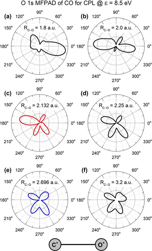
Journal of Physics B: Atomic, Molecular and Optical Physics

J. Phys. B: At. Mol. Opt. Phys. 53 (2020) 194002 (6pp)                                                                      https://doi.org/10.1088/1361-6455/aba3d3

Angular emission distribution of O 1s
photoelectrons of uniaxially oriented
methanol
L Kaiser1 , K Fehre1 , N M Novikovskiy2,3 , J Stindl1 , D Tsitsonis1 ,
G Gopakumar4, I Unger4,5 , J Söderström4, O Björneholm4, M Schöffler1 ,
T Jahnke1, R Dörner1 , F Trinter1,5,6,7 and Ph V Demekhin2,7
1
  Institut für Kernphysik, Goethe-Universität, Max-von-Laue-Str. 1, 60438 Frankfurt am Main, Germany
2
  Institut für Physik und CINSaT, Universität Kassel, Heinrich-Plett-Str. 40, 34142 Kassel, Germany
3
  Institute of Physics, Southern Federal University, 344090 Rostov-on-Don, Russia
4
  Department of Physics and Astronomy, Uppsala University, Box 516, 75120 Uppsala, Sweden
5
  Deutsches Elektronen-Synchrotron DESY, Notkestr. 85, 22763 Hamburg, Germany
6
  Molecular Physics, Fritz-Haber-Institut der Max-Planck-Gesellschaft, Faradayweg 4-6,
14195 Berlin, Germany

E-mail: trinter@atom.uni-frankfurt.de and demekhin@physik.uni-kassel.de

Received 24 April 2020, revised 8 June 2020
Accepted for publication 8 July 2020
Published 25 August 2020

Abstract
The angular distribution of O 1s photoelectrons emitted from uniaxially oriented methanol is
studied experimentally and theoretically. We employed circularly polarized photons of an
energy of hν = 550 eV for our investigations. We measured the three-dimensional
photoelectron angular distributions of methanol, with the CH3 –OH axis oriented in the
polarization plane, by means of cold target recoil ion momentum spectroscopy. The
experimental results are interpreted by single active electron calculations performed with the
single center method. A comparative theoretical study of the respective molecular-frame
angular distributions of O 1s photoelectrons of CO, performed for the same photoelectron
kinetic energy and for a set of different internuclear distances, allows for disentangling the role
of internuclear distance and the hydrogen atoms of methanol as compared to carbon monoxide.
Keywords: photoionization, methanol, molecular-frame photoelectron angular distributions

S Supplementary material for this article is available online

(Some figures may appear in colour only in the online journal)

1. Introduction                                                                          [2, 7–11], localization of core holes [12–16], electron corre-
                                                                                         lation [3, 17–22], multi-electron processes [17, 23–26], initial
In the emission of photoelectrons from molecules, the                                    electronic state [27–29], nuclear dynamics [30, 31], and allows
photoelectron’s angular emission distribution in the body-                               to probe Auger decay [32–34]. Such electron angular dis-
fixed frame of the molecule is an exquisitely sensitive probe                            tributions in the molecular frame of reference are typically
of molecular shape resonances [1–6], molecular structure                                 termed MFPADs (molecular-frame photoelectron angular dis-
                                                                                         tributions). From a more intuitive point of view, the MFPAD
7
    Authors to whom any correspondence should be addressed.                              arises as the outgoing photoelectron wave is multiply scattered
                  Original content from this work may be used under the terms            by the molecular potential. The observed distributions depend
                  of the Creative Commons Attribution 4.0 licence. Any further
distribution of this work must maintain attribution to the author(s) and the title       strongly on the electron energy (i.e. the electron wavelength)
of the work, journal citation and DOI.                                                   and the exact shape of the molecular potential [35, 36].

0953-4075/20/194002+6$33.00                                                          1       © 2020 The Author(s). Published by IOP Publishing Ltd   Printed in the UK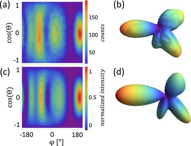
J. Phys. B: At. Mol. Opt. Phys. 53 (2020) 194002                                                                                 L Kaiser et al

    In order to access the body-fixed molecular frame, the spa-               Our present choice for studying MFPADs relates to the
tial orientation of the molecule needs to be known. In the gas             smallest alcohol, methanol (CH3 OH). Methanol is used not
phase, there are two main routes to address this experimental              only as a solvent, but it is also used in fuels and in direct-
problem. Firstly, the molecules can be aligned actively. The               methanol fuel cells, and can have potential future applications
field of active alignment can be partitioned further into several          in energy storage. Methanol has also been observed in space in
subsets. One approach utilizes the strong field of a laser to              relation to certain star-forming regions [46]. Although many
adiabatically align the molecule due to its polarizability. In             coincidence experiments on methanol have been performed
other sets of experiments, an impulsive alignment is achieved              using strong-field ionization [47–53], ion impact ionization
as a defined rotation of the molecule is induced by short laser            [54], VUV [55] and EUV [56] photoionization, only a lim-
pulses. In that case, an alignment of the molecule occurs                  ited number of soft x-ray coincidence studies on methanol
periodically after fixed time intervals, and revivals of the               exist [57–59]. In the present work, we examine the MFPADs
alignment are observable even for long times after the initial             of O 1s photoelectrons approximately 8.5 eV above the oxy-
laser pulse (i.e. the alignment reoccurs, see e.g. [37, 38]).              gen K-threshold. Although the orientation of molecules in the
Both approaches of ‘adiabatic’ and ‘dynamic’ alignment are                 gas phase is random, we are able to post-select cases where
well reviewed, for example by Stapelfeldt and Seideman [39].               the molecular CH3 –OH axis is oriented within the polariza-
Furthermore, DC fields from multi-pole electric structures                 tion plane (i.e., the plane normal to the Poynting vector) of
acting on molecular beams (see e.g. [40]) have been used as                the circularly polarized photons, using the relative momenta of
well, to actively align molecules. Experiments achieving an                the fragment ions. In contrast to linear polarization, circularly
alignment by investigating molecular adsorbates on surfaces                polarized light (CPL) does not introduce a preferable direc-
(see e.g. [41]) are also reported in the literature. The second            tion of emission in the polarization plane, but rather imprints a
chosen route, which is routinely employed in synchrotron-                  sense of its rotation on the emitted photoelectron wave. There-
related studies, consists of an a posteriori measurement of the            fore, using circular polarization has an advantage to sense
molecular orientation, and thus does not consist of an active              the molecular structure itself. In the industrially relevant pro-
spatial alignment of the molecule. In order for this approach              cess of catalytic hydrogenation to produce methanol, carbon
to be applicable, the molecule needs to dissociate during                  monoxide is the precursor. The structures of CO and CH3 OH
the ionization process. If the dissociation occurs rapidly, the            have their central C–O bond in common and differ only in the
emission direction of the ionic fragments corresponds (e.g. for            attached hydrogen atoms and in the C–O bond length. The
a diatomic molecule) to the direction of the molecular bond at             similarity between these molecules motivated us to perform
the instant of the ionization (an assumption which is known as             an extended theoretical study of the respective MFPAD of CO
the so-called ‘axial recoil approximation’ [42]). This approach            to compare the methanol data with.
is typically employed in cases, where several charges are
created after the primary ionization by Auger decay and is                 2. Methods
also the basis for Coulomb explosion imaging [43, 44].
    It is the latter approach, i.e. the extraction of molecular ori-       The experiments were carried out at the soft x-ray beamline
entation from measured data, which we chose for the exper-                 P04 of the synchrotron PETRA III (DESY, Hamburg, Ger-
imental part of our present study where we make use of the                 many) [60] in 40-bunch timing mode (bunch spacing 192 ns),
power of MFPADs to probe molecular structure. For the the-                 using circularly polarized photons of 550 eV photon energy
oretical part, we investigate by calculations, which we bench-             generated by its 5 m long APPLE-2 undulator. We used the per-
mark against our experiment on methanol, how variations in                 manently installed COLTRIMS (cold target recoil ion momen-
the molecular structure—such as bond length changes and                    tum spectroscopy) reaction microscope [61–63] for our stud-
addition of hydrogen atoms—influence the MFPADs. The                       ies. The methanol molecules were provided as a supersonic gas
photoelectron is released locally through core-level photoion-             jet, which passed two skimmers (300 μm diameter) and was
ization, and we observe the interference and scattering pat-               crossed with the photon beam at right angle. The methanol
tern of the emitted photoelectron wave. Due to the positive                reservoir was heated to 316 K (vapor pressure of approxi-
charges of the nuclei and the density distribution of electrons            mately 400 mbar), and by also heating up the gas line to
in a molecule, the photoelectron wave can be preferentially                319 K and the gas nozzle of 100 μm diameter to 328 K
emitted (focused) in the direction of their neighboring atoms              (vapor pressure of approximately 680 mbar), we achieved
(for example, in diatomic molecules the photoelectron tends                suitable conditions at the supersonic expansion of the vapor.
to an emission towards the other atom) or scattered by them                The COLTRIMS spectrometer employed consisted of an ion
[35]. These effects depend on the energy of the photoelectrons,            arm of 7 cm length and an electron arm of 15 cm length.
on the respective internuclear separations, and on the nuclear             Both were equipped with a micro-channel plate detector
charges. For instance, the MFPADs of C 1s photoelectrons                   (active area of 80 mm diameter) with hexagonal delay-line
of methane directly illustrate the three-dimensional molecular             position readout [64, 65]. For the ion detection, a funnel
structure [8, 9]. In diatomic molecules, the internuclear dis-             micro-channel plate [66] was used to achieve high coincidence
tance has been mapped by such photoelectron interference in                detection efficiency. Electrons and ions were guided by homo-
single-photon ionization [9, 10, 45] or strong-field ionization            geneous electric (21.9 V cm−1 ) and magnetic fields (6.3 G)
[11]. In summary, the emitted photoelectron wave illuminates               onto the two time- and position-sensitive detectors. These
the molecular potential from within [2].                                   fields were selected such that 4π collection solid angle for

                                                                       2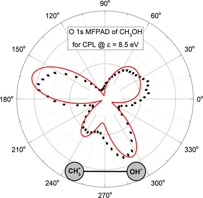
J. Phys. B: At. Mol. Opt. Phys. 53 (2020) 194002                                                                                     L Kaiser et al

electrons and ions has been achieved for electrons up to
30 eV kinetic energy and molecular fragmentation with a
kinetic energy release up to 20 eV. From the times-of-flight
and the positions-of-impact, the three-dimensional momentum
vectors of all charged fragments of the photoreaction were
retrieved. In addition, the time-of-flight measurement allowed
identifying different breakup channels. We focus on the case
of O 1s-photoionization followed by Auger decay here, and a
fragmentation of the molecule into CH3 + /OH+ . These frag-
ments were detected in coincidence with the O 1s photoelec-
trons of 8.5 eV kinetic energy. The corresponding events were
selected by gating on the time-of-flight coincidence of the
two photoions and the photoelectron and on the photoelectron
kinetic energy.
    For CPL and within the electric dipole approximation, the
MFPAD of a fixed-in-space (i.e. spatially aligned) molecule
is given by the following differential photoionization cross
section:
                                                               2
  dσ ±1


          αβγ, θ ϕ =  (−i) Dk±1 (αβγ) Aε mk Y m (θ ϕ ) ,
                                     1
   dΩ
                            mk
                                                                 (1)
here, ±1 stand for the positive and negative helicity of the CPL;
D stands for the Wigner rotation matrix, which depends on the
three molecular-orientation Euler angles{α, β, γ}; Y stands for
the spherical harmonics; and θ , ϕ are the photoelectron emis-
sion angles in the molecular frame of reference. The electron
dynamics of the photoionization process are imprinted on the
MFPAD in equation (1) through the dipole transition ampli-
tudes Aε mk for the emission of the partial photoelectron contin-
uum waves |ε m [67] with given energy and angular momen-
tum quantum numbers via the absorption of a photon of polar-
ization k. In our modeling, those amplitudes were computed by
using the stationary single center (SC) method [68, 69], which             Figure 1. Predicted angular emission distributions of O 1s
provides an accurate theoretical description of the angle-                 photoelectrons of CO, computed for CPL with negative helicity and
resolved photoemission spectra of molecules [70–84]. The                   a photoelectron kinetic energy of 8.5 eV at different internuclear
calculations were performed in the frozen-core Hartree–Fock                distances, as indicated in each panel. The light propagates out of the
                                                                           page plane (the polarization plane), and the photoelectrons are
approximation at the equilibrium internuclear geometry for                 emitted within this plane. The molecule is oriented to lie within the
methanol and at different internuclear separations for carbon              polarization plane with the oxygen ion pointing to the right, as
monoxide. The SC expansions of the occupied and continuum                  indicated at the bottom. The internuclear distances in panels (c) and
orbitals with respect to the central point between the C and O             (e) correspond to the equilibrium distances of CO and CH3 OH,
atoms were restricted to the partial harmonics with , |m|  49             respectively.
and , |m|  29, respectively. Because of the axial symmetry of
the CPL, the orientation angle γ, which describes the rotation             present theoretical approach, we firstly reproduced available
around the light propagation direction (the laboratory z-axis),            experimental MFPADs of O 1s photoelectrons from reference
is irrelevant. In order to put the molecular z -axis (chosen along        [25] (see figure 15 in reference [25]), which were measured for
the C–O bond) in the polarization plane (the laboratory xy-                linearly polarized light and somewhat different kinetic energy
plane), the orientation angle β is set to 90◦. Finally, since only         of 11.7 eV. The agreement is excellent (not shown here for
the C–O bond of methanol was fixed in space in the exper-                  brevity). In the next step, MFPADs of O 1s photoelectrons of
iment, the computed MFPADs have been integrated over the                   CO were computed for left-handed CPL and a kinetic energy
orientation angle α, which describes the rotation around the               of 8.5 eV (as in the present experiment for methanol). The cal-
molecular z -axis, and the emission angle ϕ must be reset                culations have been performed for different internuclear sepa-
accordingly.                                                               rations, from somewhat smaller than the equilibrium internu-
                                                                           clear distance of carbon monoxide (RC – O = 2.132 a.u. [85])
3. Results and discussion                                                  to somewhat larger than that of methanol (RCH3 −OH = 2.696
                                                                           a.u. [86]). The results are summarized in figure 1. As one can
We start our discussion with the theoretical results obtained              see from this figure, the lobe, which points towards the C+
for carbon monoxide. To confirm the reliability of the                     ion, develops dramatically with the increase of the internuclear

                                                                       3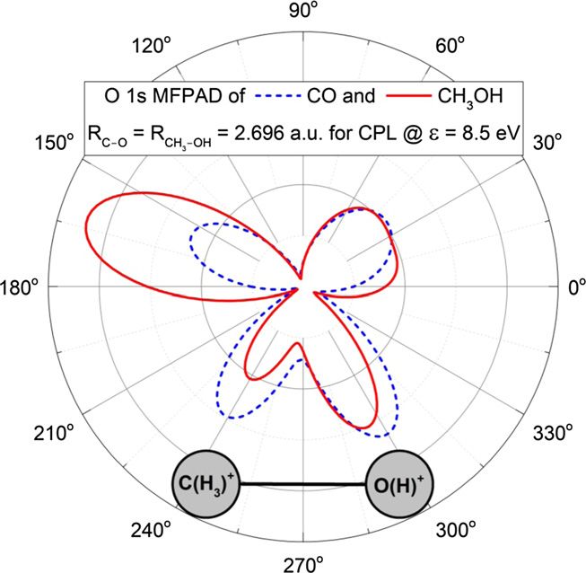
J. Phys. B: At. Mol. Opt. Phys. 53 (2020) 194002                                                                                   L Kaiser et al

Figure 2. Comparison of the MFPADs of O 1s photoelectrons of              Figure 3. Comparison of the measured (solid circles with error
CO and CH3 OH, computed for CPL with negative helicity and                bars) and computed (red curve) two-dimensional MFPADs of O 1s
photoelectron kinetic energy of 8.5 eV at the equilibrium                 photoelectrons of methanol, obtained for CPL with negative helicity
internuclear distance RC – O = RCH3 −OH = 2.696 a.u. of CH3 OH.           and photoelectron kinetic energy of 8.5 eV. The light propagates out
The MFPAD of CO is the same as in figure 1(e). The light                  of the page plane (the polarization plane). The MFPADs include all
propagates out of the page plane (the polarization plane), and the        photoelectrons, which fall in the opening angle of ±12◦ , and the
photoelectrons are emitted within this plane. The molecules are           molecular CH3 –OH axis is oriented within the opening angle of
oriented in the polarization plane with the O+ or OH+ ions pointing       ±15◦ , both around the polarization plane. The OH+ ion points to
to the right, as indicated at the bottom.                                 the right, as indicated at the bottom.

distance. At RC–O = 1.8 a.u. in figure 1(a), it is some-                  MFPAD. In particular, they recover the main lobe pointing
what suppressed, and for larger distance of RC–O = 2.0 a.u.               towards the CH3 + ion, as compared to the MFPAD of CO from
in figure 1(b), becomes strongly enhanced. The computed                   figure 1(e) (shown here for reference by the blue dashed curve),
MFPAD shows its well-known form at the equilibrium inter-                 to the form of the MFPAD of CO from figure 1(c) obtained
nuclear distance RC–O = 2.132 a.u. of CO in figure 1(c) (red              for its equilibrium internuclear distance RC – O = 2.132 a.u.
curve). Further on, in figures 1(d)–(f ), the main lobe contin-           Detailed interpretation of this effect requires an accurate anal-
uously shrinks with the increase of the internuclear distance             ysis of multiple-scattering effects of photoelectron waves in
across the equilibrium distance RC–O = 2.696 a.u. of CH3 OH               the ionic potential of methanol, which is a cumbersome task.
(blue curve in figure 1(e)). Not surprisingly increasing the              However, we suggest that the focusing effect plays a domi-
bond length at fixed photoelectron wavelength results in a sim-           nant role in this case. In particular, photoelectron waves emit-
ilar trend as decreasing the photoelectron wavelength at a given          ted in the direction of the carbon atom experience a large
bond length [17, 87].                                                     uncompensated positive charge of its nucleus, which works
    In the next step, we inspected the MFPADs of carbon                   as a lens and focuses photoelectron waves in this direction.
monoxide using the equilibrium distance RC–O = 2.696                      In methanol, the positive charge of each additional proton of
a.u. of CH3 OH. Starting from this model system, we                       the methyl group (CH3 ) introduces its own focusing, enhanc-
then built the methanol molecule by introducing one by                    ing thereby the effect of the carbon nucleus alone in CO. The
one additional hydrogen atoms, first on the oxygen site,                  CPL additionally imprints its rotational sense on the photo-
and then on the carbon site. For brevity, we compare in                   electron waves, causing thereby a slight clockwise rotation of
figure 2 the final result for the complete methanol molecule              the emission distribution along the rotation of the electric field
with all four hydrogen atoms to that computed for CO                      vector.
using an internuclear distance of RC–O = 2.696 a.u. The                      The measured results for methanol are compared to our
full account of calculations can be found in figure S1                    theoretical findings in figures 3 and 4. Figure 3 illustrates
of the supplemental material, which can be found online                   an excellent agreement between the computed and measured
at https://stacks.iop.org/JPB/53/194002/mmedia. We observe                dipole-plane MFPADs of methanol (note that the depicted data
that introducing hydrogen atoms systematically increases the              accounts for opening angles of ±12◦ and ±15◦ around the
main lobe pointing towards the carbon site in the computed                polarization plane, respectively, for the photoelectron and the
MFPAD, as compared to the MFPAD of CO at this internu-                    CH3 –OH axis, as in the experiment). A similarly excellent
clear distance (depicted in figure 1(e)). Figure 2 illustrates            agreement between the modeling and the experiment is also
the strong impact of the hydrogen atoms on the computed                   evident from figure 4. It compares the full three-dimensional

                                                                      4
J. Phys. B: At. Mol. Opt. Phys. 53 (2020) 194002 L Kaiser et al
Acknowledgments
We acknowledge DESY (Hamburg, Germany), a member of
the Helmholtz Association HGF, for the provision of exper-
imental facilities. Parts of this research were carried out
at PETRA III within the project I-20180746-EC, and we
would like to thank the staff of beamline P04 for excel-
lent support during the beam time. This research was sup-
ported in part through the Maxwell computational resources
operated at DESY. The experiments were supported by
Deutsche Forschungsgemeinschaft via Sonderforschungsbere-
ich 1319 (ELCH) and by the Bundesministerium für Bildung
und Forschung (BMBF, Federal Ministry of Education and
Research). The theoretical work was supported by the DFG
Project DE 2366/1-2. KF acknowledges support by the Ger-
Figure 4. Comparison of the measured ((a) and (b)) and computed man National Merit Foundation. GG, IU, JS, and OB acknowl-
((c) and (d)) three-dimensional MFPADs of O 1s photoelectrons of edge support from the Swedish Research Council project
methanol, obtained for CPL with negative helicity and photoelectron 2017-04162.
kinetic energy of 8.5 eV. In panels (b) and (d), the light propagates
out of the page plane (the polarization plane), and the molecule is
oriented horizontally with the OH+ ion pointing to the right. The ORCID iDs
data include the CH3 + /OH+ breakups (molecular axis orientations),
which fall in the opening angle of ±15◦ around the polarization
plane. Panels (a) and (c) show the same three-dimensional MFPADs K Fehre https://orcid.org/0000-0003-2519-5564
in a color-map representation as functions of the polar and azimuthal J Stindl https://orcid.org/0000-0002-5041-2404
angles. In this representation, the light propagation direction points D Tsitsonis https://orcid.org/0000-0002-8748-6677
from cos (θ) = −1 to cos (θ) = 1. R Dörner https://orcid.org/0000-0002-3728-4268
F Trinter https://orcid.org/0000-0002-0891-9180
MFPADs of methanol in two representations: via a color-map Ph V Demekhin https://orcid.org/0000-0001-9797-6648
as a function of the spherical angles (panels (a) and (c) in
the left column) and as three-dimensional figures where the
angular variation of the photoelectron yield is encoded in the References
distance of the surface from the origin (panels (b) and (d) in
the right column). [1] Shigemasa E, Adachi J, Oura M and Yagishita A 1995 Phys.
Rev. Lett. 74 359
[2] Landers A et al 2001 Phys. Rev. Lett. 87 013002
4. Conclusions [3] Cherepkov N A, Semenov S K, Hikosaka Y, Ito K, Motoki S
and Yagishita A 2000 Phys. Rev. Lett. 84 250
We investigated angular distributions of O 1s photoelectrons [4] Shigemasa E, Adachi J, Soejima K, Watanabe N, Yagishita A
emitted from uniaxially oriented methanol molecules. The and Cherepkov N A 1998 Phys. Rev. Lett. 80 1622
[5] Jahnke T et al 2002 Phys. Rev. Lett. 88 073002
molecular CH3 –OH axis has been fixed in the polarization [6] De Fanis A et al 2002 Phys. Rev. Lett. 89 023006
plane of the circularly polarized ionizing radiation. The exper- [7] Williams J B et al 2012 Phys. Rev. Lett. 108 233002
imental angular distributions have been recorded using the [8] Williams J B et al 2012 J. Phys. B: At. Mol. Opt. Phys. 45
COLTRIMS technique at the synchrotron radiation facility 194003
PETRA III (DESY). The measured three-dimensional photo- [9] Kreidi K et al 2008 Phys. Rev. Lett. 100 133005
[10] Schöffler M S et al 2008 Phys. Rev. A 78 013414
electron momentum distributions of methanol are in a very [11] Kunitski M et al 2019 Nat. Commun. 10 1
good agreement with those computed by the SC method and [12] Schöffler M S et al 2008 Science 320 920
code. In order to understand the findings in more detail, [13] Kreidi K et al 2008 J. Phys. B: At. Mol. Opt. Phys. 41 101002
we calculated O 1s photoelectrons of carbon monoxide at [14] McCurdy C W et al 2017 Phys. Rev. A 95 011401
the same photoelectron kinetic energy. These two molecules [15] Saito N et al 2003 J. Phys. B: At. Mol. Opt. Phys. 36 L25
[16] Zimmermann B et al 2008 Nat. Phys. 4 649
have the following main differences: firstly, the C–O bond
[17] Martin F et al 2007 Science 315 629
of methanol is about 25% longer than that of CO. Enlarg- [18] Waitz M et al 2017 Nat. Commun. 8 2266
ing this distance results in a considerable suppression of the [19] Waitz M et al 2016 Phys. Rev. Lett. 116 043001
main lobe pointing towards the carbon atom in the computed [20] Waitz M et al 2016 Phys. Rev. Lett. 117 083002
MFPAD of CO. Secondly, methanol has four additional hydro- [21] Liu X-J et al 2008 Phys. Rev. Lett. 101 023001
gen atoms as compared to CO. The additional protons of the [22] Liu X-J et al 2008 Phys. Rev. Lett. 101 083001
[23] Lebech M, Houver J C, Dowek D and Lucchese R R 2006 Phys.
methyl group seem to enlarge the focusing effect of the car- Rev. Lett. 96 073001
bon nucleus and recover thereby the corresponding main lobe [24] Golovin A V, Heiser F, Quayle C J K, Morin P, Simon M,
in the computed distribution to its experimentally observed Gessner O, Guyon P-M and Becker U 1997 Phys. Rev. Lett.
form. 79 4554
5J. Phys. B: At. Mol. Opt. Phys. 53 (2020) 194002 L Kaiser et al
[25] Jahnke T et al 2004 J. Electron Spectrosc. Relat. Phenom. 141 [60] Viefhaus J, Scholz F, Deinert S, Glaser L, Ilchen M, Seltmann
229 J, Walter P and Siewert F 2013 Nucl. Instrum. Methods Phys.
[26] Akoury D et al 2007 Science 318 949 Res. A 710 151
[27] Arasaki Y, Takatsuka K, Wang K and McKoy V 2010 J. Chem. [61] Dörner R, Mergel V, Jagutzki O, Spielberger L, Ullrich J,
Phys. 132 124307 Moshammer R and Schmidt-Böcking H 2000 Phys. Rep. 330
[28] Hockett P, Bisgaard C Z, Clarkin O J and Stolow A 2011 Nat. 95
Phys. 7 612 [62] Ullrich J, Moshammer R, Dorn A, Dörner R, Schmidt L P H and
[29] Mignolet B, Levine R D and Remacle F 2012 Phys. Rev. A 86 Schmidt-Böcking H 2003 Rep. Prog. Phys. 66 1463
053429 [63] Jahnke T et al 2011 J. Electron Spectrosc. Relat. Phenom. 183
[30] Kircher M et al 2019 Phys. Rev. Lett. 123 243201 48
[31] Sturm F P et al 2009 Phys. Rev. A 80 032506 [64] Jagutzki O, Mergel V, Ullmann-Pfleger K, Spielberger L, Spill-
[32] Cherepkov N A et al 2009 Phys. Rev. A 80 051404 mann U, Dörner R and Schmidt-Böcking H 2002 Nucl.
[33] Semenov S K et al 2010 Phys. Rev. A 81 043426 Instrum. Methods Phys. Res. A 477 244
[34] Weber T et al 2003 Phys. Rev. Lett. 90 153003 [65] Jagutzki O, Lapington J S, Worth L B C, Spillman U, Mergel V
[35] Söderström J et al 2012 Phys. Rev. Lett. 108 193005 and Schmidt-Böcking H 2002 Nucl. Instrum. Methods Phys.
[36] Travnikova O et al 2019 J. Phys. Chem. A 123 7619 Res. A 477 256
[37] Wu J et al 2011 Phys. Rev. A 83 061403 [66] Fehre K et al 2018 Rev. Sci. Instrum. 89 045112
[38] Wu J, Magrakvelidze M, Vredenborg A, Schmidt L P H, Jahnke [67] Cherepkov N A 1981 J. Phys. B: At. Mol. Opt. Phys. 14 2165
T, Czasch A, Dörner R and Thumm U 2013 Phys. Rev. Lett. [68] Demekhin P V, Ehresmann A and Sukhorukov V L 2011 J.
110 033005 Chem. Phys. 134 024113
[39] Stapelfeldt H and Seideman T 2003 Rev. Mod. Phys. 75 543 [69] Galitskiy S A, Artemyev A N, Jänkälä K, Lagutin B M and
[40] Rakitzis T P, van den Brom A J and Janssen M H M 2004 Demekhin P V 2015 J. Chem. Phys. 142 034306
Science 303 1852 [70] Demekhin P V, Petrov I D, Sukhorukov V L, Kielich W, Reiss
[41] Lablanquie P et al 1989 Phys. Rev. A 40 5673 P, Hentges R, Haar I, Schmoranzer H and Ehresmann A 2009
[42] Zare R N 1972 Mol. Photochem. 4 1 Phys. Rev. A 80 063425
[43] Vager Z, Naaman R and Kanter E P 1989 Science 244 426 Demekhin P V, Petrov I D, Sukhorukov V L, Kielich W, Reiss
[44] Pitzer M et al 2013 Science 341 1096 P, Hentges R, Haar I, Schmoranzer H and Ehresmann A 2010
[45] Fukuzawa H et al 2019 J. Chem. Phys. 151 104302 Phys. Rev. A 81 069902
[46] Pilling S, Neves R, Santos A C F and Boechat-Roberty H M [71] Demekhin P V, Petrov I D, Tanaka T, Hoshino M, Tanaka H,
2007 Astron. Astrophys. 464 393 Ueda K, Kielich W and Ehresmann A 2010 J. Phys. B: At.
[47] Okino T, Furukawa Y, Liu P, Ichikawa T, Itakura R, Hoshina Mol. Opt. Phys. 43 065102
K, Yamanouchi K and Nakano H 2006 Chem. Phys. Lett. 419 [72] Demekhin P V, Petrov I D, Sukhorukov V L, Kielich W, Knie
223 A, Schmoranzer H and Ehresmann A 2010 Phys. Rev. Lett.
[48] Okino T, Furukawa Y, Liu P, Ichikawa T, Itakura R, Hoshina 104 243001
K, Yamanouchi K and Nakano H 2006 Chem. Phys. Lett. 423 [73] Demekhin P V, Petrov I D, Sukhorukov V L, Kielich W, Knie A,
220 Schmoranzer H and Ehresmann A 2010 J. Phys. B: At. Mol.
[49] Okino T, Furukawa Y, Liu P, Ichikawa T, Itakura R, Hoshina K, Opt. Phys. 43 165103
Yamanouchi K and Nakano H 2006 J. Phys. B: At. Mol. Opt. [74] Knie A et al 2014 Phys. Rev. A 90 013416
Phys. 39 S515 [75] Antonsson E, Patanen M, Nicolas C, Benkoula S, Neville J J,
[50] Itakura R, Liu P, Furukawa Y, Okino T, Yamanouchi K and Sukhorukov V L, Bozek J D, Demekhin P V and Miron C
Nakano H 2007 J. Chem. Phys. 127 104306 2015 Phys. Rev. A 92 042506
[51] Xu H, Marceau C, Nakai K, Okino T, Chin S-L and Yamanouchi [76] Sann H et al 2016 Phys. Rev. Lett. 117 263001
K 2010 J. Chem. Phys. 133 071103 [77] Knie A, Patanen M, Hans A, Petrov I D, Bozek J D, Ehresmann
[52] Xu H, Okino T, Kudou T, Yamanouchi K, Roither S, Kitzler A and Demekhin P V 2016 Phys. Rev. Lett. 116 193002
M, Baltuska A and Chin S-L 2012 J. Phys. Chem. A 116 [78] Nandi S, Nicolas C, Artemyev A N, Novikovskiy N M, Miron
2686 C, Bozek J D and Demekhin P V 2017 Phys. Rev. A 96
[53] Fukahori S, Nakano M, Yamanouchi K and Itakura R 2017 052501
Chem. Phys. Lett. 672 7 [79] Ilchen M et al 2017 Phys. Rev. A 95 053423
[54] De S, Roy A, Rajput J, Ghosh P N and Safvan C P 2008 Int. J. [80] Tia M et al 2017 J. Phys. Chem. Lett. 8 2780
Mass Spectrom. 276 43 [81] Mhamdi A et al 2018 Phys. Rev. Lett. 121 243002
[55] Borkar S, Sztaray B and Bodi A 2011 Phys. Chem. Chem. Phys. [82] Mhamdi A et al 2018 Phys. Rev. A 97 053407
13 13009 [83] Mhamdi A, Rist J, Havermeier T, Dörner R, Jahnke T and
[56] Luzon I, Jagtap K, Livshits E, Lioubashevski O, Baer R and Demekhin P V 2020 Phys. Rev. A 101 023404
Strasser D 2017 Phys. Chem. Chem. Phys. 19 13488 [84] Hartmann G et al 2019 Phys. Rev. Lett. 123 043202
[57] Hempelmann A, Piancastelli M N, Heiser F, Gessner O, Rüdel [85] Huber K P and Herzberg G 1979 Molecular Spectra and
A and Becker U 1999 J. Phys. B: At. Mol. Opt. Phys. 32 Molecular Structure. IV. Constants of Diatomic Molecules
2677 (Princeton, NJ: Van Nostrand-Reinhold)
[58] Pilling S, Boechat-Roberty H M, Santos A C F and de Souza G [86] Venkateswarlu P and Gordy W 1955 J. Chem. Phys. 23 1200
G B 2007 J. Electron Spectrosc. Relat. Phenom. 155 70 [87] Kushawaha R K, Patanen M, Guillemin R, Journel L, Miron C,
[59] Kivimäki A, Strahlman C, Richter R and Sankari R 2018 J. Phys. Simon M, Piancastelli M N, Skates C and Decleva P 2013
Chem. A 122 224 Proc. Natl Acad. Sci. USA 110 15201
6You can also read