Approach to Cut-Up; Macroscopic Examination as the Precursor to Accurate Microscopic Interpretation - Breast - Sarah E Pinder Professor of Breast ...
←
→
Page content transcription
If your browser does not render page correctly, please read the page content below

Approach to Cut-Up; Macroscopic
  Examination as the Precursor to
Accurate Microscopic Interpretation
             – Breast
            Sarah E Pinder

     Professor of Breast Pathology
        King’s College London
     Guy’s & St Thomas’ Hospitals
https://www.rcpath.org/resourceLibrary/g
148-breastdataset-hires-jun16-pdf.html
Breast Specimen Handling
         Principles – Clinical Information
Request form with appropriate clinical information
  including:
• Name, date of birth

•   Surgical procedure – diagnostic or therapeutic
•   Side & site of lesion in the breast
•   Nature of lesion - microcalcification, mass, deformity
•   Single or multiple foci, neo-adjuvant therapy etc

Specimen:
• Appropriate orientation sutures and/or clips
• X-ray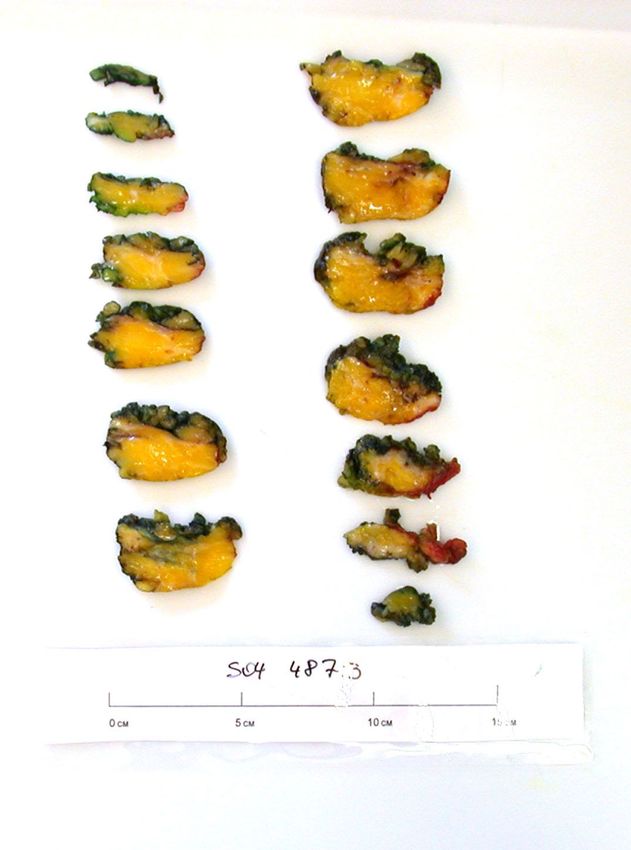
Breast Specimen Handling
        Principles – Surgical Protocols

• Lesions should be surgically resected and
    orientated according to a defined (ABS)
    protocol
•   If the surgical resection differs, this should be
    discussed
Diagnostic Surgery
Therapeutic Breast Surgery (1)

                     Anterior = skin

                     Posterior = fascia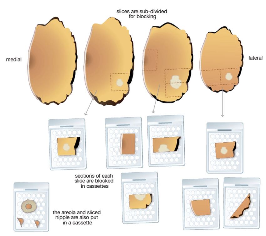
Therapeutic Breast Surgery (2)

                     Anterior = breast

                     Posterior = fascia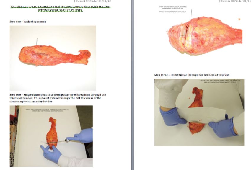
Specimen orientation
                Sutures or clips
According to local protocol
e.g.
Long – Lateral
Short – Superior
(Medium – Medial)
or
1 = Anterior
2 = Superior
3 = Nipple margin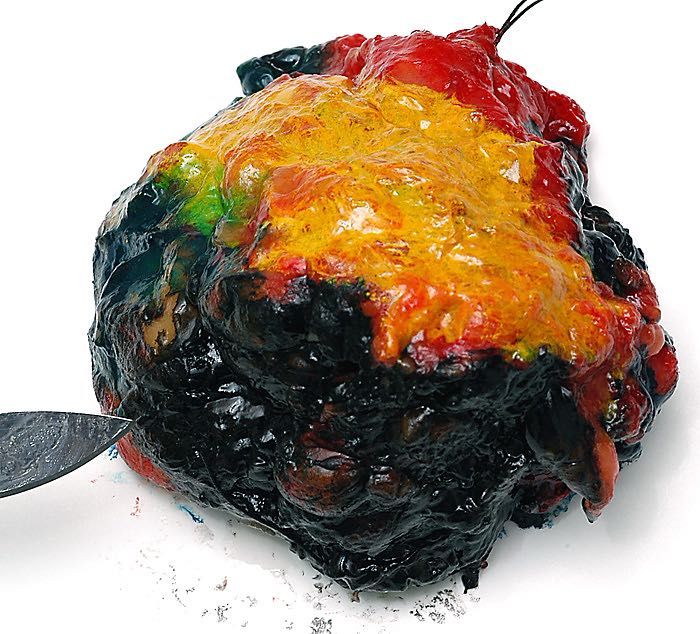
Specimen Handling
•   Measure
                           Practice
•   Weigh
•   Ink – standard protocol

     •   Anterior, red
     •   Lateral, orange
     •   Deep, black
     •   Medial, green
     •   Superior, blue
     •   Inferior, yellow
• Incise fresh for optimal fixation (& bank tissue)
• e.g. paper towels along incision(s) to act as wicks
• Wrap in something (e.g. paper towels/hair nets) to
  maintain shape
• Fix overnight
Minimum Dataset
Invasive Breast Carcinoma

• Histological grade
• Lympho-vascular invasion         Fixation
• Oestrogen receptor status

• Size
• Axillary nodes        Operator
• Excision margins
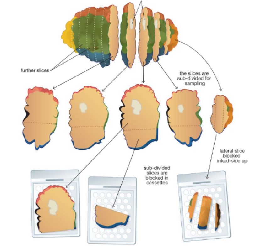
How to Slice?
        •   3 main approaches
        •   Depends on size & shape
            of specimen, lesion type &
            personal preference:
            1. “Bread-slice” - medial to
               lateral or superior to
               inferior
            2. “Bread-slice” - anterior
               to posterior
            3. Cruciate

Think before you cut………
What is lesion? Where is lesion? Nearest margin? Is there any other abnormality?
Cruciate - mass lesion
Margins – sample all, even if
 distant macroscopically
 Macroscopic   Total no.    Carcinoma within
 distance to   margins       5mm of radial
 margin
                           DCIS or invasive ca
 0-1mm         13               12 (92%)
 2-4mm         46               24 (52%)
 5-9mm         199              42 (21%)
 10-20mm       1044             67 (6%)
 21-30         439              20 (5%)
 31mm+         143               3 (2%)
 Total         1884             168 (9%)

         Hodi Z et al. Histopathol 2010;56:573-80
Excision of DCIS
Prediction of Disease Extent by Radiology
Comedo / Solid - 85% of area visible
Micropapillary / Cribriform - 50% of area visibleCalcification
Slicing from anterior to posterior
Audit
•   101 cases invasive breast cancer at STH; 63 (62%)
    examined macroscopically by bread-slicing method;
    38 cases (38%) by cruciates
•   In bread-slicing, medial to lateral measurement was
    largest plane in 44% c.f. 58% of cases with cruciates
•   Overall 50% of cases medial to lateral measurement
    was largest (average 18mm) cf superior to inferior
    (average 14mm) & anterior to posterior (average
    15mm)
    Max Whibley, unpublished
Specimen Handling
Practice - Mastectomy
• Specimen ideally arrives fresh
• Orientated
• (Ink – anterior / posterior margins)
• Slice
• Fix
• (X-ray)Fixation - Mitoses
9
8
2.6
7
2.4
6 2.2
2
5
1.8
4 1.6
1.4
3
1.2
30 minutes
60 minutes
2
No delay
1
0
Time 0 60 mins
Significant decrease after 60 mins. delay (p = 0.016)Fixation - Overall Grade
Grade
1 Hour Delay
1 2 3 Total
Grade 1 3 0 0 3
No delay 2 0 6 1 7
3 0 7 8 15
Total 3 13 9 25
7 (28%) cases lower overall grade at 1 hour delaySections of the nipple & quadrants
in mastectomy specimens
• 259 consecutive mastectomies
• New diagnosis of Paget s disease in 3 (1%)
• All 4 quadrants sampled in 230
• Unsuspected multifocality microscopically in
quadrant sections in 14, in nipple in 3, in both
in 1 (total = 8%)
• Such findings do not affect patient
management
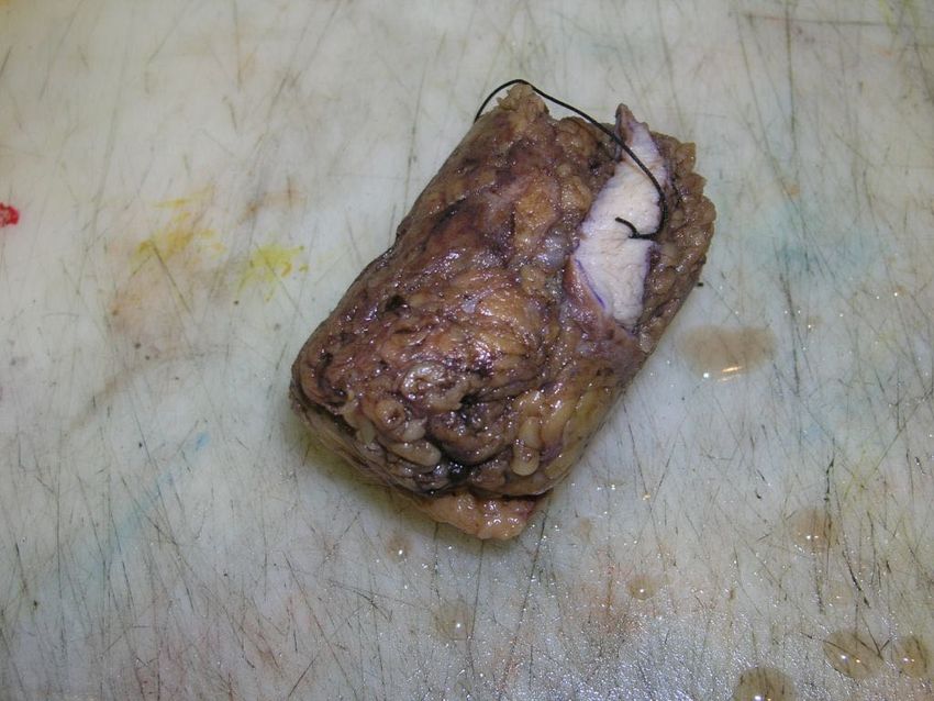
Sikand et al. J Clin Pathol. 2005;58:543-5Image courtesy of Colin Purdie
Neoadjuvant Therapy Specimens
If, on slicing, there is no obvious tumour check if
marker coil inserted, site on pre-operative core/FNAC
report, search for area of stellate fibrous scarring &
thoroughly sample, including margins

                            Image courtesy of Sami Shousha
Axillary Clearances Minimum standard • Every lymph node examined histologically • Total number of nodes assessable - at least 1 slice per node • Allows multiple nodes per block Ideal method One node per cassette - multiple slices Do not: • Bisect some nodes & include in a cassette with intact nodes
Sentinel Lymph Nodes

• UK guidelines - methodology should provide highest
  chance of finding metastases on routine H&E
• Each node sliced thinly (2mm or less) perpendicular
  to long axis, blocked separately and all embedded
• Lymph nodes 4mm or less should be bisected

• Levels not routine
• IHC if suspicious cells identified (AE1/AE3)
SLN Histopathological Handling
                  Slicing
Lymph node
 2mm slices
                  2      3     4     5
              1

Metastasis
                      Macrometastasis > or = 2mm
   4    1 2           Micrometastasis 0.2mm
       5 3            ITCs
Handling SLNs - Levels/Step Sections
2mm
Slice 2
Level 1 Level 2 Level 3
4 1 2 4 1 2 4 1
2
5 3 5 3 5 3
Identification of an isolated tumour cell would
require 312 sections of a 1cm LNSummary / Take-home messages
• Good clinical liaison and knowledge of local
protocols (e.g. margin widths)
• Think before slicing each case
• If bread-slicing consider slicing ‘horizontally’
• Sample ALL radial margins
• Sample beyond extent of lesion, particularly
for calcifications
• Concentrate on the tumour rather than
random quadrants and nipple
• Slice SLNs as thinly as possible
• Report to minimum dataset/proformasYou can also read