Artificial Intelligence for Medical Imaging and Treatment Planning
←
→
Page content transcription
If your browser does not render page correctly, please read the page content below
Artificial Intelligence for Medical Imaging
          and Treatment Planning

       Lei Xing, PhD, DABR, Jacob Haimson Professor

 Departments of Radiation Oncology & Electrical Engineering (by
     courtesy), Human-Centered Artificial Intelligence (HAI)
                      Stanford University

                                                       Stanford University
                                                  Department of Radiation Oncology
                                                                School of Medicine
Expert   Robotics
           system

 NLP       Vision

Machine
learning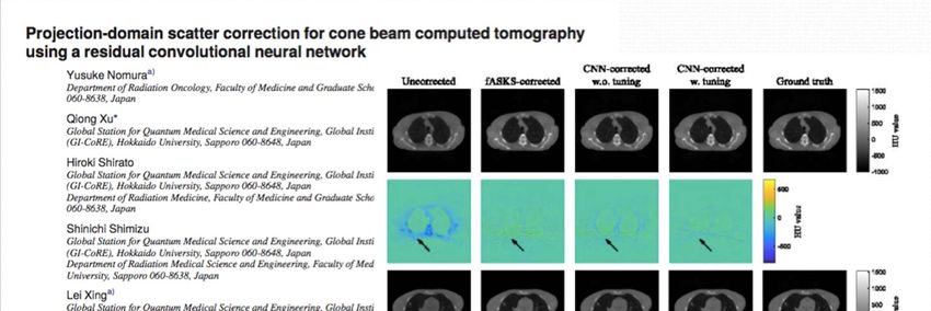
Artificial Intelligence and its Applications in Medicine

Machine Learning/          Computer Vision        Natural Language              Expert System        Robotics & Control
 Deep Learning                                    Processing (NLP)

            TECHNOLOGY
    ‐ New algorithms for improved                                                          DATA & DATABASE
     classification, detection,                                                        ‐ Data curation & augmentation
     segmentation & other image                     FUNDAMENTALS                       ‐ Data harmonization & mining
     analysis tasks.                      ‐ Data science & mathematical                ‐ Data sharing & security
    ‐ NLP tools for medical semantics     framework                                    ‐ Federated learning
      & search                                                                         ‐ Search engine (data, text,
    ‐ Enhancement & expansion of          ‐ High performance computing                  audio, video, image, etc.)
     existing AIM techniques              (GPU/TPU/multi‐core CPU, cloud
                                          computing, quantum computing)
                                          ‐ Analytics tools & algorithms (data
                                          dimensionality reduction, visualization,
                                          compression, various machine                  OTHER RELATED ISSUES
             APPLICATIONS                 learning/deep learning algorithms)
     ‐ AI augmented medical devices                                                    ‐ Training of future physicians,
     & wearables.                         ‐ Basic machine learning software              healthcare professionals, &
     ‐ Analysis of biological, imaging,   platforms                                      next generation of AI
     EMR, and therapeutic data for                                                       workforce.
     clinical decision‐making.                                                         ‐ Economic, politic, social, ethic
     ‐ Robotic interventions.                                                            and legal issues.
     ‐ Biomarker discovery& drug                                                       ‐ Workflow and clinical
     design.                                                                             implementation.

Machine Learning/Deep Learning
    Input and Output      Applications & Examples
   Data for AI Modeling

                          ‐ Superresolution
 Mapping between the        imaging
  same data domains       ‐ Image search
                          ‐ Image inpaiting

                           ‐ Image reconstruction
Data domains related by    ‐ sparse data problem
     known law(s)          ‐ Modeling physical/
                           mathematical relation

                           ‐‐Modeling of
                           therapeutic response
                           ‐ Drug design &
Data domains related by    biomarker discovery
 empirical evidence or     ‐ Translation, semantic
   measurement(s)          analysis
                           ‐ Auto‐annotation
                           ‐ Modeling correlative
                           relationship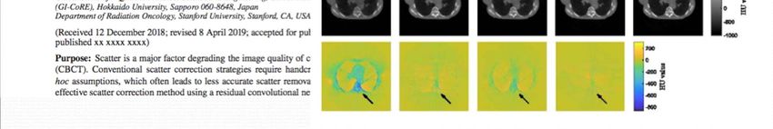
Types of learning
Modeling                  Treatment planning                  Image-guided patient                 Follow up
Imaging
                                                                                                   setup & delivery

         Image
     reconstruction
                        Different     OAR Artifacts
                        modalitiessegmentation
                                           removal         supervised  Supervised
                                                                Sparse data
                                                                        planning
                                                                                       …....
                                                                             unsupervised
                                                                                                 …....
                                                                                                   Network
                                                                                                       Monoscopic
                                                                                                   structure
                                                                                                         imaging
                                                                                                                        …....
                                                                                                                                     Therapeutic
                                                                                                                                 …....response
         High                          TumorDynamic
      dimensional
        imaging
                        Real‐time    detectioncontrast
                                               &
                                              imaging
                                    segmentation
                                                                Functional
                                                           Detection
                                                                               Muli‐ …..
                                                                     Unsupervised
                                                                 imaging     modalities          …..   Stereoscopic
                                                                                                       …..
                                                                                                         imaging                 ….. Survival
     Sparsification
     compression
                          Sparse       Image Sparse
                      representationregistration
                                              learning
                                                                  Image
                                                                    Reimforcement
                                                         Intra‐modality
                                                                compression
                                                                                 Cross‐ ….....
                                                                               dimension
                                                                        learning                  ….Inter‐
                                                                                                      Cone
                                                                                                   modalitie
                                                                                                            beam
                                                                                                           CT             ….....
                                                                                                                        ….....    Toxity
                                                                                                                    Stanford University
                                                                                                                                                    …
                                                                                                               Department of Radiation Oncology
                                                                                                                             School of Medicine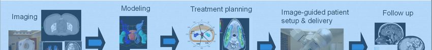
ML for Medical Image Analysis
Images reconstruction – low dose CT, fast MRI
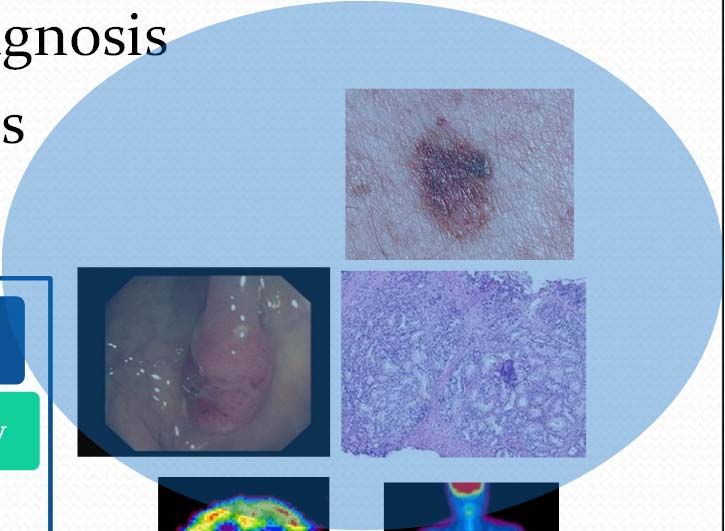
Imaging is one of the first choices for clinical diagnosis
70% clinical decisions depend on medical images
Molecular
imaging
Information technology Histology
Endoscopy
PET
fMRI
MRI
X‐CT
US
X‐
Ray
Anatomy
imaging
1895 1942 1972 1986 1993 1995 2020
& \VWRVFRS\LP DJLQJ$WWHQWLRQ 0 DS
We can directly visualize the network’s attention
when processing an input video.
The discriminative regions of tumor are highlighted,
suggesting the model works as expected and is able to
identify tumors from artifacts and background.
E. Shkolyar, X. Jia, T.C. Chang, D. Trivedi, K. E. Mach, M. Meng, L. Xing,
J. Liao, European Urology 76, 714-718, 2019
Image‐based prostate cancer classification
& virtual biopsy
Importance
Different cancer levels (Gleason score) lead to different therapy
Reduce the core needle biopsy
Modality for diagnosis
Magnetic Resonance Imaging (MRI)
T2-weighted images T2-weighted images Apparent Diffusion T1-weighted
(transaxial) (sagittal) Coefficient images Contrast images
Y. Yuan, W. Qin, B. Han, et al, Medical Physics, 2019
Stanford University
Department of Radiation Oncology
School of MedicineCT/CBCT artifacts removal
Dual-energy CT imaging using deep learning (Full 3D Meeting, 2019)
The HU difference between the predicted and original high-energy CT
images are 3.47 HU, 2.95 HU, 2.38 HU and 2.40 HU for ROIs on spine,
aorta, liver and stomach, respectively.
Stanford University
Department of Radiation Oncology
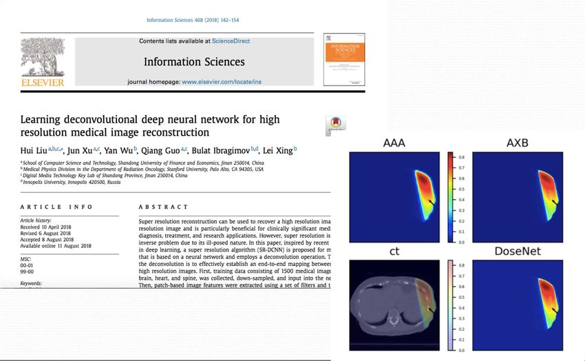
School of MedicineFrom super‐resolution imaging to super resolution dose calculation
Super‐resolution dose transformation and
machine learning‐based dose calculation
                  DL model
low resolution               high resolution
     dose                         dose

                  DL model
   low cost                    high resolution
 algorithm &                    dose, highly
low resolution               accurate algorithm

                  DL model
 ultra‐low cost                high resolution
                                                             P. Dong & L. Xing, Deep DoseNet: a deep neural
  algorithm &                   dose, highly                 network for accurate dosimetric transformation
low resolution               accurate algorithm              between different spatial resolutions and/or different
                                                             dose calculation algorithms for precision radiation
                                                             therapy, Phys. Med. Biol., 2019

                                 1.   Nomura Y, Wang J, Shirato H, Shimizu S, Xing L, Fast spot-scanning
                                      proton dose calculation method with uncertainty quantification using a
                                      three-dimensional convolutional neural network, PMB Jun. 2020
Machine learning provides a new way for small
field dosimetry and plan QA
Output factor prediction
E. Schueler, W. Zhao, et al, in preparation
J. Fan, L. Xing, Y. Yang, under reviewPushing the sparsity to the limit ‐
Stanford University
Department of Radiation Oncology
Shen
Shen L,
L, Zhao
Zhao W,
W, Xing
Xing L,
L, Nature
Nature Biomedical
Biomedical Engineering
Engineering 3,
(in880-808, 2019
press), 2019 School of MedicineShen L, Zhao W, Xing L, Nature Biomedical Engineering 3, 880-808, 2019
Sparse Data MR Image Reconstruction
Data Sampling
    Raw data are sampled point by point in Fourier domain (k-space)

Image Reconstruction
   Inverse Fourier transform is applied on the raw data to generate output in the
      image domain

                  Inverse Fourier Transform

     M. Mardani,…, L Xing, J Pauly, TMI, 2019
     Y. Wu, et al, Mag. Res. Imag., 2019
Integrated MRI‐Radiotherapy Systems: MRI Guided Localization & Delivery
#1 in the Liver Tumor
Segmentation Challenge
(LiTS‐ISBI2017)

- H. Seo, R. Xiao, L. Xing
Autonomous treatment planning for RT

                •CT                 DL
      INPUT     •Segmentation                                          Output
                                   MODEL

Clinical plan                                                      Plan predicted by
                                                                   deep learning

                                M. Ma, N. Kovalchuk, M. Buyyounouski, L. Xing, Y. Yang, Med Phys, 2019
Beam trajectory selection using reinforcement learning
Kahn, Fahimian et alBeam level imaging
Landmark detection in cephalometric analysis

                                       Stanford University
                                  Department of Radiation Oncology
                                                School of Medicine
Visualizing the invisible –
Deep learning‐augmented IGRT
Zhao et al ,IJROBP, 2019Target tracking Example of prostate motion tracking in AP direction The predict prostate position match the ground truth quite well.
• Pancreas, lung, etc.

                              Ground truth

                              Predicted

  Tracking on PTV for
  pancreas radiotherapy

   Zhao et al ,IJROBP, 2019
31
From population‐average nomogram to
deep learning‐based toxicity prediction
- B. Ibrambrov, D. Toesca, D. Chang, A Koong, L Xing
Current approach:
(i) NTCP/TCP types of modeling
Predictive
model
Problems: biological heterogeneity, spatial informationDeep dose‐toxicity prediction Multi-path network: 1) 3D CNN for dose plan; 2) fully-connected path for features
survival & toxicity results

                             Magical deep learning box
 Problem:    3D dose plan
                  +                                      Prediction
            Image analysis
Data Dimension‐Reduction Islam T & Xing L, Nature Biomedical Engineering, under revision, 2020
On‐going research Better AI models. Interpretable and trustworthy AI. General instead of task‐specific AI. Data & annotation. Clinical implementation and workflow related issues.
Modeling   Treatment planning   Image-guided patient   Follow up
Imaging
                                          setup & delivery
Acknowledgements M. Bassenne, J.‐E. Bibault, Y. Chen, D. P. Capaldi, J. Fan, C. Huang, T. Islam, M. Jia, L. Shen, H. Ren, M. Ma, H. Seo, X. Li, L.Yu, T. Liu, S. Gennatas, M. Khuzani, E. Schueler, E. Simiele, K., Sivasubramanian, H. Zhang, V. Vasudevan, Y. Wu, W. Zhao, Z. Zhang, D. P.I. Capaldi P. Dong, B. Han, Y. Yang, N. Kovalchuk, D. Hristov, L. Skinner, C. Chuang, L. Wang, J. Lewis, D. Chang, D. Toesca, Q. Le, S. Soltys, M. Buyounouski, H. Bagshaw, S. Hancock, G. Pratx, R. Li, J. Pauly, S. Boyd, .... Funding: NIH/NCI/NIBIB, DOD, NSF, ACS, Varian, & Google.
You can also read