BRIO: a web server for RNA sequence and structure motif scan
←
→
Page content transcription
If your browser does not render page correctly, please read the page content below
Published online 26 May 2021                                          Nucleic Acids Research, 2021, Vol. 49, Web Server issue W67–W71
                                                                                                      https://doi.org/10.1093/nar/gkab400

BRIO: a web server for RNA sequence and structure
motif scan
Andrea Guarracino 1,† , Gerardo Pepe 1,† , Francesco Ballesio 1 , Marta Adinolfi                                                              1
                                                                                                                                                  ,
Marco Pietrosanto 1 , Elisa Sangiovanni1 , Ilio Vitale 2,3 , Gabriele Ausiello1 and
Manuela Helmer-Citterich 1,*
1
 Department of Biology, University of Rome “Tor Vergata”, Rome, Italy, 2 IIGM - Italian Institute for Genomic Medicine,
c/o IRCSS Candiolo, Italy and 3 Candiolo Cancer Institute, FPO - IRCCS, Candiolo, Italy

                                                                                                                                                                  Downloaded from https://academic.oup.com/nar/article/49/W1/W67/6285264 by guest on 28 November 2021
Received February 21, 2021; Revised April 27, 2021; Editorial Decision April 28, 2021; Accepted May 22, 2021

ABSTRACT                                                                          GRAPHICAL ABSTRACT
The interaction between RNA and RNA-binding pro-
teins (RBPs) has a key role in the regulation of gene
expression, in RNA stability, and in many other bi-
ological processes. RBPs accomplish these func-
tions by binding target RNA molecules through spe-
cific sequence and structure motifs. The identifica-
tion of these binding motifs is therefore fundamen-
tal to improve our knowledge of the cellular pro-
cesses and how they are regulated. Here, we present
BRIO (BEAM RNA Interaction mOtifs), a new web
server designed for the identification of sequence
and structure RNA-binding motifs in one or more
RNA molecules of interest. BRIO enables the user
to scan over 2508 sequence motifs and 2296 sec-
ondary structure motifs identified in Homo sapiens
and Mus musculus, in three different types of ex-                                 INTRODUCTION
periments (PAR-CLIP, eCLIP, HITS). The motifs are
                                                                                  Molecular interactions are crucial for most biological pro-
associated with the binding of 186 RBPs and 69 pro-
                                                                                  cesses in the cell. The landscape of all possible molecular
tein domains. The web server is freely available at                               interactions depends on the actors involved in the interplay.
http://brio.bio.uniroma2.it.                                                      These actors include RNA-binding proteins (RBPs), which
                                                                                  play a central role in RNA metabolism, regulating the tran-
                                                                                  scripts throughout their life cycle, and in particular mod-
                                                                                  ulating mRNA localization, splicing, stability, and transla-
                                                                                  tion (1). Moreover, RBPs are also involved in the regula-
                                                                                  tion of non-coding RNAs. The human proteome includes
                                                                                  over 2000 RBPs (2), each one having specific functions and
                                                                                  target RNAs. This occurs through the interaction of par-
                                                                                  ticular RNA binding domains (RBDs) with specific RNA-
                                                                                  binding motifs (3). Of note, some RBPs recognize their tar-
                                                                                  get molecules via nucleotide patterns, while others favour
                                                                                  specific RNA structural motifs (4). The understanding of
                                                                                  RBP function and the identification of the binding motifs
                                                                                  are required to get insights into the regulatory mechanisms
                                                                                  in which these proteins are involved.

* To   whom correspondence should be addressed. Tel: +39 0672594324; Fax: +39 062023500; Email: manuela.helmer.citterich@uniroma2.it
†
 The authors wish it to be known that, in their opinion, the first two authors should be regarded as joint First Authors.
Present address: Marta Adinolfi, Department of Experimental Oncology, IEO, European Institute of Oncology IRCCS, Milan, Italy.

C The Author(s) 2021. Published by Oxford University Press on behalf of Nucleic Acids Research.
This is an Open Access article distributed under the terms of the Creative Commons Attribution-NonCommercial License
(http://creativecommons.org/licenses/by-nc/4.0/), which permits non-commercial re-use, distribution, and reproduction in any medium, provided the original work
is properly cited. For commercial re-use, please contact journals.permissions@oup.com
W68 Nucleic Acids Research, 2021, Vol. 49, Web Server issue
Table 1. Number of sequence and structure motifs identified in Homo sapi- human chronic myelogenous leukemia (K562) and hepato-
ens and Mus musculus cellular carcinoma (HepG2) cells. The dataset contains 186
Motif type Homo sapiens Mus musculus Total RBPs for both human and mouse, and a total of 69 unique
protein domains, 12 of which are shared between the two
Sequence 2112 184 2296 species (51 protein domains unique for human and 6 pro-
Structure 2319 189 2508
tein domains unique for mouse). The secondary structure
motifs are represented using the BEAR encoding.
The whole dataset of sequence and structure motifs is
Recent advances in high-throughput methods to assess
available for download at the BRIO website.
targets of RBPs in vitro and in vivo (5,6) have led to the de-
velopment of several resources to identify RBPs and their
binding motifs. Some tools find sequence motifs from in- Input
puts of target RNAs, while others focus on RNA secondary
Users can either input only the RNA primary sequence(s)
Downloaded from https://academic.oup.com/nar/article/49/W1/W67/6285264 by guest on 28 November 2021
structure (7). Here, we describe BRIO (BEAM RNA In-
of interest, or the sequence(s) and the corresponding sec-
teraction mOtifs), a new web server allowing users to eas-
ondary structure(s) in dot-bracket notation, all in FASTA
ily search for sequence and structure RNA-binding motifs
or multiFASTA format. Alternatively, the input can be up-
in one or more RNA molecules of interest. The dataset
loaded as a text file. The user can choose the preferred type
contains 2508 sequence and 2296 structure motifs asso-
of structural representation, e.g. Minimum Free Energy
ciated with the binding of 186 unique proteins and 69
(MFE) or centroid. Sequences submitted without the sec-
unique protein domains. The motifs were previously iden-
ondary structure are automatically folded using RNAfold
tified in Homo sapiens and Mus musculus, in three differ-
(in this case, the MFE structure is computed by default)
ent types of experiments (PAR-CLIP, eCLIP, HITS) ana-
(12). Finally, the dot-bracket notation is translated into a
lyzed in Adinolfi et al. (8). The structure motifs are encoded
BEAR string. The input RNA molecules are required to be
using BEAR (Brand nEw Alphabet for RNA), a power-
at least three nucleotides long, and shorter than 3000. To
ful context-aware structural string encoding we previously
search for structure motifs, sequences in input are requested
developed and applied in our research on RNA structural
to be at least 50 nucleotides long. At most 100 sequences can
characterization and conservation (9,10). This encoding not
be submitted at a time.
only stores information about the ’paired’ or ’unpaired’ sta-
Users can choose to compare their RNA molecules to the
tus of a nucleotide, but also takes into account the type and
whole dataset of motifs, or to only H. sapiens or M. muscu-
length of the secondary structure element (SSE) to which
lus motifs. It is also possible to select a subset of the experi-
the nucleotide belongs. This means that the BEAR encod-
ments analyzed (PAR-CLIP, eCLIP or HITS), considering
ing describes RNA secondary structure in a more compre-
that the eCLIP datasets were obtained from experiments
hensive way, but with low algorithmic complexity given the
performed only in H. sapiens.
simple string representation.
Procedure
MATERIALS AND METHODS
To identify the motifs, BRIO relies on substitution matrices:
Workflow
the Matrix of BEAR encoded RNA (MBRs) for secondary
The overall workflow of BRIO is illustrated in Figure 1. Af- structure elements (9) and a classic substitution matrix for
ter the pre-processing step (i.e. the calculation of the sec- nucleotides (with score 3 for nucleotide matching and −2
ondary structure, when not provided by the user), each in- otherwise). For each motif, the algorithm scans the cor-
put RNA molecule is scanned for the identification of se- responding Position Frequency Matrix in any single input
quence and structure motifs. The user can decide to scan RNA using a sliding-window ungapped alignment. Next,
subsets of motifs present in our database, selecting the the score of the best match is compared to the score thresh-
species and the type of experiments in which the motifs were old associated with the motif (for more information, see
identified. (8)). Finally, the one-sided Fisher’s Test is applied to deter-
Here, we provide a description of the datasets, the algo- mine if a motif is enriched in the input RNA molecules with
rithm, its input and the results provided by the web server. respect to a set of background RNAs. This test evaluates
whether the motif is identified with a significantly greater
proportion in the RNA over the background. The back-
Datasets
ground set of RNA molecules can be specified by the user.
BRIO scans the input RNA molecules using sequence and By default, all 85640 sequences from Rfam 14.3 are consid-
structure motifs (Table 1). The motifs were previously iden- ered (13).
tified in 186 RBPs and 69 protein domains analyzing 228
PAR-CLIP, eCLIP, HITS-CLIP experiments in H. sapiens
Output
and M. musculus (8,11).
The CLIP experiments are both from human (hg19; 74 BRIO returns a collection of protein binding motifs iden-
experiments performed in 13 different cell lines, principally tified in the input RNA molecules. The results are shown
HEK293 cells) and mouse (mm9; 30 experiments performed as tables, in two different views: ‘Enriched Motifs’ and ‘Se-
in 7 different cell lines, mostly brain and embryonic stem quences’ (Figure 2). The ‘Enriched Motifs’ tab shows the
cell), while eCLIP data comes from studies performed in motifs identified in the input molecules as a whole, whileNucleic Acids Research, 2021, Vol. 49, Web Server issue W69
Figure 1. The Workflow used by BRIO to search for motifs in the input RNA sequences. The input RNA molecules are folded, encoded using the BEAR
Downloaded from https://academic.oup.com/nar/article/49/W1/W67/6285264 by guest on 28 November 2021
alphabet (9), and scanned for sequence and structure motifs. Filters on species and type of experiment can also be applied. A background file can be used
as a comparison with the input sequences. The presence of enriched motifs is determined using Fisher’s exact test.
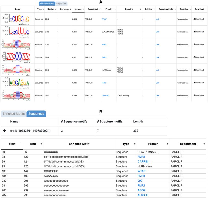
Figure 2. The output is composed of two views: (A) the ‘Enriched Motifs’ table, showing the enriched motifs found in the input RNA molecules, and (B)
the ‘Sequence’ table, showing the results for each input sequence provided. The column description is displayed in both tables when the pointer hovers over
the column name (not shown in the figure).W70 Nucleic Acids Research, 2021, Vol. 49, Web Server issue
the ‘Sequences’ tab shows the results for the different input HSURs are viral small RNAs that seem to be involved in
RNA molecules. The content of each table can be sorted in the regulation of the stability of host mRNAs. They have
ascending or descending order with respect to each column. been shown to associate, in vivo, with two proteins: het-
In the ‘Enriched Motifs’ table (Figure 2A), by default, erogeneous nuclear ribonucleoprotein D (HNRNPD) and
the motifs identified in the input RNA molecules are sorted ELAV like RNA binding protein 1 (ELAVL1, best known
according to the one-sided Fisher’s Test P-value. For each as HuR) (15). In particular, HuR is known to contribute
motif, the table reports: to the stability of mRNAs and to recognize short sequence
motifs of low statistical significance. The HSUR family con-
• the logo of the motif in the qBEAR alphabet for structure sists of four sequences, two of which share 100% sequence
motifs or in IUPAC nucleic acid notation for sequence identity. In our analysis, three different HSUR sequences
motifs; were given as input to BRIO and launched against the PAR-
• the type of the motif: sequence or structure; CLIP dataset of binding motifs. The web server identified
• the type of mapping regions from the GENCODE anno- several sequence motifs with high coverage (= 1) and low
Downloaded from https://academic.oup.com/nar/article/49/W1/W67/6285264 by guest on 28 November 2021
tation (8,14) of the RNAs datasets where the motif was P-values (in the [10–6 –10–5 ] range). After sorting the solu-
originally found. The annotation includes UTR, CDS, tions according to ascending P-values, we found in the top
and transcript for those RBPs known to act in the nu- positions sequence motifs binding the PAZ Piwi domain of
cleus on unspliced RNAs; AGO, and the HuR and ELAVL1MNASE RNA binding
• the coverage, which represents the number of input RNA domains. With a significant score, we also detected PUM2, a
molecules in which the motif is identified divided by the post-transcriptional repressor interacting with the 3 UTRs
total number of query molecules; of its target mRNAs. Of note, the structure motifs identi-
• the one-sided Fisher’s Test P-value; fied in this run did not show full coverage or a very low P-
• the type of experiment; value, although the HuR, AGO and ELAVL1MNASE pro-
• the protein associated with the RNA sequence or sec- tein binding motifs were listed with P-values in the [0.024–
ondary structure motif in the CLIP experiment reported 0.028] range and found in two out of the three input se-
in Adinolfi et al. (8); quences. The identification of structure motifs is generally
• the protein domain associated with the RNA secondary dependent on the number of input sequences (the more, the
structure motif (note that this information is not always better). The best hits can be identified also by inspecting the
available); ‘Sequences’ table in which the motifs identified in the sin-
• the cell line used in the eCLIP experiments; gle sequences are shown. In particular, this view allows the
• the link to the experiment page (for eCLIP data), or to the user to see the motifs identified according to the position in
corresponding article (for PAR-CLIP and HITS data); the sequence, but also sorted by protein name. Using this
• the organism in which the experiment was performed (H. last view, it is easy to see if single proteins or domains are
sapiens or M. musculus). repeated, and if the motifs identified are sequence and/or
structure motifs.
The last column reports the link for the download of the In the second example, we used BRIO to search for pro-
information on the motif described in each row. tein binding motifs in ten U2 spliceosomal RNA sequences
In the ‘Sequences’ table (Figure 2B), by default, the en- (the first listed in the RF00004 Rfam family) launched
tries are sorted according to the start position of the motif against the eCLIP dataset of binding motifs. In the ‘En-
in the sequence. For each entry, the table reports: riched Motifs’ Table, a structural motif associated with
• the start and the end position of the motif in the selected the SF3B1 protein showed a very low P-value. SF3B1 is
known to interact with U2 small nuclear ribonucleoprotein
sequence;
• the representation of the motif in BEAR alphabet for (snRNP), which is composed of U2 snRNAs and their asso-
structure motifs or in IUPAC nucleic acid notation for ciated polypeptide. Of note, several additional sequence and
structure motifs are listed in the BRIO output table, some of
sequence motifs;
• the type of the motif: sequence or structure; which are cytoplasmic and therefore the interaction is not
• the protein associated with the RNA sequence or sec- possible, while others can be used as suggestions for or to
confirm an experimental test.
ondary structure motif in the experiments analyzed by
Adinolfi et al. (8);
• the type of the experiment (PAR-CLIP, eCLIP, HITS). DISCUSSION
The BRIO web server allows researchers to identify se-
RESULTS
quence and structure protein binding motifs in a set of one
We describe, here, two examples of the use of BRIO web or more RNA molecules. The BRIO dataset encompasses
server to search for putative binding proteins. As input 2296 sequence and 2508 structure motifs associated with
RNA molecules, we selected RNA sequences belonging to 186 RNA binding proteins and 69 protein domains from
Rfam families whose interactors are already described in several CLIP experiments. BRIO takes advantage of the
the literature, and checked the server capability to identify BEAR encoding to represent structural motifs. This string
known motifs. These input datasets are also available in the encoding allows us to include the structural context of each
BRIO web server. nucleotide in the secondary structure representation, with-
In the first example, we analyzed sequences of the out increasing algorithm complexity. To the best of our
Herpesvirus saimiri U RNAs (HSUR) family (RF01802). knowledge, no other existing web server offers the possibil-Nucleic Acids Research, 2021, Vol. 49, Web Server issue W71
ity to search for sequence and structure motifs associated 3. Lunde,B.M., Moore,C. and Varani,G. (2007) RNA-binding proteins:
with RNA binding proteins. Indeed, few other web servers modular design for efficient function. Nat. Rev. Mol. Cell Biol., 8,
479–490.
are available to identify motifs in RNA sequences but, to 4. Cook,K.B., Hughes,T.R. and Morris,Q.D. (2015) High-throughput
our knowledge, only the RegRNA 2.0 server (16) allows the characterization of protein-RNA interactions. Brief. Funct.
user to search also in a database of sequence and structural Genomics, 14, 74–89.
motifs (such as splicing sites, polyadenylation sites, motifs 5. Hannigan,M.M., Zagore,L.L. and Licatalosi,D.D. (2018) Mapping
in 5 and 3 UTR, etc) that are not specifically associated transcriptome-wide protein-RNA interactions to elucidate RNA
regulatory programs. Quant Biol., 6, 228–238.
with RNA binding proteins. 6. Ferrè,F., Colantoni,A. and Helmer-Citterich,M. (2016) Revealing
Together with its friendly interface, BRIO can sup- protein-lncRNA interaction. Brief. Bioinform., 17, 106–116.
port scientists in their investigations on groups of RNA 7. Sasse,A., Laverty,K.U., Hughes,T.R. and Morris,Q.D. (2018) Motif
molecules of interest, their putative RBPs, and the roles models for RNA-binding proteins. Curr. Opin. Struct. Biol., 53,
115–123.
these proteins play in RNA regulation. 8. Adinolfi,M., Pietrosanto,M., Parca,L., Ausiello,G., Ferrè,F. and
Downloaded from https://academic.oup.com/nar/article/49/W1/W67/6285264 by guest on 28 November 2021
Helmer-Citterich,M. (2019) Discovering sequence and structure
landscapes in RNA interaction motifs. Nucleic Acids Res., 47,
DATA AVAILABILITY 4958–4969.
The web server is freely available at http://brio.bio. 9. Mattei,E., Ausiello,G., Ferrè,F. and Helmer-Citterich,M. (2014) A
novel approach to represent and compare RNA secondary structures.
uniroma2.it. The source code and all the data are available Nucleic Acids Res., 42, 6146–6157.
at https://github.com/helmercitterich-lab/BRIO. 10. Pietrosanto,M., Adinolfi,M., Guarracino,A., Ferrè,F., Ausiello,G.,
Vitale,I. and Helmer-Citterich,M. (2021) Relative information gain:
Shannon entropy-based measure of the relative structural
ACKNOWLEDGEMENTS conservation in RNA alignments. NAR Genom Bioinform, 3, lqab007.
11. Blin,K., Dieterich,C., Wurmus,R., Rajewsky,N., Landthaler,M. and
We acknowledge ELIXIR-IIB (elixir-italy.org), the Ital- Akalin,A. (2015) DoRiNA 2.0–upgrading the doRiNA database of
ian Node of the European ELIXIR infrastructure (elixir- RNA interactions in post-transcriptional regulation. Nucleic Acids
europe.org), and CINECA for supporting FB in the Res., 43, D160–D167.
12. Lorenz,R., Bernhart,S.H., Höner Zu Siederdissen,C., Tafer,H.,
development of this work through the ELIXIR-IIB Flamm,C., Stadler,P.F. and Hofacker,I.L. (2011) ViennaRNA
HPC@CINECA call. package 2.0. Algorithms Mol. Biol., 6, 26.
13. Gardner,P.P., Daub,J., Tate,J., Moore,B.L., Osuch,I.H.,
Griffiths-Jones,S., Finn,R.D., Nawrocki,E.P., Kolbe,D.L., Eddy,S.R.
FUNDING et al. (2011) Rfam: Wikipedia, clans and the ‘decimal’ release. Nucleic
Acids Res., 39, D141–D145.
Associazione Italiana per la Ricerca sul Cancro (AIRC) [IG 14. Harrow,J., Frankish,A., Gonzalez,J.M., Tapanari,E., Diekhans,M.,
23539 to M.H.C.]. Kokocinski,F., Aken,B.L., Barrell,D., Zadissa,A., Searle,S. et al.
Conflict of interest statement. None declared. (2012) GENCODE: the reference human genome annotation for The
ENCODE Project. Genome Res., 22, 1760–1774.
15. Cook,H.L., Mischo,H.E. and Steitz,J.A. (2004) The Herpesvirus
REFERENCES saimiri small nuclear RNAs recruit AU-rich element-binding proteins
but do not alter host AU-rich element-containing mRNA levels in
1. Glisovic,T., Bachorik,J.L., Yong,J. and Dreyfuss,G., (2008) virally transformed T cells. Mol. Cell. Biol., 24, 4522–4533.
RNA-binding proteins and post-transcriptional gene regulation. 16. Chang,T.-H., Huang,H.-Y., Hsu,J.B.-K., Weng,S.-L., Horng,J.-T. and
FEBS Lett., 582, 1977–1986. Huang,H.-D. (2013) An enhanced computational platform for
2. Corley,M., Burns,M.C. and Yeo,G.W. (2020) How RNA-binding investigating the roles of regulatory RNA and for identifying
proteins interact with RNA: molecules and mechanisms. Mol. Cell, functional RNA motifs. BMC Bioinformatics, 14(Suppl. 2), S4.
78, 9–29.You can also read